Serosurvey of
Rickettsia
spp. in small mammals
from Mato Grosso do Sul state, Brazil
Inquérito sorológico para
Rickettsia
spp. Em pequenos
mamíferos do estado do Mato Grosso do Sul, Brasil
Lina de Campos Binder
1*Felipe da Silva Krawczak
1Jonas Sponchiado
2Geruza Leal Melo
2Jonas Moraes-Filho
1Fernanda Aparecida Nieri Bastos
1Nilton Carlos Cáceres
2Marcelo Bahia Labruna
1ISSNe 1678-4596
Rickettsia rickettsii
is the etiologic agent
of a severe febrile illness in humans, known in Brazil
as Brazilian Spotted Fever (BSF) (ANGERAMI et
al.,2006). For keeping its vital cycle,
R. rickettsii
needs a
tick population and vertebrate animals, called amplifier
hosts, which develop a rickettsemia for some days or
weeks, to enable the infection of additional ticks. This
mechanism amplifies the
R. rickettsii
infection rates
among the tick population (LABRUNA, 2009).
The urban expansion process in rural
areas increases the chance of interaction of humans
and domestic animals with wild animal populations,
which may be infested by ticks, increasing the risk of
rickettsiosis (BARBIERI et al., 2014). Small mammals
are main hosts of immature stages of numerous tick
species (SARAIVA et al., 2012); hence, serosurvey of
these animals is very useful for predicting circulation
of rickettsiae in a given area.The present study aimed
to evaluate exposure of wild small mammals for
spotted fever group (SFG) rickettsiae in the state of
Mato Grosso do Sul (MS), central-western Brazil.
While BSF is endemic in many parts of
Brazil (LABRUNA, 2009), the occurrence of this
tick-borne disease in MS has been restricted to a
single, laboratory-confirmed case in Dois Irmãos do
Buriti municipality during 2013 (official data from
the Brazilian Ministry of Health, 2016). From 2012
to 2013, SPONCHIADO et al. (2015) performed a
field study in MS, in which wild small mammals were
trapped in 31 woodland fragments of Cerrado biome
distributed in the municipalities of Terenos, Anastácio,
Miranda, Bonito, and Nioaque, located in the Paraguai
River basin between the coordinates 20
o17’ - 21
o15’S
and 54
o53’ - 56
o31’W. The maximum distance between
these localities was 162km. Because these localities
are close to Dois Irmãos do Buriti (Figure 1), from
1Departamento de medicina veterinária preventiva e saúde animal (VPS), Faculdade de Medicina veterinária e Zootecnia, Universidade de São Paulo (USP), Av. Prof. Dr. Orlando Marques de Paiva, 87, 05508-270, São Paulo, SP, Brasil. E-mail: lina.binder@usp.br. *Corresponding author. 2Laboratório de Ecologia e Biogeografia, Departamento de Biologia, Universidade Federal de Santa Maria (UFSM), Santa Maria, RS, Brasil.ABSTRACT: This study aimed to evaluate exposure of wild small mammals to spotted fever group (SFG) rickettsiae in Mato Grosso do Sul State, central-western Brazil. Serum samples of 68 small mammals were analyzed by indirect immunofluorescence assay (IFA) against six Rickettsia species from Brazil. Overall, 37.5% (9/24) marsupials and 6.8% (3/44) small rodents were seroreactive to at least one of the Rickettsia species, with end point titres ranging from 64 to 512. These results suggested that wild small mammals were infected by SFG rickettsiae, and could participate in the ecology of rickettsiae in Mato Grosso do Sul, Brazil.
Keywords:Marsupial, Mato Grosso do Sul, Rodent; Brazilian spotted fever, IFA.
RESUMO:O objetivo do presente estudo foi avaliar a exposição de pequenos mamíferos silvestres para riquétsias do Grupo da Febre Maculosa (GFM) no estado do Mato Grosso do Sul, centro-oeste do Brasil. Amostras de soro de 68 pequenos mamíferos foram submetidos
à reação de imunofluorescência indireta (RIFI) frente a seis espécies de Rickettsia do Brasil. No total, 37,5% (9/24)e 6,8% (3/44) dos soros
de marsupiais e pequenos roedores, respectivamente, reagiram a pelo menos uma das espécies de Rickettsia, com títulos variando de 64 a 512. Osresultados sugerem que pequenos mamíferos silvestres foram infectados por riquétsias do GFM e poderiam participar na ecologia de riquétsias no Mato Grosso do Sul, Brasil.
Palavras-chave:marsupial, Mato Grosso do Sul, roedor; febre maculosa brasileira, RIFI.
where BSF was reported, blood samples from 68 small
mammals trapped from November 2012 to July 2013
during the study of SPONCHIADO et al. (2015) were
provided for the present study, in order to be tested for
the presence of anti-
Rickettsia
spp. antibodies. While
SPONCHIADO et al. (2015) sampled a much larger
sample of small mammals, blood samples collected from
only 68 individuals belonging to 13 different species, 4
marsupials (
Didelphis albiventris-
white-eared opossum
,
Gracilinanus agilis
- agile gracile opossum
,
Marmosa
murina
- Linnaeus’s mouse opossum
,
Thylamys
macrurus
- Paraguayan fat-tailed mouse opossum)
and 9 rodents (
Calomys callosus
- large vesper mouse
,
C. tener
- delicate vesper mouse,
Cerradomys scotti
-
Lindbergh’s rice rat
,
C. maracajuensis
- Maracaju
rice rat
,
Hylaeamys megacephalus
- Azara’s
broad-headed rice rat,
Nectomys rattus
- Amazonian water
rat,
Oecomys bicolor
- bicolored arboreal rice rat
,
O.
mamorae
- mamore arboreal rice rat
,
Rhipidomys
macrurus
-
Cerrado climbing mouse) were tested.
Overall, 75%(18/24) and 11% (5/44) of these marsupials
and rodents, respectively, were reported to be infested
by ticks, which were identified as immature stages of
Amblyomma coelebs
,
A. ovale, A. parvum, A. sculptum
and
Ornithodoros mimon
(SPONCHIADO et al.,2015).
For blood collection, all animals were
anesthetized with an intramuscular injection (dosage
25mgkg
-1) of Zoletil
®50 (mixture of tiletamine and
zolazepam, 25mgmL
-1of each), as previously described
(RIVAS et al., 2015). Blood samples were collected
by cardiac puncture and sera were separated by
centrifugation (12,000 rcf-10min). Rodent and marsupial
sera were tested by indirect immunofluorescence assay
(IFA) using crude antigens of 6
Rickettsia
isolates from
Brazil (
R. rickettsii
strain Taiaçu,
R. parkeri
strain At24,
R
.
amblyommii
strain Ac37,
R. rhipicephali
strain
HJ5,
R. felis
strain Pedreira, and
R. bellii
strain Mogi),
following previously described protocols (HORTA et
al., 2004, 2007). Fluoresce in isothiocyanate- labelled
goat anti-mouse IgG, dilution 1:400 (Sigma, St Louis,
MO, USA) and sheep anti-opossum IgG, dilution 1:500
(CCZ, São Paulo, Brazil) were used as conjugate for
rodent and marsupial sera, respectively. In each slide,
a serum previously shown to be non-reactive (negative
control) and a known reactive serum (positive control)
were tested at the 1:64 dilution.
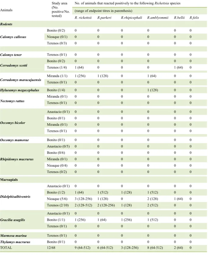
Table 1 -Results of sero reactivity of wild small mammals from MatoGrosso do Sul State, central- western Brazil to six Rickettsia species (November 2012 to July 2013).
Animals
Study area (No. positive/No. tested)
No. of animals that reacted positively to the following Rickettsia species (range of endpoint titres in parenthesis)
R. rickettsii R.parkeri R.rhipicephali R.amblyommii R.bellii R.felis
Rodents
Calomys callosus
Bonito (0/2) 0 0 0 0 0 0
Nioaque (0/1) 0 0 0 0 0 0
Terenos (0/3) 0 0 0 0 0 0
Calomys tener Terenos (0/1) 0 0 0 0 0 0
Cerradomys scotti
Bonito (0/2) 0 0 0 0 0 0
Terenos (1/4) 1 (64) 0 0 0 1 (64) 0
Cerradomys maracajuensis
Miranda (1/1) 1 (256) 1 (128) 0 1 (64) 0 0
Terenos (0/1) 0 0 0 0 0 0
Hylaeamys megacephalus Bonito (1/4) 0 0 0 1 (128) 0 0
Nectomys rattus
Miranda (0/1) 0 0 0 0 0 0
Terenos (0/1) 0 0 0 0 0 0
Oecomys bicolor
Anastacio (0/1) 0 0 0 0 0 0
Bonito (0/1) 0 0 0 0 0 0
Miranda (0/1) 0 0 0 0 0 0
Terenos (0/1) 0 0 0 0 0 0
Oecomys mamorae Bonito (0/1) 0 0 0 0 0 0
Rhipidomys macrurus
Anastacio (0/5) 0 0 0 0 0 0
Bonito (0/6) 0 0 0 0 0 0
Miranda (0/1) 0 0 0 0 0 0
Nioaque (0/4) 0 0 0 0 0 0
Terenos (0/2) 0 0 0 0 0 0
Marsupials
Didelphisalbiventris
Anastacio (0/1) 0 0 0 0 0 0
Bonito (1/2) 1 (64) 1 (512) 1 (128) 1 (512) 0 0
Nioaque (5/6) 3 (128-256) 1 (128) 0 2 (128) 1 (64) 0
Terenos (2/10) 2 (128-512) 2 (128-256) 1 (128) 2 (512) 0 0
Gracilia usagilis
Anastacio (0/1) 0 0 0 0 0 0
Bonito (1/1) 1 (256) 1 (64) 1 (256) 1 (512) 0 0
Terenos (0/1) 0 0 0 0 0 0
Marmosa murina Terenos (0/1) 0 0 0 0 0 0
Thylamys macrurus Bonito (0/1) 0 0 0 0 0 0
end point titre to a
Rickettsia
species at least 4-fold
higher than the titres exhibited to any of the other
five rickettsial antigens, precluding any inference
of which possible
Rickettsia
species infected these
animals, as described by HORTA et al. (2004).
The Fisher exact statistical test showed that these
sororeactivity for SFG rickettsiae was significantly
higher (P<0.05) among marsupials than small
rodents, what is probably related to the fact that these
marsupials had a much higher tick prevalence than
small rodents (SPONCHIADO et al., 2015). This
condition increases the likelihood of
D. albiventris
being infested by a SFG rickettsia-infected ticks,
when compared to the other small mammal species.
Interestingly, this difference was also observed by
SZABÓ et al. (2013) in the State of São Paulo. In
addition, HORTA et al.(2009) showed that
D. aurita
can act as amplifier host for
R. rickettsii
,
having an
important role in the BSF epidemiology. Moreover,
Didelphis
spp. are considered excellent sentinels for
BSF surveillance (HORTA et al., 2007).
This study is the first rickettsial sero
survey of wild small mammals in the
Central-Western region of Brazil. Our results indicate that
small mammals were exposed to SFG rickettsiae,
suggesting that animals such as
D. albiventris
participate in the maintenance of the ecological cycle
of SFG rickettsiae in these areas. The tick-borne
agents that have been reported to cause disease in
humans in Brazil are
R. rickettsii
(ANGERAMI et
al., 2006) and
Rickettsia
sp. strain Atlantic rainforest,
a
R. parkeri
-
like agent (SPOLIDORIO et al., 2010).
Even though it was not possible to infer the SFG
Rickettsia
species to which small mammals were
exposed in the present study, our results highlighted
the possibility that tick-borne zoonoses could be
circulating between small rodents and ticks in Mato
Grosso do Sul, Brazil. This assumption is supported
by the fact that these rodents were infested by at least
four tick species,
A. coelebs
,
A. ovale,
A. parvum
and
A. sculptum
(SPONCHIADO et al., 2015) that
have been reported as infected by different SFG
agents in Brazil, including the human pathogens
R.
rickettsii
and
Rickettsia
sp. strain Atlantic rainforest
(LABRUNA et al., 2004;SZABÓ et al., 2013;
NIERI-BASTOS et al., 2014; KRAWCZAK et al.
2014;WITTER et al. 2016).
ACKNOWLEDGMENTS
Fundação de Amparo a Pesquisa do Estado de
BIOETHICS AND BIOSSECURITY
COMMITTEE APPROVAL
Protocol n. 30808-2.
REFERENCES
ANGERAMI, R.N. et al. Brazilian Spotted Fever: a case series from an endemic area in Southeastern Brazil: clinical aspects.
Annals of the New York Academy of Sciences, v.1078, p.252-254, 2006. Available from: <http://onlinelibrary.wiley.com/ doi/10.1196/annals.1374.044/full>. Accessed: Nov. 04, 2015. doi: 10.1196/annals.1374.044.
BARBIERI, A.R.M. et al. Epidemiology of Rickettsia sp. strain Atlantic rainforest in a spotted fever-endemic area of southern Brazil. Ticks and Tick-borne Diseases, v.5, p.848-853,2014. Available from: <http://www.sciencedirect.com/science/article/pii/ S1877959X14001472>. Accessed: Nov. 04, 2015. doi: 10.1016/j. ttbdis.2014.07.010.
HORTA, M.C. et al. Prevalence of antibodies to spotted fever group Rickettsiae in humans and domestic animals in a Brazilian spotted
fever endemic area in the state of São Paulo, Brazil: serological
evidence for infection by Rickettsia rickettsii and another spotted fever group Rickettsia. American Society of Tropical Medicine and Hygiene, v.71, p.93-97,2004. Available from: <http://www. ajtmh.org/content/71/1/93.full.pdf+html>. Accessed: Nov. 05, 2015.
HORTA, M.C.et al. Rickettsia infection in five areas of the state of São Paulo, Brazil. Memórias do Instituto Oswaldo Cruz, v.102, p.793-801, 2007. Availablefrom: <http://www.ncbi.nlm.nih. gov/pubmed/18094887>. Accessed:Nov. 05, 2015. doi: 10.1590/ S0074-02762007000700003.
HORTA, M.C. et al.Experimental infection of opossums
Didelphisaurita by Rickettsia rickettsii and evaluation of the transmission of the infection to ticks Amblyomma cajennense.
Vector-borne and ZoonoticDiseases, v.9, p.109-118,2009. Available from: <http://online.liebertpub.com/doi/pdf/10.1089/vbz.2008.0114>. Accessed: Nov. 05, 2015. doi: 10.1089/vbz.2008.0114.
KRAWCZAK, F.S. et al. Rickettsial infection in
Amblyommacajennense ticks and capybaras (Hydrochoerus hydrochaeris) in a Brazilian spotted fever-endemic area.
Parasites & Vectors, v.7, p.1-7, 2014. Available from: <http:// www.ncbi.nlm.nih.gov/pmc/articles/PMC3892071/pdf/1756-3305-7-7.pdf>. Accessed: May 12, 2016. doi: 10.1186/1756-3305-7-7.
LABRUNA, M. B. et al. Rickettsia bellii and Rickettsia amblyommii in Amblyomma Ticks from the State of Rondônia, Western Amazon, Brazil. Journal of Medical Entomology, v.41, p.1073-1081, 2004. Available from: <http://www.ncbi. nlm.nih.gov/pubmed/15605647>. Accessed: May 14, 2016. doi: 10.1603/0022-2585-41.6.1073.
Dados. Online .Available from: <http://portalsaude.saude.gov.br/ images/pdf/2016/abril/27/tabela-casos-febre-maculosa-2016.pdf>. Accessed: May 13, 2016.
NIERI-BASTOS F.A. et al. CandidatusRickettsia andeanae, a spotted fever group agent infecting Amblyomma parvum ticks in two Brazilian biomes. Memórias do Instituto Oswaldo Cruz, v.109, p.259-261. 2014. Available from: <http://www.scielo.br/ pdf/mioc/v109n2/0074-0276-mioc-109-02-0276140283.pdf>. Accessed: May 12, 2016. doi: 10.1590/0074-0276140283. RIVAS, J.J. et al. Pathogenic potential of a Costa Rican strain of “Candidatus Rickettsia amblyommii” in guinea pigs (Cavia porcellus) and protective immunity against Rickettsia rickettsii.Ticks and Tick-borne Diseases, v.6, p.805-811, 2015. Available from: <http:// www.sciencedirect.com/science/article/pii/S1877959X15001387>. Accessed: May 01, 2016. doi: 10.1016/j.ttbdis.2015.07.008.
SARAIVA, D.G. et al. Ticks (Acari: Ixodidae) associated with
small terrestrial mammals in the state of Minas Gerais, southeastern Brazil. Experimental and Applied Acarology, v.58, p.159-166, 2012. Available from: <http://link.springer.com/article/10.1007/ s10493-012-9570-9>. Accessed: Nov. 04, 2015. doi: 10.1007/ s10493-012-9570-9.
SPOLIDORIO, M.G. et al. Novel spotted fever group rickettsiosis,
Brazil.Emerging Infectious Diseases, v.16, p.521-523,2010. Available from: <http://www.ncbi.nlm.nih.gov/pmc/articles/PMC3322033/>. Accessed: Nov. 04, 2015. doi: 10.3201/eid1603.091338.
SPONCHIADO, J.et al. Association patterns of ticks (Acari:
Ixodida: Ixodidae, Argasidae) of small mammals in Cerrado Fragments, western Brazil. Experimental and Applied Acarology, v.65, p.389-401,2015. Available from: <http://link.springer.com/ article/10.1007/s10493-014-9877-9>. Accessed: Nov. 04, 2015. doi: 10.1007/s10493-014-9877-9.
SZABÓ, M.P.J. et al. In vitro isolation from Amblyommaovale
(Acari:Ixodidae) and ecological aspects of the Atlantic Rainforest
Rickettsia, the causative agent of a novel spotted fever rickettsiosis in Brazil. Parasitology, v.140, n.6, p.719-728,2013. Available from: <http://dx.doi.org/10.1017/S0031182012002065>. Accessed: Nov. 04, 2015 .doi: 10.1017/S0031182012002065.