Copyright © 2000, American Society for Microbiology. All Rights Reserved.
Antigen Specificity of T-Cell Response to
Mycobacterium avium
Infection in Mice
TERESA F. PAIS, JOANA FEIJO´ CUNHA,ANDRUI APPELBERG*
Laboratory of Microbiology and Immunology of Infection, Institute for Molecular and Cell Biology, University of Porto, Porto, Portugal
Received 29 February 2000/Returned for modification 31 March 2000/Accepted 5 May 2000
T cells fromMycobacterium avium-infected C57BL/6 mice reacted to culture filtrate, envelope, and cytosol proteins and to fractions obtained from these proteins. Multiple targets were recognized, such as 29- to 45-kDa and <21-kDa antigens of the culture filtrate, antigens of around 30 kDa in the envelope and cytosol, and 45-to 116-kDa proteins in the envelope.
The identification of the key antigenic targets of the immune response to mycobacteria is of pivotal importance in the design and testing of new vaccine candidates against mycobacterial pathogens, most notably Mycobacterium tuberculosis. Work
from several laboratories has identified secreted/exported pro-teins fromM. tuberculosisas the major targets of a protective
immune response to experimental tuberculosis infections (2, 3, 5, 12, 13, 17, 18, 20). These antigens are also favored targets during an immune response in human patients infected with the tubercle bacilli (17). However, other proteins believed not to be excreted by the mycobacterial cells have also been iden-tified as important targets, namely in the induction of protec-tive immune responses in experimental animals (14, 23), sug-gesting that the complete picture of the immune response to mycobacterial infections may be more complex with regard to the antigenic repertoire recognized in vivo. Mycobacterium aviumis an opportunistic pathogen that is thought to interfere,
in certain areas of the world, with the efficacy of the only tuberculosis vaccine in use today, the attenuated Mycobacte-rium bovisstrain bacille Calmette-Gue´rin (BCG) (9). The
rea-son for the failure of vaccination trials is not known, but it has been speculated that sensitization of human beings to antigens from environmental mycobacteria might affect BCG efficacy, most likely because all mycobacteria would share common antigens. Sharing of antigens between nontuberculous myco-bacteria and BCG was already shown to occur at the immu-nological level in mice (15). However, except for two defined antigens, the latter study used crude extracts that combined many different antigens. The sharing of common antigens betweenM. avium, BCG, andM. tuberculosiswill be
under-stood in the near future thanks to efforts in the area of genomics. However, we still need studies in the fields of proteomics and immunology to generate functional data and therefore to be able to critically analyze the genomic informa-tion, namely studying native proteins rather than recombinant ones, as the latter may lack the immunogenicity of the former (1, 21). Thus, we initiated the characterization of the T-cell
response toM. aviumprotein antigens using a mouse model of
infection.
We isolated antigens fromM. avium2447, an AIDS patient
isolate obtained from F. Portaels (Institute of Tropical Medi-cine, Antwerp, Belgium) that forms smooth transparent colo-nies when cultured on solid media. Mycobacteria from log-phase cultures were inoculated into Sauton medium enriched with 0.5% sodium pyruvate and 0.5% glucose (Sauton P1G) (8) and with no detergent, at a final concentration of approx-imately 53 106 CFU/ml (according to the absorbance mea-sured at 600 nm), and grown at 37°C without shaking. The number of viable bacteria and the smooth transparent mor-photype were confirmed by plating serial dilutions of the cul-tures on solid Middlebrook 7H10 medium (Difco, Sparks, Md.). At the end of log phase (i.e., at day 15 as evaluated from the previous growth curves), cultures were centrifuged for pro-cessing of bacterial antigens. Culture filtrate proteins were obtained from the filter-sterilized supernatant of the culture by ultrafiltration in a stirred cell (Millipore, Bedford, Mass.) with an Amicon YM membrane (molecular weight cut-off [MWCO] of 3,000) (Millipore). The concentrate was precipitated with 80% ammonium sulfate, and the precipitate was washed in a Centriprep (MWCO of 3,000) (Millipore). Cytosolic and en-velope proteins were obtained after the pellet was washed twice with phosphate-buffered saline (PBS), resuspended in PBS containing 0.1% Tween 80 (Sigma), 1 mM MgCl2(Merck, Darmstadt, Germany) and 1 mM benzamidine (Sigma) (10), and disrupted through sonication with pulses of 1 min at maximum power, with the sample kept in ice during the whole procedure. The sonicate was centrifuged to discard intact mycobacteria (30 min at 2,700 3 g), and the
super-natant was dialyzed against PBS (MWCO of 12,000). The suspension was then ultracentrifuged for 2 h at 150,0003g.
The pellet containing the envelope proteins was resus-pended in PBS, and the supernatant enriched in cytosolic proteins was precipitated with 80% ammonium sulfate and dialyzed against PBS.
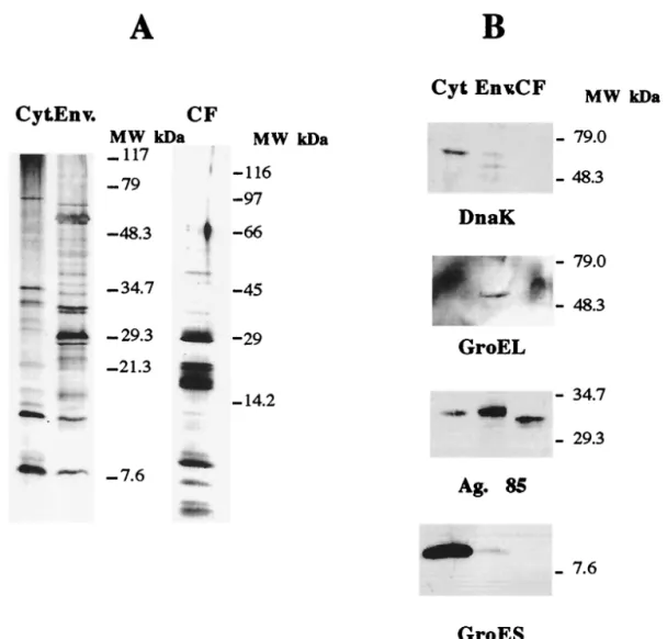
Following the preparative procedures described above, we obtained crude extracts which were analyzed to assess if they were distinct sources of antigens. Cytosolic, envelope, and culture filtrate proteins (20mg) were separated in a 10 to 20% gradient sodium dodecyl sulfate-polyacrylamide gel elec-trophoresis (SDS-PAGE) gel and analyzed either by silver staining or by Western blotting after transfer to a PROTON nitrocellulose (Schleicher and Schuell, Dassel, Germany)
* Corresponding author. Mailing address: Laboratory of Microbiol-ogy and ImmunolMicrobiol-ogy of Infection, Institute for Molecular and Cell Biology, University of Porto, Rua do Campo Alegre 823, 4150-180 Porto, Portugal. Phone: 351.226074952. Fax: 351.226099157. E-mail: [email protected].
membrane in a semidry electrophoretic transfer cell (Bio-Rad, Richmond, Calif.). During the latter procedure, the membrane was blocked in PBS containing 0.5% Tween 20 (Sigma), incu-bated for 2 h at room temperature with the antibodies diluted (1:50) in PBS–0.05% Tween–0.37 M NaCl, and washed in the dilution buffer. The primary antibodies used were specific for DnaK (clone HÅT 3), GroEL (clone HÅT 5), GroES (clone HYB 76-1), and Ag85 (clone HYT 27), and they were kindly provided by Peter Andersen (Statens Seruminstitut, Copen-hagen, Denmark). The secondary antibody, a horseradish peroxidase-coupled sheep anti-mouse antibody (Amersham-Pharmacia Biotech, Little Chalfont, United Kingdom) was incubated at a 1:500 dilution for 2 h at room temperature. The specific protein complexes were detected using the ECL re-agents (Amersham-Pharmacia Biotech).
The protein profiles observed in the silver-stained gels for the three fractions were different, with distinct, different major bands obtained with the three preparations (Fig. 1A). The immunochemical analysis by the Western blotting with the panel of monoclonal antibodies against well-defined myco-bacterial proteins (DnaK, GroEL, Ag85 complex, and GroES) showed that the three protein preparations corresponded to dis-tinct cellular compartments. Thus, the DnaK and GroES
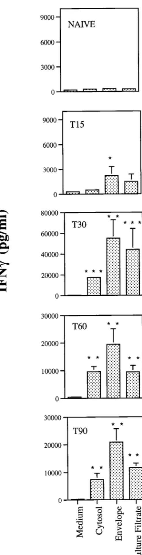
(Nunc, Roskilde, Denmark), each well containing 23105cells in a volume of 200ml with no stimulus or incubation in the presence of antigen at a final concentration of 4mg of crude extract/ml. Culture supernatants from triplicate wells were har-vested 72 h later for the detection of gamma interferon (IFN-g) as a readout of the response of those T cells to the different antigens using an enzyme-linked immunosorbent as-say (ELISA) as previously described (22). As shown in Fig. 2, the envelope proteins were strong stimulators of IFN-g pro-duction from day 15 of infection. The three preparations had antigens that stimulated T cells from mice infected for 30, 60, or 90 days, with the highest amount of IFN-gbeing produced at day 30 of infection.
To obtain panels of MW fractions from the culture filtrate, envelope, and cytosol preparations, the multielution technique (4) was used. Briefly, 7 mg of culture filtrate or cytosolic pro-teins or 9 mg of envelope propro-teins was separated by SDS-PAGE (with a gradient gel of 10 to 20% acrylamide), and the gel was prepared for electroelution as described before (4). The proteins were electroeluted (40 V) for 20 min into a 2-mM phosphate buffer in a whole-gel eluter (Bio-Rad) in a cold room. The fractions were collected and analyzed (40ml of each fraction) in a gradient SDS-PAGE (10 to 20% acrylamide) after fixation and silver staining (16). Protein concentration was quantified by the Micro BCA method (Pierce, Rockford, Ill.). The fractions were stabilized with 0.5% FCS in PBS. The SDS-PAGE analysis of the fractions obtained showed that most of them contained proteins in a very narrow range of MWs (Fig. 3), thereby greatly reducing the complexity of the crude extracts. The fractions obtained were then used to study the antigenic specificity of the T cells from infected mice, as detailed above (Fig. 4). The in vitro stimulation of spleen cells with the MW fractions obtained from the three different com-partments at a final concentration of 2mg/ml showed that the response to the culture filtrate fractions emerged at day 15, earlier than the response to the cytosol or envelope fractions. At day 30 of infection, the peak of the response, the strongest stimulators of T cells were found among fractions of culture filtrate in the 29,000-to-45,000 MW range. Fraction 13 from the culture filtrate, which was enriched in proteins in the 30-kDa region (where proteins such as Ag85 complex, a group of proteins that are highly immunogenic among mycobacteria (11, 24), are expected to locate), induced the strongest IFN-g production. Fractions 12 of the cytosol and envelope, which contain proteins in the same region of MW, were also very immunogenic. Fractions from the envelope between 45 and 116 kDa contained highly stimulatory proteins that were ab-sent from the corresponding MW fractions of the culture fil-trate. On the other hand, there was a group of fractions in the culture filtrate below 21 kDa (fractions 17 to 21), which did not include the lowest-MW fractions, that were also inducers of IFN-gproduction.
The antigen repertoire recognized by IFN-g-secreting T cells during an M. avium infection in mice revealed a highly
diverse set of protein antigens. The antigen targets were not confined to the secreted/exported proteins but rather were promiscuous among all compartments of the mycobacterial cell when grossly dissected into secreted/exported (culture fil-trate), envelope, and cytoplasmic proteins. Several groups have
FIG. 2. Antigenicity of the crude extracts ofM. avium2447 proteins. Single-cell suspensions were prepared from spleens of mice infected for 15 (T15), 30 (T30), 60 (T60), or 90 (T90) days withM. avium2447 or noninfected (naive) animals and stimulated in vitro with 4mg of cytosol, envelope, or culture filtrate
proteins per ml. IFN-grelease was quantified by ELISA in the 72-h culture
supernatants. Results are expressed as means of values of triplicate samples61
standard deviation performed on cells pooled from four mice. Statistically sig-nificant differences compared to values for nonstimulated cells are labeled:p,
favored secreted/exported proteins as the major targets of the protective immune response toM. tuberculosis(2, 3, 5, 12, 13,
17, 18, 20). In addition, cell wall-associated proteins have also been shown to evoke an immune response during infection (17), suggesting their shedding from the cell wall during growth of mycobacteria inside the vacuoles of the infected macro-phage. Finally, the response to cytoplasmic antigens would, according to some, appear later in infection as a result of the killing of the mycobacteria, and this response would be asso-ciated not with a protective IFN-g-mediated immune response but most likely with a type 2 immune response, which is puta-tively associated with removal of the dead mycobacteria (19). Our data confirm the above paradigm by showing im-portant reactivity towards secreted/exported as well as en-velope proteins. However, IFN-g-secreting T cells were also found to respond to the cytosolic proteins, namely to fractions of around 30 to 32 kDa, characteristic of the Ag85 complex. Although it is believed that this is a typical secretion antigen (24, 25), we detected its presence in both the somatic (cytosol and envelope) and secreted fractions. Curiously, the former
forms had a higher MW than the secreted form. The fact that Ag85 is found in the envelope is not surprising due to its function in the synthesis of the cell wall (7). Its presence in the cytosol may be due to contamination during preparation of the antigens, e.g., by its release from the cell wall during sonication of the bacilli, and it could justify reactivity to the cytosol. Otherwise, we are rather confident that cross-contamination represents negligible components of each fraction, since we found no traces of DnaK, GroEL, or GroES in the culture filtrates, whereas their presence in the cytoplasm or envelope was clearly detectable by Western blotting. This finding con-trasts with the data reported forM. tuberculosis, where the heat
shock proteins DnaK, GroEL, and GroES were found in the short-term culture filtrate (3). Although GroES has been de-scribed as a major T-cell antigen recognized by cells from tuberculosis patients and infected animals (6, 18), we failed to observe any reactivity to the fractions of the cytosol expected to contain this antigen. This might be explained by the fact that only the molecule isolated from culture filtrates is able to stimulate T cells, as elegantly shown by Rosenkrands and col-leagues (21).
The fractionation techniques utilized in this work are not precise enough to identify single antigens but are suitable for an initial screening of the responses against the whole pro-teome. They are adequate for the kind of kinetic study pre-sented here, which would be extremely laborious with other methods, such as those relying on two-dimensional separation procedures. They also have the advantage of using native an-tigens, which may differ from the recombinant products. The data generated here may, on the other hand, guide us to select groups of antigens separated through more potent techniques. Our data also raise interesting speculations regarding the field of vaccine development by suggesting that antigens from dif-ferent compartments may be adequate candidates for the gen-eration of protection. Also, it will be important to understand whether an improvement on the protective efficacy of subunit vaccines based on culture filtrate protein can be obtained by adding proteins from other sources of the mycobacterial cell. The fact that the responses to the antigens showed distinct kinetics raises the possibility that antigens from different com-partments may be involved in protection at distinct stages of the disease. In this context it should be mentioned that vacci-nation with HSP60-expressing DNA vaccines had a major im-pact on experimental tuberculosis when performed during in-fection as a therapeutical vaccine, whereas a similar construct expressing a secreted antigen had no effect (14); on the other hand, both antigens prevented infection when given prior to bacillary challenge (3, 23). It would be interesting to follow up the present observations and reanalyze the antigenic repertoire in experimental tuberculosis. Finally, it should be stressed that the reactivities againstM. aviumproteins observed here were
determined by the proteins expressed during culture in a par-ticular medium. Other proteins may be expressed in other media and, more importantly, other proteins may be ex-pressed in vivo and not in vitro. It is therefore still necessary to broaden this type of analysis to fully understand the nature of the immunogenic proteome of M. avium in the
context of infection.
We are indebted to P. Andersen for his support in the setting up of the analytical methods used, for the gift of reagents, and for fruitful discussions.
The work was supported by contracts ERBIC18CT970254 from the INCO/DC Programme (European Commission) and BIA247/94 from the PRAXIS Programme (Lisbon). T.F.P. and J.F.C. received fellow-ships from PRAXIS.
FIG. 4. Antigenicity of the fractions obtained from cytosol, envelope, and culture filtrate proteins (the fraction numbers on this figure correspond to the numbers in Fig. 3). Spleen cells of noninfected mice or mice infected withM. avium2447 were stimulated in vitro with 2mg of each fraction per ml. IFN-grelease was quantified
REFERENCES
1.Abou-Zeid, C., M.-P. Gares, J. Inwald, R. Janssen, Y. Zhang, D. B. Young, C. Hetzel, J. R. Lamb, S. L. Baldwin, I. M. Orme, V. Yeremeev, B. V. Nikonenko, and A. S. Apt.1997. Induction of a type 1 immune response to a recombinant antigen fromMycobacterium tuberculosisexpressed in Myco-bacterium vaccae. Infect. Immun.65:1856–1862.
2.Andersen, P. 1994. Effective vaccination of mice against Mycobacterium tuberculosisinfection with a soluble mixture of secreted mycobacterial pro-teins. Infect. Immun.62:2536–2544.
3.Andersen, P.1997. Host responses and antigens involved in protective im-munity toMycobacterium tuberculosis. Scand. J. Immunol.45:115–131. 4.Andersen, P., and I. Heron.1993. Simultaneous electroelution of whole
SDS-polyacrylamide gels for the direct analysis of complex protein mixtures. J. Immunol. Methods.161:29–39.
5.Baldwin, S. L., C. D’Souza, A. D. Roberts, B. P. Kelly, A. A. Frank, M. A. Lui, J. B. Ulmer, K. Huygen, D. M. McMurray, and I. M. Orme.1998. Evaluation of new vaccines in the mouse and guinea pig model of tuberculosis. Infect. Immun.66:2951–2959.
6.Barnes, P. F., V. Mehra, B. Rivoire, S.-J. Fong, P. J. Brennan, M. S. Voegt-line, P. Minden, R. A. Houghten, B. R. Bloom, and R. L. Modlin.1992. Immunoreactivity of a 10-kDa antigen ofMycobacterium tuberculosis. J. Im-munol.148:1835–1840.
7.Belisle, J. T., V. D. Vissa, T. Sievert, K. Takayama, P. J. Brennan, and G. S. Besra.1997. Role of the major antigen of Mycobacterium tuberculosis in cell wall biogenesis. Science276:1420–1422.
8.Collins, F. M., J. R. Lamb, and D. B. Young.1988. Biological activity of protein antigens isolated fromMycobacterium tuberculosisculture filtrate. Infect. Immun.56:1260–1266.
9.Fine, P. E. M.1988. BCG vaccination against tuberculosis and leprosy. Br. Med. Bull.44:691–703.
10. Hirschfield, G. R., M. McNeil, and P. J. Brennan.1990. Peptidoglycan-associated polypeptides ofMycobacterium tuberculosis. J. Bacteriol.172:1005– 1013.
11. Huygen, K., K. Palfliet, F. Jurion, J. Hilgers, R. ten Berg, J.-P. Van Vooren, and J. De Bruyn.1988.H-2-linked control of in vitro gamma interferon production in response to a 32-kilodalton antigen (P32) ofMycobacterium bovisbacillus Calmette-Gue´rin. Infect. Immun.56:3196–3200.
12. Leal, I. S., B. Smedegård, P. Andersen, and R. Appelberg.1999. Interleu-kin-6 and interleukin-12 participate in induction of a type 1 protective T-cell response during vaccination with a tuberculosis subunit vaccine. Infect. Im-mun.67:5747–5754.
13. Lindblad, E. B., M. J. Elhay, R. Silva, R. Appelberg, and P. Andersen.1997. Adjuvant modulation of immune responses to tuberculosis subunit vaccines.
Infect. Immun.65:623–629.
14. Lowrie, D. B., R. E. Tascon, V. L. D. Bonato, V. M. F. Lima, L. H. Faccioli, E. Stavropoulos, M. J. Colston, R. G. Hewinson, and C. L. Silva.1999. Therapy of tuberculosis in mice by DNA vaccination. Nature (London) 400:269–271.
15. Lozes, E., O. Denis, A. Drowart, F. Jurion, K. Palfliet, A. Vanonckelen, J. de Bruyn, M. de Cock, J.-P. van Vooren, and K. Huygen.1997. Cross-reactive immune responses againstMycobacterium bovisBCG in mice infected with non-tuberculous mycobacteria belonging to the MAIS-group. Scand. J. Im-munol.46:16–26.
16. Morrissey, J.1981. Silver stain for proteins in polyacrylamide gels, a modi-fied procedure with enhanced uniform sensitivity. Anal. Biochem.117:307– 310.
17. Orme, I. M., P. Andersen, and W. H. Boom.1993. T cell response to Myco-bacterium tuberculosis. J. Infect. Dis.167:1481–1497.
18. Orme, I. M., E. S. Miller, A. D. Roberts, S. K. Furney, J. P. Griffin, K. M. Dobos, D. Chi, B. Rivoire, and P. J. Brennan.1992. T lymphocytes mediating protection and cellular cytolysis during the course ofMycobacterium tuber-culosisinfection. Evidence for different kinetics and recognition of a wide spectrum of protein antigens. J. Immunol.148:189–196.
19. Orme, I. M., A. D. Roberts, J. P. Griffin, and J. S. Abrams.1993. Cytokine secretion by CD4 T lymphocytes acquired in response toMycobacterium tuberculosisinfection. J. Immunol.151:518–525.
20. Pal, P. G., and M. A. Horwitz.1992. Immunization with extracellular proteins ofMycobacterium tuberculosisinduces cell-mediated immune responses and substantial protective immunity in a guinea pig model of pulmonary tuber-culosis. Infect. Immun.60:4781–4792.
21. Rosenkrands, I., K. Weldingh, P. Ravn, L. Brandt, P. Højrup, P. B. Ras-mussen, A. R. Coates, M. Singh, P. Mascagni, and P. Andersen.1999. Differential T-cell recognition of native and recombinantMycobacterium tuberculosisGroES. Infect. Immun.67:5552–5558.
22. Silva, R. A., T. F. Pais, and R. Appelberg.1998. Evaluation of interleukin 12 in immunotherapy and vaccine design in experimentalMycobacterium avium
infections. J. Immunol.161:5578–5585.
23. Tascon, R. E., M. J. Colston, S. Ragno, E. Stavropoulos, D. Gregory, and D. B. Lowrie.1996. Vaccination against tuberculosis by DNA injection. Nat. Med.2:888–892.
24. Wiker, H. G., and M. Harboe.1992. The antigen 85 complex: a major secretion product ofMycobacterium tuberculosis. Microbiol. Rev.56:648– 661.
25. Wiker, H. G., M. Harboe, and S. Nagai.1991. A localization index for distinction between extracellular and intracellular antigens ofMycobacterium tuberculosis. J. Gen. Microbiol.137:875–884.