Natália Luísa Vasconcelos Pereira
setembro de 2015
Combining neuroprotective agents:
effect of riluzole and magnesium in a
rat model of thoracic spinal cord injury
Combinação de agentes neuroprotetores:
efeito do riluzole e magnésio num modelo
animal de lesão medular torácica
UMinho|20 15 N atália Luísa V asconcelos P er eir a Combining neuropro tective agents: ef
fect of riluzole and magnesium in a rat model of t
horacic spinal cord injur
y
Universidade do Minho
Escola de Ciências da Saúde
Trabalho efetuado sob a orientação do
Doutor Nuno A. Silva
e do
Doutor António J. Salgado
Natália Luísa Vasconcelos Pereira
setembro de 2015
Dissertação de Mestrado
Mestrado Ciências da Saúde
Combining neuroprotective agents:
effect of riluzole and magnesium in a
rat model of thoracic spinal cord injury
Combinação de agentes neuroprotetores:
efeito do riluzole e magnésio num modelo
animal de lesão medular torácica
Universidade do Minho
ii
DECLARAÇÃO Nome: Natália Luísa Vasconcelos Pereira
Endereço eletrónico: [email protected] Telefone: 938 02 72 62
Cartão do Cidadão: 11744007
Título da dissertação: Combining neuroprotective agents: effect of riluzole and magnesium in a rat model of thoracic spinal cord injury
Orientadores:
Doutor Nuno André Martins Silva
Doutor António José Braga Osório Gomes Salgado Ano de conclusão: 2015
Mestrado em Ciências da Saúde
É AUTORIZADA A REPRODUÇÃO PARCIAL DESTA DISSERTAÇÃO APENAS PARA EFEITOS DE INVESTIGAÇÃO, MEDIANTE DECLARAÇÃO ESCRITA DO INTERESSADO, QUE A TAL SE COMPROMETE.
Universidade do Minho, Setembro de 2015 Assinatura:
iii
v
A
GRADECIMENTOS
A presente tese representa o culminar do trabalho desenvolvido no Instituto de Investigação em Ciências da Vida e da Saúde, no âmbito do Mestrado em Ciências da Saúde e através do financiamento Prémios Santa Casa Neurociências - Prize Melo e Castro for Spinal Cord Injury Research.
Desde já agradeço ao Professor Nuno Sousa, Coordenador do Domínio de Investigação em Neurociências a oportunidade de desenvolver um projeto num instituto de elevado reconhecimento e mérito científico, assim como a todos os investigadores e colaboradores do mesmo.
O meu principal agradecimento é feito ao Nuno pela orientação de Excelência que prestou. O teu saber, experiência e entusiasmo foram sem dúvida preponderantes para o êxito desta tese. Obrigada pelo apoio e incentivo constante e pela paixão contagiante que transmites pela ciência e pelo saber.
Agradeço igualmente, ao Dr. António Salgado pela oportunidade de integrar uma excelente equipa, pela constante disponibilidade e apoio, assim como a ajuda imprescindível na revisão da tese e do artigo.
À Tó Team, uma equipa fantástica sempre pronta a ajudar e a debater ideias.
A todos os colegas de mestrado. Foram tempos difíceis, mas conseguimos sobreviver! Sara (a.k.a. Beyoncé) e Duda obrigada pela vossa amizade e momentos de diversão.
À Susana, Mónica, Fábio e Nuno obrigada pelos momentos gastronómicos, pela amizade e pelos momentos de (in)sanidade e parvoíce. Sem vocês definitivamente não ia ser a mesma coisa!
Por fim, agradeço a toda a minha família e em particular ao Luís pelo carinho, apoio e incentivo incondicionais.
vii
R
ESUMO
Danos infligidos na medula espinal podem resultar em deficiências irreversíveis e perda total de funções motoras, sensoriais e autonómicas. Os fármacos riluzole e magnésio têm sido amplamente investigados como agentes neuroprotetores em modelos animais de lesão vertebro-medular. Dado que estes fármacos protegem a medula lesionada através de mecanismos diferentes, um estudo foi desenvolvido com o objetivo de determinar se a sua eficácia neuroprotetora poderia ser cumulativa. Um ensaio in vivo foi definido utilizando ratos fêmeas Wistar Han submetidos a uma contusão torácica da medula espinal (T8). Uma hora após a lesão, os animais foram distribuídos aleatoriamente para receber: 1) solução salina, 2) riluzole (2,50 mg / kg), 3) cloreto de magnésio (24,18 mg / kg) numa formulação de polietilenoglicol, ou 4) um tratamento combinado (riluzole e magnésio). Os tratamentos subsequentes foram dados em quatro injeções intraperitoneais (com espaçamento de 12 horas). Foram utilizados a escala de classificação locomotora Basso, Beattie e Bresnahan, um teste de campo aberto (open-field) e um teste de natação, para avaliar a recuperação comportamental/motora dos animais durante um período de quatro semanas. Foi também realizada uma análise histológica das medulas espinais de forma a medir a extensão do volume da lesão, preservação das fibras axonais, serotonérgicas e glutamatérgicas, sobrevivência de neurónios motores e inflamação. Os resultados mostraram que apenas o tratamento com riluzole melhorou significativamente a recuperação funcional, preservou tecidos e concomitantemente obteve volumes de lesão reduzidos, assim como aumentou a preservação das fibras serotonérgicas e axonais na parte caudal da medula espinal. O tratamento combinado, embora visando simultaneamente dois mecanismos relacionados com desequilíbrios iónicos e excitotoxicidade, não resultou em melhorias funcionais e histológicas, quando comparado com a administração isolada de riluzole.
ix
A
BSTRACT
Damage to the spinal cord can result in irreversible impairments and complete loss of motor, sensory and autonomic functions. Riluzole and magnesium have been widely investigated as neuroprotective agents in animal models of spinal cord injury. As these drugs protect the injured spinal cord through different mechanisms we aimed to investigate if their neuroprotective efficacy could be cumulative. An in vivo experiment was set using female Wistar Han rats that underwent a thoracic spinal cord contusion (T8) using a weight drop method. An hour after injury, animals were randomly distributed to receive: 1)
saline, 2) riluzole (2.50 mg/kg), 3) magnesium chloride (24.18 mg/kg) in a polyethylene glycol formulation, or 4) a combined treatment (riluzole and magnesium). Subsequent treatments were given in four intraperitoneal injections (spaced 12 h apart). The Basso, Beattie, and Bresnahan locomotor rating scale, an activity box test, and a swimming test were used to evaluate behavioral recovery for periods up to four weeks. Histological analysis of the spinal cords was performed to measure the extent and volume of the lesion, axonal preservation, serotonergic and glutamatergic fiber sparing, motor neuron survival, and inflammation. The results demonstrated that only the riluzole treatment significantly improved behavioral recovery, promoted tissue sparing, reduced lesion volume, while increasing serotonergic fiber sparing and axonal preservation in the caudal portion of the spinal cord. The combined treatment, although simultaneously targeting ionic and excitotoxic-related mechanisms, did not further improve behavioral and histological outcome, when compared with riluzole given alone.
xi
T
ABLE OF CONTENTS
Agradecimentos ... v Resumo... vii Abstract... ix Table of contents ... xiList of figures ... xiii
List of tables ... xv
List of abbreviations ... xvii
1. INTRODUCTION ... 3
1.1 Spinal cord: basic anatomy and physiology ... 5
1.2 The pathophysiology of spinal cord injury ... 10
1.2.1 Primary injury ... 10
1.2.2 Secondary injury ... 10
1.2.3 From secondary injury to chronic phase ... 13
1.3 Management of spinal cord injury: current standard practice ... 14
1.4 Emerging therapeutic strategies for spinal cord injury ... 16
1.5 Targeting excitotoxicity and ionic imbalances to protect the spinal cord ... 18
1.5.1 Riluzole ... 22
1.5.2 Magnesium chloride ... 26
2. AIMS ... 33
3. METHODS ... 39
3.1 Spinal cord injury model ... 39
3.2 Drug preparation ... 40
3.3 Experimental groups ... 40
3.4 Behavioral assessment ... 41
3.4.1 BBB locomotor rating scale ... 41
3.4.2 Activity box ... 42
3.4.3 Swimming test ... 43
xii
3.5.1 Hematoxylin-Eosin staining (H & E) ... 43
3.5.2 Immunofluorescence ... 44
3.6 Statistical analysis ... 45
4. RESULTS ... 49
4.1 Behavioral assessment ... 49
4.1.1 BBB locomotor rating scale ... 49
4.1.2 Activity box ... 50
4.1.3 Swimming test ... 50
4.2 Histological assessment ... 51
4.2.1 Percentage of injured tissue and lesion volume ... 51
4.2.2 Axonal preservation ... 52
4.2.3 Serotonergic and glutamatergic fiber sparing ... 52
4.2.4 Motor neuron survival ... 53
4.2.5 Inflammation ... 54
4.2.6 Progenitor cells and immature neurons ... 55
5. DISCUSSION ... 59
6. CONCLUSION AND FUTURE PERSPECTIVES ... 65
xiii
L
IST OF FIGURES
Figure 1. Organization of laminae in the spinal cord. ... 6
Figure 2. Location of the ascending and descending tracts of the spinal cord. ... 7
Figure 3. Glial-neuron interaction. ... 9
Figure 4. Schematic representation of the injured spinal cord. ... 14
Figure 5. Representation of neuronal injury following ionic imbalances and excitotoxicity. ... 22
Figure 6. Ionic imbalance and glutamate excitotoxicity following SCI lead to neuronal and glial death, further aggravating the initial damage. ... 33
Figure 7. Schematic representation of the approach used to explore the therapeutic efficacy of both individual and combined administration of riluzole and MgCl2. ... 41
Figure 8. Functional hind limb recovery determined by the BBB test in rats that received either riluzole, magnesium, a combination of either drugs, or saline. ... 50
Figure 9. Measurement of general motor behavior using: A) an activity box test (time=5min) and B) a swimming test (time=2min). ... 51
Figure 10. Histological recovery evaluation. ... 52
Figure 11. Axonal preservation analysis. ... 53
Figure 12. Motor neuron survival. ... 54
Figure 13. Inflammation assessment. ... 55
Figure 14. A) Nestin and B) doublecortin immunofluorescence labeling of neural stem cells and immature neurons in the central canal of the spinal cord revealed that there are no differences in the number of cells between groups. ... 56
xv
L
IST OF TABLES
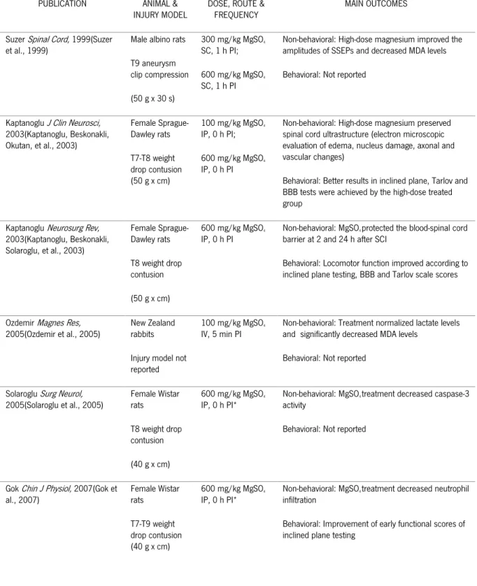
Table I. Classification of spinal cord injury severity using the American Spinal Injury Association Impairment Scale. ... 4 Table II. Mechanism of action for riluzole in preventing secondary injury following SCI... 23 Table III. Summary of in vivo studies performed in animal models of traumatic SCI using systemically administered riluzole. ... 25 Table IV. Summary of in vivo studies performed in animal models of traumatic SCI using systemically administered magnesium. ... 28 Table V. Basso, Beattie, Bresnahan Locomotor Rating Scale. ... 42
xvii
L
IST OF ABBREVIATIONS
°C - celsius 5-HT - 5-hydroxytryptamine µm - micrometer µmol - micromole AAIS - ASIA Impairment Scale ALS - Amyotrophic Lateral Sclerosis AMPA - α-amino-3-hydroxy-5-methyl-4-isoxazolepropionic acid
ANOVA - analysis of variance
ASIA - American Spinal Injury Association ATP - adenosine-5’-triphosphate
ATPases - adenosine 5'-triphosphatases AUC - area under the curve
B
BBB - Basso, Beattie and Bresnahan locomotor test
BDNF - brain-derived neurotrophic factor BSCB - blood-spinal cord barrier C
C - cervical vertebra Ca2+ - calcium
cm - centimeter Cmax - peak concentration
CNS - central nervous system CST - corticospinal tract CT - computed tomography D
d - day
DCX - doublecortin
DNA - deoxyribonucleic acid E
EAA - excitatory amino acid
EAAT - EAA transporter F
FDA - Food and Drug Administration FL - forelimb
G g - gram
GDNF - glial cell-derived neurotrophic factor H
h - hour H+ - hydrogen
H&E - hematoxylin and eosin HL - hindlimb I IL - interleukin ip - intraperitoneal iv - intravenous K K+ - potassium kdyn - kilodyne Kg - kilogram L L - lumbar vertebra M
MAP2 - microtubule associated protein 2 MDA - malondialdehyde
mg - milligram Mg2+ - magnesium
MgCl2 - magnesium chloride
MgSO4 - magnesium sulfate
min - minute ml - milliliters
xviii
mm - millimeter mM - millimolar
MP - methylprednisolone
MRI - magnetic resonance imaging N
n - total number of data points Na+ - sodium
NASCIS - National Acute Spinal Cord Injury Study NeuN - neuronal marker
NMDA - N-methyl-D-aspartate NMDAR - NMDA receptor
NGF - neurotrophin nerve growth factor NF - neurofilament
NO - nitric oxide O
OCT - optimal cutting temperature OECs - olfactory ensheathing cells P
PBS - phosphate-buffered saline PEG - polyethylene glycol
PFA - paraformaldehyde
PNS - peripheral nervous system R
ROS - reactive oxygen species RT - room temperature S
s - second
S - sacral vertebra SC - subcutaneous SCI - Spinal Cord Injury
SEM - standard error of the mean
SSEPs - somatosensory evoked potentials
T
T - thoracic vertebra TBI - Traumatic Brain Injury
TGF- β - transforming growth factor-β TNF-α - tumor necrosis factor-α TTX - tetrodotoxin
U
USA - United States of America V
VGLUT - marker for glutamatergic neurons VGSC - voltage-gated sodium channels W
1
3
1. INTRODUCTION
Trauma to the spinal cord can cause severe damage to nervous tissue leading to temporary or permanent changes in the spinal cords normal motor, sensory, or autonomic function (Hagen, 2015). Spinal cord injury (SCI) patients usually suffer permanent neurologic deficits and disability that heavily impact their physiological, psychological, and social behavior (Craig et al., 2009; 2012; Krueger et al., 2013).
In developing countries, the annual incidence of SCI was estimated in 25.5 cases per million (Rahimi-Movaghar et al., 2013). In 2012, the estimated annual incidence of SCI in the United States of America (USA) was 54 cases per million, while in Portugal, the only epidemiological study was performed by Martins et al. and set the annual incidence rate at 57.8 new cases per million inhabitants (Jain et al., 2015; Martins et al., 1998). Although SCI incidence rates are not very high, it certainly places substantial financial burden on health care systems mainly due to the long-term effects of disability and handicap that persist throughout the patient’s life. In 2006, costs related to SCI patients healthcare were estimated at 9.7 billion dollars per year in the USA alone (Thompson et al., 2015).
Injury to the spinal cord can result from traumatic events like contusion, compression and/or laceration, while non-traumatic injuries usually results from tumors, spinal stenosis and vascular events (New & Marshall, 2014; Thuret et al., 2006). Leading causes of injury include automobile crashes, falls,
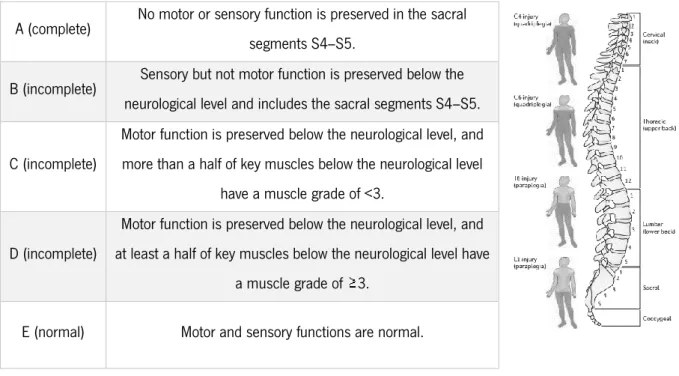
as well as sport related injuries, affecting mainly young adult males (Chen et al., 2013; Jain et al., 2015). The neurological impairment of SCI is commonly classified in different grades of severity according to the American Spinal Injury Association (ASIA) Impairment Scale (Table I). Grading relies on sensory and motor evaluation of the SCI patient at the S4-S5 sacral segment, at 72 hours (h) post-injury, allowing to determine whether the injury is complete (AIS A: no sensation or motor function at S4-S5), or incomplete (AIS B to D: motor and/or sensory function is partially preserved at S4-S5) (Kirshblum et al., 2011; Scivoletto et al., 2014). Furthermore, the level of injury is an important factor concerning survival, with a more cephalad level associated with higher rates of mortality (Wilson et al., 2012). Respiratory failure and cardiovascular dysfunction are the main cause of early mortality in complete, high-cervical (C1-C4) injuries of SCI (Shao et al., 2011).
Methylprednisolone has been widely used as the only standard therapeutic agent for SCI treatment. However, its clinical utility and role in SCI recovery remains controversial (Bydon et al., 2014). In light of this, a recent survey revealed a significant decrease in the number of surgeons using high-dose
4
steroids in 2014 as compared with 2006, clearly revealing an urgent need to develop alternative therapeutic strategies (Schroeder et al., 2014).
Table I. Classification of spinal cord injury severity using the American Spinal Injury Association Impairment Scale.
Adapted from Thuret et al., 2006.
Pathologic events following SCI can be divided into two distinct but highly interconnected phases. First, a primary injury resulting from the initial mechanical damage leads to the disruption of neural and vascular structures at the impact site. The secondary injury response follows the initial trauma and further compromises neurologic function. Several events take place during this secondary stage, ranging from ionic imbalances, free radical production, inflammation, to excitotoxic events (Oyinbo, 2011). Ultimately, these events lead to the formation of an astroglial scar that creates a physical barrier, as well as an inhibitory environment, leading to unsuccessful axonal regeneration at the lesion site (Cregg et al., 2014). Collectively, these secondary events result in severe neuronal damage. However, they also pose as an opportunity for therapeutic intervention that promotes neuroprotection. Ionic imbalances and glutamate excitotoxicity noticeably exacerbate the functional problems encountered after SCI and are, therefore, targets of current SCI therapeutic strategies (Park et al., 2004).
Riluzole and magnesium have been extensively investigated as neuroprotective agents in animal models of SCI mainly because of their ability to modulate ionic imbalances and excitotoxic events that follow SCI. Furthermore, combining drugs may lead to improved efficacy over single drug treatments as
A (complete) No motor or sensory function is preserved in the sacral segments S4–S5.
B (incomplete) Sensory but not motor function is preserved below the neurological level and includes the sacral segments S4–S5. C (incomplete)
Motor function is preserved below the neurological level, and more than a half of key muscles below the neurological level
have a muscle grade of <3. D (incomplete)
Motor function is preserved below the neurological level, and at least a half of key muscles below the neurological level have
a muscle grade of 3.
5
drug interactions may occur contributing to an additive or synergistic effect that further promotes neuroprotection. Having this in mind, we aimed to investigate the therapeutic efficacy of both individual and combined administration of riluzole and magnesium chloride in SCI.
1.1 Spinal cord: basic anatomy and physiology
Anatomically speaking, the nervous system is divided in two distinct, but highly interconnected parts: the central nervous system (CNS) and the peripheral nervous system (PNS). The CNS is the processing/control center that correlates and integrates nervous information. It receives information from and sends information to the PNS enabling the connection between the CNS and the rest of the body. The CNS is composed by the brain and the spinal cord, and while the brain controls most functions of the body, this is only possible due to the existence of the spinal cord, a major information conduit (Watson et al., 2009). However, its purpose far exceeds the simple relay of information as the spinal cord is able to integrate and modify both afferent and efferent signals allowing for the precise control of sensory, autonomic and motor functions (Nielsen, 2004).
A major difference between the CNS and the PNS lies in their different capacity to regenerate after injury. Unlike the PNS, the CNS has an extremely limited capacity to regenerate when damaged (Illis, 2012). Failure of CNS neurons to regenerate is a consequence of a surrounding inhibitory environment in combination with low intrinsic growth properties of CNS axons (Horner & Gaje, 2012).
The spinal cord, which is usually 40 to 50 centimeters (cm) long, extends from the foramen magnum, continuing with the medulla oblongata to the level of the first or second lumbar vertebrae. It comprises 31 pairs of spinal nerves composed of both motor and sensory fibers, each pair connecting a different part of the body. There are 8 cervical (C) nerves, associated with the muscles of the neck, shoulders, arms, hands, and diaphragm; 12 thoracic (T) nerves, associated with the chest and abdominal walls; 5 lumbar (L) nerves, associated with the hip, leg, and foot; and 5 sacral (S) nerves, associated with the genitals and lower digestive tract. Each spinal nerve is attached to the spinal cord by a dorsal (sensory) and a ventral (motor) root (Silva et al., 2014).
Protection to the spinal cord is provided by the vertebral column, the protective tissue of the meninges (dura, arachnoid and pia mater), and the cerebrospinal fluid.
The spinal cord, similarly to the brain, is composed of gray and white matter. The grey matter, which is centrally located and shaped like a ‘butterfly’, consists mainly in neuronal cell bodies, glial cells, and unmyelinated axons (Thuret et al., 2006).
6
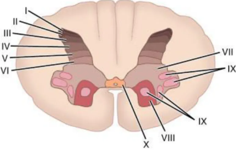
Grey matter is divided into four main columns: the dorsal horn, the intermediate column, the lateral horn and the ventral horn column. The neuronal bodies in the grey matter are organized in ten successive layers - laminae I-X from dorsal to ventral - depending on their cellular structure (Figure 1). Laminae I to VI are located in the dorsal horns and are involved in sensory input. Laminae VII to IX are found in the lateral and ventral grey matter and are related to autonomic and motor functions. Lamina X is located in the center, surrounding the central canal (Watson et al., 2009).
Figure 1. Organization of laminae in the spinal cord.
Obtained from: http://medicine.academic.ru/134796/Rexed_laminae
Surrounding grey matter we find white matter which consists mainly of longitudinally running myelinated axons as well as glial cells, the most abundant being oligodendrocytes. When a group of nerve fibers have the same origin and function, they are referred to as ‘tract’, while a group of tracts with a related function is referred to as ‘pathway’. The course of spinal tracts and pathways can be either ascending (sensorial) or descending (motor) allowing for the adequate communication between the central and the peripheral nervous systems.
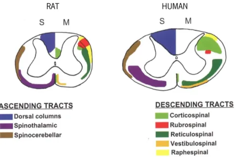
It is important to notice that differences in tract organization can be found in different species, such as rodents, cats, primates, and humans (Figure 2). This is particularly significant in spinal cord injury research because it relies on different animal models. For example rodents cortical spinal tract (associated with voluntary movement) is located dorsally, while in humans it can be found both lateral and ventrally (Watson et al., 2009).
7
RAT HUMAN
Figure 2. Location of the ascending and descending tracts of the spinal cord.
Differences in ascending sensory (S) and descending motor (M) tracts in rats and humans. Adapted from Watson et al., 2009.
At a cellular level the nervous tissue of the spinal cord is mainly comprised of neuronal and glial cells (Purves et al., 2001). It was in the late nineteenth century that Ramon y Cajal first proposed that neurons were the basic structural and functional units of the nervous system, thus paving the way for modern neuroscience (De Carlos & Borrell, 2007).
Although there are many classes of neurons, their basic structure remains the same: a cell body (soma) and two types of cell processes, axons and dendrites. The cell body contains the nucleus and is the processing center of the neuron. Dendrites receive information from other cells while the axon is the neuronal structure that allows communication between neurons. Neurons ability to generate and transmit electrochemical signals is essential for this communication to occur. The electrical signal, generated by an action potential (i.e. a transient alteration of the membrane voltage), is sent through the axon of a presynaptic neuron. Long axons, also known as nerve fibers, are insulated by fatty myelin sheets that act as electrical insulators, speeding nerve impulses.
Neurons communicate at structures called synapses. When an action potential reaches the synaptic button (the terminal end of an axon) it triggers calcium-regulated fusion of neurotransmitter-filled synaptic vesicles with the presynaptic cell membrane, ultimately releasing neurotransmitters in the synaptic cleft (Augustine, 2001). This release of neurotransmitters can result in inhibitory or excitatory signals that are conveyed to the postsynaptic neuron. Neurons, ultimately, are the tool by which the central nervous system communicates and processes information.
8
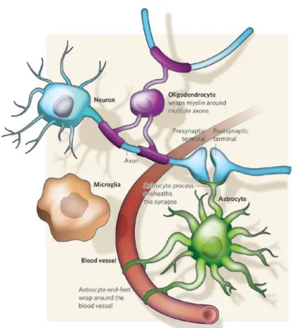
However, the tasks performed by neurons would not be possible without the support of glial cells (Figure 3). These cells differ both morphologically (lack of axons and dendrites) and functionally from neurons, and include astrocytes, microglia, and oligodendrocytes (Kriegstein & Alvarez-buylla, 2011).
Astrocytes provide trophic and metabolic support for neurons, control of extracellular osmotic pressure, local cerebral blood flow, and extracellular neurotransmitter concentration. They can be further divided in two types: protoplasmic astrocytes that can be found in the grey matter where their endfoot processes contact with blood capillaries and synapses, and fibrillary astrocytes that can be found in the white matter where their endfeet contact with blood capillaries and nodes of Ranvier (myelin sheath gaps) (Jukkola et al., 2013).
Although astrocytes are considered non-excitable cells, they are able to communicate with each other and interact with neurons through changes in intracellular calcium (Ca2+) concentration (Navarrete
et al., 2013). Typically ascribed supportive roles for proper neuronal function there is now numerous evidence that these cells play an important part in regulating synaptic transmission and plasticity, and thus are being interpreted as possible integral components of the neuronal networks and not mere bystanders (Araque & Navarrete, 2010).
It was Pío del Río Hortega in 1919, which first introduced the concept of microglia and oligodendrocytes as glial cells, a vision not shared by his mentor and employer Ramon y Cajal, that ultimately lead to his dismissal as a consequence for publishing his findings (McGeer & McGeer, 2011). Microglia serve as the CNS resident immune competent cells, and are mobilized to present antigens and become phagocytes during injury, infection, or degenerative diseases (Ousman & Kubes, 2012). These cells usually display a ‘resting’ phenotype. However, upon sensing changes in the CNS microenvironment they become ‘activated’ and undergo morphological and functional changes. Activation of these cells can range from “classical” activation, characterized by a highly pro-inflammatory profile, to “alternative” activation, associated with a less inflammatory, neuroprotective profile (Derecki et al., 2013; Giunti et al., 2014).
Oligodendrocytes are the myelin-forming cells of the CNS, providing electrical insulation of axons and consequently accelerating action potential propagation by saltatory conduction from one node of Ranvier to the next, setting the speed up to 430 km/h instead of approximately 3.6 km/h for an unmyelinated axon (Káradóttir & Attwell, 2007). Other functions performed by these cells are associated to maintenance of axonal health and provision of trophic neuronal support (Czopka et al., 2013). Loss of myelin characterizes many CNS disorders such as multiple sclerosis, Traumatic Brain Injury (TBI), and
9
SCI, and determines the disruption of neuronal signaling and axonal integrity. Thus, preventing myelin loss may be an important target for therapeutic interventions.
Ependymal cells can also be considered a specialized form of glial cells. They are responsible for the ventricular lining of the brain and spinal cord and help move cerebrospinal fluid through the ventricular system cells. Research is now focusing on their neural stem cell potential following injury to the spinal cord (Lacroix et al., 2014).
Figure 3. Glial-neuron interaction.
Neurons are responsible for the conduction of electrical currents and information relay. However, their function is highly dependent on glial cells. Oligodendrocytes myelinate CNS axons increasing the efficiency and velocity of nerve impulse conduction. Besides providing structural and chemical support neurons, astrocytes can also modulate neuronal dynamics. Microglia are the resident phagocytic cells in the brain. Reproduced from Allen & Barres, 2009.
10
1.2 The pathophysiology of spinal cord injury
As previously referred, injury to spinal cord can be caused by traumatic and non-traumatic events. For the purpose of this thesis we will focus on traumatic SCI as this is the most common cause for SCI and the most studied one.
Its pathophysiology follows a specific temporal pattern through different stages that are characterized by several specific biological and molecular events. The primary injury corresponds to the physical and mechanical trauma that occurs to the spinal cord, which is followed by a cascade of downstream events, termed secondary injury, that includes a sub-acute phase ranging from minutes to weeks, and a chronic phase, ranging from months to years after injury (Oyinbo, 2011). Furthermore, this secondary injury is not restricted to the initial lesion site and in fact it extends both radial and rostro-caudally, contributing to increased neurological dysfunction (Ek et al., 2010).
1.2.1 Primary injury
The primary injury is characterized by a mechanical insult, usually compressive, contusive and/or lacerative, that often causes vertebra fracture or disc displacement. Neuronal and endothelial cell membrane shearing occurs as a consequence of the mechanical insult, leading to tissue and blood-spinal cord barrier (BSCB) disruption, as well as vasospasm and edema (Tator & Fehlings, 1991). Hemorrhagic events immediately follow, first in the highly vascularized grey matter causing necrotic neuronal death and later expanding into the peripheral white matter(Profyris et al., 2004; Simon et al., 2009; Thuret et al., 2006).
Primary injury mechanisms that fully disrupt the anatomical continuity of the cord hardly ever occur and most often thinly myelinated or unmyelinated axons are spared and can be found in a subpial rim. However, axonal conduction of these spared axons is highly compromised (Nashmi & Fehlings, 2001; Radojicic et al., 2005).
This initial injury does not stand as promising therapeutic target as it is often uncontrollable and unpredictable. Nevertheless, primary injury highly determines the patient's neurologic grade on admission and is therefore a strong prognostic indicator (Dumont et al., 2001).
1.2.2 Secondary injury
Secondary pathophysiological cellular and molecular events that occur after the initial injury further magnify the damage to the spinal cord leading to extensive tissue loss around the injury site and in a
11
rostro-caudal manner. Secondary injury was first described in 1911 by Allen, when he observed improvement of neurological function after fluid removal from the lesion site (Allen, 1911).
Throughout this phase increase of the hemorrhagic area occurs. This expansion is characterized by the increase of petechial hemorrhages which progressively coalesce causing tissue ischemia and hypoxia. This leads to a disturbed supply of oxygen and nutrients to the damaged area and its surroundings resulting in immediate cellular death by necrosis and later on by apoptosis (Tator & Fehlings, 1991). Furthermore, the release of hemoglobin is highly toxic to CNS cells, catalyzing hydroxyl radicals production and lipid peroxidation (oxidative degradation of lipids), causing further injury to neural tissue (Gerzanich et al., 2009).
During ischemia, reactive oxygen species (ROS) including superoxide, hydroxyl radicals, and nitric oxide (NO) are generated via multiple cellular pathways including, but to limited to nitric oxide synthases, Ca2+-mediated activation of phospholipases, and inflammatory cells (Bao et al., 2005; Jia et al., 2012).
The CNS has limited antioxidant defense mechanisms as the brain and spinal cord exhibit low levels of catalase activity and only moderate levels of superoxide dismutase and glutathione peroxidase, Therefore, when oxidative stress exceeds the protective cellular antioxidant capacity, it causes oxidative damage to lipids, proteins, and nucleic acids ultimately resulting in cellular death (Christie et al., 2008; Mautes et al., 2000; Xu et al., 2005).
In turn, hypoxia and consequential oxygen deprivation hampers the cellular energy metabolism leading to adenosine triphosphate (ATP) depletion which in turn compromises cellular structural and functional integrity, triggering multiple necrotic mechanisms including loss of cell membrane permeability, release of lysosomal contents, and activation of a variety of Ca2+-activated proteases including
phospholipases, adenosine 5'-triphosphatases (ATPases), and endonucleases (Hagg & Oudega, 2006; Oudega, 2012). Furthermore, cells are driven towards anaerobic glycolysis, leading to lactic acidosis and consequential hindering of enzymatic function and deoxyribonucleic acid (DNA) damage.
Moreover, the relative immune privileged status of the spinal cord is also compromised due to BSCB disruption, as a consequence of direct mechanical disruption of the vasculature, and increased vascular permeability of endothelial cells caused by inflammatory mediators tumor necrosis factor-α (TNF-α) and interleukin-1β (IL-1β), allowing the infiltration of inflammatory cells. Permeability of the BSCB is further maintained up to two weeks post-injury by a number of vasoactive substances, such as ROS, NO, elastase, and histamines, that are released by glia and leukocytes (Donnelly & Popovich, 2008; Figley et al., 2014)
12
The immune response that follows SCI aims to remove damaged tissues and promote the healing responses of astrocytes through the release of pro-inflammatory cytokines, and is primarily mediated by resident microglia. Trauma to the spinal cord leads to sustained elevations of signaling molecules such as ATP, DNA, glutamate, growth factors and cytokines, that are released by injured neurons and glial cells, resulting in the activation of microglia and consequent release of signaling molecules such as TNF-α, IL-1β, and IL-6, which act to recruit circulating leukocytes to the injury site (Donnelly & Popovich, 2008; Loane & Byrnes, 2010; Pineau & Lacroix, 2007; Zhou et al., 2014).While the number of microglial cells remains relatively stable soon after the injury until several weeks later, neutrophils accumulate until they reach a peak at 24 h post-injury and then decline, while the number of T-lymphocytes and macrophages increases, peaking at 7 days (d) post-injury (Neirinckx et al., 2014; Rowland et al., 2008; Zhang et al., 2012).
Neutrophils have been described to promote neurotoxicity due to the activity of matrix metalloproteinase-9, generation of ROS, and secretion of TNF-α (Nguyen et al., 2007). However, their exclusively deleterious role in SCI role as been questioned by recent studies (Ghasemlou et al., 2010; Kurimoto et al., 2013; Stirling et al., 2009).
Neutrophil recruitment declines by 48 h and monocytes start to accumulate at the site of injury where they differentiate into macrophages. Several macrophage subsets have been identified, namely "classically activated" pro-inflammatory (M1) or "alternatively activated" anti-inflammatory (M2) macrophages (Kigerl et al., 2009). M1 macrophages produce pro-inflammatory cytokines such as TNF-α, IL-1, and IL-6, as well as ROS, contributing to tissue inflammation and damage. Additionally, NO generated by NO synthase activity of macrophages induces neuronal apoptosis (Satake et al., 2000; Tzekou & Fehlings, 2014).Alternatively, M2 macrophages produce anti-inflammatory factors (IL-10, transforming growth factor-β; TGF-β) contributing to wound healing and tissue-remodeling(Kigerl et al., 2009; Ren & Young, 2013).
There is still much debate on whether T-lymphocytes promote injury or recovery after SCI. While Popovich et al. have shown that myelin-reactive T-lymphocytes are activated by SCI and contribute to neurodegeneration, reports by Kipnis et al. and more recently Raposo et al. have proposed that these cells are able to mediate neuroprotection (Kipnis et al., 2002; Popovich & Jones, 2003; Raposo et al., 2014).
Overall, the inflammatory response following SCI paradoxically presents both detrimental and beneficial roles leading to the current view of inflammation as a "double-edged sword", where
13
inflammation may lead to enhanced damage and impaired regeneration, but also stands as a key regulator of tissue repair after injury (Cherry et al., 2014; Neirinckx et al., 2014).
Secondary injury is also characterized by extensive cellular death occurring as a consequence of ionic imbalances and glutamate excitotoxicity. Due to the relevant role of these mechanisms in the present thesis, they are further detailed in section 1.5.
1.2.3 From secondary injury to chronic phase
The loss of neurons and glial cells, as well as the clearance of debris by microglia and macrophages, results in the formation of a cystic cavity in the spinal cord.
Furthermore, cytokines and growth factors such as IL-1, TGF-β, neurotransmitters such as glutamate, and proteins such as fibrinogen activate astrocytes leading to changes in their morphology and molecular expression, which culminates with the formation of a glial scar (Cregg et al., 2014). Besides representing a physical barrier, the glial scar is responsible for the development of an inhibitory environment at the lesion site caused mainly by the production of molecules, including tenascin, semaphorin 3A, ephrins, and chondroitin sulphate proteoglycans, which inhibit axonal growth (Kawano et al., 2012; Rolls et al., 2009; Yiu & He, 2006).
Nevertheless, the glial scar is also known to aid SCI repair. In fact, studies where astrogliosis was ablated lead to increased lesion volume and inflammation, as well as impaired functional recovery, and did not further promote axonal regeneration (Faulkner et al., 2004; Herrmann et al., 2008; Okada et al., 2006).Reactive astrocytes can in fact promote glutamate uptake, free radical scavenging, promote BSCB repair, limit the infiltration of inflammatory cells in healthy tissue, as well as release growth factors, therefore protecting the spinal cord from deleterious secondary events (Kawano et al., 2012).
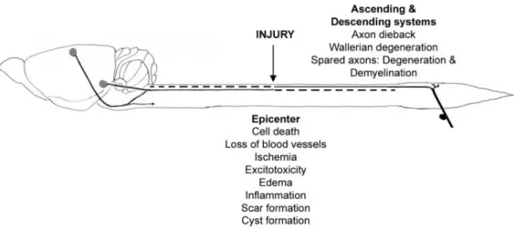
Additionally, the chronic phase is also characterized by anterograde axonal degeneration, a process known as Wallerian degeneration, and progressive loss of myelin sheaths, occurring up to extended periods of time (Hagg & Oudega, 2006). Keeping in mind that spared axons that cross the injury site are the only connection left between the brain and caudal spinal neurons, inefficient and compromised communication through these axons is a significant clinical issue (Oyinbo, 2011).
Presently, several mechanisms involved in SCI pathophysiology have been identified and extensively studied (Figure 4). Despite this increasing knowledge the fact that these injury mechanisms are intrinsically intertwined and often have both beneficial and detrimental roles may be the reason why
14
there is still no effective treatment for SCI. Nevertheless, they also present an opportunity for injury modulation and therapeutic intervention.
Figure 4. Schematic representation of the injured spinal cord.
The major degenerative processes that occur following an injury are indicated. Reproduced from Hagg & Oudega, 2006.
1.3 Management of spinal cord injury: current standard practice
The twentieth century came to revolutionize the standard medical care provided to SCI patients, namely with the implantation of SCI units and increasing focus on the importance of rehabilitation (Donovan, 2007). Currently, medical approaches to SCI aim to at minimize the progression of the initial injury and prevent secondary injury.
Patients that have suffered trauma to the spinal cord are usually immobilized during the pre-hospital setting generally resorting to a cervical collar, head immobilization, and a spinal board. On the clinical onset, imaging techniques, such as X-rays, computed tomography (CT) scans, and magnetic resonance imaging (MRI), allow for a more accurate diagnosis (Stroman et al., 2014). Peri- and postoperative airway management, namely following cervical injury, as well as hemodynamics and cardiovascular control are essential to avoid neurological compromise, morbidity, and death following SCI and are presently standard care procedures (Furlan & Fehlings, 2008; Martin et al., 2015).
Furthermore, spine stabilization and surgical decompression are performed. Decompression is associated with improved neurological outcomes when performed early (less than 24 h after injury), as supported by a recently conducted randomized multicenter trial, the Surgical Treatment of Acute Spinal Cord Injury Study. This study showed that decompressive surgery within the first 24 h confers a 2.83
15
times higher chance of a 2-grade AIS improvement when compared to surgery performed after 24 h. Additionally, differences in complication rates were not detected (Fehlings et al., 2012).
A limited number of pharmacological agents have been applied to human SCI patients including two corticosteroids (methylprednisolone and tirilazad mesylate), an opiate receptor antagonist (naloxone), and GM-1 ganglioside, However, and despite the fact that no clinical evidence exists to definitively recommend the use of any neuroprotective pharmacologic agents, methylprednisolone (MP) is still the most widely used drug therapy for acute SCI (Sharma, 2012).
The benefits of MP in SCI treatment are mainly attributed to the inhibition of lipid peroxidation, through neutralization of free-radicals, maintenance of the BSCB, and immune-modulation (Yılmaz & Kaptanoğlu, 2015). The immunomodulatory role of MP is linked to its ability to reduce microglia/macrophage activation, as well as a reduction in pro-inflammatory cytokine (TNFα, IL-6, interferon-γ) release, and increased anti-inflammatory cytokine (IL-10) secretion (Bowes & Yip, 2014). Furthermore, patients receiving high-dose MP (bolus 30 mg/kg and 5.4 mg/kg per hour over 24 h, initiated within 8 h of injury), showed decreased intramedullary spinal cord hemorrhage (Leypold et al., 2007).
The use of MP as a therapeutic strategy for SCI followed the reports of Bracken et al. in the National Acute Spinal Cord Injury Study (NASCIS) I and II. The latter revealed that a high dose administration (30 mg/kg), at time of admission and a 5.4 mg/kg/h infusion for the following 23 h, and within an 8 h range, lead to neurological improvements (light touch, pinprick sensation, and muscle strength) (Bracken et al., 1984; 1990). NASCIS III in turn, revealed that patients treated with a MP bolus administration of 30 mg/kg, followed by a 5.4 mg/kg/h infusion for a 48 h period showed improved motor recovery, namely when the first administration occurred between 3 and 8 h after SCI. However, increased adverse effects were also reported in this group, namely increased risk of severe sepsis, pneumonia and urinary tract infections (Bracken et al., 1997). The validity of these reports has been widely disputed, as well as the beneficial role of MP administration following SCI(Bydon et al., 2014; Markandaya et al., 2012; Miller, 2008).
In 2013 the American Association of Neurological Surgeons/Congress of Neurological Surgeons Guidelines for the Management of Acute Cervical Spine and Spinal Cord Injury issued a level 1 recommendation that MP not be used due to its side effects (Hurlbert et al., 2013). Nevertheless, there are also contradictory voices that state the need to carefully evaluate the administration of MP, as recent studies showed for example synergistic effects of early decompression and MP administration in patients undergoing surgical decompression after cervical SCI (Fehlings et al., 2014).
16
The systemic or local application of hypothermia has demonstrated to be neuroprotective after SCI, in both experimental and human studies, as it is able to mitigate several harmful effects caused by secondary injury (Cappuccino et al., 2010; Wang & Pearse, 2015).Therapeutic hypothermia drew significant clinical and general public notoriety as a treatment for SCI, following the recent report of an acute cervical spinal cord injury in a professional football player, Kevin Everett, who sustained a complete cervical SCI (AIS A) and received moderate systemic hypothermia for 36 h along with surgical decompression and MP administration, achieving rapid neurologic improvement that later translated into a neurological evaluation of AIS D (incomplete) (Cappuccino et al., 2010).
Rehabilitation therapies have also shown to improve recovery of locomotor function both in animal models as well as in human studies, most likely due to the reorganization of neuronal circuits that have been spared by the lesion (Edgerton et al., 2006; Engesser-Cesar et al., 2005). In an effort to maximize functional recovery after SCI, several rehabilitation strategies have been developed in order to elicit residual function bellow injury, such as over-ground training, body weight supported treadmill training, robotic-assisted step training, and functional electrical stimulation, and they are now being assessed for their efficacy in promoting functional ambulation following SCI (Lam et al., 2007).
SCI also increases the incidence of specific medical disorders such as neurogenic bladder, neuropathic pain, spasticity, and sexual dysfunction. However, progressively, this problems have been tackled and current therapies represent a huge increment in SCI patients’ lives (Cardenas et al., 2013; Nomura et al., 2002; Schmid et al., 2000; Taricco et al., 2006).
1.4 Emerging therapeutic strategies for spinal cord injury
Although SCI medical care as greatly evolved over the last few years, largely improving the chances of patients’ recovery, there is still no definitive therapeutic strategy to recover neurologic function.
Treatment strategies for SCI are mainly focused on events that follow the initial trauma, either by promoting neuroprotection or enabling neuroregeneration. The first essentially aims to prevent cellular dysfunction and death caused by secondary injury decreasing the progressive extension of the lesion and damage to the spinal cord, while the latter mainly aims to promote axonal growth and plasticity.
Several lines of strategies have been devised as possible therapies for SCI, ranging from cell transplantation, to pharmacological and combinatorial therapies, many of which showing valid potential in supporting spinal cord repair and thus standing as possible treatment strategies for SCI.
17
Several stem cells, such as neural stem cells, mesenchymal stem cells, embryonic stem cells, and induced pluripotent stem cells have been studied as possible therapeutic strategies in SCI. These cells are mainly characterized by self-renewal and the ability to differentiate in cells from different tissues. This last feature stands as an attractive feature in SCI as these cells can potentially be directed to differentiate into neurons or glia, replacing neural cells that are lost after injury, therefore supporting anatomical and functional recovery (Tewarie et al., 2009). Neuroprotective and axon regeneration-promoting effects have also been credited to transplanted stem cells through the secretion of growth factors and modulation of the inflammatory response (Baraniak & McDevitt, 2010).
A landmark stem cell clinical trial, the first in the USA involving human embryonic-derived oligodendrocyte progenitor cells, began in 2010 and was carried by the Geron Corporation. The companies’ managing director, Thomas Okarma, enthusiastically stated that the trial represented “the dawn of a new era in medical therapeutics”. However, the Geron Corporation abruptly discontinued the study in November 2011 for financial reasons, largely disappointing the scientific community (Scott & Magnus, 2014). Recently however, their embryonic stem cells technology was acquired by Asterias Biotherapeutics. After a Phase I trial (source: clinicaltrial.gov; clinical trial identifier NCT01217008) met its primary endpoints of safety and feasibility, a Phase I/IIA clinical trial (NCT02302157) has been cleared by the Food and Drug Administration (FDA), holding promise for stem cell therapies in SCI recovery.
Other cell types, such as olfactory ensheathing cells (OECs) and Schwann cells have also attracted the attention of investigators in the field. As the name suggests, OECs are glial cells that ensheath non-myelinated olfactory axons and are responsible for growth and regeneration of olfactory axons throughout the life of adult mammals. They have received the attention of the media and general population after reports of a patient that suffered a traumatic transection of the spinal cord improved from ASIA A to ASIA C, following OECs transplantation (Tabakow et al., 2014).
Schwann cells are also glial cells that are known for their roles in supporting nerve regeneration in the peripheral nervous system (Oudega & Xu, 2006). These cells are particularly interesting in SCI because of their ability to migrate to the injury site, express growth promoting factors, and myelinate regenerating axons. However, their ability to overcome the glial scar is limited revealing the need to develop combinatorial strategies, for example with biomaterials in order to maximize their regenerative potential (Assunção-Silva et al., 2015; Pêgo et al., 2012). In line with this thought, an interesting strategy is being developed by the company InVivo Therapeutics using both biomaterial scaffolds and neural stem cells with promising results obtained using a non-human primate model (Pritchard et al., 2010). The FDA has already approved a pilot study of clinical safety and feasibility of the scaffold (NCT02138110) and in
18
future trials the company plans to load the scaffold with neural stem cells in order to functionally bridge the injury site.
A number of promising molecular and pharmacological therapies are currently under investigation for neuroprotective and neuroregenerative abilities in animal models of SCI. Furthermore, several of these therapeutic agents are already in, or close to, clinical trials.
Pharmacological agents that aim to prevent secondary injury include, but are not limited to steroids (methylprednisolone, tirilazad mesylate), opiate blockers (naloxone), anti-inflammatory agents (indomethacin, ibuprofen), glutamate receptor antagonists (magnesium, MK-801), ion channel blockers (riluzole), apoptosis inhibitors (minocycline), cytokines (erythropoietin, granulocyte colony-stimulating factor), antioxidants/free radical scavengers (tirilazad mesylate, quercetin), and immune blockers (cyclosporine-A) (Kwon et al., 2011; Nagoshi & Fehlings, 2015; Priestley et al., 2012).
Furthermore, several agents have also been studied in order to promote axonal regeneration which is necessary for re-establishing connectivity across the injury site, either by targeting myelin-associated inhibitory molecules (anti-Nogo antibody), by blockade of the Rho pathway (cethrin), stimulation of axonal growth (neurotrophic factors), or by degradation of chondroitin sulfate proteoglycan inhibitory molecules (chondroitinase ABC) (Silva et al., 2014).
Albeit all the promising results of some of these therapies, when translated to a clinical context most of them have failed to prove their efficacy. Examples such as methylprednisolone and GM-1 ganglioside, are a reminder that although some of these therapies may in fact have inadequate potency there is a growing need to improve the rigor and conduct of human trials to ensure that the true effects of treatment, positive and negative, are accurately detected and reported (Lammertse, 2013).
Despite the increasing knowledge of important mechanisms regarding neuronal protection and regeneration, alongside the innumerous studies that have been, and are being performed, no gold standard therapy for SCI has yet been established and therefore identification of effective therapeutic interventions are urgently needed.
1.5 Targeting excitotoxicity and ionic imbalances to protect the spinal cord
Understanding the pathophysiology of SCI is the basis for the development of treatments for spinal cord repair. Excitotoxicity and ionic dysregulation are closely related processes that immediately follow SCI contributing to extensive neuronal damage and loss. Pharmacological protection of the spinal cord against these deleterious events is the main focus of this thesis, and therefore this section will center on
19
these mechanisms and how they individually and collectively contribute to SCI providing the rational for the use of riluzole and magnesium as therapeutic strategies.
The term ‘excitotoxicity’ was coined by Olney in 1969, when he observed that subcutaneous injection of monosodium glutamate lead to neuronal death, and refers to postsynaptic neuronal death produced by over-activation of glutamate receptors (Olney, 1969). Physiologically, the concentration of glutamate in the synaptic cleft is tightly regulated. Removal of glutamate mainly occurs by sodium (Na+)
dependent excitatory amino acid (EAA) transporters (EAAT), present in both neurons and glial cells, which maintain the extracellular concentration below toxic levels. In the spinal cord, the astrocyte glutamate transporter EAAT2 (glutamate transporter GLT-1 in rodents), is responsible for a significant percentage
of extracellular glutamate functional uptake (Lepore et al., 2011; Shigeri et al., 2004).
Glutamate acts on specific postsynaptic receptors including ionotropic N-methyl-D-aspartate (NMDA), α-amino-3-hydroxy-5-methyl-4-isoxazolepropionic acid (AMPA) and kainate receptors, as well as metabotropic glutamate receptors. Ionotropic receptors act as ion channels (permeable to Na+,
potassium; K+, and varying permeability to Ca2+), while metabotropic receptors are G protein-coupled and
act through signal transduction cascades (Stys & Lipton, 2007). Ionotropic receptors are further divided in NMDA and non NMDA-receptors, such as AMPA and kainate receptors, according to their affinity to the synthetic glutamic acid analogue N-methyl-D-aspartic acid. Non-NMDA receptors are not voltage dependent and are highly permeable to Na+ (Shigeri et al., 2004).NMDA receptors (NMDARs), which are
characterized by their voltage dependence and high permeability to Ca2+, have gained particular interest
in SCI pathology, due to their preponderant role in Ca2+-mediated excitotoxicity (Park et al., 2004).
NMDARs form a heterotetramer assembled by combination of two GluN1 subunits with two GluN2A-D or GluN3A-B subunits. These receptors require dual agonists, glutamate and glycine, for activation, and are blocked by physiological levels of magnesium (Mg2+) in a voltage dependent manner.
The sensitivity to the Mg2+ block as well as Ca2+ permeability depends on the subunit constitution, with
subunit GluN2A and GluN2B, mainly present in neurons being highly sensitive to magnesium block and Ca2+ permeability, while the subunits GluN2C, GluN2D and GluN3, mainly present in oligodendrocytes and
astrocytes having lower sensitivity to the magnesium block and Ca2+ permeability (Paoletti et al., 2013).
Subunit diversity also provides distinct pharmacological properties to NMDARs, enabling the development of specific antagonists and selective modulation of these receptors. Toxic levels of glutamate are reached within 15 min after SCI in rats at local and regional tissue surrounding the injury epicenter (McAdoo et al., 1999). The disruption of cellular membranes following SCI causes an acute burst of EAA, namely glutamate, the main excitatory neurotransmitter of the CNS (Liu et al., 1991; McAdoo et al., 1999)
20
Glutamate accumulation also occurs due to the reversal of Na+-dependent glutamate transporters leading
to synaptic glutamate release (Li et al., 1999; Li & Stys, 2001) Oxidative stress is also known to impair glutamate uptake contributing also to extracellular glutamate accumulation (Barger et al., 2007). Furthermore, activated microglial cells are also known to release glutamate. In neurons, Ca2+-dependent
postsynaptic exocytosis also leads to release of glutamate. All of these mechanisms serve as self-amplifying glutamatergic loops. Excessive activation of NMDARs due to increased extracellular glutamate accumulation plays a significant role in Ca2+-mediated excitotoxicity (Cao & Dong, 2013; Matute et al.,
2007).Pathological increases in intracellular Na+ and Ca2+ concentrations lead to excitotoxic cellular death,
via the activation of protein kinases, phospholipases, proteases, mitochondrial dysfunction, production of ROS, intracellular acidosis, edema, and cell lyses(Dong et al., 2009; Dumont et al., 2001; Matute et al., 2006; Oyinbo, 2011). Additionally, the recent studies by Salter and Micu et al. reported that NMDAR are expressed in developing oligodendrocytes processes as well as in myelin sheaths, providing compelling evidence for the involvement of these receptors in white matter injury (Micu et al., 2006; Salter & Fern, 2005).
However, it is now known that NMDARs may also play an important role in supporting neuronal survival. Furthermore, this protective role of NMDAR activity has been proposed as a reason for the failure of several clinical trials which evaluated NMDAR antagonists as potential neuroprotective drugs. NMDAR location (synaptic or extrasynaptic) and subtype composition have been suggested as two factors that may indicate its pro-survival or pro-excitotoxic profile (Besancon et al., 2008; Hetman & Kharebava, 2006).
Tightly related to excitotoxic events, severe ionic imbalances also occur after SCI. Intracellular concentrations of Na+, Ca2+ increase, while K+ and Mg2+ levels severely decrease (LoPachin et al., 1999).
Ionic imbalances mainly occur due to widespread shearing and destruction of cellular membranes occurring at the injury site, alongside energetic depletion and consequential energy-dependent electrolytic transport failure. The outcome of this deregulation is dysfunction and inappropriate propagation of action potentials along damaged axons. In particular, intracellular accumulation of Na+ has several deleterious
effects, in which voltage-gated Na+ channels (VGSC) play a pivotal role. The characteristics and function
of these channels were unraveled by work developed by Hodgkin and Huxley (Hodgkin & Huxley, 1952). Physiologically, VGSC mediate membrane depolarization and are responsible for propagating action potentials along the axonal membrane. These channels are heteromeric assemblies of one α subunit (Nav1.1 through Nav1.9) and one or more β subunits. Only five of these isoforms are expressed in the
21
produce a functional channel, β subunits are responsible for most channel functions such as voltage sensing, gating, ion permeation, and inactivation (Theile & Cummins, 2011). Additionally, the α subunit expresses six distinct neurotoxin-binding sites that when activated can lead to either pore occlusion and concomitant inhibition of Na+ conductance, or modification of gating leading to altered gating kinetics and
voltage-dependence of these channels (Caldwell et al., 2000; Stevens et al., 2011).
Following trauma, such as SCI, persistent activation of VGSC occurs due to neuronal membrane dysfunction, resulting in an increase of intracellular Na+ concentration ([Na+]
i) (Stirling & Stys, 2010).
Alongside, the activity of Na+/K+ pumps is compromised due to energetic deficits, leading to a cutback in
Na+ efflux. Several pathological effects occur as a consequence of pathological [Na+]
i, such as cytotoxic
edema and increased acidosis (via the Na+/H+ exchanger). Furthermore, increased [Na+]
i is known to
promote Ca2+-mediated cellular death. This occurs mainly due to the reverse operation of Na+/Ca2+
exchangers, causing Ca2+ to be pumped in and Na+ extruded, as well as activation of voltage-dependent
Ca2+ channels (Agrawal & Fehlings, 1996; Ates et al., 2007; Hains et al., 2004; Rosenberg et al., 1999;
Schwartz & Fehlings, 2001). Calcium overload in turn leads to mitochondrial damage, activation of Ca2+
-dependent cell death proteases (such as caspases), as well as formation of free radicals, resulting in apoptotic cell death of neurons. Additionally, membrane depolarization leads to glutamate release, through the reversal of Na+-dependent glutamate transporters and vesicular glutamate release and further
contributes to excitotoxicity (Li & Stys, 2001; Stys, 2005). In white matter, high intracellular Ca2+ levels
result in the activation of ubiquitous proteases, specifically calpains, which contribute to neurodegeneration and compromised axonal integrity through cytoskeletal degradation of neurofilaments, as well as microtubule associated protein 2 (MAP2).
Experimental evidences concerning the role of Na+ channels in secondary injury arose mainly
from studies in which the use of Na+ channel blockers, such as tetrodotoxin (TTX), a potent neurotoxin,
lead to reduced apoptotic neuronal death, while the application of veratridine, a voltage gated Na+ channel
activator, induced neuronal apoptosis and caspase-3 activation (Banasiak et al., 2004; Stys et al., 1992). Furthermore, recent studies have found that voltage-gated Na+ channels are also present on
immune cells and that these channels contribute to the activation and phagocytic function of microglia and macrophages raising the possibility that Na+ channel blockade may attenuate the inflammatory
response (Jung et al., 2013; Pappalardo et al., 2014).
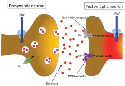
In SCI both ionic imbalances and glutamate excitotoxicity significantly contribute to the secondary injury (Figure 5). However, pharmacological treatments, if applied early, may interrupt or modulate this injurious cascade of events and consequentially improve tissue preservation and neurological outcome
22
following SCI. In this thesis we will focus on two promising neuroprotective agents that can attenuate secondary pathophysiology and reduce functional deficits, namely, the Na+ channel blocker riluzole, as
well as the NMDA receptor antagonist magnesium.
Figure 5. Representation of neuronal injury following ionic imbalances and excitotoxicity. Neuronal ionic balance is disrupted following the primary injury and the intracellular Na+ concentration
increases as a result of trauma-induced activation of voltage-sensitive Na+ channels promoting
concomitant influx of Ca2+ ions. The excessive influx of Na+ and Ca2+ triggers pathologic extracellular release
of excitatory neurotransmitter glutamate. In the postsynaptic neuron, sodium and calcium influx through NMDAR leads to cellular death and axonal edema. Adapted from (Fehlings et al., 2015).
1.5.1 Riluzole
Riluzole (2-amino-6-trifluoromethoxybenzothiazole) is a Na+ channel blocker agent approved in
1995 by the FDA for the treatment of Amyotrophic Lateral Sclerosis (ALS), a progressive neurodegenerative disorder characterized by motor neuron and corticospinal tract degeneration (Miller et al., 2012). The analysis of four placebo-controlled, randomized trials, has concluded that the administration of 100 mg daily is safe and improves overall survival of ALS patients in two or three months (Miller et al., 2012). Because neurological dysfunction due to loss of spinal motor neurons and axonal degeneration are also common in SCI, numerous preclinical studies used riluzole to demonstrate that its administration following injury improved both functional and histological outcomes in injured animals.
As previously stated, neuronal ionic balance is disrupted after SCI giving rise to an increase in intracellular Na+ concentration, resulting from constitutively activation of VGSC. This accumulation is
23
deregulation include cytotoxic edema, acidosis, and Ca2+-dependent cellular death. Riluzole exerts
neuroprotection in SCI via blocking of persistent Na+ currents, as it specifically blocks inactivated Na+
channels thus preventing the above stated pathological processes (Table II). Furthermore, the Na+ channel
blockade may also reduce energetic demands, for example through the reduction of Na+/K+ pump,
resulting in an improved resistance of cells (Theiss et al., 2007; Urbani & Belluzzi, 2000; Wahl & Stutzmann, 1999). Riluzole has also an important role as an anti-glutamatergic agent, through inhibition Ca2+-dependent release of glutamate from presynaptic terminals as well as promotion of glutamate
reuptake (Fumagalli et al., 2008; Wang et al., 2004). Additionally, riluzole is thought to preserve spinal cord white matter by preventing the disruption of the axonal Na+/H+ exchanger system (Nagoshi &
Fehlings, 2015).
Table II. Mechanism of action for riluzole in preventing secondary injury following SCI. Adapted from Wilson & Fehlings, 2014.
PRIMARY MECHANISM SECONDARY EVENTS END RESULT
Blockade of continuous posttraumatic activation of neuronal voltage
gated Na+ channels
Prevents increase in neuronal
cytosolic Na+ concentrations Prevents development of neuronal acidosis and
swelling Prevents excessive neuronal entry
of H+ through Na+/H+ exchanger Prevents excessive neuronal entry
of Ca2+ through Na+/Ca2+ exchanger
Prevents Ca2+-induced release of glutamate and excitotoxicity
Neuroprotection is also thought to be exerted by riluzole resulting from its ability to stimulate astrocyte expression of several neurotrophic factors, such as nerve growth factor (NGF), brain-derived neurotrophic factor (BDNF) and glial cell-brain-derived neurotrophic factor (GDNF) (Mizuta et al., 2001). Additionally, increased levels of BDNF and TGF-1β have also been detected in the serum of Huntington’s patients treated with riluzole (Squitieri et al., 2009).
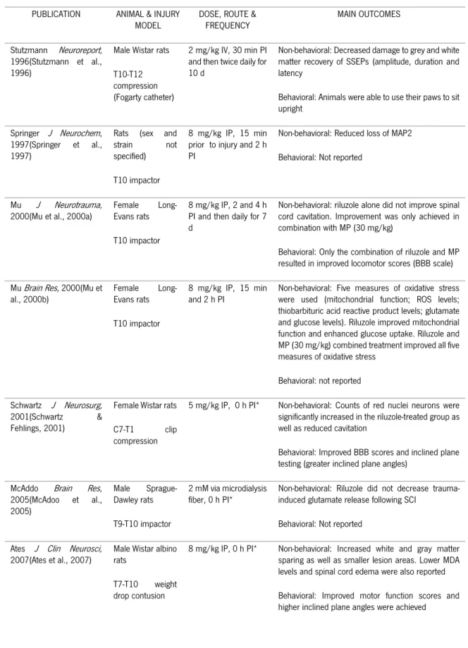
When reviewing the preclinical evidence published using riluzole in traumatic models of SCI (Table III), we find that although all eleven studies were performed using rats as animal models, several strains were used (Wistar, Long-Evans, and Sprague-Dawley) in a variety of thoracic and cervical injuries, including weight drop, clip compression, and balloon compression. Riluzole was most commonly administered intraperitoneally in doses ranging from 5 to 8 mg/kg, with higher doses associated with lethargy and locomotor ataxia as side effects. Therapeutic neuroprotective efficacy was achieved up to a
24
clinical relevant time of 4 h post-injury. Behavioral outcomes included improved Basso, Beattie and Bresnahan (BBB) locomotor scores, greater inclined plane angles, as well as beam balance and rotarod scores, while non-behavioral outcomes included increased tissue sparing, reduced MAP2 loss, decreased lipid peroxidation, capillary fragmentation, apoptosis, and inflammation, as well as improved electrophysiological recordings.
Only the study performed by Mu et al. (2000b) reported the absence of improved behavioral and non-behavioral outcomes when riluzole was administered alone. However, when combined with methylprednisolone, both motor and histological outcome was improved.
The study of Kitzman et al. employed a transection model and riluzole administration was performed 4 weeks post-injury (chronic phase). However, this was to examine the effect on established tail spasticity and not acute local tissue protection or locomotor behavior.
Based on these preclinical evidences the administration of riluzole following SCI naturally progressed on to clinical trials. A Phase I safety trial of riluzole in acute cervical SCI, Riluzole in Spinal Cord Injury Study, in which thirty-six patients with ASIA grades A-C were enrolled (28 cervical and 8 thoracic), has already established the absence of serious adverse effects and that the administration of riluzole (within 12 h of injury, 50 mg every 12 h for 28 doses) may have a beneficial effect in motor outcome, particularly for cervical SCI patients (NCT00876889) (Grossman et al., 2014). Furthermore, clinical pharmacokinetics of riluzole in patients with SCI was also studied revealing that the peak concentration (Cmax) and the 12-h area under the plasma concentration curve (AUC)(0–12h) achieved in SCI
patients were lower than those in ALS patients (on the same dose basis) due to a higher clearance and larger volume of distribution in SCI patients. Furthermore, SCI patients presented large interpatient variability in plasma concentration and an increase in the clearance and distribution of riluzole between the 3rd and 14th day after SCI, with a lower plasma concentration of riluzole on the 14th day, stressing the importance of monitoring changes in drug metabolism after SCI (Chow et al., 2012).
On the basis of these results, a multi-center, randomized, placebo controlled, double-blinded, Phase IIB/III trial of efficacy and safety of riluzole is currently recruiting (NCT01597518) (Fehlings et al., 2015).