Revina Ann Mary
maio de 2014
Validation of neuronal inclusions as a
biomarker for pre-clinical trials in a
mouse model of Machado-Joseph disease
UMinho|20
14
Re
vina Ann Mar
y
V
alidation of neuronal inclusions as a biomark
er for pre-clinical trials in a mouse model of Machado-Joseph disease
Universidade do Minho
Escola de Ciências da Saúde
Trabalho efetuado sob orientação da
Doutora Patricia Maciel
e co-orientação da
Doutora Anabela Fernandes
Revina Ann Mary
maio de 2014
Dissertação de Mestrado
Mestrado em Ciências da Saúde
Validation of neuronal inclusions as a
biomarker for pre-clinical trials in a
mouse model of Machado-Joseph disease
Universidade do Minho
III
ACKNOWLEDGEMENT
Behind every achievement there are many helping hands that aid in reaching the ultimate goal. I consider it is a great honour to express my deepest sense of gratitude and indebtness to my beloved supervisor, Pro. Patrícia Maciel., for guiding me at every stage of the project and for keeping me in high spirit. Her innovative and constructive ideas have been very invaluable, which made the work interesting and easy.
I would like to thank my co-supervisor, Dr. Anabela Fernandes. She is my dearest and humble teacher in the lab and I learned immensely from her. Her eyes see the world, people, work, in such a different and fantastic manner. Thank you for sharing them with me, thank you for trusting me with your privacy and friendship. She helps me carry out the analytical works.
I would like to thank Sara Silva, Sofia Esteves, for their guidance, collaboration and encouragement, constrictive and valuable ideas for the completion of my project work. They are very generous with their time, and always willing to talk about research, help to carry out the analytical works, for which I am grateful.
I express my sincere thanks to Andreia Carvalho, Ana Freitas, for their constant advice and support during the initial stages of my master course. They are my first inspiration in research field.
I would like to express my sincere gratitude to histology services, for helping to begin my project at earliest by for providing slides for the immunohistochemistry after micrtome cut.
My heartfelt thankfulness to my brother in law Dr. Franklin, and sister-in-law, Dr. Sheeba Franklin, for helping me during project. Their support, love, encouragement and excellent sense of humor have been truly invaluable.
It is my privilege to extend my special thanks to my dearest lovable husband Marslin, and my child, Afina, without whose unconditional love and support this process of my learning would have been incomplete. I would also like to express my regards to my rest of group mats, who directly or indirectly helped me during my work.
V
VII
ABSTRACT
Polyglutamine disorders are a class of nine inherited neurodegenerative disorders caused by expansion of a CAG repeat within otherwise unrelated genes. Polyglutamine expansion causes aberrant protein misfolding that leads to formation of protein aggregation and neuronal loss. Machado-Joseph disease (MJD) is included in this group of disorders, being caused by expansion of a CAG tract in the C-terminal region of the protein Ataxin-3 (ATXN3). MJD is characterized by the formation of intranuclear neuronal inclusions (NIs) and nervous system dysfunction, but the mechanism of this ATXN3- mediated dysfunction is still unsolved. So far, some symptoms can be controlled with specific treatments, but there is no effective therapy for MJD.
Our team previously established a new MJD mouse model expressing human ATXN3 with 135 glutamines – CMVMJD135 – that presents phenotypic, pathologic and genetic similarities with the human disorder, namely several reliable and quantifiable phenotypic markers that can be used in therapeutic trials. The NFIs are a pathological hallmark of this disease and have been found in this mouse model, as happens in human patients. Many studies related with therapeutic targets in polyQ diseases reveal phenotype amelioration without mention to neuropathology, namely inclusion load. Immunohistochemistry was performed to evaluate the relevance of inclusion load assessment in pre-clinical trials using a mouse model of MJD. We evaluated the utility of using this biomarker in therapeutic trials using three pharmacological compounds: creatine, 17-DMAG and valproic acid. We have found a significant reduction in inclusion load in the lateral reticular nucleus (LRT) region and a tendency towards a reduction of neuronal inclusions in the facial nuclei (7N) region of CMVMJD135 mice treated with creatine. The chronic treatment with creatine also lead to an amelioration of the motor phenotype observed in these animals, namely an increase in muscle strength and a better motor performance in the motor swimming test.
Another compound, 17-DMAG did not show any differences between treated and non-treated transgenic mice group regarding NIs in the LRT and 7N brain regions, although a phenotype amelioration was observed in several behavioral paradigms. CMVMJD135 mice treated with Valproic acid exhibit a tendency towards a reduction of neuronal inclusions load in the LRt but no reduction in inclusion load in the 7N brain region. Globally our results suggest that there is no direct correlation between the aggregate load in these brain regions of the CMVMJD135 mice and the phenotypic effect observed upon chronic treatment with different compounds.
IX
RESUMO
As doenças de poliglutaminas são um conjunto de doenças neurodegenerativas hereditárias causadas pela expansão de um tripleto de CAG nas regiões codificantes dos respetivos genes causadores. A expansão do tripleto CAG leva à formação de proteínas que possuem uma expansão anormal de uma sequência de poliglutamina, causando o seu desarranjo conformacional, podendo estas proteínas agregar e conduzir à morte neuronal num estadio mais avançado da doença. A doença de Machado-Joseph (DMJ) inclui-se neste grupo de doenças, sendo caracterizada por uma expansão de glutaminas localizada na região C-terminal da proteína envolvida na DMJ, a ataxina-3 (ATXN3). São características comuns a todas as doenças de poliglutaminas a presença de inclusões proteicas intranucleares nos neurónios (IINs) e a disfunção neuronal. No entanto, permanece por esclarecer a relevância das IINsna patogénese da DMJ. Até à data, embora seja possível controlar alguma da sintomatologia observada nos doentes, não existe um tratamento eficaz para a DMJ.
No presente estudo, utilizou-se um modelo de ratinho para a DMJ que foi previamente gerado no nosso laboratório, e que apresenta várias características semelhantes às da doença humana, quer a nível genético e fenotípico, quer a nível neuropatológico. Este modelo apresenta características passíveis de quantificação, permitindo o seu uso em ensaios pre-clínicos. As IINs são uma característica chave da DMJ e foram detetadas nos cérebros deste modelo. Vários ensaios pré-clínicos recorrendo a várias estratégias terapêuticas e a diferentes modelos de ratinho, demonstram uma melhoria do fenótipo observado em cada modelo, no entanto, a referência ao efeito dos compostos utilizados na quantidade de agregados proteicos não é clara. Numa tentativa de esclarecer a relação entre a melhoria no fenótipo e a quantidade de IINs nos cérebros dos ratinhos CMVMJD135, foi efetuado um estudo de quantificação das IINs em algumas regiões do cérebro relevantes na DMJ, após o tratamento crónico dos ratinhos com três compostos: creatina, 17-DMAG e ácido valpróico. Foi possível observar uma redução significativa da densidade de IINs no LRT e uma tendência para o mesmo resultado no 7N nos animais tratados com creatina, quando comparados com animais veículo. Assim, e tendo em conta que a creatina apresentou um efeito benéfico nos sintomas motores e também melhorou a perda de força muscular dos animais transgénicos, pode dizer-se que existe uma correlação direta entre densidade de IINs positivos para a ATXN3 e o efeito positivo no fenótipo destes animais. Em contraste, apesar de o 17-DMAG ter tido um efeito positivo no fenótipo dos animais, não foi observada uma redução da densidade de IINs nos núcleos LRT e 7N. Por fim, o ácido valpróico demonstrou ter um efeito muito ligeiro e tardio no fenótipo dos animais, não tendo sido observada uma redução significativa da densidade dasIINs nas áreas acima referidas. Globalmente, estes resultados demonstram que não existe uma relação direta entre densidade de IINs nestas regiões e a melhoria a nível fenotípico dos animais transgénicos para a DMJ.
XI
XIII
C
ONTENTSAbbreviations List ... XV Figures and Tables ... XIX
1. Introduction ... 1
1.1. Polyglutamine (PolyQ) diseases ... 3
1.2. Polyglutamine pathogenic mechanisms ... 4
1.2.1. Protein aggregation and misfolded proteins affect proteostasis ... 5
1.2.2.Transcriptional deregulation ... 5
1.2.3. Mitochondrial impairment/oxidative stress ... 6
1.3.Machado-Joseph disease ... 7
1.3.1. History... 7
1.3.2. MJD clinical symptoms and sub-types ... 7
1.3.3. MJD pathology ... 8
1.3.4.MJD genetics ... 9
1.3.5.MJD protein –Ataxin3 ... 9
1.4.Animal models (mouse) for the MJD ... 10
1.5.Therapeutic strategy of pre-clinical trial drug ... 11
1.5.1.Mitochondrial stabilization and reduction of oxidative stress ... 11
1.5.2.Clearance machinery: Chaperone related protein turnover ... 13
1.5.3.Transcriptional regulation ... 13
1.6.Aim of the thesis ... 15
2. Methods and Materials ... 17
2.1.Animals... 19
XIV
2.3.Immunohistochemistry ... 19
3. Results ... 21
3.1.CMVMJD135 mice treated with creatine exhibit reduced abundance of intranuclear ataxin-3 inclusion in the lateral reticular nuclei and facial nuclei of the brain………23
3.2.CMVMJD135 mice treated with 17-DMAG did not exhibit a reduction in intranuclear ataxin-3 inclusion in the lateral reticular nuclei or facial nuclei of the brain ... 24
3.3. CMVMJD135 mice treated with Valproic acid (VPA) showed less intranuclear inclusion load than the saline-treated group ... 26
4.Discussion ... 29
4.1.Future work ... 34
5.Conclusion ... 35
XV
XVII
LIST OF ABBREVIATIONS AR – Androgen receptor ATXN3 – Ataxin-3 gene ATXN3 – Ataxin-3 protein ATP – Adenosine triphosphate a.a. –Aminoacid
bp – Base pairs
CNS – Central Nervous System cDNA – complementary DNA CMVp – Cytomegalovirus promoter C. elegans – Caenorhabditis elegans CAG – Trinucleotide codon for Glutamine
CREB -cyclic AMP responsive element binding protein
CBP – CREB binding protein cyt c – Cytochrome c
CHIP- (C-terminus of Hsp70-interacting protein) Ca2+ - calcium ion
DNA – Deoxyribonucleic acid
DRPLA – Dentatorubral-Pallidoluysian Atrophy
DAB – 3, 34-biiaminobenzidine DUB- Deubiquitinating Enzymes ER – Endoplasmic reticulum HD – Huntington's Disease HDAC – Histone Deacetylase HSR- Heat-shock response Hdj2 – Constitutive form of 40-kDa heat-shock protein
Hsp – Heat shock protein HAT – Histone Acetylase Htt – Huntingtin Protein H3 – Histone 3 H4 – Histone 4 IHC-Immunohistochemistry kDa – KiloDalton(s) kb – kilobase
XVIII
MJD – Machado-Joseph Disease
MPT- mitochondrial permeability transition mtDNA – mitochondrial DNA
MT – Mitochondria
n – Number of samples in the study NI – Neuronal Inclusion
OH8dG – 8 hydroxydeoxyguanosine pCAF – Protein-associated factor PCR – Polymerase Chain Reaction PFA – Paraformaldehyde
Pn – Pontine Nucleus polyQ – Polyglutamine Q – Glutamine
RNA – Ribonucleic acid
ROS – Reactive Oxygen Species SCA – Spinocerebellar Ataxia SN – Substantia Nigra
SMBA – Spinal and Bulbar Muscular Atrophy
SAHA – Suberoylanilide Hydroxamic Acid
SOD – Superoxide dismutase SP1 – Specificity Protein 1 TFs- Transcription factor TBP- TATA-Binding Protein Th- thalamus
TAFII130 – TBP-associated factor UIM – Ubiquitin-interacting motifs UPS – Ubiquitin-proteasome system Ub- ubiquitin
Ve- vestibular nucleus WT- Wild type
XIX
XXI
FIGURES AND TABLES
Figure 1: mechanism of polyQ pathogenesis... 4 Figure 2: Brain region affected in MJD... 8 Figure 3: Intranuclear ATXN3 inclusion load in CMVMJD 135 mice treated with a normal diet and 2% creatine supplementation... 24 Figure 4: Intranuclear ATXN3 inclusion load in CMVMJD 135 mice treated with vehicle (saline) and 17-DMAG…………... 25 Figure 5: ATXN3 intranuclear inclusion load in CMVMJD 135 mice treated with vehicle and VPA. ... 27 Table 1: Molecular features of Polyglutamine disease……... 3 Table 2: Mouse models of MJD………... 10 Table 3: Therapeutic rationale of our pre-clinical trials... 16 Table 4: Illustration of experimental animal... 19 Table 5: A brief summary of the results obtained for the three compounds... 27
1
3
1. INTRODUCTION
1.1. Polyglutamine (PolyQ) diseases
Polyglutamine diseases are genetic disorders caused by an expansion of a trinucleotide (CAG) within the coding region of the corresponding genes. The first disease described was spinal bulbar muscular atrophy (SBMA) (1). Eight other disorders including HD, dentatorubropallidoluysian atrophy (DRPLA), and six types of spinocerebellar ataxia (SCA1, 2, 3, 6, 7, and 17) (2) have since been identified as associated with polyQ expansions (Table-1). With the exception of SBMA, polyQ disorders are autosomal dominant, completely penetrant and progressive. To date, the normal function of many of the genes causing polyQ disorders remains unclear and apart from the polyQ tract, these proteins share no homologies, are unrelated to each other, vary in size and contain the glutamine segment at distinct locations within the protein sequence. Commonly these proteins are expressed in central nervous system and peripheral tissues. In addition, PolyQ repeats can expand or contract , but expanded polyQ repeats show an inverse correlation between the number of CAG repeats and age of the disease onset (3-5), and a correlation was observed between the length of the CAG repeats and the number of inclusions in disease brain (6, 7).
Table-1: Molecular features of Polyglutamine disease (adapted from reference # 8)
Disease Mutated gene Gene product Protein function Protein localization Normal (CAG)n Expanded (CAG)n
Huntington Disease HD Huntingtin Signaling transcription transport, Cytoplasmic 6-35 36-121
Spinocerebellar ataxia type 1
(SCA1) ATXN1 Ataxin-1 Transcription Nuclear in neurons 6-39 39-83
Spinocerebellar ataxia type 2
(SCA2) ATXN2 Ataxin-2 RNA metabolism Cytoplasmic 14-31 32-200
Spinocerebellar ataxia type 3 (SCA3, Machado joseph
disease) ATXN3 Ataxin-3 De-ubiquitlating activity Cytoplasmic and nuclear 12-40 54-86
Spinocerebellar ataxia type 6
(SCA6) CACNA1A CACNA1A
Voltage-dependent calcium channel subunit
alpha-1A Cell membrane 4-20 20-30
Spinocerebellar ataxia type 7
(SCA7) ATXN7 Ataxin-7 Transcription Nuclear 4-35 37-306
Spinocerebellar ataxia type
17 (SCA17) TBP TBP Transcription factor Nuclear 25-42 47-63
Dentatorubral-pallidoluysian
atrophy (DRPLA) DRPLA Atrophin-1 Transcriptional regulator Cytoplasmic 3-35 49-88
spinal and bulbar muscular atrophy (SBMA, Kenedy
disease) AR
Androgen receptor
Steroid hormone
4
1.2 Polyglutamine pathogenic mechanisms
Several cellular and molecular mechanisms have been implicated in polyglutamine toxicity, most of which suggest that the expanded polyQ tract confers a toxic gain-of-function to the mutant proteins. Rarely, it has also been associated with loss-of-function mechanisms of the disease-specific proteins, which is the case of the silenced or knock out ataxin-3. The pathogenic mechanism for all polyQ diseases is believed to involve the protein folding machinery and aggregation process Transcriptional deregulation, mitochondrial dysfunction, altered calcium homeostasis, cytoskeletal abnormalities, defects in axonal transport and synaptic transmission have also been associated to pathogenesis. In the pre-clinical trials included in this thesis, we focus on three of these hypotheses: i) Protein aggregation and misfolded proteins affect proteostasis, ii) Transcriptional deregulation, iii) Mitochondrial impairment.
Figure 1. Mechanisms of polyglutamine pathogenesis (adapted from reference# 9) The pathogenic process (red arrows) begins with the synthesis of expanded polyQ tract mutant protein. The expanded polyQ tract alters the native conformation of the protein, which can be modulated by the presence of molecular chaperones and co-chaperones. Usually some part of abnormally folded protein is ubiquitylated (Ub) and degraded via the proteasome and another portion is exposed to lysosomal-dependent proteolysis. In the pathogenic condition, cleavage of the abnormally folded mutant protein produces polyQ-containing fragments that favor the aggregation process. The mutant proteins shift from a monomeric random coil or β-sheet into oligomeric β-sheets and eventually into insoluble aggregates. Some of these species might contribute to pathology through ubiquitin-proteasome system (UPS) impairment, mitochondrial dysfunction and/or dysregulation of calcium homeostasis and transcriptional deregulation. Mutant proteins located in cytoplasm and nucleus. Presence in the nucleus seems to be key for pathogenesis. To control pathogenesis, several efforts have been made to reestablish cellular proteostasis (green arrows) such as mutant gene silencing and transcriptional modulation with HDAC inhibitors, blocking cleavage and aggregation, restoration of the quality control machinery and improving neuronal function and survival (neuroprotection).
5
1.2.1 Protein aggregation and misfolded proteins affect proteostasis
Although protein aggregation is thought to be a main hallmark of many neurodegenerative diseases, the role of aggregates in neurodegeneration is not well elucidated. Formation of highly stable β-sheet structures and their subsequent stabilization by intermolecular interactions underlies the formation of oligomers and protein aggregates (10, 11). These aggregates were initially found in patients’ post-mortem brains and in the late 1990s they were also found in the brain of a HD mouse model (12). Even after two decades from their discovery in animal models, it is still controversial whether these aggregates are toxic or protective. Arrasate and colleagues suggested that in HD, Nuclear Inclusions (NI) or aggregates could provide protection to the neuronal cells, by stacking the toxic mutant proteins (13). On the contrary, protein aggregates have also been referred to harm neurons by affecting their proteostasis (8). Once misfolded, the mutant proteins are converted into insoluble aggregates which are resistant to proteolysis and they accumulate in the cell. In fact, soluble aggregates are shown to be efficiently cleared by the proteasomes (14). The mutated aggregated proteins interact with other cellular components, such as ubiquitin, ubiquitin-like proteins, proteasome subunits, molecular chaperones and other polyQ-containing proteins and disturb cellular homeostasis, causing a decline in proteostasis (8, 15-17). As a consequence, variety of normal cellular functions such as, mitochondrial dysfunction, oxidative stress, transcriptional abnormalities, clearance machinery impairment, excitotoxicity, or apoptotic pathways are affected (18, 19). The polyQ aggregates are thought to impair the function of the proteasome and interfere with the degradation of other proteins (20, 21). Proteasome inhibition might increase the intracellular load of misfolded, oxidized, or otherwise damaged proteins, thereby causing neuronal toxicity. So, activation of molecular chaperones or autophagy induction are attractive therapeutic strategies to increase clearance, overcoming the chaperone machinery and/or the ubiquitin proteasome system impairment (22). These two pathways are involved in misfolded protein degradation, and accelerating the degradation of the mutated protein aggregates is proven to produce beneficial effects in MJD and other polyQ diseases.
1.2.2 Transcriptional deregulation
Another unifying feature of polyQ disorders is transcriptional dysregulation (23-25). More than 20 reported interactors of polyglutamine disease associated-proteins are known to be involved in transcriptional regulation (24), affecting DNA methylation, histone acetylation and RNA modification. (26) The transcriptional co-activator CBP possesses HAT (histone acetyltransferase) activity and interacts with numerous transcription factors. Inhibition of HAT activity of CBP could lead to hypo-acetylation of histone
6
and suppress gene expression. Normally, the equilibrium of histone acetylation/deacetylation is controlled by histone acetyltransferases and deacetyltransferases (HDACs). Acetylation of histones relaxes the DNA structure and promote transcription, whereas hypoacetylation represses gene activity (27). PolyQ proteins by interacting with transcription regulators inhibit their acetyltransferase activity and diminish gene expression. In fact, Drosophila and mouse models of HD, treated with HDAC inhibitors showed amelioration in the disease phenotype with increased histone acetylation and consequent transcription activation and decrease cell degeneration (28-30).
The mutant ataxin-3 protein abnormally interacts with transcription factors (TFs) such as TATA-Binding Protein (TBP), CREB (cyclic AMP responsive element binding protein) Binding Protein (CBP), Specificity Protein 1 (SP1) and TAFII130 (which encodes a TBP-Associated Factor) (24, 31). Overexpression of some of these transcription regulator proteins was shown to overcome polyQ toxicity, both in vitro for MJD, SBMA, and HD cellular models (32, 33) and in vivo in a Drosophila polyQ model (25), suggesting the transcriptional deregulation role of ataxin-3 in polyQ disease pathogenesis.
1.2.3 Mitochondrial impairment/oxidative stress
Numerous studies and observation of post-mortem brain of HD led to the proposal that mitochondrial impairment may contribute to the pathogenesis of polyQ diseases. The mitochondrial respiratory chain is one of the major sources of free radicals in human and these free radicals destroy cellular macromolecules, including DNA, lipids and proteins (34). Mitochondrial dysfunction in polyQ diseases could impair respiration; produce stress-induced mitochondrial depolarization, increase ROS production there by causing oxidative damage and abnormal energy metabolism that leads to decreased ATP production and increased apoptosis (35, 36). Mitochondrial dysfunction is directly or indirectly linked with other mechanisms, like transcriptional deregulation of nuclear-encoded mitochondrial proteins, Ca2+ handling impairment and trafficking deficits (22). A gradual or dramatic decline in the energy supply due to mitochondrial impairment leads to the release of proapoptotic factors, particularly cytochrome c and initiate an apoptotic cascade, contributing to necrotic cell death. Furthermore, while the intracellular Ca2+ concentration and Ca2+ signaling are necessary to maintain the normal cellular function of astrocytes and microglia cells (37), excessive Ca2+ accumulation also trigger apoptotic cascade in the cell.
7
1.3 Machado-Joseph disease (MJD) 1.3.1 History
MJD was initially described in 1972 in a Portuguese ancestor from the Azores Islands named William Machado, by Nakano and collaborators, as an autosomal dominant ataxia and they named it as Machado disease (38). In the same year, through his study of the Thomas family, Woods and Schaumburg entitiled this disease as Nigro-spino-dentatal degeneration with nuclear ophthalmoplegia (39). Later, Antone Joseph’s family (Portuguese ethnic background, various Azores Islands) was reported to have a particular type of autosomal dominant hereditary ataxia in 1975, by Rosenberg and colleagues, and they designated it as Joseph disease (40). In 1978, Andrade and Coutinho undertook extensive studies in a large number of families in the Azores islands, and in the 1980 Coutinho research team concluded Machado and Joseph disease were same disorder, with a high clinical variability, and named it as Machado-Joseph disease or MJD (41-43). Finally, in the years 1983-84, the disease unification was achieved by Rosenberg and Barbeau who examined several patients and found siblings presenting different types of MJD. MJD has since then been described in non-Azorean families from various countries (44, 45). At present, MJD, also called spinocerebellar ataxia type 3 (SCA3) can be defined as a late onset, autosomal dominant, progressive neurodegenerative disorder (46, 47). The disease prevalence in Portugal is of 3.1:100.000 habitants, including a total of 108 MJD families (48).
1.3.2 MJD clinical symptoms and sub-types
This progressive neurodegenerative disorder is characterized by ataxia, weakness in the arms and legs, spasticity, speech and swallowing difficulty, impaired involuntary eye movements and dystonia. Motor function is highly affected in MJD. Some patients have twitching of the face or tongue, or bulging eyes (42, 45, 49, 50). Even though it is very difficult to identify the MJD types, they could be partially justified by the phenotypic variability of MJD, organization of the disease sub-types, onset of age and major disorder symptoms. MJD type I is early age at onset (10-30 years of age), faster progression and more severe pyramidal and extra-pyramidal signs. MJD type II is the most frequent, and is characterized by intermediate age at onset (20-50 years of age) and a moderate progression rate, with patients exhibiting mainly ataxia and ophthalmoplegia. MJD type III presents slow disease progression and more peripheral signs, latest age at onset (40-70 years of age). In1983, Rosenberg added a fourth type, which is the rarest and includes MJD patients exhibiting Parkinsonic symptoms associated with the most representative symptoms of MJD.
8
MJD type V is also a rare form of ataxia and includes MJD patients resembling Hereditary Spastic Paraplegia, slow progression, onset of age vary from 10 to 70 with the average age of 30 (44, 46).
1.3.3 MJD pathology
Post-mortem brains from MJD patients revealed that neuronal degeneration of specific brain regions, such as the cerebellar dentate nucleus, pallidum, substantia nigra, subthalamic, red and pontine nuclei, select cranial nerve nuclei and the anterior horn and Clarke’s column of the spinal cord (51-53). Accordingly, the majority of MJD patients’ brains with more than 15 years of disease presented reduced brain weight compared to the brain of individuals without medical histories of neurological or psychiatric diseases (51).
Figure -2. Brain region affected in MJD (red colour) STN: subtalamic nucleus, GP: globus pallidus, RN: red nucleus, SN: substantia nigra, PN: pontine nucleus, DN: dentate nucleus, LCN: lateral cuneate nucleus, CB: cerebellar cortex.
Similar results were found in histopathological analysis of MJD transgenic mice brains, that revealed cell loss and/or atrophy in the lateral dentate (LD), vestibular (Ve) and pontine (Pn) nuclei, thalamus (Th) and substantia nigra (SN) as happened in MJD patient’s brains (54). MJD transgenic mice brains also showed astrogliosis in the Ve, SN and Th. Ataxin-3 aggregates were also found in the cytoplasm (perinuclear region) of several MJD affected brain regions, namely the LD, SN and Pn.
Later on, pathoanatomical analysis of MJD brain demonstrated a widespread damage in neural cells of the cerebellum, thalamus, midbrain, pons, medulla oblongata and spinal cord. Systematic immunohistochemical study of serial thick brain section of MJD patients revealed axonal ATXN3 aggregates in fiber tracts which are known to undergo neurodegeneration in MJD (52-58). A well-known pathological sign of MJD is intranuclear neuronal inclusions that consist of ATXN3 misfolded aggregated protein, ubiquitin, molecular chaperones and proteasomal components (59, 60).
9
1.3.4 MJD genetics
Using genetic linkage analysis, MJD was mapped to the long arm of chromosome 14 (14q24.3-q32) in 1993 (61). One year later, Kawaguchi and collaborators cloned the MJD1/ATXN3 gene (8). This gene was described as containing 11 exons, spanning a genomic region of about 48 kb, the (CAG)n tract being located at exon 10 (62). Normal CAG repeat ranges in the WT alleles between 12- 44; mutated alleles, with full penetrance, range between 52-86 repeats (63). Individuals carrying Intermediate CAG repeats, with the length of 45-51, may or may not manifest the disease. As expected, the presence of CAG tract expansion (mutation) was verified only in patients (64).
1.3.5 MJD protein: Ataxin-3 (ATXN3)
The ATXN3 gene encodes ataxin-3, a 42 KDa protein. ATXN3 is evolutionarily conserved in mice (65), rat (66), chicken (67), C. elegans (68), plants and other organisms. However, the long polyQ (CAG) tract is specific to humans, since it is nearly absent in other species such as mouse (6 Q) and C. elegans (1 Q). The human ATXN3 protein has a conserved N-terminal region, named Josephin domain (1-198 aa) that contains the catalytic triad of aminoacids: cysteine (C14), histidine (H119) and asparagine (N134). This domain is followed by two or three (in isoforms 1 and 4) ubiquitin-interacting motifs (UIMs), depending on the protein isoform. ATXN3 also contains a conserved nuclear-localization signal (NLS) (69). The polyQ stretch of variable length that is associated with different protein isoforms is located at the C-terminus of the protein (70, 71), whose expansion is intrinsically related with MJD. So far, four different variants of isoforms of ataxin-3 have been described: isoform 1 (NP_004984) has 361 aa, with a hydrophilic C-terminal region and corresponds to the clones MJD1-1 and MJD5-1. Isoform 1 (3 UIMs) is the most abundant ATXN3 isoform in the brains of patients and transgenic mice. The second isoform (P54252) is formed by additional 4 a.a from isoform 1 and has a distinct C-terminal region compared with the first isoform, it is hydrophobic in nature and resembles to the clone MJD2-1 (72). Isoform 3 matches to the MJD1a variant (S50830), containing 349 a.a. The third variant particularly lacks 16 a.a due to a premature stop codon, compared with the second isoform (8). At last, isoform 4 (NP_1093376) contains less 55 a.a than isoforms 1 due to the lack of exon 2 in the H2 clone (62). This final variant lacks the catalytic amino acid (Cys14) which is essential for the deubiquitylating activity of ATXN3 against polyubiquitin chains. It remains to be defined which one of these variants is more relevant in the disease context.
10
Ataxin-3 is detected in the cytoplasm as well as nucleus and it is associated with the nuclear matrix, endoplasmic reticulum (ER) and mitochondria (60, 69, 73). Both the normal and pathogenic ATXN3 proteins were expressed throughout the body and in all regions of the brain, including areas generally spared by the disease. Since embryonic life stages, the homologous of human ataxin-3 proteins are widely expressed in C. elegans and mouse (65, 68).
A well-known function of ATXN3 is its deubiquitylating (DUB) activity which was described under in vitro conditions (71). The primary role of ATXN3 was proposed to be to deubiquitylate the substrate in order to facilitate proteasome degradation (74). Ataxin-3 also interacts with other proteins involved in (i) transcriptional regulation, (ii) cell structure and motility, and (iii) in the ubiquitin-proteasome pathway (UPP) and molecular chaperones. Moreover, histone ubiquitylation results in heterochromatin relaxation and assembly of transcription complexes on the promoter, and ubiquitylation of TF enhances their transcriptional activation function (75). Therefore, deubiquitylating enzymes like ataxin-3 can also modify transcriptional regulation through the removal of ubiquitin from histones. However, the biological consequence of this function has not been extensively characterized and the cellular substrates of ataxin-3 remain unknown.
1.4 Animal models (mouse) of MJD
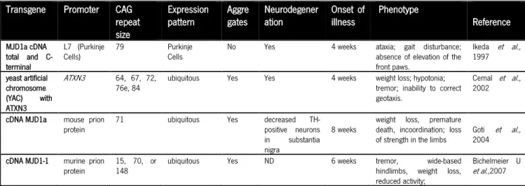
Since 1996, eight mouse animal models have been generated to understand the molecular mechanisms underlying MJD and to develop therapies (76-83), but so far, there is no treatment to delay the onset or cure the disease. Besides mouse, rat, C. elegans, cell culture and Drosophila models have also been used in MJD research. Apart from being a mammal, mouse models also have the greatest advantage of being close to humans at the genomic, anatomical, and physiological levels (84). A brief description of the mouse models generated so far is provided below (Table 2).
Table -2: Mouse models of MJD.
Transgene Promoter CAG repeat size
Expression
pattern Aggregates Neurodegeneration Onset of illness Phenotype Reference
MJD1a cDNA total and C-terminal
L7 (Purkinje
Cells) 79 Purkinje Cells No Yes 4 weeks ataxia; gait disturbance; absence of elevation of the
front paws. Ikeda et al., 1997 yeast artificial chromosome (YAC) with ATXN3 ATXN3 64, 67, 72,
76e, 84 ubiquitous Yes Yes 4 weeks weight loss; hypotonia; tremor; inability to correct
geotaxis.
Cemal et al.,
2002
cDNA MJD1a mouse prion
protein 71 ubiquitous Yes decreased positive neurons
TH-in substantia
nigra
8 weeks weight loss, premature death, incoordination; loss of strength in the limbs
Goti et al.,
2004
cDNA MJD1-1 murine prion
protein 15, 70, or 148 ubiquitous Yes ND 6 weeks tremor, hindlimbs, weight loss, wide-based
reduced activity;
Bichelmeier U et al.,2007
11
MJD1a CDNA total and C-terminal
murine prion
protein 79 ubiquitous Yes No 5 weeks incoordination; hypoactivity; ataxia; weight loss;
reduction in the pelvic elevation
Chou et al.,
2007
cDNA MJD1-1 murine prion
protein /Tet-Off
77 ubiquitous Yes Purkinje Cells 9 weeks reduced anxiety,
hyperactivity, incoordination, smaller increase in weight Boy J et al., 2009 cDNA MJD1-1 huntingtin
mouse 148 ubiquitous No Purkinje Cells 48 weeks hyperactivity in early stages of
disease, motor incoordination and amendment in motor learning in advanced disease Boy et al., 2010 cDNA MJD1-1 CMV (cytomegalov irus)
83 and 94 ubiquitous Yes astrogliosis and
neuronal atrophy
16 weeks incoordination Silva-Fernandes
et al., 2010
cDNA MJD1-1 CMV
(cytomegalov irus)
135 biquitous Yes loss of strength in the
limbs, incoordination,
weight loss, tremor, loss of
hindlimb tonus,
spontaneous locomotor and
exploratory activity
decrease
Silva-Fernandes et al., 2013
In our lab, Silva-Fernandes and colleagues generated a novel transgenic mouse model for MJD, carrying the repeat tract coding for 83 polyglutamines, using a human cDNA (isoform 1, MJD1-1 clone) under the control of the pCMV (cytomegalovirus) promoter. Recently in 2013, Silva-Fernandes, et, al. generated a novel mouse model to study MJD pathogenesis and for pre-clinical trials, using a human cDNA, under regulation of the CMV promoter, carrying a 135Q repeat in coding region of ataxin-3.This new model recapitulates the disease by mimicking disease-like features such as ubiquitious expression, protein aggregation, loss of strength in limbs from 6 weeks, balance deficit from 8 weeks, swimming incoordination and shorter step length from 12 weeks, body weight loss, tremors, limb clasping, loss of hind limb tonus, spontaneous locomotor and exploratory activity decrease from 14 weeks. This model could be used for dissecting the cellular and molecular events in the pathogenesis of MJD as well as to test therapeutic strategies for this disorder.
1.5 Rationale of pre-clinical trial drug targets
1.5.1 Mitochondrial stabilization and reduction of oxidative stress
Reduced concentrations of creatine, phosphocreatine (85) and mitochondrial electron transport enzymes (cytochrome C oxydase I) (86), and elevation in mtDNA deletion (87) have been observed in the basal ganglia and leucocytes of HD patients and in HD postmortem tissues, reinforcing the idea that oxidative
12
stress and mitochondrial dysfunction could be implicated in polyQ pathogenesis. Moreover, it was recently shown that mtDNA damage can be an early biomarker for HD. Interestingly, observations from the transgenic MJD mouse models and MJD patient samples also showed a decrease in mtDNA copy number (89) and accumulation of mtDNA 3867bp deletions (88) in the initial stages of this disease. Downregulated PGC-1α (mediator of mitochondrial biogenesis), lower lymphocyte mitochondrial membrane potential and calcium loads have also been observed in HD patients and in HD animal models (90, 91). Isolated mitochondria from striatum cells expressing mutant htt showed induced mPTP opening and disruption of mitochondrial calcium homeostasis (92, 93). Comparing mitochondrial respiratory function between R6/2 mice with 12 weeks of age and transgenic animals with 12 months, the latest showed higher respiratory control ratios and exhibited increased resistance to calcium loading; however when challenged with energy-demanding stimuli, the neurons from this old mice are more vulnerable to calcium deregulation (94). All together, these studies indicate that mitochondrial impairment and energy dysfunction might play a role in polyQ disorders, and that any treatment that might serve to buffer intracellular energy stores may delay the onset and/or progression of poly Q pathogenesis.
Increased levels of 8-hydroxydeoxyguanosine (8- OHdG), an oxidized DNA marker, (95), lipid peroxidation markers (96) and decreased levels of antioxidant enzymes SOD (97), oxidized form of glutathione, Zn/Cu-superoxide dismutase (Cu/Zn-SOD) (98) are found in HD patients’ brains and mouse model of HD. Likewise, a study in MJD also showed a significant reduction in the ratio of GSH/GSSG and total glutathione content (GSH + 2x GSSG) in mutant MJD cells expressing full length Ataxin-3 with 78Q (89). Another study from HD cellular model suggested that oxidative stress could promote aggregation and cell death by impairing proteasomal function and by overexpressing HSP27, has anti apoptotic properties; they were able to suppress the toxicity of mutant htt (99). All these evidences suggest that increased oxidative stress, metabolic deficits and mitochondrial abnormalities likely play a role in polyQ diseases. Therefore, therapeutic agents targeting these pathways could be beneficial to the patients.
Treatment with creatine, a bioenergetics supplement, stabilized the MPT, prevented ATP depletion and increased protein synthesis in HD mouse model. Creatine treatment also ameliorated brain pathology and motor symptoms, lessened brain atrophy and the formation of intranuclear inclusions in mouse models of HD. A Phase II clinical trial of creatine was performed in HD patients in which biomarkers were assessed in serum of these patients. Creatine supplementation reduced the levels of 8OH2’dG. Moreover, the dosage of creatine used (8g/day) was demonstrated to be tolerable and safe to patients (100). Based on these results, a phase III clinical trial has been approved and is currently ongoing in several centers. These results strongly support the effectiveness of creatine as a neuroprotector.
13
1.5.2 Clearance machinery: Chaperone related protein turnover
Molecular chaperones are so important to maintain the native protein conformation and most of the chaperons belong to the family of Heat Shock Proteins (HSPs) that have the ability to correct abnormal folding of expanded polyQ proteins and have a protective effect on polyQ aggregation and neurodegeneration (101, 102). It was recently shown that overexpressing HSC70 in cells and in a Drosophila model prevents polyQ toxicity by decreasing the amount of soluble oligomers and aggregate formation (103, 104) In a cellular model of SCA1, overexpression of the chaperone HDJ2/HSDJ was able to decrease ataxin-1 aggregation and overexpression of Hsp70 in a SCA1 mouse model was shown to suppress neuropathology and improve motor function (105, 106). On the other hand, genetically reducing the level of co-chaperone and ubiquitin ligase CHIP (C-terminus of Hsp70-interacting protein) in transgenic mouse models increased ataxin-3 microaggregation and MJD progression. So, increasing CHIP activity could be a possible therapy for MJD and other polyQ diseases (107).
Hsp90-containing HSF-1 (regulator of the heat-shock response (HSR)) complexes present in the unstressed cells are dissociated during stressed conditions, allowing the dislocation of this TF to the nucleus where it initiates the expression of several molecular chaperones. This includes Hsp90, which through classic negative feedback loop mechanisms, contributes to terminate the stress response (108). Therefore, Hsp90 by itself and/or associated with multichaperone complexes, is a major repressor of HSF-1. In that sense, pharmacologic inactivation of Hsp90 leads to overexpression of several molecular chaperones by activating the HSR via HSF-1 (108-109). Hsp90 is also ATP-dependent molecular chaperone and Hsp90 inhibitors mimic the unusual ATP structure that is adopted in the chaperone's N-terminal nucleotide-binding pocket, causing potent and selective blockade of ATP binding/hydrolysis and inhibit its chaperone function (110). The modification of neurodegeneration by HSF-1 has been shown in cells, Drosophila, C. elegans, mouse brain and slice culture models. Moreover, Hsp90 inhibitors, such as 17-AAG or 17-DMAG, significantly reduced aggregate load and toxicity. Despite its high potency, 17-AAG has been proved to have moderate toxicity and it also showed poor bioavailability and stability in several clinical trials (111). In contrast, 17-DMAG, a more effective analog of 17-AAG (112), is more water soluble than 17-AAG, a feature that is advantageous for clinical purposes, and it can be administered orally (113). These studies provide good evidence of the beneficial role of increasing chaperone levels in polyQ diseases.
1.5.3 Transcriptional regulation
Histone deacetylases (HDACs) regulate fundamental cellular activities such as transcription and play a crucial role in homeostasis of protein acetylation in histones and other proteins. Both acetylation and
14
deacetylation of histone proteins plays a pivotal role in the epigenetic regulation of transcription and other functions in cells, including neurons (114). Acetylation and deacetylation of histone proteins is catalyzed by histone acetyltransferases (HATs) and histone deacetylases (HDACs), respectively, at Lys (K) residues. Changes between HATs and HDACs interaction alters the balance of histone acetylation levels, resulting remodeling chromatin structure. In general, increased levels of protein acetylation facilitate transcription factor interaction with specific gene promoters and activate gene expression through relaxed chromatin conformation. HDACs often work as transcriptional repressors to silence gene expression and induce chromatin compaction through histone protein deacetylation. Consequently, inhibition of HDAC shifts the equilibrium towards enriched histone acetylation, chromatin relaxation and gene expression. A disproportion of HAT and HDAC activities could lead to disease states (115).
Several studies demonstrated the presence of transcriptional co-activators such as CREB-binding protein (CBP), p300/CBP-associated factor (PCAF), sperm-specific basic nuclear protein 1 (Sp1), and TBP-associated factor 4 (TAFII130) in inclusion bodies and their co-localization with polyglutamine proteins
(116). Mutant proteins that contain the polyQ-rich domain inhibit histone acetylase activity of CBP/p300 though protein-protein interactions, and lead to cellular toxicity (117). Counteracting hypoacetylation by deacetylase inhibitors or CBP overexpression reduces neurodegeneration (118). Histone deacetylase inhibitors such as suberoylanilide hydroxamic acid (SAHA) (119), sodium butyrate (SB) (120) and phenylbutyrate (121) have been shown to ameliorate motor phenotype in mouse models of HD, DRPLA and SBMA. Further, inhibition of histone H3 methylation by mithramycin presented increased lifespan, improved motor performance and striatal pathology in R6/2 mice (122). Rolipram also alleviated transcriptional dysregulation by increasing the phosphorylation and thus activity of CREB (123), and slower progression of neurological phenotype, increase in lifespan, and improve neuropathology in R6/2 HD mice treated with rolipram (124). All of the above evidence supports the hypothesis that an imbalance in histone acetylation may be a key process leading to transcriptional dysregulation in polyQ diseases.
Valproic acid (VPA) has been used for epilepsy and bipolar disorder and it is one of the HDAC inhibitors. It was demonstrated that VPA has the potential to regulate gene transcription and to mediate neuronal protection by inducing the apoptosis-inhibiting gene bcl-2 (124) and by inhibiting the activity of glycogen synthase kinase-3 (125). Jiping and collaborators very recently showed that VPA could increase both histone H3 and histone H4 acetylation level and reduce the early apoptotic rates of cells without inhibiting the aggregation of mutant ataxin-3 proteins in MJDtr-Q68- expressing cells (126). Based on this, we expected that VPA could serve as a new treatment strategy for SCA3 and other polyQ diseases.
15
Table- 3: Therapeutic rationale of our pre-clinical trials
Compound Target Pre -clinical trial phenotype
effect Drug effect in other polyQ disease Therapeutic strategy
Creatine Energy supplier Improvements in muscular strength parameter, motor coordination.
Improves survival and motor performance and delays neuronal atrophy, reduced NI (HD). (127)
Mitochondrial dysfunction/energy metabolism
17-DMAG Hsp-90 inhibitor Improvements in motor coordination
and balance
Improved motor phenotype,
Alleviated nuclear
localization of mutant AR; reduced muscle atrophy, body weight loss rate; prolonged life span. (SBMA) (128)
Clearance machinery:
Chaperone related
protein turnover, Autophagy
Valproic acid HDAC inhibitor, GABA
transaminase inhibitor
At late stage Prolonged improved open field activity life span;
(HD)(129) NI not discussed.
Transcription regulation
1.6 Aim of the thesis
This project aims to evaluate the utility of using ATXN3 inclusions as biomarkers in preclinical trials using a mouse model of MJD, by correlating the effect of three pharmacological compounds - creatine, 17-DMAG, and valproic acid, at the behavioral and molecular molecular level.
This will be achieved by (i) quantifying the nuclear inclusion load in facial nuclei (7N) and lateral reticular nucleus (LRT) of MJD treated vs. untreated mouse brains and (ii) correlating their effect at the level of motor phenotype (observed in preclinical trials previously performed by others from our mice team) with the effect on inclusions of the mutant protein.
To evaluate the effect of the drug at molecular level
Our team has previously developed a new MJD mouse model expressing human ataxin-3 with 135 glutamines under the regulation of the CMV promoter – CMVMJD135 – that presents phenotypic, pathologic and genetic similarities with the human disorder, namely several reliable and quantifiable phenotypic markers that can be used in therapeutic trials (Silva-Fernandes et al, 2014). For this study, we used mouse brains removed after treatment with three pharmacological compounds: creatine, 17-DMAG, and valproic acid, and with the respective vehicle solutions (control groups). We analyzed the abundance of ATXN3-positive nuclear inclusions in two specific brain regions known to be affected in MJD patients and in the CMVMJD135 mice - facial nuclei (7N) and lateral reticular nucleus (LRT) – by immunohistochemistry (IHC) and quantified the inclusion load by stereological microscopy approaches.
17
19
2. MATERIALS AND METHODS
2.1. Animals
Animals from the creatine group had been treated with 2% of supplemented creatine diet (by Sara Duarte-Silva), and sacrificed at 24 weeks of age. The second group of transgenic animals had been intraperitoneally injected with 25mg 17 DMAG /kg body weight three times per week (by Sara Duarte-Silva) and sacrificed at the age of 30 weeks. The third group CMVMJD 135 mice had been treated with valproic acid 200mg/kg (by Sofia Esteves) and sacrificed at 30 weeks of age. All control animals were fed with a normal diet or treated saline (0.9% NaCl).
Table -4: Illustration of experimental animal Compound No. Animals Tg- non Rx Tg+Rx
creatine 8 3 5
valproic acid 8 4 4
17DMAG 8 4 4
2.2. Mouse genotyping
DNA was isolated from tail biopsy using the Puregene DNA isolation kit (Gentra Systems, Minneapolis, MN). After DNA isolation, a single PCR was performed using the following primers for the human ataxin-3 cDNA (size of the product: 454 bp): TR6 (5’TAC ATC AAT GGG CGT GGA T 3’) and TR7 (5’ GAG GTC AAA ACA GCG TGG A 3’). As an internal PCR control, the mouse homologous Atxn3 gene (size of the product: 800 bp) was amplified using the following primers: mmMJD54 (5’ GAC TCC AGA GAG CAC CTG 3’) and mmMJD89 (5’ GCT AGC TAG AGC TAC TTA TTG 3’).
2.3. IMMUNOHISTOCHEMISTRY Preparation of paraffin brain sections
Transgenic mice CMVMJD135 were anaesthetized and transcardially perfused with PBS followed by 4% paraformaldehyde (PFA). Brains were immersed and embedded in paraffin block; the paraffin-embedded blocks were cut at 4µm thickness on a microtome then the sections were transferred onto glass slides
20
suitable for immunohistochemistry (IHC). Slides were selected to perform IHC based on the evaluation of sagittal section of brain morphology by cresyl violet staining (mouse brain Paxinos 2nd edition).
Immunostaining with anti-ataxin-3 antibody
Slides were pre-warmed at 700C and deparaffinized or hydrated in the staining apparatus. Sections were
incubated 20 minutes in pre boiled and cooled citrate buffer 1x in the microwave at low temperature and in 3% H2O2 solution (panreac) to block endogenous peroxidase activity. After PBS 1X washing, the sections
were immersed in to UV block (ultravision large volume detection system-thermo scientific). Slides were incubated with primary antibody 1H9 anti ataxin-3(1:1000 milipore) overnight at 4OC and then incubated
with HRP conjugated anti-mouse secondary antibody (thermo scientific) and then streptavidin reagent ((thermo scientific)). Followed by PBS T, PBS, Tris-HCL 0.05M wash, slides were incubated with DAB (sigma lifescience) substrate solution and counter stained by hematoxylin according to standard procedures and the section dehydrated; coverslip were mounted by mounting solution after wash with xilol solution. The mounted slides were observed the color of the antibody staining in the tissue sections under microscopy (17).
Quantification of ATXN3-positive intranuclear inclusions
Ataxin-3-positive inclusions in the facial nuclei and LRT either vehicle- or compound-treated animal brain (Creatine: n=3-4 for each condition, LRT- 6 to 9 slides per animal, 7N- 6-10 slides per animal),(17-DMAG: n=4- 4 for each condition, LRT- 4 to 7 slides per animal, 7N- 4 to10 slides per animal) (VPA: n=4 - 6 for each condition, LRT- 2 to 7 slides per animal, 7N- 2 to 7 slides per animal) were quantified and normalized for the total area assessed using the Olympus BX 51 stereological microscope and the Visiopharma integrator system software.
21
23
3. RESULTS
3.1 CMVMJD 135 mice treated with creatine exhibit reduced abundance of intranuclear ataxin-3 inclusion in the lateral reticular nuclei and facial nuclei of the brain
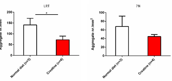
In order to analyze the ATXN3 inclusion load in the CNS, CMVMJD135 female mice treated with creatine were sacrificed at 24 weeks of age, and 4µm thickness brain section were prepared onto glass slides suitable for immunohistochemistry. By observing the presence of ataxin-3 inclusion in the facial nuclei (7N) and lateral reticular nuclei (LRT) of the brain under the microscope, brain section treated with 2% creatine transgenic mice exhibited less inclusion in the nucleus of cells when compared with transgenic littermates treated with normal diet (Figure 3A). To confirm this, inclusion load was quantified by stereological microscopy. CMVMJD135 mice treated with 2% Creatine exhibited significantly less inclusions (P=0.012) in the LRT region when compared with CMVMJD135 mice treated with normal diet (control). The facial Nuclei also showed a tendency for reduction of inclusion load (P =0.232) in neurons and massive Standard Deviation was observed in unsupplemented creatine group. (Figure 3B). This result suggests that dietary creatine supplementation is effective in decreasing aggregation of mutant human ataxin-3 in the LRT and 7N region of CMVMJD 135 mice. Normal diet Creatine LRT 7N A
24
Figure 3- : Intranuclear ATXN3 inclusion load in CMVMJD 135 mice treated with a normal diet and 2% creatine supplementation. A) ATXN3 inclusion positive cells in LRT and 7N at 24 weeks of age mice brain. The control group displayed a higher median number of inclusions than the creatine treated group in both regions. B) Stereological analysis: quantification of ATXN3 inclusions in the LRT region shows a significant (P<0.05) increase in inclusion load in the unsupplemented group (vehicle) compared with creatine supplemented group. The 7N region shows a trend for reduction in inclusion load when compared to the control group. (* P<0.05)
3.2. CMVMJD 135 mice treated with 17-DMAG did not exhibit a reduction in intranuclear ataxin-3 inclusions in the lateral reticular nuclei or facial nuclei of the brain
CMVMJD135 male mice were (previously) treated with 17-DMAG from 5 to 30 weeks of age and animals were sacrificed at 30 weeks. Brain sections were prepared for IHC in 4µm thickness and observed aggregation in 7N and LRT region under the microscope. The qualitative assessment did not reveal marked differences in ATXN3 aggregation in these two regions upon 17-DMAG treatment (Figure 4A). Consistently, the quantification of inclusion density revealed no difference between control and 17-DMAG treated transgenic mice in both regions (P=0.340 for LRT, P= 0.476 for 7N) (Figure 4B).
25
Figure 4- : Intranuclear inclusion load in CMVMJD 135 mice treated with vehicle (saline), 17-DMAG. A) ATXN3 inclusion positive cells in LRT and 7N at 30 weeks of age. Differences were not seen between the control group and treatment group in both regions. B) By stereological analysis, quantification of ATXN3 inclusion in the LRT and 7N region did not show significant differences in inclusion load in the 17-DMAG treated group compared with the saline group.
LRT Vehicle 17 DMAG Treated 7N A B
26
3.3. Transgenic mice treated with Valproic acid (VPA) showed a trend towards less inclusion load than the saline-treated group.
CMVMJD135 male mice were (previously) treated with VPA since 5 to 30 weeks of age and animals were sacrificed at 30 weeks. Brain sections were fixed with PFA and cut into 4µm thickness for IHC. Observation of inclusion in the 7N and LRT regions under the microscope did not reveal marked differences between groups (Figure 5A). Quantification of inclusion by stereological microscopy revealed a tendency for reduction of inclusion load between vehicle and VPA treated transgenic mice in the LRT region (P=0.060) and no differences in the 7N region (P=0.511) (Figure 5B).
VPA Vehicle 20 µm LRT 7N A
27
Figure 5- : ATXN3 intranuclear inclusion load in CMVMJD 135 mice treated with vehicle and VPA. A) ATXN3 inclusion positive cells in the LRT and 7N at 30 weeks of age. Marked differences were not seen by visual observation between the control and treatment group. B) Quantification of ATXN3 inclusion load showed a trens towards reduction in inclusion density in CMVMJD 135 mice treated with VPA compared to the saline treated group, but only in the LRT region.
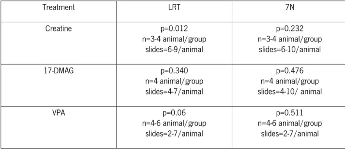
Table -5: A brief summary of the results obtained for the three compounds
Treatment LRT 7N Creatine p=0.012 n=3-4 animal/group slides=6-9/animal p=0.232 n=3-4 animal/group slides=6-10/animal 17-DMAG p=0.340 n=4 animal/group slides=4-7/animal p=0.476 n=4 animal/group slides=4-10/ animal VPA p=0.06 n=4-6 animal/group slides=2-7/animal p=0.511 n=4-6 animal/group slides=2-7/animal B
29
31
4. DISCUSSION
In this study, we have devised strategies for efficiently assessing therapeutic efficacy of compounds in molecular level in MJD mouse models. For this study we used a new mouse model expressing human ATXN3 with an increased number of glutamine residues -135Q (78). This novel model mimics several features of the human disease quite well at the behavioral and molecular level.
Since the discovery of NIs, it was thought that NI formation could be important to both pathogenesis and differential neuronal vulnerability in each polyQ disease. Sometimes, however, the connection between NIs and vulnerability is not clear, as they are not always associated with each other. For example, transgenic mice expressing a fragment of mutant htt showed very large numbers of NIs but with less neurodegeneration than mice expressing full-length mutant htt which undergo significant striatal degeneration without many NIs (54).
Our first target of pre- clinical trial compound is creatine. Generally, the mitochondrial isoform of creatine kinase and its substrates, creatine and phosphocreatine, organize a system and regulate energy homeostasis in the brain (130). The proposed role of creatine supplementation for neuroprotection is buffering intracellular energy by increasing phosphocreatine levels and inhibiting activation of mitochondrial permeability transition (MPT), thus leading to inhibition of necrotic and apoptotic cell death (131). Brdiczka and colleagues suggested that mitochondrial creatine kinase involved in a functional interaction between the inner membrane adenylate translocator, and outer membrane voltage dependent anion channel, which are thought to be components of the MPT (132). Mitochondrial creatine kinase becomes stable in an octomeric form upon creatine administration, which then inhibits activation of the MPT (133). Creatine administration also results in increased ADP concentrations, which inhibit the activation of the MPT (131). Involved in a functional interaction
The hypothesis of impairment of energy metabolism due to the mitochondrial dysfunction in polyQ diseases (34) provided the rationale for testing the therapeutic effects of creatine in CMVMJD135 mouse. Treatment of CMVMJD135 mice with creatine markedly improved limb strength at 6 weeks of age tested
32
by hanging wire test. Creatine treatment also had positive effects in the balance beam performance. At 22 and 24 weeks of age creatine-treated CMVMJD135 mice had better motor co-ordination, assed by motor swimming test. Moreover, creatine treatment was able to delay the onset of foot-dragging phenotype than unsupplemented creatine group, also delayed the disease progression by 12 weeks. (Sara Duarte-Silva, unpublished data). At the molecular level analysis, our IHC analysis showed a reduction in the nuclear aggregate load in the facial nuclei and LRT (39% less) comparable to CMVMJD135 mice fed with a normal diet. So in our pre-clinical trial with creatine, there was a direct correlation between the amelioration of the motor phenotype with the effect on the amount of inclusions of the mutant protein.
Interestingly, it has been also shown in the R6/2 HD mouse model that oral administration of creatine inhibits huntingtin aggregation in brain slice culture (134) and increases survival and ameliorates motor deficit in HD mice (127). Creatine also enhanced Purkinje cell survival in a transgenic mouse model of spinocerebellar ataxia type 1 (135). Furthermore, the efficacy of creatine in phase II clinical trials with HD patient strongly supports the effectiveness of creatine as a neuroprotector. Therefore, a phase III trial is currently ongoing in various centers. Creatine administration may therefore be useful in therapy of polyQ disease. So far the mechanism of action of creatine upon protein aggregation is not clear. We hypothesize that creatine may enhance the misfolded and aggregated protein clearance via increasing the efficiency of the proteasome and protein degradation pathways, perhaps through increased availability of energy to maintain these processes.
The second compound tested by our group in a pre-clinical trial, 17-DMAG, is proposed to reduce protein aggregation via elevated chaperone expression and proteasome activity. Chronic 17-DMAG treatment showed positive effects in the motor swimming test, rotarod and balance beam test and delayed the onset and progression of the foot-dragging phenotype CMVMJD135 mice, delaying the onset of key symptoms by approximately 8 weeks, which represents approximately 9% of the median C57Bl/6 normal lifespan 600 days), and to a lesser extent the foot-dragging (6 weeks, 7% of the lifespan) (136).
In contrast to these previous findings (136), our recent results revealed no reduction in the nuclear aggregate load in the 7N and LRT regions. . Overall, for 17-DMAG treatment in CMVMJD135 mice we did not observe a direct correlation between the amelioration of motor phenotype and intranuclear inclusions as a molecular biomarker, at least in these in these brain regions. However, administration of 17-DMAG was described to reduce the number of monomeric and nuclear-accumulated mutant AR and to improve motor performance without detectable toxicity in a SBMA mouse model (137). It has been demonstrated in vitro in SBMA that 17-DMAG significantly increases the HSP70 and HSP40 expression level and decreased mutant AR, even when induction of HSP70 was blocked. This result strongly suggests that
17-33
DMAG contributes to mutant AR degradation through the inhibition of Hsp 90, with or without induction of Hsp 70 (137). Currently, phase I clinical trials with 17-DMAG are ongoing for some types of solid tumour, to test dosage, toxicity, pharmacokinetics and pharmacodynamics, therefore this compound has some possibility of entering the market soon for these indications. Further clinical studies will be need for neurodegenerative diseases, among which MJD.
Deregulation of transcription due to the alteration of histone acetylation and deacetylation provided the rational for testing therapeutic effects of Valproic acid (VPA) in CMVMJD135 mouse. Results from our IHC analysis show that administration of VPA (200mg/kg) does not inhibit the nuclear accumulation of insoluble mutant Ataxin-3 in the LRT region. This is consistent with a previous publication in which it is described that administration of VPA increased H3 and H4 acetylation levels and reduced the early cell apoptotic rate without inhibiting the aggregation of mutant ATXN3 protein in vitro (138). CMVMJD135 mice treated with VPA showed minor phenotypic effects at late stages of the disease. No analysis was made beyond age of 24 weeks, which precludes more clear conclusions on the efficacy of the compound. Although it has been demonstrated in a SCA3 Drosophila model that VPA rescued polyQ-induced phenotypic abnormalities, no mention is made to effects on aggregation (126). However, histone acetylation and deacetylation can also regulate expression of heat shock proteins (HSPs) including HSP70. As an HDAC inhibitor, VPA could promote the expression of HSP70, that suppresses neuropathology and improve motor function in SCA1 mice, and extends the life of wild-type flies (139, 141). Based on this data and on the evidence we have for effects in aggregation, we suggest that VPA is a potential therapeutic agent for MJD, requiring further study.
As mentioned above, CMVMJD mice treated with creatine had amelioration since early disease stages; our IHC results also suggest that creatine treatment reduces the neuronal inclusion density in the LRT and 7N region, which is directly correlated with the motor phenotype amelioration. So, creatine is a potential therapeutic compound at molecular and behavioral level in MJD. Another compound, 17-DMAG, showed amelioration since an inter mediate disease stage. Molecular level analysis did not show any differences between treated and non-treated transgenic mice group regarding neuronal inclusions in the LRT and 7N brain regions, although reduction in inclusion load in the PN and a phenotypic amelioration had previously observed by our team, in several behavioral paradigms. Thus for this compound there was no direct correlation of phenotype amelioration effect with intra-nuclear inclusion load in the LRT and 7N regions.
34
CMVMJD135 mice treated with Valproic acid showed a tendency towards reduction of inclusion load in the LRT and no reduction in inclusion load in the 7N brain regions; phenotype amelioration was observed only at a late stage. So, this compound showed good effects on a molecular readout even though it exhibited a very mild phenotype amelioration only at late stages.
Globally, our results suggest that there is no direct correlation between the aggregate load in these regions of the brain of the CMVMJD135 mice and the amelioration of the phenotype observed with the chronic treatment with different compounds.
4.1. Future Perspectives
Regarding the identification of potential effects of the candidate therapeutic compounds at the molecular level in the CMVMJD135 mouse model, we need to further validate our hypothesis about creatine, 17-DMAG and VPA in other brain regions. It would be interesting to perform a similar study in another brain region, such as the dentate or pontine nuclei, for creatine and VPA. Moreover, other pre-clinical trials with additional compounds such as lithium and novel Hsp90 inhibitors provided by a pharmaceutical company are still ongoing in our lab, for which studies at molecular level will also be required.
Inclusion in non-CNS tissues were also observed in HD models. So, it would also be interesting to study inclusion in non-CNS tissues of CMVMJD135, particularly in tissues we have identified as affected, such as the skeletal muscle.
Finally, since the intranuclear inclusions we observe in the brain of patients and mouse models are thought to be a final product of a complex aggregation pathway, it would be essential to study as potential biomarkers ATXN3 aggregation species situated more upstream in this pathway, such as soluble oligomers, which may be more relvant in terms of pathogenesis.
35