Ana Paula Nunes de Lima Fernandes
I, Jessica Naiara de Medeiros Araújo
I, Fabiane Rocha Botarelli
I,
Danielly Oliveira Pitombeira
I, Marcos Antonio Ferreira Júnior
II, Allyne Fortes Vitor
II Universidade Federal do Rio Grande do Norte. Natal, Rio Grande do Norte, Brazil. II Universidade Federal do Mato Grosso do Sul. Campo Grande, Mato Grosso do Sul, Brazil.
How to cite this article:
Fernandes APNL, Araújo JNM, Botarelli FR, Pitombeira DO, Ferreira Jr MA, Vitor AF. Dry Eye Syndrome in Intensive Care Units: a concept analysis. Rev Bras Enferm [Internet]. 2018;71(3):1162-9. DOI: http://dx.doi.org/10.1590/0034-7167-2016-0582
Submission: 11-01-2016 Approval: 05-27-2017
ABSTRACT
Purpose: To analyse the concept of Dry Eye Syndrome in patients admitted to Intensive Care Units (ICU). Method: This is a concept analysis, according to Walker’s and Avant’s method, conducted using an integrative review, through search in the database. Science Direct, Scopus, Cinahl, Pubmed, Lilacs, Cochrane and Web of Science. The following keywords were used: “Keratoconjuntivite Sicca”, “Risk Factors”, “Dry eye Syndromes” and “Intensive Care Units”. After selection, 85 articles have been kept. Results: Antecedents found: age, lagophthalmos, environmental factors, use of medications, systemic diseases, mechanical ventilation and eye surgeries. Attributes: Tear Break-up Time < 10 s, Schirmer’s test I < 10 mm, Schirmer’s test II < 5 mm and signs and symptoms. Consequents: eye damage and discomfort; unstable vision. The Model Case and the Contrary Case were used to illustrate it. Conclusion: The research provided clarifi cation of the concept and consequent understanding of the Dry Eye Syndrome, which is preventable especially in ICU.
Descriptors: Keratoconjunctivitis Sicca; Risk Factors; Dry Eye Syndrome; Intensive Care Units; Nursing.
RESUMO
Objetivo: Analisar o conceito de Olho Seco em pacientes internados em Unidade de Terapia Intensiva (UTI). Método: Trata-se de uma análise de conceito, segundo método de Walker e Avant, operacionalizada mediante revisão integrativa por meio da busca nas bases de dados: Science Direct, Scopus, Cinahl, Pubmed, Lilacs, Cochrane e Web of Science. Foram utilizados os descritores: “Keratoconjuntivite Sicca”, “Risk Factors”, “Dry eye Syndromes” e “Intensive Care Units”. Após seleção, resultaram 85 artigos. Resultados: Identifi caram-se como antecedentes: idade, lagoftalmia, fatores ambientais, uso de medicamentos, doenças sistêmicas, ventilação mecânica e cirurgias oftálmicas. Atributos: Tear Break-up Time < 10 s, teste de Schimer I < 10 mm, teste de Schimer II < 5 mm e sinais e sintomas. Consequentes: dano e desconforto à superfície ocular, instabilidade visual. Como ilustração apresentaram-se o Caso Modelo e o Caso Contrário. Conclusão: O estudo promoveu clarifi cação do conceito e consequente entendimento do fenômeno, o qual é evitável, sobretudo na UTI.
Descritores: Ceratoconjuntivite Seca; Fatores de Risco; Síndromes do Olho Seco; Unidades de Terapia Intensiva; Enfermagem.
RESUMEN
Objetivo: Analizar el concepto del Ojo Seco en pacientes hospitalizados en Unidades de Terapia Intensiva (UTI). Método: Esto es un análisis de concepto, según el método de Walker y Avant, operacionalizado mediante revisión integrativa por medio de la búsqueda en las bases de datos: Science Direct, Scopus, Cinahl, Pubmed, Lilacs, Cochrane e Web of Science. Fueron utilizados los descriptores: “Queratoconjuntivitis Seca”, “Factores de Riesgo”, “Síndromes del Ojo Seco” y “Unidades de Terapia Intensiva”. Después de la selección, resultaron 85 artículos. Resultados: Identifi camos como antecedentes: edad, lagoftalmia, factores ambientales, uso de medicamentos, enfermedades sistémicas, ventilación mecánica y cirugías oftalmológicas. Atributos: Tear Break-up Time < 10 s, test de Schirmer I < 10 mm, test de Schirmer II < 5 mm y señales y síntomas. Consecuencias: daño e incomodidad a la superfi cie ocular, inestabilidad visual. Como representación se presentó el Caso Modelo y el Caso Contrario.
Dry Eye Syndrome in Intensive Care Units: a concept analysis
INTRODUCTION
The Dry Eye Syndrome (DES), also known as Keratocon-junctivitis sicca (KCS), is a multifactorial disease caused by the inadequate tear production and/or fast evaporation of tear. The DES can result from inflammatory diseases, environmen-tal factors, hormonal changes or age(1).
There are two causes for developing the Dry Eye Syndrome. One is due to a deficiency in the production of the tear film and the other is due to increased evaporation. Regarding In-tensive Care Units (ICU), the tear film is compromised due to a disorder in the responsible mechanisms for lubricating and protecting the eyes(2).
In a study conducted, the DES was the most frequent eye affection. It presented 72.2% cases in Intensive Care Units, which shows a high incidence of the phenomenon in this sec-tor(3). The ICU are care sectors for patients who are more
de-pendent on account of a serious or risky health condition. The units have health professionals from diverse areas and special-ties, who use a great range of technologies for diagnosis and therapy in the hospital environment(4).
Hospitalized patients in ICU are in critical health condi-tions, often depending on technological devices and using several medication to continue living. Nurses are the profes-sionals who provide the most assistance to these patients and perform fundamental activities in this sector(5).
Therefore, knowing what the Dry Eye condition is, its char-acteristics and factors that happen before and after this phe-nomenon, is extremely important to perform a qualified nurs-ing care, directed to preventnurs-ing this potential adverse event. Exposure and eye dryness may result in different complica-tions, including superficial keratopathy and inflammatory dis-eases in the cornea. Moreover, involvement of the epithelial surface and subsequent corneal exposure, resulting in ulcer-ation or perforulcer-ation. All these consequences hurt and reduce the patient’s life(6).
In this context, patients admitted to ICU present a high risk for the development of Dry Eye, since they present, in most cases, severe clinical conditions, such as the use of numer-ous medication, sedation and ventilatory support. Some other technologies to maintain vital signs and promote comfort and therapeutic aid are also used. These patients are predisposed to lose their natural mechanisms of eye protection and there-fore need effective assistance from the health team, with em-phasis on nursing(7).
Among the many responsibilities of nurses, there is a clear prioritisation of immediate procedures for critical patients to the detriment of certain easily accessible care, such as eye care. This is related to lack of knowledge of the nurse and the multi-professional team about anatomy, physiology, eye examination
and care to be implemented, besides the consequences that may arise from an inadequate or inexistent eye care(6).
Hence, clarifying this concept is crucial to develop a theo-retical support that bases the practice of nursing destined to preventing the DES in patients admitted to ICU.
We found imperative to analyse this concept given the abun-dance of information in literature and the need to stablish con-sensus about the incidence of this phenomenon in ICU.
OBJECTIVE
Analysing the concept of Dry Eye in patients admitted to Intensive Care Units (ICU).
METHOD
Concept analysis based on the framework proposed by Walker and Avant, performed through an integrative review of literature according to Whittemore and Knalf(9). They will be
described separately for better understanding.
Integrative Review of Literature
We performed the review from April to June 2015 and followed the steps recommended in the literature by Whit-temore and Knalf(9), which allowed us to order the knowledge
produced on the Dry Eye concept. The steps were identifica-tion of research quesidentifica-tions; literature search; data assessment; results analysis and presentation of review. We drew up these research questions: What is the concept of Dry Eye? What are the attributes of Dry Eye in ICU? Which aspects precede and proceed the Dry Eye Syndrome?
We selected studies using the relevance test(10), respecting the
following inclusion criteria: articles fully available in the select-ed databases, in Portuguese, English or Spanish; studies that re-spond to the proposed guiding questions. We implemented pre-vious notes, protocols, ongoing research, reviews, editorials and letters to the editor as exclusion criteria. We also implemented secondary studies, e.g., bibliographical reviews in general to un-derstand definitions and concepts about the phenomenon stud-ied. Excluding this type of publication could mean a significant reduction of essential information to be used in this analysis.
The search was performed from April to July 2015 by a pair of researchers in these databases: Science Direct, Sco-pus, Cinahl (Cumulative Index to Nursing and Allied Health Literature), Pubmed (Public Medline), Lilacs (Latin American and Caribean Health Science Literature Database), Cochrane, Web of Science. The access to these databases was carried out through the Brazilian portal of Coordination for the Improve-ment of Higher Education Personnel (Coordenação de Aper-feiçoamento de Pessoal de Nível Superior - CAPES).
Ana Paula Nunes de Lima Fernandes E-mail: [email protected] CORRESPONDING AUTHOR
Conclusión: El estudio posibilitó clarificación del concepto y consecuente entendimiento del fenómeno, lo cual es evitable, sobre todo en la UTI.
The search was performed in an uncontrolled way, using keywords indexed in the MeSH (Medical Subject Headings) and the Keywords in Health Sciences (Descritores em Ciên-cias da Saúde - DeCS), in Portuguese, English and Spanish: “Keratoconjuntivite Sicca”, “Risk Factors”, “Dry eye Syn-dromes” and “Intensive Care Units”.
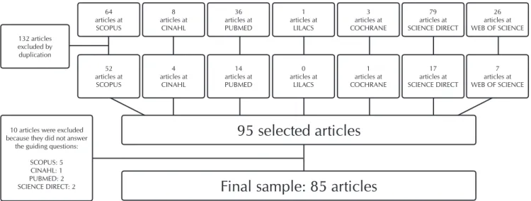
There were 19.856 titles found in different databases. When the relevance test was performed, 191 publications were se-lected for the next phase. After reading the abstracts, 132 were excluded by duplication and 95 selected. Due to the exclusion of ten articles after full reading, the study had a sample of 85 articles, as shown in Figure 1.
After obtaining the sample, data extraction was started through a form that had methodological information of the study and items related to concept analysis. When extracted, the data were summarised in tables and later grouped into categories by similar ideas and concepts.
Model of concept analysis of Walker and Avant
This model clarifies the concept in eight steps: (1) Selection of the Dry Eye concept in Intensive Care Units; (2) Purpose of the analysis, which was to analyse the Dry Eyes in patients admitted to ICU; (3) Use of the Dry Eye concept in ICU; (4) Establishment of the attributes that define Dry Eye in ICU; (5) Identification of a Model Case; (6) Identification of a Contrary Case; (7) Identification of antecedents and consequents of the phenomenon; (8) Definition of the empirical references.
The authors propose in the sixth step that cases examples be put together, to exemplify the use of the concept in its different perspectives; although, they see these cases as supplementary. Therefore, according to the reading of 85 articles, the attributes of the concept of Dry Eye and its antecedents and consequents were identified. We point out that, for the reason set above, only one model case and one contrary case were constructed to ex-emplify the contrary elements to the concept studied.
Firstly, we will detail the description of the articles, and then we will show the results according to the steps defined for the concept analysis. No submission to the Ethics Committee
in Research was sent, since this is a study with search only in literature.
RESULTS
Among the 24 different countries found in the articles that composed the sample, the United States (US) stands out with 34 articles written (39.53%). We noticed a high occurrence of Asian countries such as: Japan, India, Taiwan, Turkey, China, Korea, Indonesia, Iran, Israel and Thailand. Together, they ac-count for 23 articles and represent 26.74% of the sample.
As for the year of publication, the articles date from 1989 to 2015, of which 56.98% were published over the last five years. This express a greater recent interest in the matter. Regarding lan-guage, 96.51% of publications were in English and were not pub-lished in specific nursing journals, but rather in medical journals.
The methods used by the studies constituting the final sam-ple were 7: 43.02% were based on narrative review studies; 29.07% on cross-sectional studies; 10.45% on experimen-tal studies; 8.14% on case control studies; 5.81% on cross-cutting studies; 2.33% on systematic review; and 1.16% on methodological study. Regarding the type of approach, the majority (56.98%) consisted in quantitative research.
From the analysis of the articles, we identified the set of components of the concept, as described below.
Identification of the use of the concept
According to the theoretical and methodological frame-work adopted, the use of dictionaries, encyclopedias and all available literature is recommended to verify the possibilities of identification of the various attributes of the concept. As proposed, this initial phase should not be limited to only one aspect of the concept. All uses of the word must be consid-ered. Therefore, here are the main and most used definitions of the Dry Eye concept, as shown in Chart 1.
Hence, for this study, the description of the core concept is that Dry Eye Syndrome in Intensive Care Units is a multifacto-rial disease caused by the inadequate production of tears and/ or fast evaporation of the tear film.
Figure 1 – Diagram of results by phase of selection of articles, 2015 64
articles at SCOPUS 132 articles
excluded by duplication
10 articles were excluded because they did not answer
the guiding questions:
SCOPUS: 5 CINAHL: 1 PUBMED: 2 SCIENCE DIRECT: 2
95 selected articles
Final sample: 85 articles
52articles at SCOPUS
8 articles at
CINAHL
4 articles at
CINAHL
36 articles at PUBMED
14 articles at PUBMED
1 articles at
LILACS
0 articles at
LILACS
3 articles at COCHRANE
1 articles at COCHRANE
79 articles at SCIENCE DIRECT
17 articles at SCIENCE DIRECT
26 articles at WEB OF SCIENCE
volume. This test consists of placing small strip of sterile filter pa-per under the eyelid, in the lower fornix near the lateral corner, away from the cornea. The eyelid is then closed for five minutes and the wet portion of the strip is measured in millimetres(5).
There are two types of Schirmer’s Test, I and II. In Schirm-er’s test I, reflex tear secretion is evaluated in response to nasal and conjunctival stimulation, which makes a Schirmer I less than 10 mm positive for Dry Eye. Schirmer II is conducted after instilling a topical aesthetic. This test allows the mea-surement of the basal component of the tear film. The reflex component is considered absent. This results in a Schirmer II less than 5 mm positive for Dry Eye(12).
Fluorescein, Rose Bengal and Lissamine Green Test
The fluorescein dye is useful to assess the Dry Eye disease. This dye checks the integrity of the epithelium of the cornea and conjunctiva, in which an intact epithelium presents no spots, due to the presence of the mucin layer of the tear film. The assessment must be systematically performed using the cobalt illumination after two minutes using the dye, and the identification of spots highlights the absence of the protective layer and consequent presence of the Dry Eye disease(13).
The rose bengal stain derives from fluorescein. The stain is used in strips, moistened with artificial tears and has a simi-lar function, but unlike fluorescein it is a test of low sensitiv-ity and causes great eye irritabilsensitiv-ity. It can capture devitalised cells of both the conjunctiva and the cornea, resulting from the absence of the mucin layer of the tear film(14).
Lissamine green is a stain used to assess the anterior seg-ment of the eye and dye degenerated or dead cells. It is ap-plied the same way rose bengal is but causes less irritation that the latter(13).
Ocular assessment of signs and symptoms
The ocular assessment of signs and symptoms is conducted according to a set of strategies to verify the changes that can occur at the ocular level. A record analysis should be the first step so that predisposing factors are identified at the moment of admission.
Once all data are collected, signs must then be verified according to their corresponding method; symptoms must be questioned regarding existence, periodicity, intensity and level of limitation caused. This way, the most common signs and symptoms are: decreases lacrimal production; incomplete palpebral closure; burning feel in eyes; hyperaemia; sensation of ocular pruritus; sandy feeling in eyes; foreign body sensa-tion; eye pain; excessive tearing; blurred vision; mucous se-cretion; light sensitivity; ocular fatigue; and diminished blink-ing mechanism(15).
Critical attributes
Attributes are components that define a concept, i.e., char-acteristics that will determine the Dry Eye condition in Inten-sive Care Units. The four critical attributes of “Dry Eyes in ICU” identified through concept analysis are: (1) Precipitate tear film rupture; (2) Insufficient volume; (3) Spots in the use of stain in the ocular surface; (4) Eye anamnesis.
Chart 1 – An overview of the main definitions of the Dry Eye concept, 2015
Definition Authors/Year Research Sources
The Dry Eye syndrome is a disease with multiple etiologies. The common characteristic of its different manifestations is an abnormal tear film. Abnormalities in the tear film linked to the Dry Eye disease are tear deficiencies, due to insufficient supply or excessive loss, and abnormal tear composition.
(Johnson & Murphy
2004)
Pubmed
Dry Eye is a multifactorial disease of the tear film and ocular surface, which may be due to reduced tear production or excessive tear evaporation. It results in discomfort, disturbances in vision and unstable tear film, with potential for damage to the ocular surface.
(Rege, Kulkami; Puthran & Khandgave
2013)
Scopus
The Dry Eye Syndrome (DES) is a multifactorial disease caused by the inadequate production of tears and/or fast evaporation of the tear film. It is a result of inflammatory diseases, environmental factors, hormonal changes, age, etc.
(Yeh et al.
2015) Pubmed
Identification of empirical references
This step consisted in determining the empirical references for the attributes, considered classes or categories of real phe-nomena that, by their existence, show the incidence of the concept(8). Although this step of the concept analysis normally
takes place at the end, being the eighth step of the Walker and Avant method, we chose to perform it before in this study, because it describes the assessment methods of each attribute. There are four main ways to determine the Dry Eye: (1) The rupture time of the tear film; (2) Schirmer’s Test I and II; (3) The Fluorescein, Rose Bengal and Lissamine Green Test; (4) Ocular assessment of signs and symptoms.
The rupture time of the tear film
Widely known as Tear Break-up Time (TBUT), the rupture time of the tear film is a test to assess the evaporation of the tear film, that results in determining the tear quality. The pro-cedure consists of instilling a 2% fluorescein strip, moistened with a drop of lubricant, into the lower conjunctival fornix between the external and the middle third of the lower eyelid for one minute. After instilling the strip, patients must blink naturally and keep their eyes open. The tear film is observed under a beam of cobalt blue illumination until dark spots ap-pear. A timer must be turned on when patients stop blinking and turned off when the first dark spot appears(11).
Schirmer’s Test I and II
Precipitate tear film rupture
The time between the last blinking frequency and the ap-pearance of the first spot will be the time of tear film rupture, which is considered Dry Eye when it is less than 10 seconds(9).
Insufficient volume
To identify the volume of the tear film we must conduct Schirm-er’s test I or II. Values lower than 10 mm in SchirmSchirm-er’s test I repre-sent eye dryness, as well as values lower than 5 in Schirmer’s test II.
These tests measure the volume/amount of the tear film and make it possible to identify the insufficient volume of this eye component(5).
Spots in the use of stains in the ocular surface
The main vital stains used to assess the ocular surface and help to detect the DES are: fluorescein, rose bengal and lissa-mine green. The appearance of spots when performing these three tests means the person has DES.
All vital stains present similar functions and point to eye dryness, especially regarding the reduction of the lipid layer of the tear film. However, as evidenced, the isolated use of one of these tests cannot infer the diagnosis of DES. The combina-tion with the following attribute is needed(13).
Eye anamnesis
Ocular assessment, made of anamnesis and physical ex-amination, is a primordial step to identify DES. It should be conducted together with the tests to conclude this detection.
Previous symptoms are analysed along with the patient’s re-cords, work environment, anxiety, systemic diseases, automune diseases, infections such as hepatitis and the human im-munodeficiency virus (HIV), vascular history and medications(16).
Because of the characteristics of the admitted patients and the ICU’s environment, some elements require the professional to perform recurrent assessments, e.g.: sedation, lagophthalmia, low humidity, multi-drug therapy, basic systemic diseases. According to evidences, people exposed to these environments present ac-celeration in the process of the phenomenon’s development(5).
On physical examinations it is possible to identify ocular signs and symptoms(15).
We observed that other tests are suggested, such as tear film osmolarity, measurement of corneal sensitivity, cytology printing, biopsy of the conjunctival tissue, assays of fluids of detachable proteins and tear drainage by fluorescein dye. Despite the existence of these common tests in research, we highlight that the attributes currently exposed to determine DES in ICU are the most applied in clinical practice.
All the attributes listed can be measured by nurses using the empirical references described above, either autonomous-ly or in a team(17).
Antecedents and consequences
The next step proposed by Walker and Avant(8) is to identify
the antecedents and consequents. According to this model, antecedents are events commonly occurring before the iden-tification of the phenomenon and contribute to its consolida-tion. On the other hand, the consequents are identified or
even foreseen whenever there is the combination of factors of different origins in the presence of the phenomenon, if devel-oped and left untreated(8).
Antecedents
On the matter, some intrinsic factors predispose to DES re-gardless of the environment in which the patient is, e.g.: age over 40 years old, female over 50 years old, young men, anxiety, hypovitaminosis, hormonal imbalance, autoimmune diseases (Rheumatoid arthritis, Lupus, Myasthenia Gravis, Sjögren’s Syn-drome), systemic diseases (Stroke, Hyperlipidemia, Diabetes Mellitus, Systemic Arterial Hypertention); and infections such as HIV and Hepatitis(5,13).
Important precursors of eye dryness are environmental fac-tors involved in increasing the rate of evaporation of the tear film. They may take place isolated in combination with the in-trinsic factors, namely: high altitude, temperature, smoke, strong winds, environmental pollution, low humidity, sunlight and ra-diation. Another significant antecedent, common in ICU, is the use of medication such as diuretics, beta blockers, antidepres-sants, antihistamines, anxiolytics and eye drops for glaucoma.
Also related to ICU, there is a private environment with spe-cific therapy for patients who need intermittent care. In this field, intrinsic and extrinsic factors exist and can precede the occurrence of Dry Eyes, e.g.: presence of systemic drugs; associ-ated use of various drugs; lagophthalmia; sedation; insufficient blinking periodicity, length of admission; frequent use of health technological devices, use of mechanical ventilation and envi-ronmental factors such as low humidity and low temperature.
Consequents
Since consequents are considered resulting from the occur-rence of the concept, this analysis revealed the existence of severe consequences from Dry Eye in ICU. We identified the following consequents: eye damage, loss of vision, decreased quality of life and specific signs and symptoms of DES.
In ICU patients, ocular lubrification and protection mecha-nisms may be inefficient or even compromised, and they in-crease the proneness to develop the DES. Once the phenom-enon occurs, the cornea is the first ocular structure to suffer from the inefficiency of the tear film, with consequent damage to the ocular surface and possible serious impairment to the patient’s vision, depending on the increase and extension of the damage(6). To assess this damage, the vital fluorescein, rose
bengal and lissamine green stains are used.
The loss of vision is a serious consequence of DES. It only occurs when the person has impairment of visual acuity and it can reach the point of making the individual can no longer perform routine activities. Both ways of measurement loss of vision are conducted using specific ophthalmologic examina-tions to assess the reduction of acuity and visual field(18).
survey with relevant and comprehensive ques-tions, that can analyse the effective impact of this phenomenon(19).
IDEEL is an instrument with 27 items and consists of three main domains: limitations of daily activities, emotional well-being and work-ing limitations. All domains offer a score, which goes from zero (representing total impairment) to 100 (representing no impairment)(20).
Identification of a Model Case and a Con-trary Case
The Model Case is an example of the use of the concept, where all defining attributes are demonstrated, i.e., a pure case (the instance of the concept is eminent(8)). For this stage, the
following fictitious case was created:
Model Case
Ms. Maria, 56, on the 5th day of admission
in an Intensive Care Unit (ICU) for stroke, pres-ents Systemic Arterial Hypertension (SAH). The patient is intubated under mechanical
in-vasive ventilation, sedated and presents lagophthalmia. She has been using diuretics and beta blockers. In the ocular assess-ment, the presence of hyperaemia, palpebral edema and excess of mucous secretion were verified. The rupture time of the tear film was 5 seconds; Schirmer’s test I showed a result of 6 mil-limetres. The ocular surface was assesses using fluorescein and lissamine green and random spots were detected.
The Contrary Case is an example of denial to the concept. According to Wilson(21), it is a case to verify and conclude that,
certainly, whatever the concept is, the opposite case is not an example of this, as it follows:
Contrary Case
Mr. João, 37, on the 1st day of admission in the Intensive
Care Unit in the postoperative period of bariatric surgery, had no associated comorbidities, was conscious, oriented, breathing ambient oxygen, using analgesics, gastric protector and prophy-lactic antibiotic therapy. There is no change in the surface of the eye in the ocular assessment. The rupture time of the tear film was 15 seconds; Schirmer’s test I showed a result of 25 millime-tres. The ocular surface was assesses using the vital fluorescein and lissamine green stains. No spots were found.
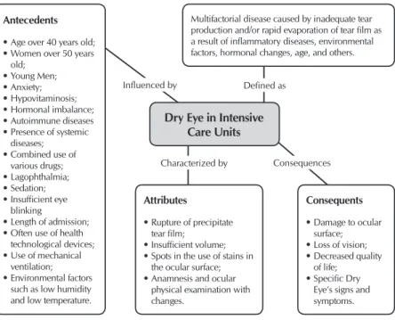
Therefore, the figure below is the representation of the concept studied, its definition, antecedents, consequents and attributes. It illustrates the information found and assembled.
DISCUSSION
The Dry Eye phenomenon is widely discussed and, as evi-dence in this study, research is often carried out by establishing studies of prevalence, experimental investigations, systematic re-views with meta-analysis; setting guidelines and protocols. There has been a more frequent production over the last five years and application of several approaches to the same problem.
All this progress made it possible to recognize the phenom-enon’s risk factors, ways of detection, measurement and con-sequences. Our goal was to answer a question about the clar-ity of the Dry Eye concept in the intensive care environment and explore the diverse predisposing factors to its occurrence. Unlike many organs of our body, and despite the eye is eas-ily available and accessible for clinical assessment, research still exposes different connotations about the development of DES, its risk factors and consequences. Therefore, it became necessary to produce a synthesis that would subsidize the clarification of this concept.
Factors such as sedation, use of drug combinations, mechani-cal ventilation, prolonged admission time, low humidity, low temperature and lagophthalmia are examples of risk factors often present in ICU and thus favour a prevalence of more than 70% of patients with eye dryness up to five days of hospitalization(3).
Although there is not a clear consensus to define the best way of treatment and which is the most used, the applica-tion of some substances such as topical cyclosporine, lin-oleic acid, omega-3 fatty acids, androgens, some types of tet-racyclines and steroids are prescribed by physicians(22), and
recommended.
The role of nursing clearly refers to its prevention and de-tection. As for prevention, the best alternative relates to ocular care procedures through the creation of a tear film with the use of eye drops, lubricants, artificial tears or the constitution of a moisture chamber by the occlusion with polyethylene film. Another prevention option is ocular closure using gauzes, ad-hesives or even sutures(6,23). As for detection, the nurse can
autonomously or in teams conduct tests that allow generat-ing guides to identify the phenomenon. Once trained, nurses are capable of conducting TBUT tests, Fluorescein Test, Rose Bengal and Lissamine Green, besides the usual Schirmer’s test I and II and Ocular Physical Examination(17).
Consequences Influenced by
Dry Eye in Intensive Care Units
Antecedents
• Age over 40 years old; • Women over 50 years
old; • Young Men; • Anxiety; • Hypovitaminosis; • Hormonal imbalance; • Autoimmune diseases • Presence of systemic
diseases; • Combined use of
various drugs; • Lagophthalmia; • Sedation; • Insufficient eye
blinking
• Length of admission; • Often use of health
technological devices; • Use of mechanical
ventilation; • Environmental factors
such as low humidity and low temperature.
Attributes
• Rupture of precipitate tear film;
• Insufficient volume; • Spots in the use of stains in
the ocular surface; • Anamnesis and ocular
physical examination with changes.
Consequents
• Damage to ocular surface; • Loss of vision; • Decreased quality
of life; • Specific Dry
Eye’s signs and symptoms. Multifactorial disease caused by inadequate tear production and/or rapid evaporation of tear film as a result of inflammatory diseases, environmental factors, hormonal changes, age, and others.
Defined as
Characterized by
As we have observed, the prevention options are easy to access procedures, simple handling and low cost. This is why the high prevalence of DES in intensive care is questioned, because patients admitted to this environment are cared in a global and intermittent manner.
Studies to investigate the performance of ocular care should be carried out in order to diagnose which elements of nursing care may be revised due to the relevance of this phenomenon.
Study limitations
In this study, we discussed the concept analysis of DES in In-tensive Care Units and we found two limitations during the con-struction and finishing procedure. The first regards the method of integrative review chosen to conduct the concept analysis.
This method allows the researcher to summarize the re-quired information through articles located in databases. Therefore, the material used was restricted to that of the bases, published in previously selected languages, and not consid-ered grey literature like dissertations and theses.
Another limitation relates to the non-evaluation of method-ological quality, allowing the inclusion of studies considered of low scientific evidence, like narrative reviews. Neverthe-less, we had to admit them in view of the need to synthesize definitions and identify diverse concepts.
Contributions to the Nursing, health or public policy sectors This study provided clarification of the concept and, conse-quently, a better understanding of the phenomenon. In addition, it is in line with the World Health Organisation’s Global Action Plan on Universal Eye Health 2014-1029, aimed at reducing avoidable visual impairment as a global public health problem(24).
Hence, its results evidence and influence the development of policies aimed at prevention and assessment of Dry Eye in
ICU. Moreover, it allows the foundation of the knowledge of nursing practice in relation to this phenomenon, and it spe-cially highlights the urgent need for research and professional education when considering the analysis of antecedents, at-tributes and consequents of Dry Eye in ICU.
CONCLUSION
This analysis of the concept of Dry Eye in Intensive Care Units proposed a clear meaning of the selected concept. It allowed us to verify this is an evitable phenomenon, despite recurring, mainly when the condition is acquired in ICU.
The antecedents and consequents found after the integra-tive review and analysis of the concept show that ICU are pre-disposing environments to the development of Dry Eye. After considering the consequents, we found that this is a limiting condition in physical and psychological aspects, intervening in the reduction of the quality of life of its patients, besides the possibility of causing irreversible damage.
This analysis is extremely important since it investigates a harmful condition, which can be prevented with the use of lubrication procedures considered simple and easily acces-sible. In conclusion, this study may represent a progress in literature on the matter by synthesizing, defining and analys-ing the Dry Eye phenomenon in a specific and highly prone environment.
FUNDING
This research was financed by the National Council for Sci-entific and Technological Development of Brazil (Conselho Nacional de Desenvolvimento Científico e Tecnológico do Brasil) under the Protocol CNPq/444290/2014-1.
REFERENCES
1. Yeh P, Chien H, Kwong NG, Tseng S, Chen W, Wang I, et al. Concordance between patient and clinician assessment of dry eye severity and treatment response in Taiwan. Cornea [Internet]. 2015 [cited 2015 Nov 14];34:500-5. Available from: https://www. ncbi.nlm.nih.gov/pubmed/25782401
2. Fonseca EC, Arruda GV, Rocha EM. Olho Seco: etiopatogenia e tratamento. Arq Bras Oftalmol[Internet]. 2010 [cited 2015 Nov 14];73(2):197-203. Available from: http://www.scielo.br/pdf/abo/v73n2/v73n2a21.pdf
3. Oh EG, Lee WH, Yoo JS, Kim SS, Ko IS, Chu SH, et al. Factors related to incidence of eye disorders in Korean patients at intensive care units. J Clin Nurs [Internet]. 2009 [cited 2015 Nov 14];18:29-35. Available from: http://onlinelibrary.wiley.com/ doi/10.1111/j.1365-2702.2008.02388.x/epdf
4. Brasil. Ministério da Saúde. Secretaria de Atenção à Saúde. Portaria GM/MS N° 2.918 de 09 de Junho de 1998 [Internet]. Brasília: MS; 1998[cited 2015 Nov 13]. Available from: http://dtr2001.saude.gov.br/sas/portarias/port98/GM/GM-2918.htm
5. Alavi, NM, Sharifitabar Z, Shaeri M, Hajbaghery MA. An audir of eye dryness and corneal abrasion in ICU parientes in Iran [Internet]. British Association of Critical Care Nurses. 2013 [cited 2015 Nov 14];19:73-7. Available from: http://onlinelibrary.wiley. com/doi/10.1111/nicc.12052/epdf
6. Werli-Alvarenga A, Ercole FF, Botoni FA, Oliveira JADMM, Chianca TC. Corneal injuries: incidence and risk factors in the Intensive Care Unit. Rev Latino-Am Enfermagem [Internet]. 2011 [cited 2015 Nov 14];19(5):1088-95. Available from: http://www.scielo.br/ pdf/rlae/v19n5/05.pdf
8. Walker L, Avant KC. Concept analysis. In: Walker L, Avant KC. Strategies for theory construction in nursing. California: Appleton & Lange, p. 63-84, 2011.
9. Whittemore R, Knafl K. The integrative review: updated methodology. J Adv Nurs[Internet]. 2005 [cited 2015 Nov 16];52(5):546-53. Available from: http://onlinelibrary.wiley.com/doi/10.1111/j.1365-2648.2005.03621.x/epdf
10. Olsen J. Meta-analysis or Collaborative Studies. J Occup Environ Med[Internet]. 1995 [cited 2015 Nov 16];37(8):897-902. Available from: http://www.ncbi.nlm.nih.gov/pubmed/8520950
11. Onwubiko SN, Eze BI, Udeh NN, Arinze OC, Onwasigwe EN, Umeh RE. Dry eye disease: prevalence, distribuition and determinants in a hospital-based population. Cont Lens Anterior Eye [Internet]. 2014 [cited 2015 Nov 19];37(3):157-61. Available from: http://www.sciencedirect.com/science/article/pii/S1367048413002798?via%3Dihub
12. Zeev MS, Miller DD, Latkany R. Diagnosis of dry eye disease and emerging technologies. J Clin Ophthalmol [Internet]. 2014 [cited 2015 Nov 19];8:581-90. Available from: https://www.ncbi.nlm.nih.gov/pmc/articles/PMC3964175/pdf/opth-8-581.pdf
13. Kastelan S, Tomic M, Salopek-Rabatic J, Novak B. Diagnostic procedures and management of dry eye. BioMed Res Int [Internet]. 2013 [cited 2014 Nov 14];1-17. Available from: https://www.ncbi.nlm.nih.gov/pmc/articles/pmid/24024186/
14. Javadi M, Feizi S. Dry Eye Syndrome. J Ophthalmic Vis Res[Internet]. 2011 [cited 2014 Nov 14];6(3):192-8. Available from: http:// www.ncbi.nlm.nih.gov/pmc/articles/PMC3306104/pdf/jovr-6-3-192.pdf
15. Yao W, Davidson RS, Durairaj VD, Gelston CD. Dry eye syndrome: an update in office management. Am J Med [Internet]. 2011 [cited 2015 Nov 14];124:1016-8. Available from: http://www.amjmed.com/article/S0002-9343(11)00498-0/pdf
16. Mesmer EM. The pathophysiology, diagnosis, and treatment of dry eye disease. Dtsch Arztebl Int [Internet]. 2015 [cited 2015 Nov15];112(5):71-82. Available from: https://www.ncbi.nlm.nih.gov/pmc/articles/PMC4335585/
17. Câmara VG, Araújo JNM, Fernandes APNL, Botarelli FR, Silva AB, Medeiros RAC, et al. Methods for detection of dry eye in critically ill patients: an integrative review. Int Arch Med [Internet]. 2016 [cited 2017 Apr 25];9(58):1-10. Available from: http:// imed.pub/ojs/index.php/iam/article/view/1504
18. Brasil. Conselho Brasileiro de Oftalmologia. Sociedade Brasileira de Oftalmologia. As condições da saúde ocular no Brasil: 2012 [Internet]. São Paulo: SBO; 2012[cited 2017 Apr 25]. 35p. Available from: http://www.cbo.com.br/novo/medico/pdf/01-cegueira.pdf
19. Abetz L, Rajagopalan K, Mertzanis P, Begley C, Barnes R, Chalmers R. Development and validation of the impact of dry eye on everyday life (IDEEL) questionnaire, a patient-reported outcomes (PRO) measure for the assessment of the burden of dry eye on patients. Health Qual Life Outcomes [Internet]. 2011 [cited 2015 Nov15];9:111. Available from: https://www.ncbi.nlm.nih.gov/ pmc/articles/PMC3269387/
20. Camp A, Wellik SR, Tzu JH, Feuer W, Arheart KL, Sastry A, et al. Dry eye specific quality of life in veterans using glaucoma drops. Cont Lens Anterior Eye [Internet]. 2015 [cited 2015 Nov 16];38:220-5. Available from: http://www.contactlensjournal.com/article/ S1367-0484(15)00020-X/fulltext
21. Wilson J. Pensar com conceitos. São Paulo: Martins Fontes, 2005.
22. Alves JS. Olho seco: uma abordagem didática. Rio de Janeiro: E-papers, 2010.
23. França CSFM, Fernandes APNL, Carvalho DPSRP, Xavier SSM, Ferreira Júnior MA, Boatrelli FR, et al. Evidence of interventions for the risk of dry eye in critically ill patients: an integrative review. App Nurs Res [Internet]. 2016 [cited 2017 Apr 25];29(2016):e14-e17. Available from: https://linkinghub.elsevier.com/retrieve/pii/S0897-1897(15)00118-4