DOENÇAS INFLAMATÓRIAS INTESTINAIS NA REGIÃO SUDESTE DO
BRASIL: UM ESTUDO RETROSPECTIVO
KAMILA ROSA MARTINS
DOENÇAS INFLAMATÓRIAS INTESTINAIS NA REGIÃO SUDESTE
DO BRASIL: UM ESTUDO RETROSPECTIVO
Tese apresentada ao Programa de Pós-
Graduação em Ciências da Saúde da
Faculdade de Medicina da Universidade
Federal de Uberlândia, como requisito
parcial para a obtenção do título de
Mestre em Ciências da Saúde.
Área de concentração: Ciências da
Saúde.
Orientador: Anderson Luiz Ferreira
M386d
2017 Martins, Kamila Rosa, 1988Doenças inflamatórias intestinais na região Sudeste do Brasil: um estudo retrospectivo / Kamila Rosa Martins. - 2017.
70 f.
Orientador: Anderson Luiz Ferreira.
Dissertação (mestrado) - Universidade Federal de Uberlândia, Programa de Pós-Graduação em Ciências da Saúde.
Inclui bibliografia.
1. Ciências médicas - Teses. 2. Proctocolite - Teses. 3. Doença de Crohn - Teses. 4. Intestinos - Doenças inflamatorias - Teses. I. Ferreira, Anderson Luiz. II. Universidade Federal de Uberlândia. Programa de Pós-Graduação em Ciências da Saúde. III. Título.
Kamila Rosa Martins
Doenças Inflamatórias Intestinais na região sudeste do Brasil: um
estudo retrospectivo
Presidente da banca:
Prof. Dr. Anderson Luiz Ferreira.
Tese apresentada ao Programa de Pós-
Graduação em Ciências da Saúde da
Faculdade de Medicina da Universidade
Federal de Uberlândia, como requisito
parcial para a obtenção do título de
Mestre em Ciências da Saúde.
Área de concentração: Ciências da
Saúde.
Banca Examinadora:
Prof. Dr. Anderson Luiz Ferreira (Orientador)
Prof. Dr. Carlos Augusto Real Martinez (Unicamp)
A minha família pelo apoio à
minha formação profissional.
Em especial, ao meu filho
Nicolas, que sempre enche
À Deus pelo dom da vida e a pelas oportunidades de estudos.
À minha família, em especial meus pais Edilene Rosa e Davi José
Martins e ao meu irmão Davi José Martins Filho, pelo apoio e incentivo dedicados
em todos os momentos.
Ao meu filho Nicolas Davi Martins de Oliveira por ser luz no meu caminho.
Ao Professor Doutor Anderson Luiz Ferreira, que confiou e investiu nesse
trabalho, obrigado pela orientação, dedicação, paciência e incentivos constantes
ao longo de todo o mestrado.
Aos professores da Pós-Graduação em Ciências da Saúde pela
compreensão devido as minhas limitações durante a gestação.
Aos profissionais do arquivo médico do Hospital de Clínicas de
Uberlândia pela disponibilização dos dados para a pesquisa.
À equipe de trabalho do Setor de Epidemiologia e aos amigos do Hospital de
Clínicas de Uberlândia pela compreensão durante a realização do curso.
ainda pensou sobre aquilo que todo
mundo vê.”
O ingresso no Mestrado Acadêmico do Programa de Pós-Graduação em
Ciências da Saúde (PPGCS) da Faculdade de Medicina (FAMED) da
Universidade Federal de Uberlândia (UFU) possibilitou consolidar e aprofundar
os conhecimentos relacionados às metodologias da pesquisa científica. Durante
a execução do projeto várias outras atividades foram realizadas no intuito de
enriquecer a formação profissional.
1. Disciplinas cursadas
Disciplinas cursadas no Programa de Pós-Graduação em Ciências da Saúde
da Universidade Federal de Uberlândia.
Disciplinas Créditos
Bioestatística 03
Fisiopatologia I 02
Fisiopatologia II 01
Metodologia e Ética em Pesquisa na Área Saúde 03
Seminários de Pesquisa 04
Total de créditos 13
2. Artigo Submetido
MARTINS, K.R., ARAÚJO, J.M., LUIZ-FERREIRA, A. Inflammatory bowel
disease in Brazil: is geographic area yet with low occurrence? Scandinavian
Journal of Gastroenterology. (SGAS-2017-0404) - Fator de impacto: 2,199.
3. Participação em Eventos
Introdução:
A doença inflamatória intestinal (DII) vem sendo cada vez mais
diagnosticada na América do Sul. Apesar de serem extensivamente investigadas
nos últimos anos, são raros os dados epidemiológicos da doença no Brasil.
Objetivo:
Traçar o perfil clínico e epidemiológico dos pacientes portadores de DII
atendidos no Hospital de Clínicas de Uberlândia durante o período de 1999 a
2014.
Material e métodos:
Foi realizado um estudo retrospectivo, descritivo e
quantitativo realizado nos prontuários dos pacientes com DII a partir da
confirmação diagnóstica por exame endoscópico, sendo analisadas as variáveis
faixa etária, sexo, raça, hábito tabágico, diagnóstico principal, bem como a
localização das doenças, principais manifestações clínicas, tratamento instituído,
complicações relacionadas às DII, manifestações extraintestinais e tratamento
medicamentoso e/ou cirúrgico utilizado.
Resultados:
Foram avaliados 183 casos
de DII, sendo 91 pacientes diagnosticados com Retocolite Ulcerativa (RCU) e 92
com Doença de Crohn (DC). As proporções da incidência da DII no sexo feminino
e masculino foram de 1,7 para RCU e 1,8 para DC. A média de idade dos
pacientes com DII foi de 35 anos. A etnia caucasiana foi predominante para
ambas. Os principais sintomas foram diarreia, dor abdominal e cólica intestinal. As
complicações mais vistas foram fístulas, enterorragias e obstruções intestinais, e a
principal manifestação extraintestinal foram as articulares. A terapêutica mais
usada foi a medicamentosa, sendo mais frequente os aminosalicilatos e os
imunomoduladores.
Conclusão
: Encontramos um perfil bem próximo daqueles
constatados pela literatura. A pesquisa permitiu concluir a baixa prevalência de DII
na população estudada quando comparado à América do Norte, mas alta em
relação a outras regiões consideradas com baixa incidência como a Ásia.
Introduction:
Inflammatory bowel disease (IBD) has been increasingly diagnosed
in South America. Although it has been extensively investigated in recent years,
epidemiological data on the disease in Brazil are rare.
Objective:
To describe the
clinical and epidemiological profile of patients with IBD treated at Hospital of
Uberlândia clinics during the period from 1999 to 2014.
Material and methods:
A
retrospective, descriptive and quantitative study was performed in the medical
records of patients with IBD from confirmation Diagnosis, endoscopic examination
and the variables age, sex, race, smoking habit, main diagnosis, as well as the
location of diseases, main clinical manifestations, treatment instituted,
complications related to IBD, extra-intestinal manifestations and drug treatment and
/ or Used.
Results:
183 cases of IBD were evaluated, of which 91 patients were
diagnosed with ulcerative colitis (RCU) and 92 with Crohn's disease (CD). The
proportions of the incidence of IBD in females and males were 1.7 for RCU and 1.8
for DC. The mean age of patients with IBD was 35 years. The Caucasian ethnicity
was predominant for both. The main symptoms were diarrhea, abdominal pain and
intestinal colic. The most frequent complications were fistulas, enterorrhages and
intestinal obstructions, and the main extraintestinal manifestations were the
articular ones. The most commonly used therapy was medication, with
aminosalicylates and immunomodulators being the most frequent.
Conclusion:
We
found a profile very close to those found in the literature. The research allowed to
conclude the low prevalence of IBD in the studied population when compared to
North America, but high in relation to other regions considered with low incidence
like Asia.
Figura 1 -
Prevalência global de DII em 2015... 15
Figura 2 -
Fatores ambientais associados a D II... 17
Figura 3 -
Fatores genéticos, imunológicos, ambientais e microbianos
associados às D II... 1 8
Figura 4 -
Biomarcadores da mucosa intestinal envolvidos na D II... 19
Figura 5 -
Sinais, sintomas e classificação da Colite Ulcerativa...
2 1
Figura 6 -
Sinais, sintomas e classificação da Doença de Crohn... 22
Figura 7 -
Controle Médico da Doença de Crohn...25
Figura 8 -
Controle Médico da Colite Ulcerativa Leve e Moderada... 26
DC
Doença de Crohn
1 INTRODUÇÃO ...13
2 FUNDAMENTAÇÃO TEÓRICA... 15
2.1 Epidemiologia...15
2.2 Etiopatogenia... 16
2.3 Quadro Clínico e Sintomas...20
2.4 Manifestações Extraintestinais... 22
2.5 Diagnóstico e tratamento...23
3 OBJETIVOS...28
3.1 Objetivo Geral... 28
3.2 Objetivos Específicos... 28
REFERÊNCIAS...29
1 INTRODUÇÃO
As doenças inflamatórias intestinais (DII) são doenças crônicas que
caracterizam-se pelo conjunto de modificações clínicas responsáveis pelo
processo inflamatório constante e idiopático das alças intestinais, representadas
principalmente pela Doença de Crohn (DC) e pela Retocolite Ulcerativa (RCU)
(BAUMGART et al., 2009). São doenças que se distinguem pela fisiopatogenia,
comprometimento das camadas intestinais e também pela localização no sistema
gastrointestinal (SCHIRBEL; FIOCCHI, 2010).
A doença de Crohn foi descrita pela primeira vez no ano de 1932, pelo
Doutor Burril B. Crohn, em Nova York sendo caracterizada por uma inflamação
crônica do intestino delgado, acarretando cicatrizes na parede do intestino
(STEVENS; LOWE, 2002). Já a RCU, doença descrita por Samuel Wilks, foi
apresentada em 1859, sendo publicado o primeiro caso no
Times Gazette, que
evidenciava comprometimento exclusivo da mucosa do cólon (MAGRO et al.,
2007).
A DC é um processo inflamatório crônico, recidivante, idiopático e
transmural que atinge uma ou mais partes do tubo digestório, geralmente o íleo e o
cólon, de forma segmentar e assimétrica (BAUMGART; SANDBORN, 2012).
Segundo Stevens e Lowe (2002), no início da DC o intestino é caracterizado por
uma hipertrofia da mucosa e submucosa com danos das pregas e aparecimento
de áreas de ulceração hemorrágica que, ao decorrer da doença, transformam-se
em fissuras.
A RCU também é um processo inflamatório crônico idiopático, que
atinge apenas a camada mucosa e submucosa, de modo contínuo, afetando
somente o reto e o cólon (KOTZE et al., 2011). A inflamação apresenta
prevalência de 40 a 50% limitada ao retossigmóide, com possível extensão por
todo o cólon (MAGALHÃES, 1993).
DII, porém as alterações emocionais podem exacerbar essas doenças, bem como
alguns alimentos podem propiciar alguns sintomas.
Nos últimos anos, nota-se um aumento considerável no número de
pessoas portadoras de DII. Estudos apontam um crescimento acentuado nos
países do hemisfério sul, apesar de permanecer prevalente no hemisfério norte
(RAPOSO, 2008).
Apesar do Brasil ser considerado área de baixa prevalência de DII,
observa-se o aumento significativo da incidência destas doenças nos registros da
literatura nacional (SOUZA; BELASCO; AGUILAR-NASCIMENTO, 2008;
CAMPOS; CAVALCANTE, 2012). Tal fato é comprovado através da quantidade de
internações registradas nas diferentes regiões brasileiras (MATTE, 2014).
2 FUNDAMENTAÇÃO TEÓRICA
2.1 Epidemiologia
Mundialmente as DII são distribuídas de forma heterogênea,
apresentando nos últimos anos um aumento significativo nas populações
ocidentais (BRAEGGER et al., 2011), principalmente nos países desenvolvidos e
naqueles onde as condições socioeconômicas vêm sendo melhoradas (EKBOM
et al.,1991; ZWI; MILLS, 1995; BOURREILLE et al., 2009).
Tradicionalmente, devido à escassez de dados de populações não
ocidentais, a DII foi considerada doença ocidental e pouco frequente nos países
do Oriente (ANANTHAKRISHNAN, 2015).
Em regiões do hemisfério Norte as incidências são mais elevadas
(LOFTUS et al., 2004), já em outras regiões, consideradas de baixa prevalência,
observa-se um aumento no número de casos, como é o caso do Japão, Coréia do
Sul, Cingapura, norte da Índia e América Latina (APPLEYARD; HERNANDEZ;
RÍOS- BEDOYA, 2004) (Figura 1).
Figura 1- Prevalência mundial de DII em 2015.
A ocorrência de DII tornou-se um problema de saúde pública em
algumas regiões da América do Norte e da Europa. Os Estados Unidos e o Canadá
apresentam uma prevalência de aproximadamente 1,7 milhões de pessoas com DII,
com uma média de 511 doentes a cada 100 mil habitantes (LOFTUS, 2007).
Existe uma ampla variação de dados em relação à incidência de DII.
Anos atrás, a RCU era considerada mais comum porém, com o aumento do
número de casos de DC, esta tendência mudou. A incidência anual na América do
Norte, tanto de DC como de RCU, está agora bastante equilibrada (MOLODECKY,
2012).
Na América Latina, são escassos os estudos epidemiológicos que
abordam as DII, embora algumas pesquisas apontem um crescimento nas
frequências de ambas doenças, RCU e DC (FIGUEROA, 2005).
Não diferente no Brasil, país considerado de baixa prevalência de DII,
são raros os estudos que permitem avaliar as ocorrências dessas doenças,
embora, estas não sejam tão incomuns como se imaginava há anos atrás
(GABURRI et al., 1998; FARIA; FERRARI; CUNHA, 2004). Vargas (2010) destaca
uma incidência de 20 a 100 doentes em 100 mil habitantes no Brasil, com novos
casos principalmente nas regiões sul, sudeste e centro-oeste. Algumas questões,
como as financeiras, dificultam a realização de estudos epidemiológicos em todas
as regiões brasileiras (ADORNE, 2016).
Rook (2012) apresenta uma correlação entre a redução de doenças
infecciosas, a falta de micoorganismos parasitas, o uso de medicações
(antibióticos), vacinações e um avanço das condições alimentares e sanitárias com
um aumento no índice de doenças auto-imunes e inflamatórias crônicas.
Vários autores descrevem que as DII atingem preferencialmente um
público jovem e apresentam um curso clínico longo e redicivante, o que acarreta
danos à educação, no desenvolvimento laboral, no relacionamento social e na
qualidade de vida do doente (LOVASZ et al., 2013).
2.2 Etiopatogenia
alterações psicológicas, dietas, alergia gastrointestinal, entre outros (Figura 2).
Não há uma etiologia conhecida para as DII, entretanto sabe-se que a
inflamação é consequência da associação entre os fatores do conteúdo do tubo
digestivo e da alteração na resposta imunológica da mucosa (FIOCHI, 1998;
KORZENICK; PODOLSKY, 2006).
Figura 2- Fatores ambientais associados às DII.
Localização geográfica e social
Estresse
q
£HP
w*
Microorganismos
Farmacos
Flora entérica
Permeabilidade
Fatores Ambientais
Apendectomia Tabagismo
Sistema Imune Geneticos
nflamatoria
Fonte: Adaptado de Danese; Sans; Fiocchi, 2004.
Alguns autores acreditam que os fatores genéticos, juntamente com os
fatores ambientais, são responsáveis pelo desencadeamento e modulação, entre
eles, hábitos alimentares, estilo de vida, condições de higiene e sanitárias e a
composição da microbiota intestinal (CATAPANI, 2009; CARDOZO; SOBRADO,
2012; KEYASHIAN et al., 2012).
(2012), nenhum dos fatores de risco por si só é capaz de desenvolver a DII, mas
as interações entre eles levam ao desenvolvimento da doença.
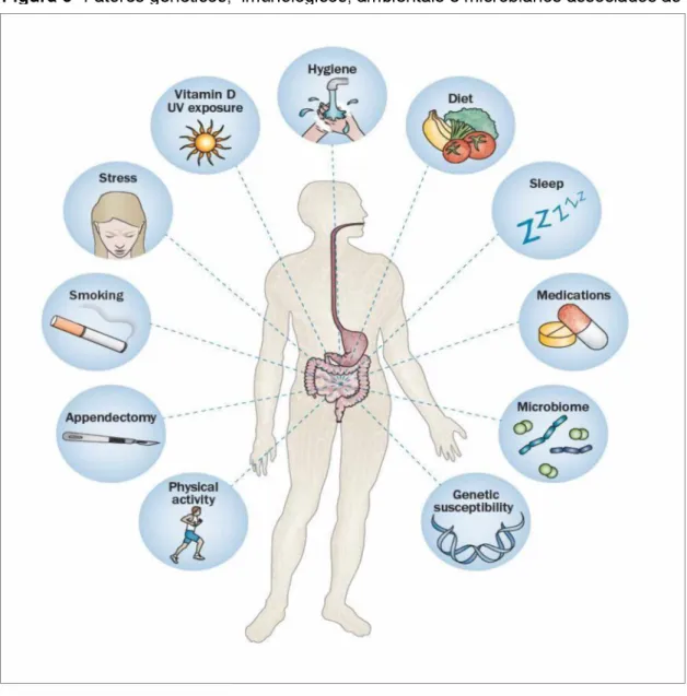
Figura 3- Fatores genéticos, imunológicos, ambientais e microbianos associados às DII.
Segundo Maranhão, Vieira e Campos (2015):
Não se sabe como uma pessoa geneticamente predisposta desenvolve
o processo inflamatório crônico. Sugere-se que a alteração genética transforma a
barreira epitelial intestinal, influenciando nas respostas imunológicas e na
composição da microbiota intestinal (ESBERARD, 2012).
Segundo Pinho (2008) nas DII instaura-se o desequilíbrio imunológico,
constatado pelo aumento dos níveis de várias citocinas. Na DC ocorre a liberação
de TNF-a, IFN- a, IL- 12 e IL-17, enquanto que na RCU ocorre um crescimento de
IL-13. Algumas hipóteses vêm sendo estudadas como possíveis origens do
desequilíbrio imunológico, entre elas estão o distúrbio da flora bacteriana, fatores
imunológicos e defeitos na barreira mucosa.
Figura 4- Biomarcadores da mucosa intestinal envolvida na DII.
Mesenchymal
IL-17 IL-10 / Í L - 2 1
IFN-y II-lp IL_12 IL-3 2 \
TNF-a IL-6 i L.4
A: Mucosal cytokines and chem okines
CL-8 Rantes . r rR 7 Fractalkine,
v MCP-1 CCR7 /
\ C X C L - 1 0 CXCL2 ' --- MIP-1
Integrins Selectins
lAdCAM
Pro-inflàmmatory gene/xpression Intestinal
and food antigens Intestinal bacteria epithelial cells
Dendritic Endothelial
X cells
OOc
cellsa a
Macrophages
Platelets
Plasma
D: Non im m une cells
cells PM Ns
C: Im m une cells
T ce s cells
Nk cells
/ I V ®
Extracellularmatrix Regulatory
Activated Th2 ce s
T h l
leukocyte Th2
Blood vessels
Mesenchymal cells
B: A dhesion m olecules and E: O ther factors
TLRS
m arkers o f activation
APK
IkBu
MUC2
MK
NLRsContraregulators
(PPARs')
A20
Tollip,
G6PD 1 OCS1
A partir da patogênese das DII, a Figura 4 reproduz as categorias
principais de biomarcadores da mucosa que podem ser preceptores da gravidade
da doença e da resposta ao tratamento.
Muito se debate sobre os fatores ambientais e quais seriam de maior
impacto nas DII. Esberard (2012) enfatiza a maior prevalência das DII em centros
urbanos e por trabalhadores com atividades mais intelectualizadas, apontando
ainda que o fumo e a "hipótese da higiene” são os fatores ambientais de maior
destaque.
Loftus (2004) concluiu que apesar de não haver uma conexão entre o
tabagismo e a inflamação intestinal, o uso do tabaco piora o quadro clínico da DC
enquanto age como um efeito protetor da RCU.
Muitos estudos contribuíram com grandes avanços no desenvolvimento
do mecanismo fisiopatológico das DII. Até hoje, sabe-se que essas doenças se
caracterizam por um processo inflamatório crônico acompanhado por uma
resposta imunológica inadequada contra agentes intraluminais. A suscetibilidade
às DII se dá pela genética e estímulo ambiental (ESBERARD, 2012).
2.3 Quadro Clínico e Sintomas
As DII são doenças crônicas intermitentes que envolvem uma grande
variedade de sintomas. Durante as recidivas a severidade dos sintomas oscila de
leve a severa, e durante as remissões muitos desses sinais podem desaparecer
ou atenuar. Normalmente, os sintomas dependem do segmento do trato intestinal
afetado (BERNSTEIN et al., 2015).
Figura 5- Sinais, sintomas e classificação da Colite Ulcerativa.
Fonte: Associação Brasileira de Colite Ulcerativa e Doença de Crohn, 2014.
Na DC os sintomas vão depender da área intestinal afetada. A dor
abdominal é a queixa predominante na maioria dos pacientes e é referida para
todo o abdômen ou somente na fossa ilíaca direita. Outro sintoma de destaque é o
quadro de diarreia, contínua ou intermitente. Assim como na RCU, outros sinais
clínicos podem acontecer com menor frequencia como anorexia, náusea, vômitos,
emagrecimento, déficit de crescimento e doença perianal (BARBIERI, 2000).
Figura 6- Sinais, sintomas e classificação da Doença de Crohn.
Fonte: Associação Brasileira de Colite Ulcerativa e Doença de Crohn, 2014
2.4 Manifestações Extraintestinais (MEIs)
As MEIs são comuns tanto na RCU quanto na DC e ocorrem em
aproximadamente 25-40% dos pacientes. Essas manifestações podem ocorrer em
quase todos os sistemas e promovem um grande desafio para a equipe
assistencial desses doentes.
apresentam piora do quadro durante a exacerbação da doença. Já nas
manifestações que envolvem o sistema dermatológico, nota-se o aparecimento de
eritema nodoso e pioderma gangrenoso, sendo, o primeiro, observado
principalmente em pacientes com DC e o pioderma gangrenoso em pacientes com
RCU (LONGO; FALCY, 2014).
A incidência de complicações oftalmológicas nos pacientes com DII é de
1 a 10%, sendo as mais comuns a conjuntivite, uveíte, e episclerite (LEVINE;
BURAKOFF, 2011; LONGO; FALCY, 2014).
A colangite esclerosante primária, a colelitíase, a trombose da veia
porta, a pancreatite e a hepatotoxicidade (provocada por medicamentos) são
destaque nas manifestações hepatobiliares. Quanto às manifestações urológicas,
destaca-se a possibilidade de nefrolitíase, uropatia obstrutiva e fístulas do trato
urinário. Por fim, são comuns as alterações da função pulmonar, embora o
surgimento de patologias significativas seja raro (LEVINE; BURAKOFF, 2011).
2.5 Diagnóstico e Tratamento
A confirmação do diagnóstico de DII deve ser realizado a partir da
correlação entre as manifestações clínicas e as evidências laboratoriais,
histológicas, endoscópicas e radiológicas (DAMIAO; SIPAHI, 2004).
A realização de exames de imagem como a enteróclise por tomografia
computadorizada,
cápsula
endoscópica,
colonoscopia,
tomografia
computadorizada de abdômen e ressonância magnética, tem favorecido o
diagnóstico e a avaliação da resposta ao tratamento, tornando-se essencial no
manejo da doença (HARA et al., 2005).
Quadro 1- Características diferenciais entre a Retocolite Ulcerativa e a Doença de Crohn. Características típicas da
RCU Características típicas da DC
Clínica
Diarreia frequente de pequeno volume com urgência
Predominantemente diarreia sanguinolenta
Diarreia acompanhada de dor abdominal e desnutrição Tumoração
abdominal Lesões perianais
Endoscópico e Radiológico
Inflamação cólica superficial difusa
Envolvimento do reto, que pode ser em placas
Erosões pouco profundas e úlceras
Sangramento espontâneo
Lesões assimétricas transmurais descontínuas Envolve principalmente íleo e lado direito do cólon Aspecto empedrado Úlcera longitudinal e fissuras profundas
Histopatológico
Inflamação difusa na mucosa ou submucosa
Distorção da arquitetura das criptas
Inflamação granulomatosa Fissuras ou úlceras aftóides observáveis, muitas vezes inflamação transmural.
Marcadores
séricos Anticorposcitoplasmáticos antineutrofilos
Anticorpos anti-Saccharomyces Cerevisiae e outros anticorpos contra antígenos microbianos
Fonte: Bernstein et al. , 2015.
Atualmente, a terapêutica das DII envolve o tratamento
farmacológico, nutricional e cirúrgico. O tratamento medicamentoso tem por
finalidade reduzir os sintomas clínicos na fase aguda e promover a remissão da
doença. Os principais fármacos envolvidos no tratamento das DII incluem os
aminosalicilatos, corticóides, antibióticos, imunossupressores e a terapia biológica.
O tratamento cirúrgico se faz necessário em situações refratárias ao tratamento
clínico (CATAPANI, 2009).
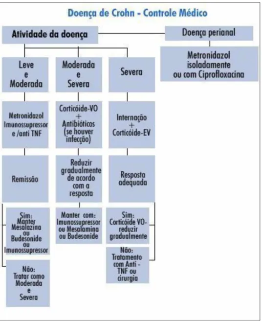
De acordo com a ABCD, o controle médico é realizado de acordo com a
atividade da doença na DC (Figura 7) e de acordo com a severidade na Colite
Ulcerativa (Figuras 8 e 9).
Figura 7- Controle Médico da Doença de Crohn.
Fonte: Associação Brasileira de Colite Ulcerativa e Doença de Crohn, 2014.
Figura 9- Controle Médico da Colite Ulcerativa Grave.
3 OBJETIVOS
3.1 Objetivo Geral
A pesquisa teve como objetivo traçar o perfil clínico e epidemiológico dos
pacientes portadores de DII atendidos no Hospital de Clínicas da Universidade
Federal de Uberlândia durante o período de 1999 a 2014.
3.2 Objetivos Específicos
• Identificar o perfil dos pacientes portadores de DII a partir das variáveis: faixa
etária, sexo, raça, estado civil, hábito tabágico;
• Traçar o diagnóstico principal, bem como a localização da doença, tempo de
diagnóstico da doença;
• Descrever as principais manifestações clínicas, bem como o tratamento
instituído;
• Levantar as complicações relacionadas às DII, inclusive as manifestações
extraintestinais;
REFERÊNCIAS
ADORNE, E. F.
Avaliação do Perfil Lipídico em Pacientes com Doença
Inflamatória Intestinal.
2016. 66 f. Dissertação (Mestrado) - Pontifícia
Universidade Católica do Rio Grande do Sul, Faculdade de Medicina, Pós
Graduação em Medicina e Ciências da Saúde, 2016.
ANANTHAKRISHNAN, A. N. Environmental risk factors for inflammatory bowel
diseases: a review.
Dig. Dis. Sci.
v.60, p.290-298, 2015.
APPLEYARD, C. B.; HERNÁNDEZ, G.; RIOS-BEDOYA, C. F. Basic epidemiology of
inflammatory bowel disease in Puerto Rico.
Inflammatory bowel diseases
, v. 10,
n. 2, p. 106-11, mar. 2004.
ASSOCIAÇÃO BRASILEIRA DE COLITE ULCERATIVA E DOENÇA DE CROHN.
Sobre a Doença de Crohn e Colite Ulcerativa
, 2014. Disponível em: <
http://abcd.org.br/>. Acesso em 09 jan. 2017.
BARBIERI, D. Inflammatory bowel diseases.
Jornal de Pediatria
, v. 76, n. 7, p.
173- 80, 15 jul. 2000.
BAUMGART, D. C. et al. Exaggerated inflammatory response of primary human
myeloid dendritic cells to lipopolysaccharide in patients with inflammatory bowel
disease.
Clinical & Experimental Immunology
, v. 157, n. 3, p. 423-436, set. 2009.
BAUMGART, D. C.; SANDBORN, W. J. Cronh’s disease.
The Lancet
, v.
380, n. 9853, p. 1590-1605, nov. 2012.
BERNSTEIN, C. et al.
Doença inflamatória intestinal:
practice guidelines.
Canadá: World Gastroenterology Organisation, 2015. Disponível em:
<http://www.worldgastroenterology.org/UserFiles/file/guidelines/inflammatory-
bowel-disease-portuguese-2015.pdf>. Acesso em: 06 de Maio 2017.
BOURREILLE, A. et al. Role of small-bowel endoscopy in the management of
patients with inflammatory bowel disease: an international OMED-ECCO
consensus.
Endoscopy
, v. 41, n. 7, p. 618-637, 8 jul. 2009.
BRAEGGER, C. P. et al. Epidemiology of Inflammatory Bowel Disease.
Journal of
Pediatric Gastroenterology and Nutrition
, v. 53, n. 2, p. 141-144, ago. 2011.
CAMPOS, M.O.B; CAVALCANTE, T.B. Smoking etiologic factor for Crohn’s
diasease.
Revista de Enfermagem da UFPI
, v.1, n. 3, p. 201-204, 2012.
CARDOZO, W.; SOBRADO, C.
Doença inflamatória intestinal
. 1. ed. São Paulo:
Editora Manole, 2012.
DAMIÃO, A. O. M. C.; SIPAHI, A. M. Doença inflamatória intestinal.
Gastroenterologia. Rio de Janeiro: MEDSI Editora Médica e Científica Ltda
, p.
1105-1149, 2004.
DANESE, S.; SANS, M.; FIOCCHI, C. Inflammatory bowel disease: the role of
environmental factors.
Autoimmunity Reviews
, v. 3, n. 5, p. 394-400, 2004.
EKBOM, A. et al. The epidemiology of inflammatory bowel disease: a large,
population-based study in Sweden.
Gastroenterology
, v. 100, n. 2, p. 350-8, fev.
1991.
ESBERARD, B.C. Etiopatogenia das doenças inflamatórias intestinais.
Revista
Hospital Universitário Pedro Ernesto
, v. 11, n. 4, p. 13-16, 2012.
FARIA, L.; FERRARI, M.; CUNHA, A. Aspectos clínicos da doença de Crohn em um
centro de referência para doenças intestinais.
GED: Gastroenterologia
Endoscopia Digestiva
, v. 23, n. 4, p. 151-164, 2004.
FIGUEROA C, C. et al. Inflammatory bowel disease: experience of two Chilean
centers.
Revista Medica de Chile
, v. 133, n. 11, p. 1295-304, nov. 2005.
FIOCCHI,
C. Inflammatory bowel disease: etiology and pathogenesis.
Gastroenterology
, v. 115, n. 1, p. 182-205, jul. 1998.
GABURRI, P. et al. Epidemiologia, aspectos clínicos e evolutivos da doença de
Crohn: um estudo de 60 casos.
Arquivos de Gastroenterologia
, v. 34, n. 4, p.
240-246, 1998.
HARA, A. K. et al. Imaging of Small Bowel Disease: Comparison of Capsule
Endoscopy, Standard Endoscopy, Barium Examination, and CT.
RadioGraphics
,
v. 25, n. 3, p. 697-711, maio 2005.
KEYASHIAN, K. et al. Management of inflammatory bowel disease: past, present
and future.
Expert Review of Clinical Immunology
, v. 8, n. 4, p. 303-305, 10 maio
2012.
KORZENIK, J. R.; PODOLSKY, D. K. Evolving knowledge and therapy of
inflammatory bowel disease.
Nature Reviews Drug Discovery
, v. 5, n. 3, p. 197
209, mar. 2006.
KOTZE, L.; KOTZE, P.; KOTZE, L. Doença de Crohn. In: DANI, R.; PASSOS, M.
(Eds.).
Gastroenterologia Essencial
. 4. ed. Rio de Janeiro: Guanabara Koogan,
2011. p. 347-379.
LEVINE, J.; BURAKOFF, R. Extraintestinal Manifestations of Inflammatory Bowel
Disease.
Gastroenterology & Hepatology
, v. 7, n. 4, p. 235-241, 2011.
LOFTUS, E. V. Clinical epidemiology of inflammatory bowel disease: Incidence,
prevalence, and environmental influences.
Gastroenterology
, v. 126, n. 6, p. 1504
17, maio 2004.
LONGO, D. L.; FAUCY, A. S.
Gastrenterologia e Hepatologia de Harrison
. 2. ed.
New York: Editora Artmed, 2015.
LOVASZ, B. D. et al. New trends in inflammatory bowel disease epidemiology and
disease course in Eastern Europe.
Digestive and Liver Disease
, v. 45, n. 4, p.
269-276, abr. 2013.
MAGALHÃES, A. Doença de Crohn (CH). In: DANI, R.; CASTRO, L.
(Eds.).
Gastroenterologia Clínica
. 3. ed. Rio de Janeiro: Guanabara Koogan, 1993. p.
765-777.
MAGRO, F. et al. Avaliação nacional dos doentes com doença de Crohn.
GE
Jornal Portugues de Gastroenterologia
, v. 14, n.24, 2007.
MARANHÃO, D. D. DE A.; VIEIRA, A.; CAMPOS, T. DE. Características
e
diagnóstico diferencial das doenças inflamatórias intestinais.
Jornal Brasileiro de
Medicina
, v. 103, n. 1, p. 9-15, 2015.
MATTE, C. A. S.
Repercussões bucais de doenças gastrointestinais de
pacientes dos serviçoes de gastroenterologia e endoscopia da HU/UFSC
.
2014. 121 f. Trabalho de conclusão de curso (Graduação em Odontologia).
Universidade Federal de Santa Catarina, Florianópolis, 2014.
MOLODECKY, N. A. et al. Increasing incidence and prevalence of the inflammatory
bowel diseases with time, based on systematic review.
Gastroenterology,
v. 142,
p. 46-54 e 42, 2012.
MOTA, A. J. M. P. DA.
Avanços no Tratamento da Doença Inflamatória
Intestinal
. Maio de 2012. 52p. Dissertação de mestrado. Universidade da Beira
Interior, Covilhã, 2012.
OLIVEIRA, F. M.; EMERICK, A. P. DO C.; SOARES, E. G. Aspectos
epidemiológicos das doenças intestinais inflamatórias na macrorregião de saúde
leste do Estado de Minas Gerais.
Ciência & Saúde Coletiva
, v. 15, p. 1031-1037,
jun. 2010.
PINHO, M. A Biologia molecular das doenças inflamatórias intestinais.
Revista
Brasileira de Coloproctologia
, v. 28, n. 1, mar. 2008.
RAPOSO, F. A. Q.
Doença inflamatória intestinal
. Junho de 2008. 122p.
Dissertação de mestrado. Universidade da Beira Interior, Covilhã, 2008.
SCALDAFERRI, F. Mucosal biomarkers in inflammatory bowel disease: Key
pathogenic players or disease predictors?
World Journal Of Gastroenterology
, v.
16, n. 21, p.2616-2625, 2010.
SCHIRBEL, A.; FIOCCHI, C. Inflammatory bowel disease: Established and evolving
considerations on its etiopathogenesis and therapy.
Journal of Digestive
Diseases
, v. 11, n. 5, p. 266-276, 28 jul. 2010.
SOUZA, M. M. DE; BELASCO, A. G. S.; AGUILAR-NASCIMENTO, J. E. DE. Perfil
epidemiológico dos pacientes portadores de doença inflamatória intestinal do
estado de Mato Grosso.
Revista Brasileira de Coloproctologia
, v. 28, n. 3, set.
2008.
STEVENS, A.; LOWE, J.
Patologia
. 2. ed. São Paulo: Editora Manole, 2002.
VARGAS, R. D. Epidemiology of inflammatory bowel disease (IBD): Why are there
differences between North America and Latin America?
Revista Colombiana de
Gastroenterologia
, v. 25, n. 2, p. 103-105, 2010.
ZALTMAN, C. Doença inflamatória intestinal: qual a relevância da doença no Brasil?
Cadernos de Saúde Pública
, v. 23, n. 5, p. 992-993, maio 2007.
ANEXO
Artigo 1
Scandinavian Journal /Q Taylor & Francis
of Gastroenterology ^
! H om e ” A u th o r
A uthor Dashboard / Submission Confirmation
Submission Confirmation
Thank you for your submission
Submitted to Scandinavian Journal of Gastroenterology
Manuscript ID SGAS-2017-0404
Title INFLAMMATORY BOWEL DISEASE IN BRAZIL: IS GEOGRAPHIC AREA YET WITH LOW OCCURRENCE?
Authors Martins, Kamila de Araujo, Joniel Luiz-Ferreira, Anderson
Date Submitted 10-May-2017
Author D
SCHOLARONE™
© Thom son Reuters | © ScholarO ne, Inc., 2017. A ll Rights Reserved.
ScholarOne Manuscripts and ScholarOne are registered trademarks of ScholarOne, Inc. ScholarOne Manuscripts Patents #7,257,767 and #7,263,655.
INFLAMMATORY BOWEL DISEASE IN BRAZIL: IS
GEOGRAPHIC AREA YET WITH LOW OCCURRENCE?
Jo u rn a l: Scandinavian Journal o f G astroenterology
M a n u sc rip t ID S G A S -2 0 1 7 -0 4 0 4
M a n u sc rip t T ype: O rig in a l A rtic le
D ate S u b m itte d by th e A u th o r: 1 0 -M a y -2 0 1 7
C o m p le te List of A u th o rs: M a rtin s, K a m ila ; U n iv e rs id a d e Fed eral de U b e rla n d ia , P ro g ra m a de Pós- G ra d u a ç ã o em C iê n c ia s da S a ú d e
de A ra u jo , Jo n ie l; U n iv e rs id a d e Fed eral de G o iá s, C iê n c ia s B io ló g ica s L u iz-F e rre ira , A n d e rso n ; U n iv e rsid a d e Fed eral de G o iá s , C iê n c ia s B io ló g ica s
K eyw o rd : E p id e m io lo g ica l p ro file , C ro h n 's D ise a se , U lc e ra tiv e C o litis
1 2 3 4 5 6 7 8 9 10 11 12 13 14 15 16 17 18 19 2 0 21 2 2 2 3 2 4 2 5 2 6 2 7 2 8 2 9 3 0 31 3 2 3 3 3 4 3 5 3 6 3 7 3 8 3 9 4 0 41 4 2 4 3 4 4 4 5 4 6 4 7 4 8 4 9 5 0 51 5 2 5 3 5 4 5 5 5 6 5 7 5 8 5 9 6 0
INFLAMMATORY BOWEL DISEASE IN BRAZIL: IS GEOGRAPHIC AREA YET WITH LOW OCCURRENCE?
Kamila Rosa Martinsa, Joniel Mendes de Araújob, Anderson Luiz-Ferreirab*
aUniversidade Federal de Uberlândia, Faculdade de Medicina, Pós Graduação em Ciências da Saúde, Uberlândia, Minas Gerais, Brasil.
bUniversidade Federal de Goiás, Instituto de Biotecnologia, Departamento de Ciências Biológicas, Catalão, Goiás, Brasil.
Correspondence author Dr. Anderson Luiz-Ferreira
Federal University of Goiás, Biotechnology Institute, Biological Sciences Department, CEP 75704-020 Catalão, GO, Brazil. e-mail: luiz_ferreira@ufg.br
1 2 3 4 5 6 7 8 9 10 11 12 13 14 15 16 17 18 19 2 0 21 2 2 2 3 2 4 2 5 2 6 2 7 2 8 2 9 3 0 31 3 2 3 3 3 4 3 5 3 6 3 7 3 8 3 9 4 0 41 4 2 4 3 4 4 4 5 4 6 4 7 4 8 4 9 5 0 51 5 2 5 3 5 4 5 5 5 6 5 7 5 8 5 9 6 0 ABSTRACT
Objective: Crohn’s Disease (CD) and Ulcerative Colitis (UC), two of the main conditions of Inflammatory Bowel Disease (IBD), have been increasingly
diagnosed within South America. Although IBDs were intensively studied in the last years, there are still few data on the epidemiology of these diseases in Brazil.
Methods: We performed a retrospective study of the medical records of patients diagnosed with IBDs according to the International Classification of Diseases (ICD): ICD K50 for CD and ICD K51 for UC, with diagnosis confirmed through endoscopic examination for both diseases. We analyzed the following
variables: age; sex; ethnicity; smoking behavior; primary diagnosis; location of the disease manifestation; main clinical manifestations; instituted treatment; IBDs-related complications; extraintestinal manifestations; and drug and/or surgical treatment instituted.
Results: We evaluated 183 IBDs cases (91 UC and 92 CD cases). The prevalence ratio was of 13.6/100,000 inhabitants for UC and 13.7/100.000 for CD. The incidence proportions of IBDs in both female and male patients were 1.7 for UC and 1.8 for CD. The average age of the patients at diagnosis was
39.4 years for UC and 31.1 for CD. White-skinned people were the predominant ethnical group affected by both UC and CD (66.0% and 69.0%, respectively). Few patients were submitted to surgical procedures as treatment alternatives.
1 2 3 4 5 6 7 8 9 10 11 12 13 14 15 16 17 18 19 2 0 21 2 2 2 3 2 4 2 5 2 6 2 7 2 8 2 9 3 0 31 3 2 3 3 3 4 3 5 3 6 3 7 3 8 3 9 4 0 41 4 2 4 3 4 4 4 5 4 6 4 7 4 8 4 9 5 0 51 5 2 5 3 5 4 5 5 5 6 5 7 5 8 5 9 6 0
Conclusions: The IBDs prevalence within this population is low compared to that of populations within North America, but high compared to other regions considered to be of low incidence, like Asia.
KEYWORDS: Epidemiological profile; Crohn’s Disease; Ulcerative Colitis
1 2 3 4 5 6 7 8 9 10 11 12 13 14 15 16 17 18 19 2 0 21 2 2 2 3 2 4 2 5 2 6 2 7 2 8 2 9 3 0 31 3 2 3 3 3 4 3 5 3 6 3 7 3 8 3 9 4 0 41 4 2 4 3 4 4 4 5 4 6 4 7 4 8 4 9 5 0 51 5 2 5 3 5 4 5 5 5 6 5 7 5 8 5 9 6 0 INTRODUCTION
The Inflammatory Bowel Diseases (IBD) are a group of chronic diseases of still unknown causes [1,2] that unchain a chronic inflammatory reaction of the digestive mucosa [3,4]. The physiopathology responsible for the IBDs
development comprises a chronic uncontrolled inflammation process of the colic mucosa as a response to a potential aggressor agent. Despite the unknown etiology, genetic and environmental factors (e.g., feeding habits, lifestyle, sanitary conditions and gut microbiota composition) are believed to be
responsible for the disease’s development [5-7].
The main types of the IBDs are the Crohn’s Disease (CD) and the Ulcerative Colitis (UC) [8,9]. Both diseases are considered to be great public health issue in many countries [10] due to their progressive tendencies and to
the impacts they have over the patients’ life quality, affecting them socially, psychologically and professionally [11].
CD is chronic and recurrent, and can affect any part of the digestive track (from the mouth to the anus). Its inflammation is most often classified as of the transmural type and can be characterized by discontinuous injuries [12]. These
injuries can usually be seen on endoscopic examination, where it is possible to evaluate their characteristics, extension and severity, and to collect biopsies for microscopic analysis [13]. On the other hand, UC is limited to the colon and rectum regions, develops in a retrograde continuous way, and is strictly involved
to the mucosal layer [14,15].
Over the last years, the number of IBDs cases has significantly risen in occidental populations [16], especially in developed countries [17], with higher occurrence within the Northern hemisphere [18-20]. However, IBDs incidence
1 2 3 4 5 6 7 8 9 10 11 12 13 14 15 16 17 18 19 2 0 21 2 2 2 3 2 4 2 5 2 6 2 7 2 8 2 9 3 0 31 3 2 3 3 3 4 3 5 3 6 3 7 3 8 3 9 4 0 41 4 2 4 3 4 4 4 5 4 6 4 7 4 8 4 9 5 0 51 5 2 5 3 5 4 5 5 5 6 5 7 5 8 5 9 6 0
has also increased in regions where they were previously considered to be of low prevalence, such as Japan, South Korea, Singapore, northern India and Latin America [21]. Such increase might be following the enhance of socioeconomic conditions in those places [22], a fact that may be explained by
the hygiene hypothesis. This hypothesis states that the improvement of health conditions coupled with the reduction of exposure to infections during childhood could result in an immune system less prepared to face antigens throughout life [23].
Epidemiologic studies approaching IBDs in Latin America are scarce. Some researches, however, have shown the increase of UC and CD incidences, like a study conducted in Chile [24]. Brazil is a country considered to be of low prevalence for these disorders, and studies evaluating their
occurrence in it are particularly rare [25]. Some studies, however, argue that they are not as infrequent in the country as it was thought years ago [26, 27]. The lack of studies and scientific divulgation focusing on this group of diseases contributes to their diagnosis delay and the increase of their morbidity [1].
Thus, the main goal of this research was to know the epidemiologic
profile of IBDs patients treated in the Clinical Hospital of the Federal University of Uberlândia (HC-UFU).
1 2 3 4 5 6 7 8 9 10 11 12 13 14 15 16 17 18 19 2 0 21 2 2 2 3 2 4 2 5 2 6 2 7 2 8 2 9 3 0 31 3 2 3 3 3 4 3 5 3 6 3 7 3 8 3 9 4 0 41 4 2 4 3 4 4 4 5 4 6 4 7 4 8 4 9 5 0 51 5 2 5 3 5 4 5 5 5 6 5 7 5 8 5 9 6 0 METHODS Study location
The study took place in the municipality of Uberlândia (Figure 1), in Minas Gerais state, Brazil’s Southeast region. The city presented a Municipal
Human Development Index (MHDI) of 0.789 in 2010, a higher index than those of both Minas Gerais state and Brazil, which were of 0.731 and 0.727, respectively [28].
The research was conducted in the Clinical Hospital of the Federal
University of Uberlândia (HC-UFU). The hospital is a reference for medium and high-complexity cases’ attendance by Brazil’s Unified Health System (UHS) in the Triângulo Mineiro e Alto Paranaíba region.
Sampling Design
The study aimed to delineate the clinical and epidemiological profile of IBDs patients attended in HC-UFU from 1999 to 2014. We performed a retrospective study of descriptive nature and quantitative analysis based on the medical records of patients in the hospitalization and ambulatory care units of
the institution in the aforementioned period. Their diagnoses were obtained from endoscopic examination (colonoscopy) and classified after the International Classification of Diseases (ICD) - K50 for CD and K51 for UC. We excluded from analysis those patients presenting colitis caused by other factors, such as:
parasitic infections; granulomatous diseases, like tuberculosis; neoplastic diseases; and Acquired Immunodeficiency Syndrome (AIDS); and those patients that, despite being codified, were not diagnosed with any IBD.
1 2 3 4 5 6 7 8 9 10 11 12 13 14 15 16 17 18 19 2 0 21 2 2 2 3 2 4 2 5 2 6 2 7 2 8 2 9 3 0 31 3 2 3 3 3 4 3 5 3 6 3 7 3 8 3 9 4 0 41 4 2 4 3 4 4 4 5 4 6 4 7 4 8 4 9 5 0 51 5 2 5 3 5 4 5 5 5 6 5 7 5 8 5 9 6 0
We analyzed the following variables: age; sex; ethnicity; marital status; smoking behavior; primary diagnosis; location of the disease manifestation; main clinical manifestations; instituted treatment; IBDs-related complications; extraintestinal manifestations; drug and/or surgical treatment instituted; and
amount of hospitalizations after the IBD diagnosis.
Statistical Analysis
The ratios were expressed as the number of patients per 100,000
inhabitants. We performed the descriptive statistical analysis through the use of the absolute frequency and the relative frequency. For the inferential analysis we performed a Chi-squared test (X2) in order to compare the qualitative variables (sex, marital status, ethnicity and location of the manifestation). We
considered to be significant the results presenting a p-value equals to or below 5% (< 0.05). We performed the statistical tests in the IBM® SPSS® Statistics software version 23.0 and in Microsoft Excel® 2011.
Ethical Considerations
The study was approved by the Federal University of Uberlandia’s Research Ethics Committee (CAAE: 45724715.6.0000.5152), and the patients’ confidentiality was guaranteed in all the phases of the ethical aspects. The dismissal of the informed consent form authorizing was accepted as our data
were obtained from medical records filed in HC-UFU, which are instruments already widely recognized for their usage.
1 2 3 4 5 6 7 8 9 10 11 12 13 14 15 16 17 18 19 2 0 21 2 2 2 3 2 4 2 5 2 6 2 7 2 8 2 9 3 0 31 3 2 3 3 3 4 3 5 3 6 3 7 3 8 3 9 4 0 41 4 2 4 3 4 4 4 5 4 6 4 7 4 8 4 9 5 0 51 5 2 5 3 5 4 5 5 5 6 5 7 5 8 5 9 6 0 RESULTS
We analyzed 183 medical records from 1999 to 2014, all of which were confirmed diagnoses of IBDs. We found a total of 91 (49.7%) UC cases and 92 (50.3%) CD cases. The prevalence of the disorders was of 13.6/100,000
inhabitants for UC and 13.7/100,000 inhabitants for CD.
The IBDs cases incidence increased progressively per year during the sampled period, and a greater number of new cases was evidenced from 2008 to 2010 (Figure 2).
The age at diagnosis varied from 1 to 80 years old, with an average of 35.2 years and a standard deviation (s.d.) of 15.3 years. The average ages for the UC and CD appearance were 39.4 (s.d. = 15.3) and 31.1 (s.d. = 15.0) years, respectively. Our results show a greater CD incidence in individuals from 21 to 30 years, while UC incidence is greater from 31 to 40 years. Figure 3 shows the distribution of UC and CD patients by age at diagnosis.
CD and UC occurrences were predominant in women, with a ratio between women and men of 1.9/1.0 for CD nd 1.7/1.0 for UC. Both associations were statistically significant (p=0.007 and p=0.016, respectively).
In relation to the patients’ ethnical groups, 66.0% of all UC cases belonged to white-skinned people, 29.0% to brown-skinned people and 5.0% to black-skinned people. For the CD disorder, 69.0% of the patients were white skinned, 30.0% were brown-skinned and 1.0% were black-skinned. As for the
marital status, 95 among all patients (51.9%) were married, 74 (40.4%) were single, 10 (5.5%) were divorced and 4 (2.2%) were widowed (Table 1).
1 2 3 4 5 6 7 8 9 10 11 12 13 14 15 16 17 18 19 2 0 21 2 2 2 3 2 4 2 5 2 6 2 7 2 8 2 9 3 0 31 3 2 3 3 3 4 3 5 3 6 3 7 3 8 3 9 4 0 41 4 2 4 3 4 4 4 5 4 6 4 7 4 8 4 9 5 0 51 5 2 5 3 5 4 5 5 5 6 5 7 5 8 5 9 6 0
We analyzed some of the factors that could influence the IBDs pathogenesis in this group of patients, including the recent population migration to urban centers. All the CD patients and 88 UC patients (97.0%) are residents in urban areas, while only 3 (3.0%) UC patients reside in rural areas (Table 1).
Only 102 medical records presented information regarding the patients’ smoking habits. Among these, 62 (29 UC and 33 CD) patients reported they had never smoked, 22 patients (14 UC and 8 CD) used to smoke but had quit the habit, and 13 patients (7 UC and 6 CD) were regular tobacco smokers (Figure 4).
The patients’ clinical characteristics can be seen in Table 2. Regarding the location of the IBDs, 39.5% of the UC patients presented proctitis/proctosigmoiditis, 25.3% presented left colitis, and 35.2% presented pancolitis. For the CD cases, 42.4% a ected the ileocolonic region, 32.6% of them affected the terminal ileum region and ' 5.0% were restricted to the colon.
The fistulizing clinical behavior is consi^.ed the most aggressive clinical course and was observed in 37.0% of all CD patient .
The main clinical manifestations presented by UC patients were abdominal pain (64.8%), bloody diarrhea (60.4%), intestinal colic (52.7%) and weight loss (21.9%). These symptoms were also present in CD patients, despite the bloody diarrhea events (38.0%) being less common than the bloodless diarrhea ones (51.0%) within this group.
Joint manifestations were the most frequent extraintestinal manifestations, affecting 25.0% of the CD patients and 34.0% of the UC patients.
1 2 3 4 5 6 7 8 9 10 11 12 13 14 15 16 17 18 19 2 0 21 2 2 2 3 2 4 2 5 2 6 2 7 2 8 2 9 3 0 31 3 2 3 3 3 4 3 5 3 6 3 7 3 8 3 9 4 0 41 4 2 4 3 4 4 4 5 4 6 4 7 4 8 4 9 5 0 51 5 2 5 3 5 4 5 5 5 6 5 7 5 8 5 9 6 0
For the surgical procedures, appendectomy was the most used for CD patients (11.9%), while cholecystectomy was the most common procedure used for UC patients (6.5%). However, 85.7% of all IBD patients hadn’t been through any previous surgical procedure.
As for the instituted treatments, 118 (64.5%) of all cases were drug- treated (22.4% for CD and 42.1% for UC), while 65 (35.5%) involved both drug and surgical treatments (27.9% for CD and 7.6% for UC).
Among the drug interventions used by physicians, the use of aminosalicylates is the most common treatment (88% of all IBDs patients), followed by the use of immunomodulators (48%). Besides these, the use of corticosteroids is common for UC patients (37.7%), while the use of biological therapy (Infliximab®) is common for CD patients (42.3%).
Of all patients attended by HC-UFU, 132 (72.0%) were hospitalized, and 51 (28.0%) were in ambulatory care only.
1 2 3 4 5 6 7 8 9 10 11 12 13 14 15 16 17 18 19 2 0 21 2 2 2 3 2 4 2 5 2 6 2 7 2 8 2 9 3 0 31 3 2 3 3 3 4 3 5 3 6 3 7 3 8 3 9 4 0 41 4 2 4 3 4 4 4 5 4 6 4 7 4 8 4 9 5 0 51 5 2 5 3 5 4 5 5 5 6 5 7 5 8 5 9 6 0 DISCUSSION
The Inflammatory Bowel Diseases (IBDs), which include the Crohn’s Disease (CD) and the Ulcerative Colitis (UC), are chronic diseases that present a big challenge for global health [30,31].
The world incidence ratios for both CD and UC have risen over the last decades and, due to their high immunity complexity, epidemiologic tendencies suggest a link to occidental world culture and lifestyle.
The highest IBDs incidence and prevalence are in the United States and
in Europe, although they are fast expanding in Asia, especially in Japan, India and Middle East [32]. In South America, a region that still presents a very low incidence, the number of IBDs cases has slowly increased according to the scarce reports in literature [19].
In this study we found a UC prevalence of 13.6/100,000 inhabitants and a CD prevalence of 13.7/100.000 inhabitants, both of them lower than those from North America (19.2/100,000 for UC and 20.2/100,000 for CD). However, our results show a higher frequency than studies from Asia and Middle East (6.3/10,000 for UC and 5.0/100,000 for CD) [33, 34].
Although few studies report the incidence of IBDs among men and women, some data suggest a stronger prevalence over women [33]. Despite the equal diagnosis between both sexes, IBDs chronicity presents a unique impact over women’s lives [35]. Our study shows a significant prevalence of
these diseases in women, what can highly affect their lives, including their sexuality and family planning [36]. Moreover, the drug interventions and surgical procedures can also negatively affect their self-image and life quality, and broader discussion regarding differentiated treatment for women is necessary.
1 2 3 4 5 6 7 8 9 10 11 12 13 14 15 16 17 18 19 2 0 21 2 2 2 3 2 4 2 5 2 6 2 7 2 8 2 9 3 0 31 3 2 3 3 3 4 3 5 3 6 3 7 3 8 3 9 4 0 41 4 2 4 3 4 4 4 5 4 6 4 7 4 8 4 9 5 0 51 5 2 5 3 5 4 5 5 5 6 5 7 5 8 5 9 6 0
Brazilian population presents a heterogeneous ethnical phenotype represented by miscigenous ethnical characteristics. Uberlândia municipality population is mostly composed by white-skinned people (55.8%), followed by
brown-skinned (34.6%), black-skinned (8.3%), yellow-skinned (1.1%) and Amerindian people (0.1%) [37]. Our results agree with those in literature showing that IBDs are more common in caucasian populations than in non caucasian ones [38]. According to the National Health Interview Survey, an
American agency that provides estimates of health measures, there is a higher prevalence of IBDs in white people than in black people [39].
Besides the great variety of environmental factors involved with the pathophysiology of IBDs, the geographic variations also contribute to the rising
of their incidence [40].
The recent population migration towards urban centers has brought a positive association between the incidence o' CD and UC in urban and rural areas [41]. Our results show that 98% of the IBDs patients (100% of the CD patients and 97% of UC patients) reside in urban areas, what corroborates the data of a study from Piauí state, where most IBDs patients were born and resided within an urban area [42]. Other studies report a higher incidence and prevalence of CD cases, but not UC, in very distinct urban areas across the globe [43, 44].
The analysis of the relation between the previous or current use of tobacco and the appearance of IBDs in our patients points to a similar frequency of both CD and UC groups. Thus, we didn’t notice any protector
1 2 3 4 5 6 7 8 9 10 11 12 13 14 15 16 17 18 19 2 0 21 2 2 2 3 2 4 2 5 2 6 2 7 2 8 2 9 3 0 31 3 2 3 3 3 4 3 5 3 6 3 7 3 8 3 9 4 0 41 4 2 4 3 4 4 4 5 4 6 4 7 4 8 4 9 5 0 51 5 2 5 3 5 4 5 5 5 6 5 7 5 8 5 9 6 0
effect for UC nor an increase in the susceptibility to CD as already reported in literature [45, 46].
The most frequent inflammations for UC were proctitis/proctosigmoiditis and pancolitis, a result that was already found in Brazil’s Central-West region
[25].
For CD, inflammations in the ileum and colon region are the most common. This result was also reported for a sample of IBDs patients from a miscigenous population in Bahia state [47]. This information agrees to the results of a study performed in São José de Joinville Municipal Hospital, in Brazil’s South region, in which the location of the IBDs manifestation were the same found in the present study [48].
The main symptoms described n literature include diarrhea, abdominal pain, gastrointestinal bleeding, weigh, loss, bad nutrition, and fatigue [9]. In the studied casuistry, diarrhea episodes were th most reported symptom, although UC presented more bloody diarrhea episodes while CD presented more bloodless diarrhea episodes. Symptoms as abdominal pain, intestinal colic and weight loss were here reported for both diseases. A more precise correlation between the symptoms and the severity of IBDs may bring a definitive diagnosis, since the approach for suspected IBDs patients is different from that for CD diagnosed patients [49].
Over a third of the IBDs patients are affected by extraintestinal
manifestations besides the regular intestinal manifestation of the disease. The most common ones include arthropaties, mucocutaneous and ophthalmologic manifestations, as well as hepatobiliary system affecting conditions for both CD and UC [50]. Our results show that most UC and CD patients had joint
1 2 3 4 5 6 7 8 9 10 11 12 13 14 15 16 17 18 19 2 0 21 2 2 2 3 2 4 2 5 2 6 2 7 2 8 2 9 3 0 31 3 2 3 3 3 4 3 5 3 6 3 7 3 8 3 9 4 0 41 4 2 4 3 4 4 4 5 4 6 4 7 4 8 4 9 5 0 51 5 2 5 3 5 4 5 5 5 6 5 7 5 8 5 9 6 0
manifestations. Autoimmune disorders, like the rheumatoid arthritis, can result in a greater morbidity than the subjacent IBD, besides being initial IBDs symptoms. In this regard, the early recognition of such manifestations could guide to the best suiting therapy to reduce the general morbidity of the affected
patients [51].
During the I B Ds development there could occur one or more complications. Among them one could cite fistulas, which are intestinal obstructions caused by the thickening of the wall in the affected local. In our research, fistulas stood out as complications related to CD. Studies already point to their prevalence and suggest the endoscopic treatment as an important multidisciplinary approach for IBDs [52, 53].
In the present study, most of fh , patients were treated using medication only (64.5%), with no need for surgica intervention for the IBDs treatment. This results are probably due to the growth of the use of immunosuppressant and/or biological therapy over surgical procedures %r both CD and UC [54, 55]. Despite of this, the patients with more surgical proc dures were those affected by CD (27.9%).
Although there is a rising capacity to control the inflammation with new drugs, few progress has been made to prevent the evolution of intestinal inflammation to fibrosis, a complication that forces many CD patients to go through surgical procedures [56].
Antibiotics, salicylate derivates, steroids, immunosuppressants and biological therapy are part of the therapeutic arsenal for IBDs [31]. Among the drug interventions used by the patients in this study, the most common were aminosalicylates and immunomodulators (88 and 48%, respectively). Besides
1 2 3 4 5 6 7 8 9 10 11 12 13 14 15 16 17 18 19 2 0 21 2 2 2 3 2 4 2 5 2 6 2 7 2 8 2 9 3 0 31 3 2 3 3 3 4 3 5 3 6 3 7 3 8 3 9 4 0 41 4 2 4 3 4 4 4 5 4 6 4 7 4 8 4 9 5 0 51 5 2 5 3 5 4 5 5 5 6 5 7 5 8 5 9 6 0
these, corticosteroids were common for UC and biological therapy (anti-TNF) for CD patients.
The treatment of IBDs has improved over the last years, mainly after the emergence of biological drugs directed to the inflammatory chain block. Despite
the importance of conventional therapy for the treatment of the IBDs, these new drugs are necessary to treat IBDs moderate and severe forms [31].
Some limitations regarding the profile design of IBDs patients need to be highlighted, like the lack of available data, poorly filled medical records and the
correct codification of the cases diagnosis. These events hindered the research, but were overcome through the analysis of the executed endoscopic examinations and biopsies.
1 2 3 4 5 6 7 8 9 10 11 12 13 14 15 16 17 18 19 2 0 21 2 2 2 3 2 4 2 5 2 6 2 7 2 8 2 9 3 0 31 3 2 3 3 3 4 3 5 3 6 3 7 3 8 3 9 4 0 41 4 2 4 3 4 4 4 5 4 6 4 7 4 8 4 9 5 0 51 5 2 5 3 5 4 5 5 5 6 5 7 5 8 5 9 6 0 CONCLUSION
In summary, we found a patient profile close to those found in literature. The IBDs evolved/developed progressively and didn’t present any prevalence in the studied group. There was predominance in middle-aged, female, married
and white-skinned people. The main presented symptoms were diarrhea, abdominal pain, intestinal colics and weight loss. The most noticed complications were fistulas, enterorrhagias and intestinal obstructions, and the main extraintestinal manifestation were joint manifestations. The most common
therapy was based on drugs and aminosalicylates and immunomodulators were the most commonly used medications. Despite some limitations, this study allows for an analysis of the epidemiologic data, which may be helpful to implement clinical conducts that are more appropriate to the reality of the
attended patients.
Besides this, the present study shows that IBDs prevalence has risen, being now greater in Brazil than in regions considered to be of low incidence, like Asia.
1 2 3 4 5 6 7 8 9 10 11 12 13 14 15 16 17 18 19 2 0 21 2 2 2 3 2 4 2 5 2 6 2 7 2 8 2 9 3 0 31 3 2 3 3 3 4 3 5 3 6 3 7 3 8 3 9 4 0 41 4 2 4 3 4 4 4 5 4 6 4 7 4 8 4 9 5 0 51 5 2 5 3 5 4 5 5 5 6 5 7 5 8 5 9 6 0 REFERENCES
I. Bellaguarda E, Chang EB. IBD and the gut microbiota—from bench to personalized medicine. Curr Gastroenterol Rep. 2015; 17:15.
2. Weigmann B, Neurath MF. Th9 cells in inflammatory bowel diseases. Semin Immunopathol. 2017; 39:89-95.
3. Hartman C, Eliakim R, Shamir R. Nutritional status and nutritional therapy in inflammatory bowel diseases. World J Gastroenterol. 2009;15:2570-2578. 4. Latella G, Papi, C. Crucial steps in the natural history of inflammatory bowel
disease. World J Gastroenterol. 2012;18:3790-3799.
5. Sheehan D, Shanahan F. The gut microbiota in inflammatory bowel disease: Gastroenterol Clin North Am. 2017;46:143-154.
6. Blumberg RS. Environmentand Genes: What Is the Interaction? Dig Dis.
2016;34:20-26.
7. Keyashian K, Annunziata ML, Sakuraba A, et al. Management of inflammatory bowel disease: past, present and future. Expert Rev Clin Immunol. 2012;8:303-5.
8. Podolsky DK. Inflammatory bowel disease. N Engl J Med. 2002;347:417-
429.
9. Torres J, Colombel JF. Genetics and phenotypes in inflammatory bowel disease. Lancet 2016; 387:98-100.
10. Floyd DN, Langham S, Severac HC, et al. The economic and quality of life
burden of Crohn's disease in Europe and the United States, 2000 to 2013: a systematic review. Dig Dis Sci. 2015;60:299-312.
I I . Kawalec P. Indirect costs of inflammatory bowel diseases: Crohn's disease and ulcerative colitis. A systematic review: Arch Med Sci. 2016;12:295-302.
1 2 3 4 5 6 7 8 9 10 11 12 13 14 15 16 17 18 19 2 0 21 2 2 2 3 2 4 2 5 2 6 2 7 2 8 2 9 3 0 31 3 2 3 3 3 4 3 5 3 6 3 7 3 8 3 9 4 0 41 4 2 4 3 4 4 4 5 4 6 4 7 4 8 4 9 5 0 51 5 2 5 3 5 4 5 5 5 6 5 7 5 8 5 9 6 0
12. Baumgart DC, Sandborn WJ. Crohn's disease. Lancet 2012; 380:1590-605. 13. Dejaco C, Osterreicher C, Algenberger S, et al. Diagnosing colitis: a
prospective study on essential parameters for reaching a diagnosis. Endoscopy 2003;35:1004-1008.
14. Assche GV, Dignass A, Panes J, et al. The second European evidence- based Consensus o n the diagnosis and management of Crohn's disease: Definitions and diagnosis. J Crohns Colitis 2010; 4: 7-27.
15. Dignass A, Eliakim R, Magro F, et al. Second European evidence-based consensus on the diagnosis and management of ulcerative colitis Part 1: Definitions and diagnosis. J Crohns Colitis 2012;6:965-990.
16. Braegger CP, Ballabeni P, Rogler D, et al. Epidemiology of inflammatory bowel disease: is there a shift towards onset at a younger age? J Pediatr
Gastroenterol Nutr. 2011;53:141-144.
17. Bourreille A, Ignjatovic A, Aabakken L, et al. Role of small-bowel endoscopy in the management of patients with inflammatory bowel disease: an international OMED-ECCO consensus. Endoscopy. 2009;41:618-637.
18. Freitas D. Hemorragia Digestiva. In: Gastroenterologia Semiologia Clínica &
Laboratorial, pp117-141. Coimbra: edição com apoio AstraZeneca.
19. Loftus EV. Clinical epidemiology of inflammatory bowel disease: incidence, prevalence and environmental influences. Gastroenterology 2004;126:1504- 1517.
20. Picco, FM, Sandborn WJ, Lashner BA. Advances in the epidemiology of inflammatory bowel disease. Medscape gastroenterology 2017.