81 81 81 81 81 Mem Inst Oswaldo Cruz, Rio de Janeiro, Vol. 93(1): 81-90, Jan./Feb. 1998
Eimeria peltocephali
n. sp., (Apicomplexa:Eimeriidae) from
the Freshwater Turtle
Peltocephalus dumerilianus
(Chelonia:Pelomusidae) and
Eimeria molossi
n. sp
.,
from
the Bat,
Molossus ater
(Mammalia:Chiroptera)
R Lainson/
+, RD Naiff*
Departamento de Parasitologia, Instituto Evandro Chagas, Caixa Postal 691, 66017-970 Belém, PA, Brasil *Instituto Nacional de Pesquisas da Amazonia, Caixa Postal 478, Manaus, AM, Brasil
The oocyst is described of Eimeria peltocephali n.sp. from faeces of the freshwater turtle Peltocephalus dumerilianus from Barcelos, State of Amazonas, Brazil. Sporulation is exogenous and fully developed oocysts are elongate, ellipsoidal or cylindrical, frequently curved to a bananashape, 54.4 x19.1 (37.5 -68.7 x 18.7-20.0 µm), shape-index 2.8 (1.8 -3.9). The oocyst wall is a single thin, colourless layer about 1 µm thick, with no micropyle. There is a bulky oocyst residuum, at first spherical to ellipsoidal, 19 x 16 (16. 2 -26.2 x 16 - 21.5µm) , but becoming dispersed on maturation. There are no polar bodies. The sporocysts, 19.1 x 6.8 ( 17.5 -21.2 x 6.2 -7.5 µm), shape- index 2.8 (2.3 -3.2), are usually disposed in pairs at each end of the oocyst, and bear an inconspicuous Stieda body in the form of a flat cap. The sporozoites are elongate and slightly curved around the residuum. No refractile bodies were seen.
Eimeria molossi n.sp., is described from the molossid bat Molossus ater. Sporulation is exogenous and the mature oocysts are predominantly broadly ellipsoidal, 23.4 x 17.5 (18-30 x 15-22.5 µm), shape-index 1.3 (1-1.6). The oocyst wall is about 2 µm thick, and of three layers: an inner thin, colourless one and two outer layers which are thicker, yellowish-brown, prominently striated and in close apposition. There is no micropyle or oocyst residuum, but one and occasionally two polar bodies are usually present. Sporocysts are ellipsoidal, 10.2 x 7.5 (10-12.5 x 7.5 µm), shape-index 1.4 (1.3-1.7) with an inconspicu-ous Stieda body. Endogeninconspicu-ous stages are described in the epithelial cells of the small intestine.
Key words: Eimeria peltocephali n. sp. - Eimeria molossi n. sp. - turtles - Peltocephalus dumerilianus - bat
-Molossus ater
During a scientific expedition to Barcelos, on the river Rio Negro, State of Amazonas, north Bra-zil (0.58’ S: 62.57’ W) in January l996, one of us (RDN) had the opportunity to collect material from a number of freshwater turtles when these were being killed and sold in the local market A hith-erto unrecordedspecies of Eimeria was encoun-tered in the faeces of 9 out of 18 adult specimens of the “cabeçudo”, Peltocephalus dumerilianus
(Schweigger, 1812),and faecal material from a number of “irapucas”, Podocnemis erythrocephala
(Spix, 1824) was found to contain scanty coccid-ian oocysts which failed, however, to sporulate. The parasite from P. dumerilianus is described below.
Coccidial oocysts found in faecal samples from 17 of 38 adult specimens of the bat Molossus ater
Geoffroy 1805,captured in the suburbs of Manaus, State of Amazonas, Brazil, are considered to be those a new species of Eimeria. Descriptions are given of the immature and mature oocysts, and of endogenous stages of the parasite seen in sections of the small intestine.
MATERIALS AND METHODS
Faecal material removed from the rectum of each animal was gently triturated in 2% (w/v) aque-ous potassium dichromate (K2Cr2O7) and main-tained at room temperature (23 -24°C). Fifty oo-cysts and 30 sporooo-cysts of the Eimeria sp. from
Peltocephalus, and 100 oocysts and 50 sporocysts of the parasite from M. ater were measured by normal light microscopy with a x100 neofluar ob-jective, x 8 eyepieces and an ocular micrometer. Photomicrographs were prepared using a Zeiss Photomicroscope III and Kodak TMX 100 film. All measurements are in µm and are given as means, with the range in parentheses, followed by the shape-index (ratio of length/width).
Work supported by a grant from the Wellcome Trust, London (RL) and financed in part by the Instituto Nacional de Pesquisas da Amazônia, Brasil (RDN). +Corresponding author. Fax: +55.91.226.1284 Received 26 June 1997
83 83 83 83 83 Mem Inst Oswaldo Cruz, Rio de Janeiro, Vol. 93(1), Jan./Feb. 1998 82
82 82 82
82 Eimeria peltocephali n. sp. R Lainson, RD Naiff
The small intestine of each bat was fixed in buffered 10% formaldehyde for the subsequent preparation of histological sections, cut at 4 µm and stained with haematoxylin and eosin. Unfor-tunately, work conditions and the large size of the turtles did not permit a similar treatment for the intestines of these animals.
RESULTS
Eimeria peltocephali n. sp. (Figs 1-4 and 24)
Description: with the characters of the genus
Eimeria Schneider, 1875. Oocysts elongate, fre-quently in the form of a gently curved cylinder with rounded ends, 54.4 x 19.1 (37.5-68.7 x 18.7-20), shape-index 2.8 (1.8-3.9). Oocyst wall a single, colourless layer approximately 1 thick and with no micropyle. Oocyst residuum at first a semi-spherical mass of globules which measures a mean of 19 x 16 (16.2-26.2 x 16-21.5) in the unsporulated oocyst, and frequently becomes dispersed when this is mature: no polar bodies are produced. Spo-rocysts 19.1 x 6.8 (17.5-21.2 x 6.2-7.5), shape index 2.8 (2.3-3.2), very frequently located in pairs towards the ends of the oocyst. Sporocyst residuum bulky and composed of a mixture of fine and slightly larger granules. Sporocyst wall very deli-cate and bearing an inconspicuous cap-like Stieda body, apparently without a sub-Stieda body. The sporozoites occupy almost the entire length of the sporocyst and are slightly recurved around the re-siduum. No refractile bodies were visible under ordinary light-microscopy.
Type host: the freshwater turtle, Peltocephalus dumerilianus (Schweigger, 1812)(Reptilia: Che-lonia: Pelomusidae). Local name “cabeçudo”.
Location in host: uncertain, but with the fail-ure to detect parasites in the gall-bladder of infected animals, it is most probably in the intestine. The oocysts were described from the faeces.
Sporulation: exogenous. Sporulation time not recorded.
Type locality: Barcelos, State of Amazonas, north Brazil (0.58’ S: 62.57’ W).
Prevalence: of 18 turtles examined, 9 were in-fected.
Pathogeniciy: unknown.
Etymology: the specific name is derived from the generic name of the host, Peltocephalus.
Eimeria molossi n. sp. (Figs 5-23 and 25)
Description: with the characters of the genus. Oocyst predominantly broadly ellipsoidal, some-times subspherical, 23.4 x 17.5 (18-30 x 15-22.5), shape index 1.3 (1-1.6). Intact oocyst wall about 2, and with three layers. An inner one which is
thin, colourless and unstriated, and two outer lay-ers which are thicker, yellowish-brown, promi-nently striated and closely contiguous. The two outer layers may be lost, so that the oocyst then appears smooth, colourless and thin-walled. There is no micropyle. Formation of the four sporocysts leaves no oocyst residuum, but most oocysts have a conspicuous spherical to ellipsoidal polar body of about 1.9; on rare occasions two polar bodies may be present. Sporocysts broadly ellipsoidal, 10.25 x 7.5 (10-12.5 x 7.5), shape-index 1.4 (1.3-1.7), with the very fine wall bearing a small, nipple-like Stieda body. No sub-Stieda body could be detected. Sporocystic residuum composed of from 4 to 12 relatively large spherules lying between the two sporozoites, which lay in “head-to-tail” fashion, occupy the entire sporocyst, and are usu-ally recurved on themselves. At least one refrac-tile body is present (seen with difficulty).
Host: the bat Molossus ater Geoffroy 1805 (Chiroptera:Molossidae).
Location in host: epithelium of the ileum, with all stages positioned between the brush-border and the host cell nucleus, which is usually grossly dis-placed or destroyed by the larger parasites.
Endogenous stages: in histological sections, the six mature meronts seen had a mean measure-ment of 12.3 x 9.3 (11-14 x 8-10) and produced an estimated 8-12 merozoites measuring 6 x 1 (Fig.18). Young macrogametocytes are at first spherical (Figs 14-16), becoming ellipsoidal with growth and finally reaching about 18 x 14, when the wall-forming glycoprotein granules become very conspicuous and may measure up to 2 in di-ameter (Fig. 17). The oocyst wall is fully devel-oped before the oocysts are shed into the gut lu-men (Fig. 18).
Mature microgametocyes seen in sections (Figs 19-21) averaged 15.8 x 11.8 (15.5-17 x 11-12), and shed > 50 microgametes measuring about 3 x 0.5. There is a bulky residual body of about 10 x 8.
Sporulation: exogenous. Sporulation time was not determined, but it was noted that many oocysts (sometimes as many as 70% of a given faecal speci-men) failed to sporulate.
Locality: Manaus, State of Amazonas.
Prevalence: 17 of the 38 bats examined (44.7%) were infected.
Pathogenicity: there were no outward signs of disease in the infected animals. Histological sec-tions of the ileum of heavily infected bats, how-ever, showed epithelial damage presumed to be caused by the parasite (Fig. 22), and endogenous stages were commonly seen together with sloughed epithelial cell débris in the gut lumen (Fig. 23).
Etymology: the specific name is derived from the generic name of the host, Molossus.
85 85 85 85 85 Mem Inst Oswaldo Cruz, Rio de Janeiro, Vol. 93(1), Jan./Feb. 1998 84
84 84 84
84 Eimeria peltocephali n. sp. R Lainson, RD Naiff
Photomicrographs of oocysts of Eimeria molossi n. sp., in faeces of the bat Molossus ater: bright-field microscopy. Fig. 5: intact, immature oocyst. Figs 6-9: crushed, immature oocysts, with the two outer, striated layers (L2 & L3) separated from the third smooth, inner layer (L1); in Fig. 8 the line of contact between the two outer layers is clearly visible. Figs 10-13: mature oocysts, some partially broken (12,13). PB: polar bodies; RB: refractile body; S: Stieda body; SP: sporocyst; SR: sporocyst residuum; Z: zygote. µm bar in Fig. 5 also applies to Figs 7-13.
DISCUSSION
Of the 44 previously named species of Eime-ria in chelonids (Table I), the oocysts of E.
peltocephali n.sp. most resemble those of E. texana
and E. cooteri (McAllister & Upton, 1989), which are also elongate-cylindrical. They are, however, very much larger (mean 54.4 x 19.1 versus 20.5 x
Endogenous stages of Eimeria molossi n. sp., in epithelial cells of the ileum of the bat Molossus ater. Sections stained with haematoxylin and eosin. Figs 14-15: young macrogametocytes (ma). Fig. 16: older macrogametocytes with developing wall-forming bodies. Fig. 17: mature macrogametocyte with peripherally disposed wall-wall-forming bodies (w). Fig. 18: young oocyst (o)
and mature meront (m). Fig. 19: nearly mature microgametocyte (mi). Figs 20-21: mature microgametocytes shedding gametes and leaving a large residuum (r). Fig. 22: destruction of villus epithelium in an area of parasite development (d). Fig. 23: epithelial cell-débris and developing macrogametocytes (ma)sloughed into the gut lumen. The µm bar in Fig.15 applies to Figs 14-21; that in Fig. 22 also applies to Fig. 23.
8.4 for E. texana and 25.9 x 10.9 for E. cooteri).
The sporocysts of E. peltocephali are elongate (19.1 x 6.8), while those E. texana are ovoid (8.1 x 4.7). Although elongate, the sporocysts of E. cooteri
(14.9 x 5.3)differ in the possession of a strangely
elongated Stieda body capped by tiny, knob-like thickenings.
From their own and other authors’ studies, McAllister and Upton (1989) concluded that
87 87 87 87 87 Mem Inst Oswaldo Cruz, Rio de Janeiro, Vol. 93(1), Jan./Feb. 1998 86
86 86 86
86 Eimeria peltocephali n. sp. R Lainson, RD Naiff
Fig. 24: line-drawing of a mature oocyst of Eimeria peltocephali
n. sp. from the Amazonian turtle Peltocephalus dumerilianus.
Fig. 25: Line-drawing of a mature oocyst of Eimeria molossi
n.sp., from the bat Molossus ater.
aqueous environments in North America arenot particularly species specific”, and that “....most species of coccidia in the Testudines arespecific at the family level” (McAllister et al. 1990). A glance at Table I certainly supports this view, with some Eimeria species recorded in three (E. marginata and E. tetradacrutata), four (E. graptemydos and E. lutotestudinis)or even an as-tonishing eight different genera of chelonians (E. mitraria). For records of such multiplicity of hosts, reference may be made to McAllister and Upton (1988, 1989b, 1992), McAllister et al. (1990a, 1991) and Wacha and Christianson (1976, 1979). Much less is known about the host range and preva-lence of the coccidia of chelonians in the neotropics and the Old World. It would be strange, however, if a similar situation does not exist in these re-gions.
As far as we are aware, 13 different specific names have been allocated to the genus Eimeria
found in bats. Of these, however, E. viridis (Labbe
1893) Reichenow 1921was clearly considered as a nomen nudum by Pellerdy (1974) due to what appears to be a confused description of more than one parasite, while E. myotis and E. plecoti
Gottschalk 1969 must also be regardedas nomina nuda, because their description was restricted to unsporulated oocysts, the true nature of which is clearly questionable.
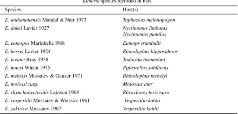
Of the remaining ten species (Table II), Eime-ria molossi n.sp.,is readily differentiated from E. andamanensis, E. hessei, E. levinei, E. mehelyi, E. rhynchonycteridis, E. vespertilii and E. zakirica,
which all have a smooth, unstriated oocyst wall, and from E. dukei which has a large oocystic re-siduum.
Morphologyof the oocyst of E. molossi n.sp., most closely approaches that of E. eumopos and
E. macyi, both of which have a roughish, striated oocyst wall. The oocysts of E. eumopos, how-ever, are substantially larger (35 x 28, range 34-36 x 27-28 versus 23 x 17, range 18-30 x 15-22), and the oocyst wall has only two layers. Mature meronts of E. molossi n.sp. are small and produce only from 8-12 merozoites, whereas Marinkelle de-scribed those of E. eumopos as measuring up to 98 x 62 (globidial schizogony?), with a thick wall and
TABLE I
Eimeria species recorded from chelonians
Species Recorded host(s) Shapea and mean measurements
and locality of oocysts (µm)
E. amydae Roundabush 1937 Apalone (=Amyda)) E. 21.8 x 14.6 b
spinifer pallidus (U.S.A) (18-24 x 14-16)
E. apalone McAllister et al. 1990 Apalone s. pallidus (U.S.A.) E-P-SS. 16.8 x 13.2 (12-19 x 10-16)
E. brodeni Cerruti 1930 Testudo graeca (Sardinia) O. 28-32 x 18-20
E. caretta Upton et al.1990 Caretta caretta (U.S.A.) SS-E. 24.5 x 22 (21.4- 28 x 18.4-24)
E. carinii Lainson et al. 1990 Geochelone denticulata (Brazil) S-SS. 19.2 x 18.6 (15-20 x 14-19)
E. carri Ernst & Forrester 1973 Terrapene c. carolina (U.S.A) SS. 16 x 15 (12-20 x 12-16)
E. chelydrae Ernst et al. 1969 Chelydra s. serpentina (U.S.A) S-SS-E. 15.2 x 14.4 (13-17 x 12-17)
E. chrysemydis Deeds & Jahn 1939 Chrysemys picta bellii Elongate P. 27.6 x 17 c
Trachemys scripta elegans, (23.8-30.4 x 14.5-18.5)
Graptemys caglei (U.S.A.)
E. cooteri McAllister & Upton 1988 Pseudemys texana Curved C-E. 25.9 x 10.9
(U.S.A.) (23-28 x 10-13)
E. delagei (Labbé 1893) Emys orbicularis P. 20-22 x 16-17 Reichenow 1921 (France)
E. dericksoni Roundabush 1937 Apalone (= Trionyx) spinifera SS. 14.5 x 12.9
hartwegi (U.S.A.) (12.3-16.7 x 10.6-15.8)
E. filamentifera Wacha & Chelydra s. serpentina O-E. 23.2 x 18.6
Christiansen 1979 (U.S.A.) (19-27 x 14.5-21)
E. graptemydos Wacha & Trachemys s. elegans SS. 12.6 x 11.4 Christiansen 1976 Graptemys caglei, (9.9-15.8 x 8.6-14.5)
Graptemys versa, Graptemys geographica, Kinosternon f. flavescens, Chrysemys p. bellii (U.S.A.)
E. harlani Upton et al. 1992 Macroclemys temminckii S-SS. 13 x 12.6
(U.S.A.) (10.4-14.4 x 10.4-13.8)
E. innominata Kar 1944 Lissemys punctata SS. 17.6 x 13.2
(India) (16.5-18.8 x 11.5-14.3)d
E. irregularis Kar 1944 Lissemys punctata S. 15.4
(India) (14.6-16.5) d
E. jaboti Carini 1942 Geochelone (=Testudo) S-SS. 17
tabulata (Brazil) (17-19 x 15-17)
E. juniataensis Pluto & Graptemys geographica S-SS. 14 x 13
Rothenbacher 1976 (U.S.A.) (12-19 x 12-17)
E. koormae Das Gupta 1938 Lissemys punctata S. 14
(India) (13.5-15.8) d
E. lagunculata Lainson et al. 1990 Podocnemis expansa E. 19.2 x 12.8
Podocnemis unifilise (17-20.7 x 11.8-14.1)
E. legeri (Simond 1901) Emyda granosa S. 16-18 Reichenow 1921 (India)
E. lutotestudinis Wacha & Trachemys s. elegans, E-SS. 11.9 x 10.8 Christiansen 1976 Graptemys caglei (9.8-13.2 x 9.3-11.8)
Kinosternon f. flavescens Pseudemys texana (U.S.A.)
E. mammiformis Lainson et al 1990 Podocnemis expansa E. 30 x 19.4
(Brazil) (23-37 x 16.3-21.5)
E. marginata (Deeds & Jahn 1939) Trachemys s. elegans, P. 22.1 x 17.6c Pellerdy 1974 Chrysemys p. bellii (18.5-25.1 x 15.8-19.8)
Graptemys geographica, G. pseudogeographica (U.S.A.)
E. mascoutini Wacha & Apalone s. hartwegi E-SS. 14 x 11.9
89 89 89 89 89 Mem Inst Oswaldo Cruz, Rio de Janeiro, Vol. 93(1), Jan./Feb. 1998 88
88 88 88
88 Eimeria peltocephali n. sp. R Lainson, RD Naiff
containing up to 350 merozoites. Finally, the oo-cysts of E. macyi are smaller than those of E. molossi n. sp., (19 x 17.6 versus 23 x 17), more inclined to a spherical shape (shape-index 1 ver-sus 1.3), and have a wall of only one layer. Fur-thermore, its sporocysts have a much more promi-nent Stieda body and possesses a very conspicu-ous sub-Stieda body, not seen in the sporocysts of
E. molossi.
ACKNOWLEDGEMENTS
To Constância Maia Franco and Walter M Campos, Instituto Evandro Chagas, Belém, for technical assis-tance; to Dr Suely Marques, Museu Paraense Emílio Goeldi, Belém, for the identification of bats and advice regarding chiropteran taxonomy; and to Dr WE Magnusson, Instituto Nacional de Pesquisas da Amazônia, Manaus, for identification of the turtles.
REFERENCES
Bray RS 1958. On the parasitic protozoa of Liberia. I. Coccidia of some small mammals. J Protozool 5:
81-83.
Carini A 1942. Sobre uma Eimeria da “Testudo tabulata”. Arq Biol (São Paulo) 26: 163-164. Cerruti A 1930. Su di un coccidio parassita di Testudo
graeca Linn. Arch Ital SciMed Col 11: 328-331.
Chakravarty M, Kar AB 1943. Observations on two coccidia, Eimeria trionyxae n.sp. and Eimeria tri-angularis n.sp., from the intestine of the turtle
Trionyx gangeticus Cuv. J Roy Asiat Soc Bengal Sci 9: 49-54.
Das Gupta M 1938. On a new coccidium, Eimeria koormae n.sp. from the intestine of the Indian tor-toise, Lissemys punctata Smith. Arch Protistenkd 90: 410-413.
Deeds OJ, Jahn TL 1939. Coccidian infections of
west-ern painted turtles of the Okoboji region. Trans Am Micr Soc 58: 249-253.
Doflein F 1909. Lehrbuch der Protozoenkunde, Verlag Gustav Fischer, Jena.
Ernst JV, Forrester DJ 1973. Eimeria carri sp.n. (Proto-zoa: Eimeriidae) from the box turtle Terrapene carolina. J Parasitol 59: 635-636.
Ernst JV, Stewart TB, Sampson JR, Fincher GT 1969.
Eimeria chelydrae n.sp (Protozoa: Eimeriidae) from the snapping turtle, Chelydra serpentina. Bull Wildl Dis Assoc 5: 410-411.
Ernst JV, Fincher GT, Stewart TB 1971. Eimeria paynei
sp.n. (Protozoa: Eimeriidae) from the gopher tortoise,
Gopherus polyphemus. Proc Helminthol Soc Washington 38: 223-224.
Gottschalk VC 1969. Kokzidien aus Thüringen und der Oberlausitz. Angew Parasitol 10: 229-232. Kar AB 1944. Two new coccidia from pond turtles,
Lissemys punctata (Bonnaterre). Indian Vet J 20:
231-235.
Labbé A 1893. Coccidium delagei coccidie nouvelle parasite des tortues d’eau douce. Arch Zool Exp Gen 1: 267-280.
Lainson R 1968a. Parasitological studies in British Hon-duras. IV. Some coccidial parasites of reptiles. Ann Trop MedParasitol 62: 260-266.
Lainson R 1968b. Parasitological studies in British Honduras III. Some coccidial parasites of mammals.
Ann Trop Med Parasitol 62: 252-259. Lainson R, Costa AM, Shaw JJ 1990. Eimeria species
(Apicomplexa: Eimeriidae) of Podocnemis expansa
(Schweigger) and Geochelone denticulata (Linn.) from Amazonian Brazil. Mem Inst Oswaldo Cruz 85: 383-390.
Laveran A, Mesnil F 1902. Sur quelques protozoaires parasites d’une tortue d’Asie (Damonia reevesii). C R Seances Acad Sci Ser 3 135: 609-614. Lavier G 1924. Eimeria hessei n.sp., coccidie parasite
Species Recorded host(s) Shapea and mean measurements
and locality of oocysts (µm)
E. megalostiedai Wacha & Clemmys insculpta SS. 14 x 13
Christiansen 1974 (U.S.A.) (12-16 x 10-15)
E. mitraria (Laveran & Mesnil 1902) Chinemys reevesii, E. 10 x 8 Doflein 1909 Kinosternon f. flavescens, (8-12 x 6-9)
Pseudemys texana, “mitre-shaped”with 3-4
Trachemys s. elegans protrusions at flat end
Chrysemys p. bellii, Chelydra s. serpentina Graptemys geographica, Graptemys versa,
Graptemys caglei
G. pseudogeographica,
Emydoidea blandingii
(Asia; U.S.A.)
E. ornata McAllister & Upton 1989 Terrapene o. ornata E. 17.9 x 15.7
(U.S.A.) (16-21 x 14-18)
E. pallidus McAllister et al. 1990 Apalone s. pallidus S-SS. 23.4 x 21.6
(U.S.A.) (18-27 x 17-25)
E. paynei Ernst et al. 1971 Gopherus polyphemus E. 23.2 x 18.6
(U.S.A.) (19-26 x 16-20)
E. peltocephali n.sp Peltocephalus dumerilianus elongate-C. 54.4 x 19.1 (37.5-68.7 x 18.7-20)
E. podocnemis Lainson et al. 1990 Podocnemis expansa E. 17 x 12.8
(Brazil) (14.8-19.2 x 11.8-14.1)
E. pseudemydis Lainson 1968 Pseudemys ornata, S-SS. 19 x 17.5
Pseudemys texana, (18-20 x 16.5-18.2) Trachemys s. elegans
(Belize, C. Am.; U.S.A.)
E. pseudographica Wacha & Trachemys s. elegans, O-E. 19.5 x 13.5 Christiansen 1976 Chrysemys p. bellii, (17.2-20.8 x 11.9-15.2)
Graptemys pseudographica,
Graptemys caglei (U.S.A.)
E. scriptae Sampson & Ernst 1969 Trachemys s. elegans O-E. 24.2 x 13.7
(U.S.A.) (22-27 x 12-16)
E. serpentina McAllister et al 1990 Chelydra s. serpentina E. 12.8 x 8.1
(U.S.A.) (11-15 x 7-10)
E. somervellensis McAllister Pseudemys texana, P. 21.2 x 16.1 & Upton 1992 P. concinna metteri (U.S.A.) (16.8-23.2 x 13.6-17.2)
E. spinifera McAllister et al 1990 Apalone s. pallidus E-P-SS. 16.3 x 14
(U.S.A.) (14-19 x 12-18)
E. stylosa McAllister & Upton 1989 Trachemys s. elegans O. 16.5 x 13.1
(U.S.A.) (14.4-17.6 x 12-14.4)
with conical projectionsat each end
E. tetradacrutata Wacha & Trachemys s. elegans S-SS. 19.5 x 19.2 Christiansen 1976 Chrysemys p. bellii, (16.6-23 x 16-21.8)
Pseudemys texana (U.S.A.)
E. texana McAllister & Upton 1988 Pseudemys texana curved-C. 20.5 x 8.4
(U.S.A.) (17.6-23.2 x 7.2-9)
E. trachemydis McAllister & Upton Trachemys s. elegans, E. 25 x 13.6 1988 Graptemys caglei (U.S.A.) (20.8-30.4 x 12-16)
E. triangularis Chakravarty & Kar Trionyx gangeticus Triangular, 1943 (India) 10.3-14.4 (longest side)
E. trionyxae Chakravarty & Kar 1943 Trionyx gangeticus S. 16.5
(India) (14.45-19.5)d
E. vesicostieda Wacha & Apalone s. hartwegi O-E. 23.4 x 18.6
Christiansen 1977 (U.S.A.) (22-25.5 x 16.5-20.5)
a: C: cylindrical; E: ellipsoidal; O: ovoid; P: pear-shaped; S: spherical; SS: subspherical. b: McAllister et. al.1990.
c:Wacha & Christianson 1976. d:Mandal 1976. e: Lainson (unpublished observations).
TABLE II
Eimeria species recorded in bats
Species Host(s)
E. andamanensis Mandal & Nair 1973 Taphozous melanopogon E. dukei Lavier 1927 Nyctinomus limbatus
90 90 90 90
90 Eimeria peltocephali n. sp. R Lainson, RD Naiff
intestinale de Rhinolophus hipposideros. Ann Parasitol 2: 335-339.
Lavier G 1927 Eimeria dukei n.sp., coccidie parasite intestinale de chéiroptère. C R Soc Biol 97: 1707-1709.
Mandal AK 1976. Coccidia of Indian vertebrates. Rec Zool Surv India 70: 39-120.
Mandal AK, Nair KN 1973. A new species of coc-cidium from Taphozous melanopogon Temminck (Mammalia: Chiroptera) from Andaman Islands.
Proc Ind Acad Sci Sect B 77: 243-246. Marinkelle CJ 1968. Eimeria eumopos n.sp. from a
Co-lombian bat Eumops trumbulli. J Protozool 15: 57-58.
McAllister CT, Upton SJ 1988. Eimeria trachemydis
n.sp. (Apicomplexa: Eimeriidae) and other eimerians from the red-eared slider, Trachemys scripta elegans
(Reptilia: Testudines), in northcentral Texas. J Parasitol 74: 1014-1017.
McAllister CT, Upton SJ 1989a. Eimeria ornata n.sp. (Apicomplexa: Eimeriidae) from the ornate box turtle, Terrapene ornata ornata (Reptilia: Testudines), in Texas. J Protozool 36: 131-133. McAllister CT, Upton SJ 1989b. The coccidia
(Apicomplexa: Eimeriidae) of testudines, with de-scriptions of three new species. Can J Zool 67: 2459-2467.
McAllister CT, Upton SJ 1992. A new species of Eime-ria (Apicomplexa:Eimeriidae) from Pseudemys texana (Testudines: Emydidae), from north-central Texas. Tex J Sci 44: 37-41.
McAllister CT, Upton SJ, McCaskill 1990a. Three new species of Eimeria (Apicomplexa: Eimeriidae) from Apalone spinifera pallidus (Testudines: Trionychidae) in Texas, with a redescription of E. amydae. J Parasitol 76: 481-486.
McAllister CT, Upton SJ, Trauth 1990b. Coccidian para-sites (Apicomplexa: Eimeriidae) of Chelydra serpentina (Testudines:Chelydridae) from Arkansas and Texas, U.S.A, with descriptions of Isospora chelydrae sp.nov. and Eimeria serpentina sp.nov.
Canad J Zool 68: 865-868.
Mussaiev MA 1967. [ A new species of coccidia, Eime-ria zakirica, from the Mediterranean bat Vespertilio kuhlii Kuhl] (In Russian). Izv Akad Nauk Azerb SSR Ser Biol Nauk No 5: 37-38.
Mussaiev MA, Gauzer ME 1971. Eimeria mehelyi -novyi vid koktsidii iz podkovonosa megeli (Rhinolophus mehelyi). Izv Akad Nauk Azerb SSR Ser Biol Nauk No 2: 94-96.
Mussaiev MA, Veisov AM 1961. [A new species of coccidium from the Mediterranean bat, Vespertilio kuhli Kuhl] (In Russian). Dok Akad Nauk Azerb SSR 17: 741-744.
Pellerdy LP 1974. Coccidia and Coccidiosis, 2nd ed., Paul Parey, Berlin and Hamburg, Akadémiai Kiadó, Budapest, 959 pp.
Pluto TG, Rothenbacher H 1976. Eimeria juniataensis
sp.n. (Protozoa: Eimeriidae) from the map turtle,
Graptemys geographica, in Pennsylvania. J Parasitol 62: 207-208.
Reichenow E 1921. Die Coccidian, p. 1136-1277. In SJM Prowazek Handbuch der Pathogenen Protozoen, JA Barth, Leipzig.
Roundabush RL 1937. Some coccidia of reptiles found in North America. J Parasitol 23: 354-359. Sampson JR, Ernst JV 1969. Eimeria scriptae n.sp.
(Sporozoa: Eimeriidae) from the red-eared turtle
Pseudemys scripta elegans. J Protozool 16: 444-445. Simond P-L 1901. Note sur une coccidie nouvelle, Coc-cidium legeri, parasite de Cryptopus granosus (Emyda granosa). C R Seances Soc Biol (Paris) 53:
485-486.
Upton SJ, McAllister CT, Trauth SE 1992. Description of a new species of Eimeria (Apicomplexa: Eimeri-idae) from the alligator snapping turtle, Macroclemys temminckii (Testudines: Chelydridae). J Helminthol
Soc Washington 59: 167-169.
Upton SJ, Odell DK, Walsh MT 1990. Eimeria caretta
sp. nov. (Apicomplexa:Eimeriidae) from the logger-head sea turtle, Caretta caretta (Testudines). Can J Zool 68: 1268-1269.
Wacha RS, Christiansen JL 1974. Eimeria megalostiedai
sp.n. (Protozoa: Sporozoa) from the wood turtle,
Clemmys insculpta, in Iowa. Proc Helminthol Soc Washington 41: 35-37.
Wacha RS, Christiansen JL 1976. Coccidian parasites from Iowa turtles: systematics and prevalence. J Protozool 23: 57-63.
Wacha RS, Christiansen JL 1977. Additional notes on the coccidian parasites of the soft-shell turtle, Trionyx spiniferus Le Sueur, in Iowa, with a description of
Eimeria vesicostieda sp.n. J Protozool 24: 357-359. Wacha RS, Christiansen J 1979. Eimeria filamentifera
sp.n. from the snapping turtle, Chelydra serpentina
(Linné), in Iowa. J Protozool 26: 353-354. Wheat BE 1975. Eimeria macyi sp.n. (Protozoa: