1 Dual antibiotherapy of tuberculosis mediated by inhalable locust bean gum 1
microparticles 2
Susana Rodrigues1,2§, Ana D Alves1§, Joana S Cavaco1, Jorge F Pontes1,2, Filipa
3
Guerreiro1,2, Ana M Rosa da Costa3, Francesca Buttini4and Ana Grenha1,2* 4
5
1Center for Biomedical Research (CBMR), Faculty of Sciences and Technology,
6
University of Algarve, Faro, Portugal; 2Centre for Marine Sciences (CCMar), 7
Faculty of Sciences and Technology, University of Algarve, Faro, Portugal; 8
3Algarve Chemistry Research Center (CIQA) and Department of Chemistry and
9
Pharmacy, Faculty of Sciences and Technology, University of Algarve, Faro, 10
Portugal; 4Department of Food and Drug Science, University of Parma, Parma, 11
Italy; §Authors with equal contribution 12 13 *Corresponding author: 14 University of Algarve 15
CBMR and CCMAR, Faculty of Sciences and Technology 16 Campus de Gambelas 17 8005-139 Faro, Portugal 18 Tel.: +351 289800100 – Ext. 7441 19 Fax: +351 289818419 20
E-mail address: [email protected] 21
22 23
2 Abstract
24
Despite the existence of effective oral therapy, tuberculosis remains a deadly 25
pathology, namely because of bacterial resistance and incompliance with 26
treatments. Establishing alternative therapeutic approaches is urgent and 27
inhalable therapy has a great potential in this regard. As pathogenic bacteria are 28
hosted by alveolar macrophages, the co-localisation of antitubercular drugs and 29
pathogens is thus potentiated by this strategy. This work proposes inhalable 30
therapy of pulmonary tuberculosis mediated by a single locust bean gum (LBG) 31
formulation of microparticles associating both isoniazid and rifabutin, 32
complying with requisites of the World Health Organisation of combined 33
therapy. Microparticles were produced by spray-drying, at LBG/INH/RFB mass 34
ratio of 10/1/0.5. The aerodynamic characterisation of microparticles revealed 35
emitted doses of more than 90% and fine particle fraction of 38%, thus 36
indicating the adequacy of the system to reach the respiratory lung area, thus 37
partially the alveolar region. Cytotoxicity results indicate moderate toxicity (cell 38
viability around 60%), with a concentration-dependent effect. Additionally, rat 39
alveolar macrophages evidenced preferential capture of LBG microparticles, 40
possibly due to chemical composition comprising mannose and galactose units 41
that are specifically recognised by macrophage surface receptors. 42
43
Keywords: inhalation, locust bean gum, microparticles, polysaccharides, spray-44
drying, tuberculosis therapy 45
3 1. Introduction
47
Tuberculosis (TB) remains a leading cause of death, with particular incidence 48
and prevalence in developing countries (WHO, 2016 ). Drug resistance is a 49
major problem, but therapeutic incompliance is also a great limitation (McBryde 50
et al., 2017). Albeit the commercial availability of several effective oral and 51
parenteral drugs and the existence of international treatment guidelines (NICE, 52
2016; Wells et al., 2009), new therapeutic approaches are demanded not only to 53
improve compliance but also to reduce the severe side effects associated with 54
conventional therapy (Kaur et al., 2016; Lee et al., 2015). Inhalable therapy has 55
great potential in this context, enabling the direct administration of drugs to the 56
alveoli, where macrophages hosting pathogenic bacteria are located (Gupta et 57
al., 2016; Parumasivam et al., 2016). 58
In a previous work, we proposed the use of spray-dried microparticles based on 59
locust bean gum (LBG) as inhalable formulation for the treatment of pulmonary 60
tuberculosis. The individual association of first-line antitubercular drugs was 61
effective and a preliminary assay further suggested high affinity of LBG 62
microparticles for macrophages (Alves et al., 2016), which we explained by 63
specific recognition of the mannose and galactose residues of microparticles by 64
macrophage surface receptors (Ahsan et al., 2002). LBG was proposed in that 65
work for the first time for lung delivery applications. It is a galactomannan and 66
its biodegradability has been ascribed to the presence of β-mannosidase in the 67
lung (Alkhayat et al., 1998). 68
This work intends to produce a dry powder that delivers two antitubercular drugs 69
upon inhalation while providing improved internalisation by macrophages, 70
mediated by LBG. Furthermore, the respirability of LBG microparticles was 71
4 studied in order to determine their inhalability when used as carriers for 72
antitubercular drugs. The microparticle aerosolisation profile was experimentally 73
determined. The uptake by macrophages was assessed and compared with that of 74
microparticles composed by a polymer devoid of specific moieties recognizable 75
by macrophage receptors. Finally, combined therapy is proposed by the 76
association of two first-line antitubercular drugs (isoniazid and rifabutin) in a 77
single microparticle formulation, to meet the World Health Organisation (WHO) 78
requirements of combined tuberculosis therapy (WHO, 2014). 79
80
2. Materials and Methods 81
2.1. Materials 82
Locust bean gum (LBG, C30H50O26, Mw 860 kDa (Pollard et al., 2008)), poly
83
(vinyl alcohol) (PVA, [-CH2CHOH-]n, Mw 89-98 g/mol), isoniazid (INH,
84
C6H7N3O, Mw 137.14 g/mol), Tween 80®, phosphate buffer saline (PBS) tablets
85
pH 7.4, Dulbecco’s modified Eagle’s medium (DMEM), L-glutamine solution 86
(200 mM), non-essential amino acids solution and penicillin/streptomycin 87
(10000 units/mL, 10000 g/mL), trypsin-EDTA solution (2.5 g/L trypsin, 0.5 g/L 88
EDTA), trypan blue solution (0.4%), phorbol 12-myristate 13-acetate (PMA, 89
C36H56O8), thiazolyl blue tetrazolium bromide (MTT),
N-(3-90
dimethylaminopropyl)-N′-ethylcarbodiimide hydrochloride (EDAC), Triton-X 91
100, sodium dodecyl sulfate (SDS), dimethylformamide (DMF), dimethyl 92
sulfoxide (DMSO) and HCl were purchased from Sigma-Aldrich (Germany). 93
Lactate dehydrogenase (LDH) kit was purchased from Takara Bio (Tokyo, 94
Japan). Rifabutin (RFB, C46H62N4O11, Mw 847.00 g/mol) was supplied by
95
Chemos (Germany) and fetal bovine serum (FBS) by Gibco (Life Technologies, 96
5 USA). RPMI 1640 and Ham’s F12 media were obtained from Lonza Group AG 97
(Switzerland). Ultrapure water (MilliQ, Millipore, UK) was used throughout. All 98
other chemicals were reagent grade. 99
100
2.2. Cell lines 101
A549 cells (human alveolar epithelium) and NR8383 cells (rat alveolar 102
macrophages) were obtained from the American Type Culture Collection 103
(ATCC, USA) and used in passages 27-37 and 9-18, respectively. THP-1 cells 104
(human monocytes) were obtained from the Leibniz-Institut DSMZ (Germany) 105
and used in passages 10-20. Cell cultures were grown in humidified 5% 106
CO2/95% atmospheric air incubator at 37 ºC (HerAcell 150, Heraeus, 107
Germany). Cell culture medium (CCM) for A549 cells was DMEM 108
supplemented with 10% (v/v) FBS, 1% (v/v) L-glutamine solution, 1% (v/v) 109
non-essential amino acids solution and 1% (v/v) penicillin/streptomycin. For 110
NR8383 cells, CCM consisted of Ham’s F12 supplemented with 15% (v/v) FBS, 111
1% (v/v) L-glutamine and 1% (v/v) penicillin/streptomycin, while THP-1 cells 112
were grown in RPMI 1640 medium supplemented with 10% (v/v) FBS, 1% (v/v) 113
L-glutamine and 1% (v/v) penicillin/streptomycin. 114
THP-1 cells were grown in suspension and cell culture was maintained between 115
0.2 x 10⁶ and 0.8 x 10⁶ cells/mL. When reaching this higher concentration, cells
116
were sub cultivated in new passage at the concentration of 0.2 x 10⁶ cells/mL. 117
Differentiation of THP-1 monocytes to provide the macrophage phenotype was 118
performed using PMA (0.2 x 10⁶ cells/mL, 50 nM, 48 hours exposure), after 119
which the medium was replaced by fresh medium without PMA for 24 hours 120
before the experiments. NR8383 cells grow in mixed culture (half population 121
6 keeps adherent and half suspended). Adherent cells were those used to perform 122
the assays described below and their harvesting was made by scraping. 123
124
2.3. Preparation of microparticles 125
LBG microparticles, without drug and containing an association of the 126
antitubercular drugs INH and RFB, were prepared by spray-drying, according to 127
a previously reported protocol (Alves et al., 2016). The preparation of unloaded 128
LBG microparticles involved grinding LBG in a glass mortar for 10 min, after 129
which 5 mL HCl 0.1 M were slowly added and grinding continued until 130
complete mixture of powder and HCl solution was obtained. This was followed 131
by the addition of purified water previously heated to 85 ºC, up to a final volume 132
of 50 mL. The concentration of LBG in the final solution was 2% (w/v). The 133
solution was maintained under magnetic stirring for 30 min and subsequently 134
placed on a water bath at 85 ºC under slow stirring for additional 30 min. At the 135
end, the solution was kept under stirring at room temperature overnight, until the 136
moment of spray-drying. 137
For the production of drug-loaded microparticles, a solid dispersion of LBG and 138
RFB was first prepared, by trituration in a mortar. After grinding, the same 139
procedure used to prepare the LBG solution described above was applied. In 140
parallel, INH was triturated in a mortar, being then solubilized with purified 141
water under mild stirring for 10 minutes. The resulting solution was slowly 142
added to the previously formed LBG/RFB solution immediately before spray-143
drying. 144
Microparticles were produced at LBG/INH/RFB mass ratio of 10/1/0.5 (final 145
concentration of solids is 2.3%, w/v) using a laboratory mini spray dryer (Büchi 146
7 B-290, Büchi Labortechnik AG, Switzerland) operating in open mode and 147
equipped with a high-performance cyclone. Protection from light was ensured 148
for the whole process. The operating parameters were: inlet temperature: 160 ± 2 149
ºC; aspirator setting: 85%; feed rate: 0.7 ± 0.1 mL/min; and spray flow rate: 473 150
L/h. These conditions resulted in outlet temperature of 100 ± 2 ºC. After spray-151
drying, microparticles were collected, placed in a dark flask and stored inside a 152
desiccator until further use. 153
The spray-drying yield was calculated by gravimetry, comparing the total 154
amount of solids initially added with the resultant weight of microspheres after 155
spray-drying (Grenha et al., 2005). 156
Fluorescent (unloaded) microparticles of LBG or PVA were also prepared, to be 157
used in the assay of macrophage capture. Covalent binding between fluorescein 158
and the polymeric molecules was performed before microparticle production. To 159
do so, LBG or PVA (1.0 g) were dissolved in HCl solution (10-4 M) at a 160
concentration of 1% (w/v). LBG solution was applied the same treatment 161
described above for the preparation of LBG microparticles. PVA solution was 162
maintained at 80 ºC under stirring overnight. Fluorescein (43 mg) was dissolved 163
in ethanol 96% (v/v) and added to the previously formed LBG or PVA solutions. 164
EDAC (ca. 33 mg) was dissolved in milli-Q water and added to the solution. 165
This was kept under stirring for 72 h and then dialysed (2000 Da Mw cut off) 166
against water. Light protection was ensured in the whole process. The resulting 167
suspension was frozen and freeze-dried (FreeZone Benchtop Freeze Dry System, 168
Labconco, USA). The fluorescently-labelled polymers were stored in a 169
desiccator until further use, under light protection. Fluorescent microparticles 170
were produced by spray-drying. The used conditions were the same described 171
8 above for LBG, while the following were used for PVA: inlet temperature: 155 ± 172
2 ºC; aspirator setting: 80%; feed rate: 1.0 ± 0.1 mL/min; and spray flow rate: 173
473 L/h. These conditions resulted in outlet temperature of 96 ± 2 ºC. 174 175 2.4. Characterisation of microparticles 176 2.4.1. Morphology 177
The surface morphology of LBG microparticles was characterised by field 178
emission scanning electron microscopy (FESEM; FESEM Ultra Plus, Zeiss, 179
Germany). Dry powders were placed onto metal plates and 5 nm thick iridium 180
film was sputter-coated (model Q150T S/E/ES, Quorum Technologies, UK) on 181
the samples before viewing. 182
183
2.4.2. Feret’s diameter
184
Microparticle size was estimated as the Feret’s diameter and was directly 185
determined by optical microscopy (Microscope TR 500, VWR international, 186
Belgium) from the manual measurement of 300 microparticles (n = 3). 187
188
2.4.3. Density
189
Real density (g/cm3) was determined using a Helium Pycnometer (Micromeritics
190
AccuPyc 1330, Germany) (n = 3). Tap density (g/cm3) was determined using a 191
tap density tester (Densipro 250410, Deyman, Spain), by measuring the volume 192
of a known weight of powder before and after tapping, respectively (n = 3). The 193
determination of tap density involved tapping the sample until no further 194
reduction of powder volume was observed (average of 180 taps). 195
9
2.4.4. Drug association efficiency and loading
197
The determination of microparticle drug content was performed by UV-Vis 198
spectrophotometry (Pharmaspec UV-1700, Shimadazu, Japan), at 265.5 nm for 199
INH and 500 nm for RFB. A screening of the matrix material (LBG) revealed no 200
interference at the selected wavelengths. 201
In order to determine the drug content, a certain amount of drug-loaded 202
microparticles was incubated with HCl 0.1 M, under magnetic stirring for 60 203
min, which ensures complete dissolution of the carriers. Samples were then 204
centrifuged (8000 rpm, 30 min; 5810 R, Eppendorf, Germany) and filtered (0.45 205
µm) before quantification. Calibration curves were performed at 265.5 nm for 206
INH and at both this wavelength and at 500 nm for RFB, using the medium of 207
dissolution of unloaded microparticles. The latter curve was used to determine 208
the concentration of RFB and the former allowed the determination of the 209
fraction of the absorbance at 265.5 nm that is due to RFB, the remainder being 210
attributable to INH, thus allowing the determination of its concentration through 211
the corresponding calibration curve. Drug association efficiency (AE) and 212
microparticle (MP) loading capacity (LC) were estimated as follows (n = 3): 213
AE (%) = (Real amount of drug on MP / Theoretical amount of drug on MP) x 214
100 (Eq. 1) 215
LC (%) = (Real amount of drug on MP/ Weight of MP) x 100 (Eq. 2) 216
217
2.5. Aerodynamic characterisation of microparticles 218
HPMC size 3 capsules (Quali-V-I, Qualicaps, Spain) were filled with 22.5 mg of 219
LBG/INH/RFB powder. The content of four capsules was discharged in each 220
aerodynamic test using the medium resistance RS01® inhaler (Plastiape Spa,
10 Italy) and experiments were performed in triplicate. The device was connected to 222
the Andersen cascade impactor (ACI, Copley Scientific, UK) operated at 223
60 L/min, ensuring a pressure drop of 4 kPa through the device. This was 224
activated for 4 s in order to let 4 L of air passing through the system, thus 225
complying with the standard procedure described by USP 38 and Ph.Eur.8 226
(Ph.Eur., 2014; USP, 2015). 227
ACI separates particles according to their aerodynamic diameter and it was 228
assembled using the appropriate adaptor kit for the 60 L/min air flow test. Cut-229
offs of the stages from -1 to 6 are the following: 8.60, 6.50, 4.40, 3.20, 1.90, 230
1.20, 055, 0.26 µm. A glass fiber filter (Whatman, Italy) was placed right below 231
stage 6 in order to collect particles with diameter lower than that of stage 6 cut-232
off. 233
The plates of the impactor were coated with a thin layer of ethanol containing 234
1% (w/v) Tween 20to prevent particle bounce. The drugs were recovered from 235
the apparatus with water/acetonitrile mixture (50/50, v/v) and quantified by 236
HPLC (Agilent 1200 series, Germany). A LiChrospher® 100 RP-18 (5 µm) 237
column of 4 mm i.d.×250 mm length with security guard cartridge was used. 238
The eluent was a mixture of phosphate buffer pH = 7 (A) and acetonitrile (B) at 239
a flow rate of 1 mL/min, starting with A/B = 95:5 and kept for 5 min, followed 240
by a 7 min step gradient, until a 20:80 A/B ratio was reached, which was then 241
kept for 16 min. Detection was performed by a diode array detector at 275 nm. 242
The used chromatographic conditions were: gradient flow (Phosphate buffer pH 243
= 7 (A), acetonitrile (B); A/B from 95:5 in the first 5 min it is changed to 20:80 244
in 7 min and kept in this 20:80 during other 16 min). A linear calibration plot for 245
INH and RFB was obtained over the range 10-400 μg/mL (n= 3). Under these 246
11 conditions, the retention times of INH and RFB were 4.7 and 17.4 min, 247
respectively. 248
The quantification of drug deposited inside the impactor allows calculating 249
different aerodynamic parameters. The emitted dose (ED) is the amount of drug 250
ex-device, considered the total amount of drug collected in the impactor, 251
quantified by HPLC (induction port, stages -1 to 6 and filter). The mass median 252
aerodynamic diameter (MMAD) was determined by plotting the cumulative 253
percentage of mass less than the stated aerodynamic diameter on probability 254
scale versus aerodynamic diameter on logarithmic scale. The fine particle dose 255
(FPD) corresponds to the mass of drug particles with aerodynamic diameter 256
lower than 5 µm calculated using the particle size distribution equation obtained 257
from the ACI analysis. The fine particle fraction (FPF) is the ratio between the 258
FPDs and the MD. 259
The drug recovery ranged between 77-91% in all the experiments, being thus 260
coincident with the requisites of the pharmacopeia (Buttini et al., 2013). 261
262
2.6. Determination of drug release profile 263
The determination of drug release was performed in PBS pH 7.4 added of 1% 264
(v/v) Tween 80®. The assay respected sink conditions, as the maximum amount
265
of drug was always below 30% of its maximum solubility (EMA, 2014). INH 266
solubility was considered 274 ± 4.79 mg/mL (Hiremath and Saha, 2008), while 267
that of RFB was determined experimentally to be 0.496 mg/mL (Alves et al., 268
2016). A determined amount of microparticles (20 mg) was incubated with the 269
medium (10 mL), at 37 °C, under mild shaking (100 rpm, orbital shaker OS 20, 270
Biosan, Latvia). Samples (1 mL) were periodically collected and the amount of 271
12 each drug quantified as indicated above (n = 3). Direct quantification applied for 272
RFB, while INH required dilution (1:10). A calibration curve was performed 273
using the medium resulting from the incubation of unloaded LBG 274
microparticles, after centrifugation (8000 rpm, 60 min) and filtration (0.45 μm). 275
276
2.7. In Vitro Biocompatibility Study 277
2.7.1. Cell viability evaluation by MTT test 278
The evaluation of cell viability upon exposure to LBG/INH/RFB microparticles 279
was performed by the MTT assay, using two cell lines of high relevance within 280
the scope of tuberculosis infection, A549 and macrophage-differentiated THP-1 281
cells. A549 cells were seeded at a density of 1 x 10⁴ cells/well on 96-well plates 282
(Orange Scientific, Belgium), in 100 µL of complete DMEM. Cells were 283
incubated for 24 h at 37 ºC in 5% CO2 atmosphere before use. THP-1 cells were
284
differentiated with PMA to obtain the macrophage-phenotype before the 285
experiments, according to the procedure described above, with the necessary 286
adaptations. THP-1 cells were seeded on 96-well plates (0.035 x 10⁶ cells/well) 287
in 100 µL of RPMI supplemented with 50 nM of PMA and incubated for 48 h at 288
37 ºC in 5% CO2 atmosphere. After that time, CCM was renewed for other 24 h,
289
before the experiments. 290
Microparticles (unloaded or drug-loaded) were exposed in the form of a 291
suspension prepared in pre-warmed CCM without FBS and evaluated at the 292
concentrations of 0.1 and 0.5 mg/mL (includes polymers and drugs), for 3 and 293
24 h. INH and RFB were also tested as free drugs, at concentrations representing 294
their loading in microparticles (0.01 and 0.05 mg/mL for INH; 0.005 and 0.025 295
mg/mL for RFB). 296
13 MTT solution (0.5 mg/mL in PBS, pH 7.4) was added after the exposure time (in 297
A549 cells, samples were previously removed; in THP-1 cells no removal was 298
applied) and incubated for 2 h, after which formazan crystals were solubilised 299
with DMSO (A549 cells) or 10% SDS in a 1:1 mixture of DMF:water (THP-1 300
cells) and the absorbance measured by spectrophotometry (Infinite M200, 301
Tecan, Austria). The viability of untreated cells was assumed to correspond to 302
100% of cell viability, and viability of treated cells was compared to this control. 303
The assay was replicated at least three times, each with six replicates. 304
305
2.7.2. Determination of cell membrane integrity 306
The integrity of cell membrane after exposure to LBG microparticles was 307
determined by the quantification of LDH released by cells. This was performed 308
in A549 and macrophage-differentiated THP-1 cells upon 24 h exposure to a 309
concentration of 0.5 mg/mL of unloaded or drug-loaded LBG microparticles. 310
The chosen concentration corresponds to the maximum concentration tested in 311
the MTT assay. Free INH and RFB were also tested as controls (0.05 mg/mL for 312
INH; 0.025 mg/mL for RFB). 313
Cells were cultured in 96-well plates in the conditions described before for the 314
MTT assay (the assays were performed simultaneously). Upon exposure, cell 315
culture supernatants were collected, centrifuged (16 000 x g, 5 min, 4 ºC) and 316
processed using a commercial kit. Absorbances were measured by 317
spectrophotometry (Infinite M200, Tecan, Austria) at a wavelength of 490 nm 318
(background correction at 690 nm). 319
A negative control of LDH release was performed incubating cells with CCM 320
only and Triton-X 100 (10%) was used as positive control, being assumed as 321
14 100% LDH release. Released LDH upon incubation with each sample was 322
determined by comparison with the positive control. All measurements were 323
performed in triplicate. 324
325
2.8. Macrophage capture of LBG microparticles 326
The determination of macrophage ability to capture microparticles was 327
determined in vitro in two cell lines, differentiated THP-1 cells and NR8383 328
cells. The latter were seeded (1.0 x 10⁶ cells per well) in 6-well plates for 329
adhered cells, with 2 mL of Ham’s F12 medium. This procedure was performed 330
24 h before the test to ensure the adhesion of 50 to 75% of the original 331
population. THP-1 cells (2.0 x 10⁵ cells/mL) were suspended in RPMI medium 332
supplemented with 50 nM PMA and seeded at 5 mL/well in 6-well plates for 333
cells in suspension. 334
The evaluation of microparticle uptake by macrophages was performed by flow 335
cytometry (FacScalibur cell analyser, BD Biosciences, Belgium) upon exposure 336
to LBG and PVA microparticles (50 µg/cm2), both fluorescently-labelled. PVA 337
microparticles were used as control. Microparticles were aerosolised onto the 338
macrophage layer using the Dry powder Insufflator™ (Model DP-4, Penn-339
CenturyTM, USA) and 2 hours incubation at 37 ºC was allowed, without CCM 340
(only a residual amount of medium was kept to ensure the hydration of cell 341
surface). The phagocytic process was stopped by the addition of a cold solution 342
of PBS.3% FBS (5 mL, two applications), which also provided washing. Cells 343
were scraped and centrifuged (1500 rpm, 2 min, room temperature, centrifuge 344
MPW – 223e, MedInstruments, Poland) in 2 mL of PBS.3% FBS. The cycle of 345
resuspension in PBS.3% FBS and centrifugation was repeated thrice. At the end, 346
15 cells were re-suspended in 1 mL of PBS.3% FBS, transferred to cytometry tubes 347
(BD Biosciences, Belgium) and maintained at 4 ºC until the analysis. 348
In flow cytometry, FSC-H and SSC-H channels were used, respectively, to 349
measure size and granularity of cells, while side scatter light was used to identify 350
cell viable population. The amount of cells exhibiting a fluorescent signal was 351
considered to have phagocytosed microparticles. The assay for each dose was 352
replicated at least three times. 353
354
2.9. Statistical analysis 355
The student t-test and the one-way analysis of variance (ANOVA) with the 356
pairwise multiple comparison procedures (Holm-Sidak method) were performed 357
to compare two or multiple groups. For the analysis of results of in vitro release 358
assay, a two-way ANOVA with Bonferroni’s method for multiple comparison 359
test was used. All analysis were run using the GraphPad Prism (version 6.07) 360
and differences were considered to be significant at a level of p < 0.05. 361
362
3. Results and Discussion 363
3.1. Preparation and characterisation of LBG/INH/RFB microparticles 364
LBG microparticles loaded with a combination of the first-line antitubercular 365
drugs INH and RFB were successfully obtained by spray-drying, with yields of 366
60-70%. The used concentration of LBG (2%, w/v) was chosen from previous 367
experiments (unpublished data) in order to provide suitable microparticle size 368
for the purpose of lung delivery, along with acceptable spray-drying yield. 369
Theoretical drug loadings of 8.70% (INH) and 4.35% (RFB) were selected 370
because they provide microparticles without the formation of aggregates, in 371
16 micron-size range, which are capable to deliver the two drugs in combination. 372
LGB was selected because of its potential to enhance macrophage uptake and the 373
amount of polymer was kept purposely high in order to favour internalisation. 374
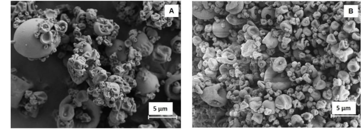
The morphological observation of LBG-based microparticles, unloaded or drug-375
loaded, revealed irregular shapes with convoluted surface (Figure 1), without 376
any evident effect resulting from drug association. The latter was actually 377
expected because loadings are relatively low. Similar observations on the 378
morphology and the absence of effect of drug association were reported in a 379
previous work with LBG microparticles associating either INH or RFB 380
separately (Alves et al., 2016). 381
382
Figure 1. Microphotographs of LBG-based microparticles viewed by scanning 383
electron microscopy: A) Unloaded LBG microparticles; B) LBG/INH/RFB 384
(10/1/0.5, w/w) microparticles. INH: isoniazid, LBG: locust bean gum, RFB: 385
rifabutin. 386
387
The determined Feret’s diameters were rather low, 1.35 ± 0.7 μm for unloaded 388
LBG microparticles and 1.14 ± 0.51 μm for drug-loaded microparticles. Real 389
densities were around 1.4 g/cm3 and tap densities varied between 0.2 and 0.37
390
g/cm3. These values are in the same range of others reported for spray-dried 391
17 polysaccharide microparticles (Dalpiaz et al., 2015; Pai et al., 2015; Rassu et al., 392
2015). 393
The encapsulation of both antibiotics was very high, 94% for INH and 102% for 394
RFB, as indicated in Table 1. The resulting loading capacities were, thus, close 395
to theoretical maximum, being of 8.2% and 4.4% for INH and RFB, 396
respectively. Spray-drying is a method usually reported to lead to high efficiency 397
of drug association (Peltonen et al., 2010), as also corroborated in the present 398
work. Moreover, the obtained results are in line with the high association 399
efficiencies reported for each drug when associated individually (Alves et al., 400
2016). 401
Table 1. Drug association efficiency and loading capacity of 402
LBG/INH/RFB (10/1/0.5, w/w) microparticles (mean ± SD, n = 3). 403
Drug Association efficiency (%) Loading capacity (%)
INH 94.4 ± 3.3 8.2 ± 0.3
RFB 102.1 ± 1.1 4.4 ± 0.1
INH: isoniazid; RFB: rifabutin 404
405
3.2. Aerodynamic behaviour of LBG/INH/RFB microparticles 406
Considering the intended application in lung delivery, the determination of 407
aerosolisation properties stands as the most important aspect in the design of 408
inhalable dry powders. The in vitro aerosol performance was determined using a 409
RS01® dry powder inhaler and the aerodynamic properties determined upon
410
assessment in the ACI are displayed in Table 2. 411
412 413
18 Table 2. Aerodynamic characteristics of LBG/INH/RFB (10/1/0.5, w/w) 414
microparticles (mean ± SD, n = 3). Loaded powder amount = 22.5 mg, 415
containing 7.8 mg INH and 3.9 mg RFB, respectively. 416
Drug Emitted dose (%)
MMAD (μm) GSD (μm) FPD (mg) FPF (%)
INH 92.6 ± 0.9 6.2 ± 0.6 2.4 ± 0.7 2.7 ± 0.6 38.0 ± 1.6 RFB 92.0 ± 0.5 5.8 ± 0.3 2.8 ± 0.2 1.3 ± 0.1 38.1 ± 1.8 FPD: fine particle dose; FPF: fine particle fraction; GSD: geometric standard deviation; INH: isoniazid; 417
MMAD: mass median aerodynamic diameter; RFB: rifabutin 418
419
The dose emitted from the inhaler was very satisfactory, reaching 92%. This is 420
indicative of the favourable properties of the material LBG to produce 421
microparticles by spray-drying with good flowing capacity. However, a 422
consistent amount of powder impacted on the high stages of the impactor, 423
leading to MMAD value equal to 5.8 and 6.2 μm for RFB and INH, respectively. 424
This is due to incomplete deaggregation of microparticle clusters during the 425
product aerosolisation. It is well known that the aerodynamic performance of a 426
dry powder inhaler (DPI) is strongly affected by both the device and the 427
formulation characteristics. However, the spinning movement of the capsule 428
inside the inhaler used in this study has demonstrated to be the most efficient in 429
powder deaggregation in comparison with other capsule-based DPI (Martinelli et 430
al., 2015). Hence, the optimisation of microparticles, with size, shape and 431
density promoting their aerodynamic behavior will be addressed in the future, in 432
order to increase the amount of LBG/INH/RFB fine particles capable of reaching 433
the target site of alveoli. Nevertheless, LBG/INH/RFB microparticles showed a 434
FPF of 38%, indicating that 38% of the microparticles have aerodynamic 435
diameter below 5 μm, thus having the necessary conditions to reach the 436
respiratory zone. This value is in agreement with those usually determined for 437
19 high doses antibiotic powder formulated without lactose as carrier (Belotti et al., 438
2015; Maretti et al., 2016). 439
Figure 2 shows the stage-by-stage deposition profiles of both drugs encapsulated 440
in the tested microparticles. The similarity of the profiles indicates that the two 441
drugs were equally co-deposited on the different stages. This supports the 442
decision of developing a carrier with drug combination, as the microparticles 443
demonstrate to have homogeneous composition, leading to a co-deposition of 444
drugs. 445
446
Figure 2. Stage-by-stage deposition profiles of isoniazid and rifabutin inside the 447
Andersen cascade impactor after RS01 aerosolisation at 60 L/min, inhalation 4L 448
(values are mean ±SD, n = 3). 449
450
3.3. In vitro drug release from LBG microparticles 451
Release studies were performed in PBS pH 7.4 added of 1% Tween 80. In this 452
way, the local pH of the alveolar zone is resembled, along with the content of 453
surfactant (Kyle et al., 1990), and the dissolution of RFB is ensured (Alves et al., 454
2016). The release profile determined for each drug is depicted in Figure 3. 455 0 5 10 15 20 25 Drug d ep os ition (% ) INH RFB
20 456 457 458 459 460 461 462 463 464
Figure 3. In vitro release of isoniazid (INH) and rifabutin (RFB) from 465
LBG/INH/RFB (10/1/0.5, w/w) microparticles, in PBS pH 7.4 - 1% Tween 80 466
at 37 ºC (LBG: locust bean gum; mean ± SD, n = 3). *p < 0.05 comparing 467
release of two drugs. 468
469
As can be observed, the release of the drugs is rapid, at 30 min 40% (RFB) – 470
50% (INH) of the antibiotics being already available. At 60 min, the values 471
reach 73% for RFB and 84% for INH. Although the profile is very similar for 472
both antibiotics, RFB release is somewhat slower than that of INH, with 473
statistically significant differences at some time-points (30, 90 and 120 min, p < 474
0.05). The higher release of INH is a consequence of its higher solubility in 475
aqueous media (O'Neil, 2006). Considering the conditions of the assay, the rapid 476
release was expected, as LBG is a hydrophilic polymer and rapidly dissolved, 477
releasing the associated drugs. Despite this observation, slower drug release is 478
expected to occur in vivo, as has been reported (Bur et al., 2010; Haghi et al., 479
2014). When reaching the alveoli, microparticles will deposit on an epithelium 480
*
* *
21 covered by the lung lining fluid (Fröhlich et al., 2016), which is estimated to 481
have 0.01 – 0.1 μm. In this manner, deposited particles will not be immersed, 482
only a small part being in direct contact with fluid instead and, therefore, erosion 483
and dissolution will initiate from underneath the microparticles (Bur et al., 2010; 484
Haghi et al., 2014). Ultimately, this will result in slower release and is also a 485
relevant observation towards the objective of having particle uptake by 486
macrophages before complete dissolution and drug release. 487
488
3.4. In vitro cytotoxicity of LBG microparticles 489
Two complementary cell viability assays were used to test the effect of 490
LBG/INH/RFB microparticles, the metabolic assay MTT and the LDH release 491
assay, which assesses cell membrane integrity. Considering the environment 492
underlying tuberculosis pathogenesis, alveolar epithelial cells (A549) and 493
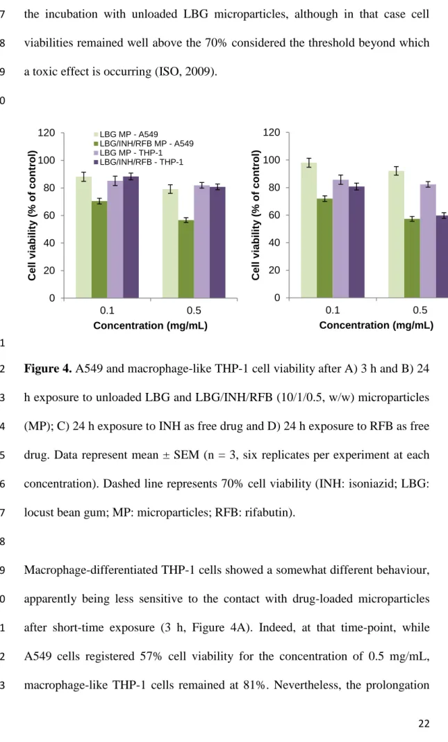
macrophage-like cells (macrophage-differentiated THP-1 cells) were used. 494
Microparticle concentrations of 0.1 and 0.5 mg/mL were tested. These are 495
concentrations typically reported in the assessment of lung drug carriers, despite 496
being possibly overestimated if an alveolar area of 100 m2 is considered 497
(Fröhlich et al., 2016). Unloaded microparticles and free drugs were tested as 498
controls. 499
Regarding the MTT assay, as can be observed in Figure 4A and 4B, the 500
exposure of A549 cells to drug-loaded microparticles induced very similar 501
results after 3 h or 24 h, evidencing an absence of time-dependent effect. 502
However, there is a clear concentration-dependent effect (p < 0.05), visible at 503
both time-points, as the resulting cell viability decreases from 72% to 57% (at 24 504
h) as the concentration of microparticles increases from 0.1 to 0.5 mg/mL. 505
22 Similar observations regarding the effect of time and concentration resulted from 506
the incubation with unloaded LBG microparticles, although in that case cell 507
viabilities remained well above the 70% considered the threshold beyond which 508
a toxic effect is occurring (ISO, 2009). 509
510
511
Figure 4. A549 and macrophage-like THP-1 cell viability after A) 3 h and B) 24 512
h exposure to unloaded LBG and LBG/INH/RFB (10/1/0.5, w/w) microparticles 513
(MP); C) 24 h exposure to INH as free drug and D) 24 h exposure to RFB as free 514
drug. Data represent mean ± SEM (n = 3, six replicates per experiment at each 515
concentration). Dashed line represents 70% cell viability (INH: isoniazid; LBG: 516
locust bean gum; MP: microparticles; RFB: rifabutin). 517
518
Macrophage-differentiated THP-1 cells showed a somewhat different behaviour, 519
apparently being less sensitive to the contact with drug-loaded microparticles 520
after short-time exposure (3 h, Figure 4A). Indeed, at that time-point, while 521
A549 cells registered 57% cell viability for the concentration of 0.5 mg/mL, 522
macrophage-like THP-1 cells remained at 81%. Nevertheless, the prolongation 523 0 20 40 60 80 100 120 0.1 0.5 Cell v iability ( % o f con tr o l) Concentration (mg/mL) LBG MP - A549 LBG/INH/RFB MP - A549 LBG MP - THP-1 LBG/INH/RFB - THP-1 0 20 40 60 80 100 120 0.1 0.5 Cell v iability ( % o f con tr o l) Concentration (mg/mL)
23 of the exposure to 24 h (Figure 4B) decreased cell viability to 60%, which is 524
similar to the 57% registered for A549 cells. 525
The results obtained on both cell lines are comparable to those reported for RFB-526
loaded LBG microparticles in a previous work and the opposite of those 527
obtained for INH-loaded microparticles (Alves et al., 2016). This suggests that 528
the negative effect of drug-loaded microparticles on cell viability is certainly 529
due, at least in part, to the RFB content. The pH of microparticle suspension in 530
cell culture medium that is incubated with cells is around 7.2, so that is not 531
expected to have a negative contribution. As shown in Figure 4C, INH has no 532
effect on cell viability in any of the tested conditions (concentrations, cell lines) 533
at 24 h when exposed as free drug. An exposure of 3 h similarly generated 534
viabilities around 90-100%, not only for INH, but also for RFB (data not 535
shown). In turn, free RFB (Figure 4D) was observed to induce a decrease of 536
A549 cell viability to 65% after 24 h exposure to the higher concentration, 537
which correlates well with the results observed in the same cell line for drug-538
loaded microparticles, although in that case viability was even lower (57%). 539
However, a different behaviour was observed in THP-1 cells, which viability 540
remained at 85% upon 24 h exposure to the same concentration of free RFB. 541
Several reports on the literature indicate higher susceptibility of A549 cells 542
comparing with THP-1 cells (Lankoff et al., 2012; Singh et al., 2015). This is 543
generally observed in our results, where THP-1 cells frequently show higher cell 544
viability in the same testing conditions. The fact that THP-1 cells have 85% cell 545
viability when exposed to 0.5 mg/mL RFB and then register 60% when exposed 546
to the same amount of antibiotic encapsulated in LBG microparticles, is possibly 547
attributed to the phagocytic capacity of these cells (Lankoff et al., 2012). This 548
24 characteristic certainly mediates a more intense contact of the cells with RFB, 549
leading to a reduction of cell viability (Lanone et al., 2009). As referred above 550
for A549 cells, unloaded LBG microparticles were also tested as control in 551
macrophage-differentiated THP-1 cells (Figure 4A and B), resulting in cell 552
viabilities above 80% in all cases. Overall, this is a general indication on the 553
absence of any deleterious effect of the polysaccharide on cell viability under the 554
tested conditions. 555
The amount of the cytoplasmic enzyme LDH released after 24 h contact with the 556
higher concentration of microparticles and free drugs was also determined 557
(Figure 5). The results essentially corroborate those of the MTT, with 558
LBG/INH/RFB microparticles having the more intense effect on released LDH, 559
which is similar in both cell lines. The incubation with CCM generated 21% 560
LDH release in A549 cells and 38% in macrophage-differentiated THP-1 cells. 561
The free drugs (INH and RFB) and LBG MP revealed an effect similar to that of 562
the CCM in each cell line, with no significant differences in released LDH. On 563
the contrary, LBG/INH/RFB microparticles induced significantly higher release 564
of LDH in both cell lines, 58% in A549 cells and 60% in THP-1 cells (p < 0.05). 565
25 566
Figure 5. LDH released from A549 and macrophage-like THP-1 cells after 24 h 567
exposure to 0.05 mg/mL isoniazid (INH), 0.025 mg/mL rifabutin (RFB), 0.5 568
mg/mL of unloaded LBG and LBG/INH/RFB (10/1/0.5, w/w) microparticles 569
(MP). Cells incubated with cell culture medium (CCM) are the negative control 570
and Triton-X 100 is the positive control. Data represent mean ± SEM (n = 3, six 571
replicates per experiment at each concentration). *p < 0.05 compared to 10% 572
Triton X-100 573
574
A comparison of results obtained from the MTT and LDH assays shows that the 575
latter was more sensitive than the former. Drug-loaded LBG microparticles had 576
stronger impact in released LDH (Figure 5) compared with the effect on 577
mitochondrial dehydrogenase activity assessed in MTT assay (Figure 4B). Other 578
studies report similar observations (Braz et al., 2017; Wang et al., 2009) and 579
several justifications may apply. In fact, the two assays evaluate different aspects 580
of the interaction between cells and particles. A possible explanation is that 581
microparticles act as metabolic enhancers (Braz et al., 2017), thus accelerating 582
MTT conversion into formazan in spite of the lower number of cells (as 583 0 20 40 60 80 100 120
Triton X-100 CCM INH RFB LBG MP LBG/INH/RFB
MP LDH rel ea s e (% of c on tr ol
26 indicated by the LDH assay), resulting in the overestimation of cell viability. It 584
may also happen that the interaction occurs mostly at the plasma membrane 585
level, causing cell lysis but without reaching intracellular mitochondria (Wang et 586
al., 2009). The latter is however possibly not applicable at least to THP-1 cells, 587
given the phagocytic capacity of these cells. 588
The overall observation of cell viability results indicates that the formulation still 589
needs some refinement to improve its toxicological profile. The results are no 590
longer severe as those exhibited with higher amount of rifabutin (Alves et al., 591
2016), but there is still room for improvement, perhaps by working out a 592
solution that eliminates the use of HCl in the preparation of microparticles. 593
Additionally, the differences observed in the two assays reinforce the need to 594
diversify the range of tests used to obtain a more realistic view of the true impact 595
of the envisioned application. 596
597
3.5. Uptake of LBG microparticles by macrophage-like cells 598
Considering the aim of this work, it is very important to evaluate the ability of 599
alveolar macrophages to uptake the produced microparticles. A preliminary 600
study evidenced an uptake around 100% of LBG microparticles independently 601
of the tested dose (50 and 220 µg/cm2) and used cells (Alves et al., 2016). 602
Considering this high affinity of LBG microparticles for macrophages, the 603
lowest dose (50 µg/cm2) was selected to provide a comparison of behaviour 604
between LBG and PVA microparticles. The latter were used as control because 605
PVA is not reported to undergo specific recognition by macrophages. Moreover, 606
in order to avoid interference of microparticle size in the uptake, PVA 607
27 microparticles were tailored to have size similar to LBG microparticles (Feret’s 608
diameter was calculated as 1.5 ± 1.0 μm). 609
As depicted in Figure 6, very high macrophage uptake (95-100%) was observed 610
for LBG microparticles in both cell lines. In turn, PVA microparticles induced 611
high uptake (92%) in macrophage differentiated THP-1 cells, but this value 612
decreased significantly to 60% (p < 0.05) in rat alveolar macrophages (NR 8383 613
cells). In that case, the uptake was significantly lower than that induced by LBG 614
microparticles (p < 0.05), showing a higher affinity of the cells for LBG. 615
616
Figure 6. Uptake of fluorescently-labelled locust bean gum (LBG) and 617
polyvinyl alcohol (PVA) microparticles by macrophage-differentiated THP-1 618
cells and NR8383 cells upon 2 h exposure to 50 μg microparticles/cm2, at 37 ºC. 619
Results are expressed as mean ± SEM (n 3). 620
621
Macrophages have a natural ability to uptake particulate matter (Pacheco et al., 622
2013; Patel et al., 2015) and, thus, the uptake of a certain amount of particles 623 0 20 40 60 80 100 120 THP-1 NR8383 P er ce n tage o f ce lls p h ago cy to sing micr o p ar ticl es Cell line PVA MP LBG MP
28 was expected in any case, independently of the particle composition. LBG is, 624
however, a galactomannan, being composed of mannose and galactose units. 625
These are reported to mediate favourable recognition by macrophage surface C-626
type lectin receptors (Chavez-Santoscoy et al., 2012; Coombs et al., 2006; East 627
and Isacke, 2002). 628
NR8383 cells are reported to naturally express a functional mannose receptor in 629
culture (Vigerust et al., 2012). Therefore, the different response of these cells to 630
the two formulations of microparticles is possibly due to a higher affinity for 631
LBG, mediated by the specific receptor recognition of LBG residues. On the 632
contrary, THP-1 cells differentiated by PMA adopt an activation state of M0 633
which has been reported to not express the mannose receptor (Daigneault et al., 634
2010). The inability to differentiate between both polymers is, therefore, the 635
possible reason for the similar capture of the two microparticle types. 636
637
4. Conclusions 638
In this work, LBG microparticles loaded with a combination of the first-line 639
antitubercular drugs isoniazid and rifabutin were proposed as inhalable carriers 640
for tuberculosis therapy. The co-encapsulation of the drugs in a single carrier 641
meets WHO requirements regarding combined tuberculosis therapy. Drug 642
release from microparticles was fast, but this is expected to be counterbalanced 643
by the reduced amount of fluid in the alveolar zone in in vivo conditions. The 644
experimental assessment of aerosolisation properties of LBG microparticles 645
demonstrated a favorable respirable dose lower than 5 µm, albeit the extrafine 646
dose potentially capable of reaching the target alveolar zone should be enhanced 647
in order to maximise macrophage uptake. A preferential ability of rat 648
29 macrophages to uptake LBG microparticles in comparison with a control was 649
observed in vitro, an effect attributed to the presence of mannose and galactose 650
units in LBG. The cytotoxic evaluation of these microparticles demonstrated 651
moderate decrease of cell viability to around 60%, indicating the need to 652
improve this aspect. Overall, the proposed strategy of dual antibiotherapy of 653
tuberculosis mediated by inhalable LBG microparticles is believed to be a 654
promising approach in the treatment of the disease. 655
656
Acknowledgements 657
This work was supported by National Portuguese funding through FCT - 658
Fundação para a Ciência e a Tecnologia, through projects PTDC/DTP-659
FTO/0094/2012, UID/BIM/04773/2013, UID/Multi/04326/2013, 660
UID/QUI/00100/2013, and PEst-OE/QUI/UI4023/2011. The studentship of 661
Susana Rodrigues is also acknowledged (SFRH/BD/52426/2013). 662
The authors also would like to thank Plastiape Spa (Lecco, Italy) and Qualicaps 663
(Madrid, Spain) for kindly donating the RS01 dry powder inhaler and HPMC 664 capsules, respectively. 665 666 References 667
Ahsan, F., Rivas, I.P., Khan, M.A., Torres Suárez, A.I., 2002. Targeting to 668
macrophages: role of physicochemical properties of particulate carriers— 669
liposomes and microspheres—on the phagocytosis by macrophages. Journal of 670
Controlled Release 79, 29-40. 671
Alkhayat, A.H., Kraemer, S.A., Leipprandt, J.R., Macek, M., Kleijer, W.J., 672
Friderici, K.H., 1998. Human β-mannosidase cDNA characterization and first 673
30 identification of a mutation associated with human β-mannosidosis. Human 674
Molecular Genetics 7, 75-83. 675
Alves, A., Cavaco, J., Guerreiro, F., JLourenço, J., Rosa da Costa, A., Grenha, 676
A., 2016. Inhalable antitubercular therapy mediated by locust bean gum 677
microparticles Molecules 21, 1-22. 678
Belotti, S., Rossi, A., Colombo, P., Bettini, R., Rekkas, D., Politis, S., Colombo, 679
G., Balducci, A.G., Buttini, F., 2015. Spray-dried amikacin sulphate powder for 680
inhalation in cystic fibrosis patients: The role of ethanol in particle formation. 681
European Journal of Pharmaceutics and Biopharmaceutics 93, 165-172. 682
Braz, L., Grenha, A., Ferreira, D., Rosa da Costa, A.M., Gamazo, C., Sarmento, 683
B., 2017. Chitosan/sulfated locust bean gum nanoparticles: In vitro and in vivo 684
evaluation towards an application in oral immunization. Int J Biol Macromol 96, 685
786-797. 686
Bur, M., Huwer, H., Muys, L., Lehr, C.-M., 2010. Drug transport across 687
pulmonary epithelial cell monolayers: Effects of particle size, apical liquid 688
volume, and Deposition technique. Journal of Aerosol Medicine and Pulmonary 689
Drug Delivery 23, 119-127. 690
Buttini, F., Colombo, G., Kwok, P.C.L., Wui, W.T., 2013. Aerodynamic 691
Assessment for Inhalation Products: Fundamentals and Current Pharmacopoeial 692
Methods, Inhalation Drug Delivery. John Wiley & Sons, Ltd, pp. 91-119. 693
Chavez-Santoscoy, A.V., Roychoudhury, R., Pohl, N.L.B., Wannemuehler, M.J., 694
Narasimhan, B., Ramer-Tait, A.E., 2012. Tailoring the immune response by 695
targeting C-type lectin receptors on alveolar macrophages using “pathogen-like” 696
amphiphilic polyanhydride nanoparticles. Biomaterials 33, 4762-4772. 697
31 Coombs, P., Taylor, M., Drickamer, K., 2006. Two categories of mammalian 698
galactose-binding receptors distinguished by glycan array profiling. 699
Glycobiology 16, 1C-7C. 700
Daigneault, M., Preston, J., Marriott, H., Whyte, M., Dockrell, D., 2010. The 701
identification of markers of macrophage differentiation in PMA-stimulated 702
THP-1 cells and monocyte-derived macrophages. PLoS ONE 5, e8668. 703
Dalpiaz, A., Fogagnolo, M., Ferraro, L., Capuzzo, A., Pavan, B., Rassu, G., 704
Salis, A., Giunchedi, P., Gavini, E., 2015. Nasal chitosan microparticles target a 705
zidovudine prodrug to brain HIV sanctuaries. Antiviral Research 123, 146-157. 706
East, L., Isacke, C.M., 2002. The mannose receptor family. Biochimica et 707
Biophysica Acta (BBA) - General Subjects 1572, 364-386. 708
EMA, 2014. Guideline on quality of oral modified release products. European 709
Medicines Agency, pp. 1-16. 710
Fröhlich, E., Mercuri, A., Wu, S., Salar-Behzadi, S., 2016. Measurements of 711
deposition, lung surface area and lung fluid for simulation of inhaled 712
compounds. Frontiers in Pharmacology 7, 181. 713
Grenha, A., Seijo, B., Remuñán-López, C., 2005. Microencapsulated chitosan 714
nanoparticles for lung protein delivery. European Journal of Pharmaceutical 715
Sciences 25, 427-437. 716
Gupta, A., Meena, J., Sharma, D., Gupta, P., Gupta, U.D., Kumar, S., Sharma, 717
S., Panda, A.K., Misra, A., 2016. Inhalable particles for “Pincer Therapeutics” 718
targeting nitazoxanide as bactericidal and host-directed agent to macrophages in 719
a mouse model of tuberculosis. Molecular Pharmaceutics 13, 3247-3255. 720
Haghi, M., Ong, H.X., Traini, D., Young, P., 2014. Across the pulmonary 721
epithelial barrier: Integration of physicochemical properties and human cell 722
32 models to study pulmonary drug formulations. Pharmacology & Therapeutics 723
144, 235-252. 724
Hiremath, P.S., Saha, R.N., 2008. Controlled release hydrophilic matrix tablet 725
formulations of isoniazid: design and in vitro studies. AAPS PharmSciTech 9, 726
1171-1178. 727
ISO, 2009. Biological evaluation of medical devices Part 5: Tests for in vitro 728
cytotoxicity, in: Standardization, I.O.f. (Ed.), 10993-5. 729
Kaur, M., Garg, T., Narang, R.K., 2016. A review of emerging trends in the 730
treatment of tuberculosis. Artificial Cells, Nanomedicine, and Biotechnology 44, 731
478-484. 732
Kyle, H., Ward, J., Widdicombe, J., 1990. Control of pH of airway surface liquid 733
of the ferret trachea in vitro. Journal of Applied Physiology 68, 135-140. 734
Lankoff, A., Sandberg, W.J., Wegierek-Ciuk, A., Lisowska, H., Refsnes, M., 735
Sartowska, B., Schwarze, P.E., Meczynska-Wielgosz, S., Wojewodzka, M., 736
Kruszewski, M., 2012. The effect of agglomeration state of silver and titanium 737
dioxide nanoparticles on cellular response of HepG2, A549 and THP-1 cells. 738
Toxicology Letters 208, 197-213. 739
Lanone, S., Rogerieux, F., Geys, J., Dupont, A., Maillot-Marechal, E., 740
Boczkowski, J., Lacroix, G., Hoet, P., 2009. Comparative toxicity of 24 741
manufactured nanoparticles in human alveolar epithelial and macrophage cell 742
lines. Particle and Fibre Toxicology 6, 1-12. 743
Lee, W.-H., Loo, C.-Y., Traini, D., Young, P.M., 2015. Nano- and micro-based 744
inhaled drug delivery systems for targeting alveolar macrophages. Expert 745
Opinion on Drug Delivery 12, 1009-1026. 746
33 Maretti, E., Rustichelli, C., Romagnoli, M., Balducci, A.G., Buttini, F., 747
Sacchetti, F., Leo, E., Iannuccelli, V., 2016. Solid lipid nanoparticle assemblies 748
(SLNas) for an anti-TB inhalation treatment: A Design of Experiments approach 749
to investigate the influence of pre-freezing conditions on the powder 750
respirability. International Journal of Pharmaceutics 511, 669-679. 751
Martinelli, F., Balducci, A.G., Rossi, A., Sonvico, F., Colombo, P., Buttini, F., 752
2015. “Pierce and inhale” design in capsule based dry powder inhalers: Effect of 753
capsule piercing and motion on aerodynamic performance of drugs. International 754
Journal of Pharmaceutics 487, 197-204. 755
McBryde, E.S., Meehan, M.T., Doan, T.N., Ragonnet, R., Marais, B.J., 756
Guernier, V., Trauer, J.M., 2017. The risk of global epidemic replacement with 757
drug resistant M. tuberculosis strains. International Journal of Infectious 758
Diseases 56, 14-20. 759
NICE, 2016. NICE guideline for tuberculosis, in: Excellence, N.I.f.H.a.C. (Ed.), 760
London. 761
O'Neil, M., 2006. The Merck Index: An encyclopedia of chemicals, drugs, and 762
biologicals, 14 ed, New Jersey. 763
Pacheco, P., White, D., Sulchek, T., 2013. Effects of microparticle size and Fc 764
density on macrophage phagocytosis. PLoS ONE 8, e60989. 765
Pai, R.V., Jain, R.R., Bannalikar, A.S., Menon, M.D., 2015. Development and 766
evaluation of chitosan microparticles based dry powder inhalation formulations 767
of rifampicin and rifabutin. Journal of Aerosol Medicine and Pulmonary Drug 768
Delivery 29, 179-195. 769
34 Parumasivam, T., Chang, R.Y.K., Abdelghany, S., Ye, T.T., Britton, W.J., Chan, 770
H.-K., 2016. Dry powder inhalable formulations for anti-tubercular therapy. 771
Advanced Drug Delivery Reviews 102, 83-101. 772
Patel, B., Gupta, N., Ahsan, F., 2015. Particle engineering to enhance or lessen 773
particle uptake by alveolar macrophages and to influence the therapeutic 774
outcome. European Journal of Pharmaceutics and Biopharmaceutics 89, 163-775
174. 776
Peltonen, L., Valo, H., Kolakovic, R., Laaksonen, T., Hirvonen, J., 2010. 777
Electrospraying, spray drying and related techniques for production and 778
formulation of drug nanoparticles. Expert Opinion on Drug Delivery 7, 705-719. 779
Ph.Eur., 2014. European Pharmacopoeia, in: Medicines, E.D.f.t.Q.o. (Ed.). 780
Pollard, M., Kelly, R., Fischer, P., Windhab, E., Eder, B., Amadò, R., 2008. 781
Investigation of molecular weight distribution of LBG galactomannan for flours 782
prepared from individual seeds, mixtures, and commercial samples. Food Hyd. 783
22, 1596–1606. 784
Rassu, G., Soddu, E., Cossu, M., Brundu, A., Cerri, G., Marchetti, N., Ferraro, 785
L., Regan, R.F., Giunchedi, P., Gavini, E., Dalpiaz, A., 2015. Solid 786
microparticles based on chitosan or methyl-β-cyclodextrin: A first formulative 787
approach to increase the nose-to-brain transport of deferoxamine mesylate. 788
Journal of Controlled Release 201, 68-77. 789
Singh, M., Bhatnagar, P., Mishra, S., Kumar, P., Shukla, Y., Gupta, K.C., 2015. 790
PLGA-encapsulated tea polyphenols enhance the chemotherapeutic efficacy of 791
cisplatin against human cancer cells and mice bearing Ehrlich ascites carcinoma. 792
International Journal of Nanomedicine 10, 6789-6809. 793
35 USP, 2015. United States Pharmacopeia USP-38, in: Convention, U.S.P. (Ed.), 794
Rockville. 795
Vigerust, D., Vick, S., Shepherd, V., 2012. Characterization of functional 796
mannose receptor in a continuous hybridoma cell line. BMC Immunology 13, 1-797
13. 798
Wang, C., Muttil, P., Lu, D., Beltran-Torres, A., Garcia-Contreras, L., Hickey, 799
A., 2009. Screening for potential adjuvants administered by the pulmonary route 800
for tuberculosis vaccines. The AAPS Journal 11, 139-147. 801
Wells, B., Dipiro, J., Schwinghammer, T., Dipiro, C., 2009. Tuberculosis, 802
Pharmacotherapy Handbook, 9 ed. McGraw-Hill, p. 1066. 803
WHO, 2014. The End TB Strategy. World Health Organization, Geneva. 804
WHO, 2016 Global Tuberculosis Report 2016. World Health Organization, 805
Geneva. 806
807