of the Cell-Free Layer (CFL)
in a Microchannel Network
D. Bento1, C.S. Fernandes1, A.I. Pereira1,2, J.M. Miranda3, and R. Lima3,4(&)
1
Polytechnic Institute of Bragança, ESTiG/IPB, C. Sta. Apolonia, 5301-857 Bragança, Portugal
2
Algoritmi R & D Centre, University of Minho, Campus de Gualtar, 4710-057 Braga, Portugal
3
CEFT, Faculdade de Engenharia da Universidade do Porto (FEUP), Ruas Dr. Roberto Frias, 4200-465 Porto, Portugal
4 MEtRiCS, Mechanical Engineering Department, University of Minho,
Campus de Azurém, 4800-058 Guimarães, Portugal
Abstract. In the past years, in vitroblood studies have revealed several sig-nificant hemodynamic phenomena that have played a key role in recent devel-opments of biomedical microdevices for cells separation, sorting and analysis. However, the bloodflow phenomena happening in complex geometries, such as microchannel networks, have not been fully understood. Thus, it is important to investigate in detail the blood flow behavior occurring at microchannel net-works. In the present study, by using a high-speed video microscopy system, we have used two workingfluids with different haematocrit (1% Hct and 15% Hct) and we have investigated the cell-free layer (CFL) in a microchannel network composed by asymmetric bifurcations. By using the Z Project method from the image analysis software ImageJ, it was possible to conclude that the successive bifurcations and confluences influence the formation of the CFL not only along the upper and lower wall of the microchannel but also at the region immediately downstream of the confluence apex.
1 Introduction
Blood is a multiphase biofluid composed by deformable red blood cells (RBCs) sus-pended in plasma. The study of RBCs flowing in microfluidic channels is very important to provide a better understanding on the blood rheological properties and disorders in microvessels [1,2].
The cell-free layer is a well-known physiological phenomenon that was studied both inin vivo[3–7] andin vitro[8–13] studies. This phenomenon results on the RBCs axial migration toward the center of a simple and straight microchannel. However, this
©Springer International Publishing AG 2018
J.M.R.S. Tavares and R.M. Natal Jorge (eds.),VipIMAGE 2017,
phenomenon in microvascular and microchannel networks still remains incompletely understood. Thus, it is important to investigate in detail the behaviour of the CFL occurring in a microchannel network with divergent and convergent bifurcations, which mimics the irregular vessel segments linked by numerous diverging and con-verging bifurcations.
Past studies made in in vitro in microchannels with a simple divergent and con-vergent bifurcation that showed a pronounced CFL immediately downstream of the apex of the convergent bifurcation [14]. This interesting result led us to the present work, where the CFL in a microchannel network is investigated by using a high-speed video microscopy system in order to further understand the bloodflow behaviour in microvessel networks.
2 Materials and Methods
2.1 Experimental Set-up
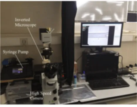
The experimental set-up comprises an inverted microscope (IX71, Olympus, Japan) combined with a high-speed camera (see Fig.1). The PDMS microchannel network, fabricated by a soft lithography method [9], was placed on the stage of the microscope where a syringe pump (PHD ULTRA, Harvard Apparatus) was used to control theflow rate of the workingfluids.
The blood samples used were collected from a healthy adult sheep, and ethylenediaminetetraacetic acid (EDTA) was added to prevent coagulation. The RBCs were separated from the blood by centrifugation and washed twice with saline. The washed RBCs were suspended in Dextran 40 (Dx 40) to make up the required RBCs concentration by volume. All blood samples were stored hermetically at 4 °C until the experiment was performed at a room temperature of about 22 °C.
In this study, we have analysed the CFL of two working fluids with different haematocrit (1% Hct and 15% Hct) with aflow rate of 30µl/min.
2.2 Geometry of the Microchannel Network
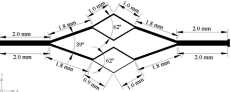
The geometry of the microchannel network used in this study is complex as it contains several bifurcations and confluences (see Fig.2).
2.3 Image Processing
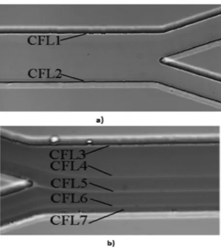
The images were recorded at the center plane of the microchannel using a frame rate of 2000 frames/second. All of frames were analyzed using the Z Project method from the ImageJ software [15]. The number of analyzed frames ranges from 300 to 500 per movie. The recorded sequence of images was replaced by an image that represents the median of neighboring pixel values. This way allows distinguishing two different regions with different intensities. The high intensity region is the CFL and the low intensity region correspond the RBCs, as it is possible to see in Fig.3(b).
Fig. 2. Geometry of the microchannel network and regions where the CFL was measured. The depth of this microchannel is 100µm.
3 Results and Discussion
After the application of the Z Project method, the width of the CFL at the inlet and outlet region of the microchannel was determined. Figure4 shows the different CFLs that we have observed at both inlet and outlet region of the microchannel network.
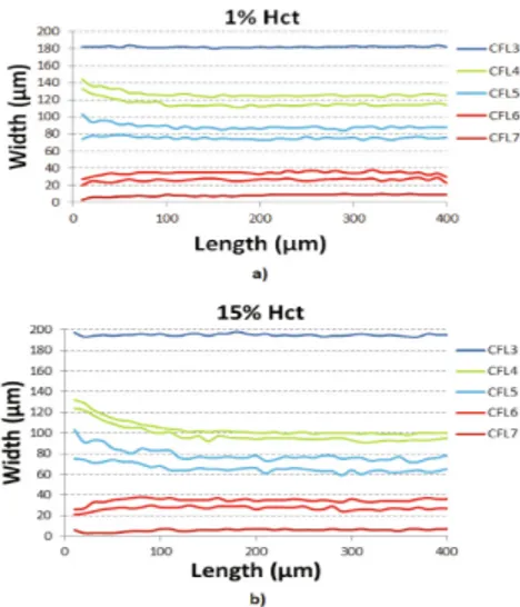
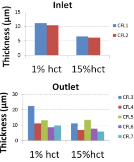
Figure5shows the behavior of the CFL at the inlet of the microchannel for 1% Hct (a)) and 15% Hct (b)) with aflow rate of 30 µl/min.
In Fig. 5it is possible to observe that the width of CFL1 and CFL2 are independent of the Hct.
In Fig. 6it is possible to see the behavior of the CFL at the outlet region of the microchannel for 1% Hct (a)) and 15% Hct (b)) with aflow rate of 30µl/min.
Fig. 4. The CFLs at the (a) inlet; and (b) outlet region of the microchannel network.
From these preliminary results, it is possible to observe that there is an increase of the CFL along the microchannel network. For instance, at the outlet region it was possible to visualize and measure three additional CFLs around the center of the microchannel (see Figs.6and7). The existence of these new CFLs is derived from the confluence geometries that influence the RBCs distribution and trajectories, as was shown by a past study performed in a simple microchannel composed by a single bifurcation followed by a confluence [14].
Due to the asymmetric bifurcations along the microchannel network theflow rate in each branch is affected and consequently influences the concentration of RBCs that flows for each branch. This phenomenon is known as bifurcation law [8]. Briefly, when the RBCs reach an asymmetric bifurcation they tend to prefer the channel with the higher flow rate, i.e., the channel with the highest hydraulic diameter. This phe-nomenon leads to an enrichment of RBCs at the wider channel. The results shown in this study suggest that the bifurcation law may have a little influence on the behavior and width of the CFL along a microchannel network composed by asymmetric bifurcations. More detailed results need to be performed to clarify the role of the bifurcation law in a microchannel with complex geometries such as the channel investigated in this study.
Recently, we have performed CFL measurements in a microchannel network with a depth of about 58µm [16]. However, in this study we have only observed one CFL at center of the channel downstream the confluences. This result suggests that the geometry of the microchannel network plays an important role on the CFL formation at the center of the microchannel. Hence, we are currently investigating in detail the effect of the microchannel network geometry on the CFL formation in order to better understand the blood phenomena that happen in both in vivo and in vitro microcirculation.
In Fig.7it is possible to observe that the thickness of CFL1 and CFL2 are similar for both studiedfluids. And, there is a decrease in the thickness of the CFLs with an increase in the percentage of hematocrit. Comparing the values of the thickness of the CFLs in inlet and outlet it is verified that, in addition to the appearance of new CFLs there is an increase in the thickness of the CFLs that appear in the wall of the microchannel.
4 Conclusion and Future Directions
In this study we have investigated the CFL in a microchannel network composed by asymmetric bifurcations. The results suggest that the geometry of bifurcations and confluences influence the formation of the CFL along the upper and lower wall of the microchannel, as well as the formation of new CFLs at the center of channel after the confluences.
In the near future we expect to deeply understand the effect of multiple asymmetric bifurcations on the CFL width and consequently use this knowledge to develop and optimize lab-on-chips for cells separation, sorting and analysis.
Ackowledgements. The authors acknowledge the financial support provided by the project POCI-01-0145-FEDER-016861 (with associated reference PTDC/QEQ-FTT/4287/2014), UID/EMS/00532/2013 and UID/CEC/00319/2013 funded by FCT (Foundation for Science and Technology), through national funds (PIDDAC), and FEDER through COMPETE2020 - Pro-grama Operacional Competitividade e Internacionalização (POCI). D. Bento acknowledges the PhD scholarship SFRH/BD/91192/2012 granted by FCT.
The authors also acknowledge the financial support provided by the project Nos. UID/EMS/00532/2013 and UID/EMS/04077/2013 and the project Nos. UID/EMS/00532/2013, UID/EMS/04077/2013, POCI-01-0145-FEDER-007043, UID/CEC/00319/2013.
References
1. Popel, A.S., Johnson, P.C.: Microcirculation and Hemorheology. Annu. Rev. Fluid Mech.
37, 43–69 (2005)
2. Lima, R., Ishikawa, T., Imai, Y., Yamaguchi, T.: Blood flow behavior in microchannels: past, current and future trends. In: Dias, R., et al. (ed.) Single and two-Phase Flows on Chemical and Biomedical Engineering, pp. 513–547. Bentham Science (2012)
3. Tateishi, N., Suzuki, Y., Soutani, M., Maeda, N.: Flow dynamics of erythrocytes in microvessels of isolated rabbit mesentery: cell-free layer andflow resistance. J. Biomech.27, 1119–1125 (1994)
4. Kim, S., Kong, R.L., Popel, A.S., Intaglietta, M., Johnson, P.C.: A computer-based method for determination of the cell-free layer width in microcirculation. Microcirculation 13, 199–207 (2006)
5. Dietzel, S., Pircher, J., Nekolla, A.K., et al.: Label-free determination of hemodynamic parameters in the microcirculaton with third harmonic generation microscopy. PLoS ONE9, e99615 (2014)
6. Namgung, B., Liang, L.H., Kim, S.: Physiological significance of cell-free layer and experimental determination of its width in microcirculatory vessels. In: Lima, R., Ishikawa, T., Imai, Y., Oliveira, M.S.N. (eds.) Visualization and Simulation of Complex Flows in Biomedical Engineering, vol. 12, pp. 75–87. Springer, New York (2014)
7. Ong, P.K., Jain, S., Kim, S.: Spatio-temporal variations in cell-free layer formation near bifurcations of small arterioles. Microvasc. Res.83, 118–125 (2012)
8. Tripathi, S., Bala Varun Kumar, Y.V., Prabhakar, A., Joshi, S.S., Agrawal, A.: Passive blood plasma separation at the microscale: a review of design principles and microdevices. J. Micromech. Microeng.25, 083001 (2015)
9. Faustino, V., Catarino, S.O., Lima, R., Minas, G.: Biomedical microfluidic devices by using low-cost fabrication techniques: a review. J. Biomech.49(11), 2280–2292 (2016)
10. Pinto, E., Faustino, V., Rodrigues, R., et al.: A rapid and low-cost nonlithographic method to fabricate biomedical microdevices for blood flow analysis. Micromachines 6, 121–135
(2014)
11. Lopes, A.R., et al.: Low cost microfluidic device for partial cell separation: micromilling approach. In: Proceedings of the IEEE International Conference on Industrial Technology. Seville, Spain (2015)
12. Pinho, D., Yaginuma, T., Lima, R.: A microfluidic device for partial cell separation and deformability assessment. Bio. Chip. J.7, 367–374 (2013)
13. Bento, D., Sousa, L., Yaginuma, T., Garcia, V., Lima, R., Miranda, J.M.: Microbubble moving in blood flow in microchannels: effect on the cell-free layer and cell local concentration. Biomed. Microdevice19(1), 6 (2017)
14. Leble, V., Lima, R., Dias, R., et al.: Asymmetry of red blood cell motions in a microchannel with a diverging and converging bifurcation. Biomicrofluidics5, 44120 (2011)
15. Abramoff, M., Magelhaes, P., Ram, S.: Image processing with image. J. Int. J. Biophotonics
11, 36–42 (2004)