2018
UNIVERSIDADE DE LISBOA
FACULDADE DE CIÊNCIAS
DEPARTAMENTO DE QUÍMICA E BIOQUÍMICA
Changes in synaptic plasticity and lipid raft composition in a rat
model of temporal lobe epilepsy
Andreia Bento de Oliveira
Mestrado em Bioquímica
Especialização em Bioquímica
Dissertação orientada por:
Doutora Diana Cunha-Reis
Doutor Rodrigo de Almeida
II
The most exciting phrase to hear in science, the one that heralds new discoveries, is not “Eureka!” but “That’s funny…”.
IV Table of contents
Acknowledgments/Agradecimentos ... VI Abstract ... VII Resumo ... IX List of Figures ... XII List of Tables ... XIV Abbreviations and Symbols... XV
1. Introduction ... 1 1.1. Epilepsy ... 1 1.2. Synaptic plasticity ... 2 1.3. AMPA receptors ... 3 1.3.1. Receptor phosphorylation ... 4 1.4. Kv4.2 channel ... 6
1.5. Pre-synaptic protein: SNAP-25 ... 8
1.6. Post-synaptic density proteins: PSD-95 and gephyrin ... 9
1.7. Summary of the changes found in TLE regarding the proteins mentioned ... 10
1.8. Lipid rafts ... 12
1.8.1. Membrane model systems ... 14
2. Aims ... 16
3. Methods ... 17
3.1. Animals ... 17
3.2. Brain dissection and hippocampus isolation ... 17
3.3. Long-term potentiation studies ... 18
3.3.1. Hippocampal slices preparation ... 18
3.3.2. LTP induction and electrophysiological recordings ... 18
3.4. Lithium-Pilocarpine temporal lobe epilepsy rat model ... 19
3.4.1. Lithium-Pilocarpine treatment ... 19
3.4.2. Histochemistry ... 19
3.4.3. Isolation of total hippocampal membranes for western-blotting ... 20
3.4.4. Protein assays ... 20
3.4.4.1. Determination of protein concentration ... 20
3.4.4.2. Protein quantification using Western-blot ... 21
3.4.4.2.1. Gel preparation... 21
3.4.4.2.2. Sample preparation and loading ... 21
3.4.4.2.3. Protein transfer to PVDF membranes and incubation with antibodies ... 21
3.4.4.3. Membrane stripping and re-probing ... 23
3.4.5. Lipid assays ... 23
3.4.5.1. Lipid dosing ... 23
3.4.5.2. Preparation of giant unilamellar vesicles ... 23
3.4.5.3. GUV visualization with confocal microscopy... 24
3.4.5.3.1. 3D viewing and determination of the mole fraction of each domain in the GUVs .. 24
3.5. Statistics ... 26
4. Results ... 27
4.1. Long-term potentiation by θ-burst stimulation in vitro ... 27
V
4.1.2. PSD-95 and SNAP-25 levels following LTP induction ... 29
4.2. Lithium-pilocarpine model of TLE ... 30
4.2.1. Neuronal survival and hippocampus organization ... 30
4.2.1.1. NeuN immunolabelling ... 30
4.2.1.2. Nissl-staining ... 31
4.2.1.3. Timm-staining ... 32
4.2.2. Proteins involved in synaptic plasticity and in lipid raft structure ... 33
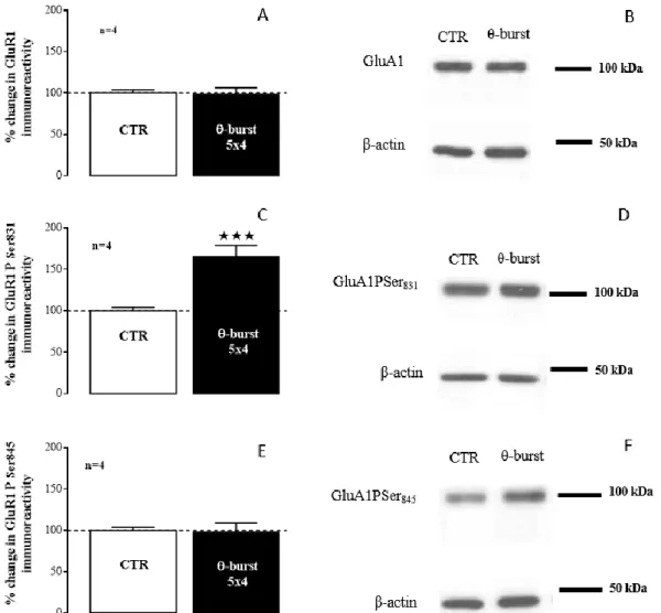
4.2.2.1. AMPA receptor subunits GluA1 and GluA2 ... 33
4.2.2.2. AMPA receptor subunit GluA1 phosphorylation ... 34
4.2.2.3. Pre and post-synaptic markers ... 36
4.2.2.4. Kv4.2 channel ... 37
4.2.2.5. Lipid rafts proteins: flotillin-1 and caveolin-1 ... 38
4.3. Imaging and quantification of lipid domains in GUVs ... 38
5. Discussion ... 43
5.1. Molecular changes in LTP induced with moderate θ-burst stimulation ... 43
5.1.1. Kv4.2 channel association with LTP modulation ... 43
5.1.2. PSD-95 and SNAP-25 expression was not affected by θ-burst stimulation ... 44
5.2. Changes in lithium-pilocarpine model of TLE ... 44
5.2.1. Neuronal death and reorganization in the dentate gyrus ... 44
5.2.2. AMPA receptor subunit regulation leads to hyperexcitability that characterizes TLE . 45 5.2.3. PSD-95 and gephyrin expression is altered in the chronic period of TLE: a new adaptive mechanism that guarantees animal survival? ... 46
5.2.4. Kv4.2 channel is not changed in the chronic period of TLE ... 46
5.2.5. Temporal lobe epilepsy and lipid rafts ... 47
5.3. GUV preparation by electroformation was optimized ... 47
6. Conclusion and future perspectives ... 49
7. References ... 50
8. Appendix ... 57
8.1. Calibration curves for the Bradford method ... 57
8.2. Calibration curve for the Rouser method ... 58
VI Acknowledgments/Agradecimentos
Em primeiro lugar quero agradecer à minha orientadora, Doutora Diana Cunha-Reis, por me ter aceite para trabalhar neste seu projeto, no qual espero não ter desiludido. Um obrigada por acompanhar os meus primeiros passos rumo à investigação e por toda a ajuda, disponibilidade, e por todos os conselhos profissionais. Trabalhar consigo permitiu-me saber o que é ser uma boa investigadora e colega de laboratório, algo ao qual espero ser fiel no futuro. Um muito obrigada por tudo.
Nunca esquecendo o Professor Rodrigo de Almeida, por toda a ajuda e simpatia. Principalmente por me ter apresentado à Biofísica de Biomembranas que se tornou uma paixão. Ainda, por me ter aceite no seu grupo sem qualquer reserva. Não me poderia ter sentido mais incluída, e devido a isso estar-lhe-ei para sempre grata.
Ao restante grupo de Biofísica Molecular, Carla, António, Sr. Doutor Joaquim e Carolina, por toda a ajuda e acolhimento que prestaram a esta naba. Principalmente por todos os momentos de descontração (musical ou baseboliana) entre trabalho. Não poderia ter encontrado um grupo melhor para partilhar a minha loucura científica. Um carinho especial à Filipa, por todas as gargalhadas, amizade e inúmeras boleias (físicas e principalmente emocionais) sem as quais não teria conseguido manter a minha saúde.
Aos bioquímicos do meu coração (que pirosice): Ana, Rita e Guilherme por entenderem melhor que ninguém os desabafos em relação a pessoas que dizem “a” enzima. Por partilharem comigo noites de verdadeira loucura, nada científicas, cheias de intriga, ficção e revelações extraordinárias enquanto jogávamos os bons jogos de tabuleiro.
Às minhas meninas Joana e Iara, pela amizade e gargalhadas partilhadas já há 5 anos (bolas isso é muito tempo!). Obrigada por estimularem o meu intelecto com as vossas conversas sobre ciência, voluntariado e anatomia humana bicuda.
À minha árvore genealógica que apesar da dificuldade em perceberem o que é ser investigador, foi a crescer ao vosso lado que moldou o meu espírito crítico, e o empenho pelo trabalho que caracteriza a nossa família, e a mim também.
Por fim, ao meu pilar nisto tudo, que apesar de estar a passar por stresses próprios, sempre esteve lá para me ajudar, apoiar, ouvir. Percorremos a adversidade em conjunto, a comer bem, a dormir pouco, a fingir que conseguíamos emagrecer. Dedico este último parágrafo à minha Sushi, a gata do meu namorado. Ah, e a ele também claro, coitado. Obrigada Fernando, obrigada por me respeitares como mulher, amiga, cientista e cozinheira.
VII Abstract
Temporal lobe epilepsy (TLE) is the most common form of epilepsy. In many cases, patients with TLE are resistant to antiepileptic drugs. Therefore, it is imperative to unravel the underlying molecular mechanisms that are the cause and/or consequence of the disease to find new strategies to fight it.
Synaptic plasticity is the ability of synapses to strengthen or weaken the communication efficiency in response to transient variations in their activity. Studies in the pilocarpine rat model of TLE indicate that neuronal hyperactivity in epilepsy is associated with abnormal synaptic plasticity, involving molecular changes in α-amino-3-hydroxy-5-methyl-4-isoxazole propionic acid receptors (AMPARs), in voltage-gated potassium channels Kv4.2, and post-synaptic proteins, post-synaptic protein 95 (PSD-95) and gephyrin.
Remodelling of lipid raft composition is believed to contribute to altered cognition and synaptic plasticity observed upon ageing and neurodegenerative diseases. However, it is still unknown if such changes contribute to the cognitive decline and altered synaptic plasticity observed in TLE.
In this thesis, an initial study was performed to evaluate changes in Kv4.2. channel levels and its phosphorylation in Thr602, Thr607 and Ser438 observed in non-pathological long-term-potentiation (LTP;
a type of synaptic plasticity) induced by a moderate θ-burst stimulation and their relation to PSD-95 and synaptosomal nerve-associated protein 25 (SNAP-25) levels was evaluated by western-blot in hippocampal membranes obtained from stimulated and controlslices. As expected, PSD-95 and SNAP-25 levels did not change, but the levels of Kv4.2 channel and its phosphorylation at Ser438 and Thr602
increased following LTP induction, while phosphorylation at Thr607 did not change. These results
suggest a possible role for Kv4.2 channels in LTP expression even with mild θ-burst stimulation patterns, through phosphorylation at Ser438 and Thr602 residues.
The main objective of this work was to unravel molecular mechanisms underlying TLE regarding the role of lipid raft changes in abnormal synaptic plasticity observed in TLE. For that, several approaches were used to characterize the chronic period of the Li2+-pilocarpine model of TLE in the rat.
First, histochemistry was performed in hippocampal slices from 12-month-old Wistar rats exhibiting long-term spontaneous recurrent seizures (SRSs) and Sham controls to evaluate anatomical changes in this tissue. In SRS animals, NeuN immunolabelling revealed a decrease in neurons in the pyramidal cell layer of CA1 and CA3 and in the hilus of the dentate gyrus (DG). Changes in the architecture of the granule cell layer of the DG were also detected for NeuN immunolabelling and Nissl-staining. Finally, using Timm-staining it was possible to detect mossy fibre sprouting. These findings show that the DG is the hippocampal region that suffers more damage in this model.
Molecular changes related to synaptic plasticity were studied by performing western-blot in hippocampal membranes isolated from SRS and Sham rats to evaluate: GluA1 and GluA2 AMPARs subunit levels, GluA1 phosphorylation in Ser831 and Ser845 and Kv4.2 channel expression; in addition,
molecular changes related to profound changes in synaptic architecture were studied by targeting postsynaptic proteins PSD-95 and gephyrin and the presynaptic marker SNAP-25; finally, changes in planar lipid rafts and caveolae scaffolding proteins, flotillin-1 and caveolin-1 respectively, were monitored. The GluA1 and GluA2 subunit and PSD-95 levels decreased while the GluA1/GluA2 ratio, GluA1 phosphorylation in Ser831 and Ser845, along with gephyrin and caveolin-1 levels increased in SRS
animals. SNAP-25, Kv4.2 channel and flotillin-1 levels did not change.
To initiate the biophysical study of lipid rafts in model membranes obtained from synaptic lipids from the Li2+-pilocarpine rat model of TLE, the biophysical imaging and quantification of lipid rafts in
giant unilamellar vesicles obtained from binary and ternary artificial lipid mixtures was performed. Giant unilamellar vesicles preparation by electroformation was optimized with success.
VIII
Changes found for AMPARs subunit stoichiometry and phosphorylation contribute to an increase in synaptic transmission that surely contributes to enhancement of neuronal excitability, and to progression of the disease. A decrease in PSD-95 and gephyrin suggests that excitatory transmission is down-regulated and inhibitory transmission is up-regulated, contributing to lower neuronal excitability, hinting on an adaptive molecular mechanism that is trying to counteract the hyperexcitability that characterizes epilepsy. The increase found for caveolin-1 suggests a new role for this protein in TLE, that is probably neuroprotection, as it occurs in other diseases.
IX Resumo
A epilepsia do lobo temporal (do inglês, TLE) é o tipo de epilepsia mais comum. Na maioria dos casos os pacientes com TLE são resistentes ao tratamento com fármacos antiepiléticos. É importante compreender os mecanismos moleculares subjacentes à doença, de modo a encontrar novas estratégias para combatê-la.
Estudos no modelo da pilocarpina em rato indicaram que a hiperexcitabilidade na TLE experimental está associada a plasticidade sináptica anómala, através de alterações moleculares que consequentemente afetam a excitabilidade neuronal.
A plasticidade sináptica é a capacidade que as sinapses apresentam de reforçar ou atenuar o seu rendimento em resposta a variações transientes na sua actividade que resultam de estímulos fisiológicos. A nível molecular, este mecanismo é caracterizado por alterações bidirecionais no número e actividade de alguns receptores de neurotransmissores e canais. Na potenciação de longo prazo (do inglês, LTP), o tipo de plasticidade sináptica mais estudado e conhecido no hipocampo, algumas destas alterações incluem o aumento dos receptores de ácido α-amino-3-hidroxi-5-metil-4-isoxazolo propiónico (Rs AMPA) no neurónio pós-sináptico, que aumenta a eficiência da comunicação glutamatérgica, e diminuição da corrente K+ do tipo A (I
A), mediada por canais de potássio dependentes de voltagem 4.2
(Kv4.2), que reduz a excitabilidade dendrítica.
A modulação dos Rs AMPA pode afetar a excitabilidade neuronal através de alterações na estequiometria das subunidades e da fosforilação das mesmas. Estas alterações estão ambas implicadas na expressão da LTP no hipocampo assim como nas alterações patológicas em modelos de TLE em roedores. O aumento da excitabilidade neuronal está relacionado com a presença de Rs AMPA sem a subunidade GluA2, permeáveis ao ião Ca2+, e com a fosforilação da subunidade GluA1 na Ser
831 e na
Ser845, através do aumento dos Rs AMPA com maior condutância, na região sináptica.
Alterações na expressão e modulação do canal Kv4.2 foram observadas tanto na LTP como na TLE. Sendo o principal mediador da corrente IA, a sua regulação afeta a excitabilidade dendrítica, que
dela depende. Uma forma de modular o canal é através da fosforilação nos resíduos Thr602, Thr607 e
Ser438.
A densidade pós-sináptica (do inglês, PSD) tem um importante papel na regulação de cascatas de sinalização que ajustam a composição molecular das entidades pós-sinápticas necessárias para manter a plasticidade sináptica. Duas proteínas da PSD associadas à TLE são a proteínas pós-sináptica 95 (PSD-95), que é expressa exclusivamente em sinapses glutamatérgicas (excitatórias), e a gefirina que se encontra exclusivamente em sinapses glicinergicas e GABAergicas (inibitórias).
No sistema nervoso podem ser encontradas tanto jangadas lipídicas planares como caveolas, associadas às proteínas específicas, flotilinas e caveolinas, respectivamente. Estas jangadas são microdomínios membranares ordenados, ricos em colesterol e esfingolípidos e que estão associados a diferentes funções celulares. Estudos recentes sugerem que estes microdomínios podem estar envolvidos na plasticidade sináptica, fornecendo a estrutura necessária à organização e compartimentalização de diferentes eventos moleculares na neurotransmissão, tendo também um papel na sua regulação dinâmica de forma a aumentar ou diminuir a eficiência da comunicação sináptica. A remodelação da composição das jangadas lipídicas ocorre durante o desenvolvimento e contribui para alterações cognitivas e de plasticidade sináptica observadas durante o envelhecimento e doenças neurodegenerativas. No entanto, ainda não é claro se tais alterações contribuem para o declínio cognitivo e para as alterações na plasticidade sináptica encontradas na TLE.
O principal objectivo deste trabalho foi avaliar as alterações nos marcadores da plasticidade sináptica e na composição sináptica de jangadas lipídicas num modelo de TLE induzida por lítio-pilocarpina no rato, de forma a desvendar os mecanismos moleculares subjacentes à doença.
X
Num primeiro passo avaliaram-se por western-blot as alterações nos níveis e na fosforilação do canal Kv4.2 nos resíduos de Thr602, Thr607 e Ser438, relativamente à PSD-95 e à proteína pre-sináptica
SNAP-25, em membranas de hipocampo obtidas de fatias nas quais se induziu LTP com um estímulo θ-burst moderado, que mimetiza a actividade neuronal fisiológica na região CA1 do hipocampo durante a aprendizagem e memória.
Apesar dos níveis da proteína pós-sináptica, PSD-95 e da pré-sináptica, SNAP-25 não se alterarem em resposta à estimulação com θ-burst moderado, como era esperado, os níveis do canal Kv4.2 e da sua fosforilação nos resíduos de Ser438 e Thr602 aumentaram. Estes resultados sugerem um papel da regulação
do canal Kv4.2 na LTP, através da fosforilação nos resíduos de Ser438 e Thr602.
Para a caracterização do modelo de Li2+-pilocarpina no período crónico da TLE, foram primeiro
realizados ensaios de histoquímica em fatias de hipocampo de ratos Wistar com 12 meses que exibiam convulsões espontâneas recorrentes (SRSs) induzidas pelo método do Li2+-pilocarpina e com controlos
Sham. Nos ratos modelos da TLE de Li2+-pilocarpina, usando o marcador NeuN foi possível observar
uma diminuição de neurónios na camada de células piramidais da região CA1 e CA3 e do hilus do giro dentado (DG). Foi vista também uma alteração de disposição dos neurónios da camada de células granulares do DG também observadas em resultados da coloração de Nissl. Por fim, com a coloração de Timm foi possível detetar sprouting axonal no DG nos ratos modelo da TLE, concordante com o observado em trabalhos anteriores. Neste estudo confirmamos então que o DG é a região do hipocampo que sofre maior dano neste modelo, o que é concordante com o observado no DG de pacientes com TLE.
As alterações moleculares associadas à plasticidade sináptica foram avaliadas por western-blot em membranas de hipocampo obtidas de ratos modelo da TLE e Sham tendo como alvo: a subunidade GluA1 e GluA2 dos Rs AMPA, a fosforilação da subunidade GluA1 nos resíduos de Ser831 e na Ser845
e a expressão do canal Kv4.2. Para avaliar alterações estruturais nas sinapses estudaram-se as proteínas da PSD, PSD-95 e gefirina e o marcador pré-sináptico SNAP-25. Por fim, estudou-se a expressão das proteínas específicas de jangadas lipídicas, flotilina-1 e caveolina-1.
Os níveis de subunidade GluA1 e GluA2 diminuíram em ratos modelo da TLE, enquanto que a razão GluA1/GluA2 e a fosforilação da subunidade GluA1 na Ser831 e na Ser845 aumentaram. Estas
alterações contribuem para um aumento da transmissão glutamatérgica que certamente contribui para o aumento da excitabilidade neuronal, e para a progressão da doença.
Os níveis de SNAP-25 não se alteraram nos ratos modelo da TLE de Li2+-pilocarpina. Face aos
níveis desta proteína a expressão relativa de PSD-95 diminuiu enquanto que a da gefirina aumentou. Uma vez que o PSD-95 e a gefirina são específicos para sinapses glutamatérgicas e GABAérgicas/glicinérgicas, respetivamente, estes resultados evidenciam que há mecanismos associados à transmissão glutamatérgica que estão diminuídos neste modelo enquanto que outros associados à transmissão GABAérgica/glicinérgica estão aumentados. Isto, por sua vez, sugere um aumento da transmissão inibitória e diminuição da excitatória, o que na totalidade diminui a excitabilidade neuronal. Esta alteração das proteínas da PSD poderá ser um mecanismo de adaptação molecular para contrariar a hiperexcitabilidade causada em parte pelas alterações descritas para os Rs AMPA, garantindo a sobrevivência do animal.
A expressão do canal Kv4.2 não se alterou nos animais modelo da TLE, no entanto observa-se uma tendência para aumento e o estudo de um número mais elevado de animais poderá tornar mais clara esta questão.
Finalmente, os níveis de flotilina-1 não se alteraram no modelo de TLE em rato enquanto que os níveis de caveolina-1 aumentaram, sugerindo que a caveolina-1 e alterações nas jangadas lipídicas têm um papel na fisiopatologia da TLE. Face à sua ação noutros modelos de doença no SNC, este papel será provavelmente neuroprotector.
XI
Para iniciar o estudo biofísico de jangadas lipídicas em membranas modelo preparadas com lípidos sinápticos do rato modelo da TLE de Li2+-pilocarpina, foram realizadas a deteção e quantificação
biofísica de jangadas lipídicas em vesículas gigantes unilamelares preparadas com misturas de lípidos artificiais. A otimização da preparação de vesículas gigantes unilamelares por eletroformação foi efetuada com sucesso.
Este trabalho revela alterações moleculares nunca descritas tanto para a LTP como para o período crónico da TLE no modelo de Li2+-pilocarpina em rato. No entanto, serão necessários estudos adicionais
para melhor compreender o que é causa ou consequência da doença e, mais importante, o que pode ser feito para tratar ou prevenir a TLE.
XII List of Figures
Figure 1.1. Magnetic resonance imaging (MRI) brain scan from a patient with mesial TLE (MTLE). 1
Figure 1.2. Induction of LTP depends on NMDA receptors and AMPARs. ... 3
Figure 1.3. Overview of the AMPARs structure (A) and subunit composition (B). ... 4
Figure 1.4. GluA1 levels and phosphorylation following LTP induction with moderate TBS. ... 5
Figure 1.5. Overview of Kv4.2 channel structure. ... 7
Figure 1.6. Regulation of dendritic Kv4.2 channels by physiological activity and in epilepsy. ... 8
Figure 1.7. The PSD, opposing presynaptic terminals forming glutamatergic synapses with two dendritic spines. ... 9
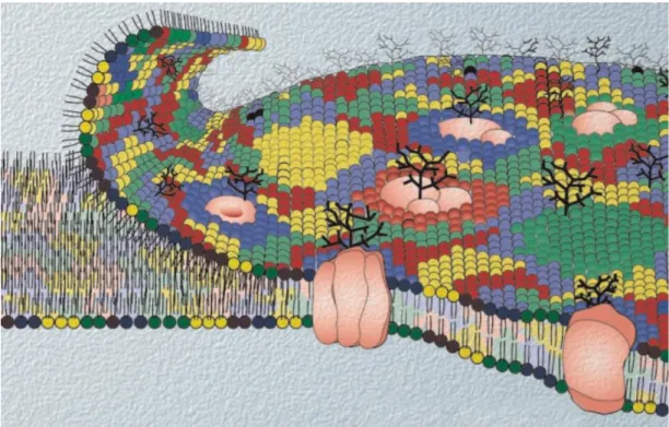
Figure 1.8. Schematic representation of the fluid mosaic membrane proposed by Singer and Nicolson modified to highlight the diversity of lipid components and their heterogeneous transversal and lateral distribution. ... 12
Figure 1.9. Lipids raft organization and possible roles in neurotransmitter signaling. ... 13
Figure 1.10. Miscibility phase diagram for DPPC, DOPC and cholesterol at 24°C. ... 15
Figure 3.1. Schematic of the representation of the LTP induction procedure in rat hippocampal slices. The illustration shows the different hippocampal areas. ... 18
Figure 3.2. Procedure for the Li2+-Pilo treatment performed in this work. ... 19
Figure 3.3. GUV representation for determination of domain area fraction. ... 25
Figure 4.1. LTP induced by moderate TBS. ... 27
Figure 4.2. LTP induction with moderate TBS enhanced the levels of Kv4.2 channels and its phosphorylation on Ser438, but not on Thr602 and Thr607. ... 28
Figure 4.3. LTP induction with moderate TBS enhanced the percentage variation ratio to Kv4.2 channel of phosphorylation on Ser438, but not on Thr607. ... 29
Figure 4.4. LTP induction with moderate TBS did not change PSD-95 and SNAP-25 expression. .... 29
Figure 4.5. Loss and dispersion of CA1 and CA3 pyramidal cells in the hippocampus of Epi rats. .... 31
Figure 4.6. Epi rats have less neurons in the DG than Sham rats. ... 31
Figure 4.7. Loss of CA1 pyramidal cells in the hippocampus of Epi rats. ... 32
Figure 4.8. Axon sprouting in the hippocampus of Epi rats. ... 33
Figure 4.9. Epi rats have lower levels of GluA1 and GluA2 but higher GluA1/GluA2 ratio then Sham rats. ... 34
Figure 4.10. GluA1 phosphorylation in Ser831 and in Ser845 did not change between Sham and Epi rats. ... 35
Figure 4.11. Epi rats have higher levels of GluA1 phosphorylated in Ser831 and in Ser845 when total amount of GluA1 subunit is considered. ... 35
Figure 4.12. PSD-95 levels in Epi rats is lower than in Sham rats, and gephyrin levels are increased in Epi rats, while SNAP-25 levels are not changed. ... 36
Figure 4.13. The ratio of PSD-95 and gephyrin with SNAP-25 decreased and increased respectively in Epi rats when compared with Sham rats. ... 37
Figure 4.14. Kv4.2 channel levels did not change between Epi and Sham rats. ... 37
Figure 4.15. Flotillin-1 levels did not change, and caveolin-1 levels are increased in Epi rats when compared with Sham rats. ... 38
Figure 4.16. Confocal microscopy of GUVs prepared with DPPC/DOPC (equimolar) and Rhod-DOPE as a fluorescent probe reveals the coexistence of so and ld phases. ... 39
Figure 4.17. Confocal microscopy of GUVs prepared with DPPC/DOPC/chol (equimolar) and Rhod-DOPE as a fluorescent probe reveals the presence of ld and lo, i.e., raft-like domains. ... 40
Figure 8.1. Calibration curve for the Bradford method120, for determination of protein concentration of C and θ samples. ... 57
XIII
Figure 8.2. Calibration curve for the Bradford method120 for determination of protein concentration of
Sham and Epi samples. ... 57 Figure 8.3. Calibration curve for the phospholipid quantification by Rouser method 122, used to quantify
DPPC and DOPC. ... 58 Figure 8.4. Absorption spectrum of Rhod-DOPE in chloroform at room temperature. ... 58
XIV List of Tables
Table 1.1. Protein changes found in patients with TLE. ... 10
Table 1.2. Protein changes found in rodent models of TLE. ... 11
Table 3.1. Dilution of primary and secondary antibodies used and according blocking agent. ... 22
XV Abbreviations and Symbols
aCSF artificial cerebrospinal fluid
AMPAR α-amino-3-hydroxy-5-methyl-4-isoxazole propionic acid receptors
APS ammonium persulfate
BSA bovine serum albumin
CA1 cornu Ammonis area 1
CA3 cornu Ammonis area 3
Ca2+ calcium ion
CaMKII Ca2+/calmodulin-dependent protein kinase II
Chol cholesterol
CNS Central nervous system
CTD carboxy-terminal domain
CTR control
DG dentate gyrus
DOPC 1,2-dioleoyl-sn-glycero-3-phosphocholine (DOPC), DPPC 1,2-dipalmitoyl-sn-glycero-3-phosphocholine EDTA ethylenediamine tetraacetic acid
EEG electroencephalograms
EPSP excitatory post-synaptic potential ERK extracellular signal-regulated kinases fEPSP field excitatory post-synaptic potential GABA γ-aminobutyric acid
Glu glutamate
GluA1 Ser818 Serine residue 818 of the GluA1 subunit
GluA1PSer831 GluA1 phosphorylated on Serine 831
GluA1PSer845 GluA1 phosphorylated on Serine 845
GPHN gephyrin gene
GPI glycosylphosphatidylinositol GUV giant unilamellar vesicle
HEPES 4-(2-hydroxyethyl)-1-piperazineethanesulfonic acid HRP horseradish peroxidase
IA A-type K+ current
i.m. intramuscular i.p. intraperitoneal
KA kainic acid
Kv4.2 voltage-gated potassium channel 4.2 Kv 4.2 Thr38 Threonine residue 38 of the Kv4.2 channel
Kv4.2PSer438 Kv4.2 phosphorylated on Serine 438
Kv 4.2 Ser552 Serine residue 552 of the Kv4.2 channel
Kv 4.2 Ser616 Serine residue 616 of the Kv4.2 channel
Kv4.2PThr602 Kv4.2 phosphorylated on Threonine 602 Kv4.2PThr607 Kv4.2 phosphorylated on Threonine 607 ld liquid-disordered Li2+-Pilo Lithium-pilocarpine lo liquid-ordered LTD long-term depression
XVI LTP long-term potentiation
MAPK mitogen-activated protein kinases
Mg2+ magnesium ion
mPFC medial prefrontal cortex MRI magnetic resonance imaging MTLE mesial temporal lobe epilepsy N.A. numerical aperture
Na+ sodium ion
NeuN Neuronal nuclear protein NMDA N-methyl-D-aspartate
NTD N-terminal domain PKA protein kinase A PKC protein kinase C
PMSF Phenylmethanesulfonyl fluoride
PSD post-synaptic density
PSD-95 post-synaptic density protein 95 PVDF polyvinylidene fluoride
Rhod-DOPE N-(lyssamine Rhodamine B sulfonyl)-1,2-dioleoyl-sn-glycero-3-phosphoethanolamine
SDS sodium dodecyl sulfate
SE status epilepticus
SEM Standard error of the mean
SNAP-25 synaptosomal-associated protein 25
SNARE soluble n-ethylmaleimide-sensitive factor attachment protein receptor
so solid-ordered
SRS spontaneous recurrent seizures TBS θ-burst stimulation
TBST Tris-buffered saline with Tween 20 TEMED tetramethylethylenediamine TLE temporal lobe epilepsy
TM transmembrane domain
1 1. Introduction
1.1. Epilepsy
Epilepsy is one of the most common neurological diseases1, and is characterized by recurrent
unpredictable seizures involving the synchronized firing of multiple neurons. It is associated with a chronic imbalance between excitatory and inhibitory transmission. Besides genetic changes, which make up a minor cause for the disease, epilepsy is believed to be a secondary consequence of brain insults such as acute seizures caused by toxic or febrile stimuli, stroke, head trauma, or cerebral infections2. According to the Epilepsy Foundation, approximately 60% of all living people with epilepsy
have temporal lobe epilepsy (TLE), making it the most common form of focal epilepsy in humans3,4.
Some of the changes found in patients with TLE include abnormal electroencephalograms (EEGs), memory impairment, hippocampal atrophy and sclerosis, as seen in Figure 1.1. At the cellular level changes include neuronal loss, gliosis and axonal sprouting, but also abnormal morphology. With the progression of the disease, alterations can spread to the rest of the brain, as seen in Figure 1.1 where besides the hippocampus, the whole right temporal cortex shows atrophy.
Figure 1.1. Magnetic resonance imaging (MRI) brain scan from a patient with mesial TLE (MTLE).MTLE is associated with hippocampal atrophy and sclerosis, which includes neuronal loss and gliosis, but also loss of internal architecture. In this case an asymmetry of the brain is detected which is due to the temporal cortex atrophy.4
Epilepsy is a major public health concern since 50 million people worldwide are epileptic1, and
currently there are no disease-modifying therapies. Possible treatments include antiepileptic drugs, ketogenic diet, neurosurgical resection, and electrical stimulation of the nervous system5. In the
particular case of TLE, about one third of the patients do not respond to therapy with antiepileptic drugs having to resort to hippocampal and/or amygdala resection surgery, from which 70% of the patients become seizure-free4. This drug resistance and the attempt to avoid cerebral surgery are the main reasons
why it is imperative to explore the molecular mechanisms underlying TLE and find new strategies to fight it.
Multiple experimental approaches have been developed to study TLE disease mechanisms. In vitro models usually consist of mammalian neuronal cell cultures or brain slices from animals, that are subjected to epileptic crisis (usually monitored using electrophysiological recordings), and can be acutely isolated or organotypic slices6. A wide range of species have been used to study epilepsy in an
in vivo fashion, but the most common animals used to model TLE are the rodents Rattus norvegicus or
Mus musculus (see ref 7 for review), due to their small size, rapid breeding and the possibility to use
advanced genetic tools. Several procedures can be used to induce chronic seizures in these animals, such as: 1) electrical stimulation which includes the kindling model; 2) administration of chemoconvulsants
2
such as kainic acid (KA) or pilocarpine; and, less frequently, 3) brain hypoxia or ischaemia models. In addition, genetic models of seizure-prone rodents have been used.
The properties of some antiepileptic drugs available today, e.g. phenobarbital, to which some patients are resistant, were found using simple acute seizure models5. One possible hypothesis is that
the drug resistance found in epileptic patients is due to the fact that the acute models do not mimic the chronic state of the patients, characterized by spontaneous recurrent seizures (SRSs)5. Therefore, chronic
models, also called SRS models are being used to better understand the disease and search for novel therapeutic approaches.
In the lithium-pilocarpine (Li2+-Pilo) rat model of SRSs, pathological features including
histological, behavioral, electroencephalographic (EEG) and pharmacological responses to antiepileptic and other drugs, resemble those observed in patients with MTLE, making it a good model of the human disease. The injection of pilocarpine induces an initial crisis of epileptic seizures or status epilepticus (SE), that lasts for up to a few hours. After typically 15 to 44 days with no observable seizures, a time called latent period, the animal starts to exhibit SRSs8,9.
Abnormal synaptic plasticity has been found in animal models of epilepsy3,10,11 and in epileptic
patients12, not only during epileptogenesis (the process by which the brain develops epilepsy13) but also
in the chronic period of the disease. This altered plasticity relies on changes in α-amino-3-hydroxy-5-methyl-4-isoxazole propionic acid receptors (AMPARs) and the voltage-gated potassium channels 4.2 (Kv4.2 channel), among others.
To better understand this association between disease and synaptic plasticity, it is important to first define synaptic plasticity itself.
1.2. Synaptic plasticity
Synaptic plasticity is a cellular process that confers synapses the ability to strengthen or weaken their communication in response to transient variations in their activity. These changes can endure for short or long periods and are considered crucial cellular mechanisms in learning and memory formation. Synaptic plasticity can involve changes at the molecular level, such as bidirectional scaling in the number and activity of some neurotransmitters receptors and channels14, or more profound structural
cellular changes, like the formation of new synaptic contacts.
In vitro, synaptic plasticity involves cellular changes in synaptic communication that can occur in
a broad range of time frames and through different molecular processes depending on the stimulation pattern15–17. The two most studied forms of synaptic plasticity in the hippocampus are long-term
potentiation (LTP) and long-term depression (LTD), since the lasting duration of the molecular and cellular changes observed suggests that these are crucial cellular mechanisms in hippocampal memory storage14.
Using 2-amino-5-phosphonopentanoate, a competitive N-methyl-D-aspartate (NMDA) receptor antagonist, it was found that, LTP at hippocampal glutamatergic synapses is triggered by the activation of NMDA receptors18. The sustained depolarization of the postsynaptic membrane releases magnesium
ions (Mg2+) that block NMDA receptors channels, allowing calcium ions (Ca2+) to enter the cell (Figure
1.2 - B)19. The influx of Ca2+ ions leads to autophosphorylation of enzyme Ca2+/calmodulin-dependent
protein kinase II (CaMKII) causing its persistent activation15,20. This enzyme, in turn, catalyzes the
phosphorylation of AMPARs21,22.
The increase of intracellular Ca2+ and the consequent activation of CaMKII triggers the molecular
events that drive synaptic plasticity (Figure 1.2) such as the recruitment of AMPARs to the synaptic region of the postsynaptic neuron, which then becomes easier to stimulate showing an increase in excitatory post-synaptic potentials (EPSPs)23. Simultaneously, there is an increase in dendritic
excitability caused by the decrease of A-type K+ current (I
3
1.4)24. Other changes also include an increase in neurotransmitter release, an increase in postsynaptic
ribosomes, changes in calcium compartmentalization, and growth of the actin cytoskeleton25,26.
Other structural synaptic changes were found following LTP induction with θ-burst stimulation (TBS). The neck of dendritic spines (input neuronal protrusions) becomes wider and shorter, while the volume of the spine head increases, which facilitates protein traffic from dendrites into the spines, and enhances the synaptic contact area, leading to increased efficacy of synaptic transmission27. These spine
changes are accompanied by modifications in the postsynaptic density (PSD)28. The PSD suffers a
dynamic reorganization that modulates glutamate receptors (excitatory transmission) through linking these receptors to downstream signaling molecules that guarantee LTP expression29.
Figure 1.2. Induction of LTP depends on NMDA receptors and AMPARs. In normal synaptic transmission (A), NMDA
receptors have Mg2+ that blocks Ca2+ influx through the channel of this receptors. Sustained depolarization of the postsynaptic
membrane (caused by high-frequency or TBS) drives Mg2+ out of the channel allowing Ca2+ influx (B), triggering synaptic
changes such as the increase of AMPARs in the postsynaptic membrane (C).30
Although many studies have focused on LTP, there is still a lot to uncover. One distinct problem is the fact that when this type of synaptic plasticity is being investigated the procedures followed, specifically stimulation that induces LTP, are not kept the same between studies, making it hard to learn how this mechanism is expressed and maintained physiologically.
1.3. AMPA receptors
AMPARs (Figure 1.3) mediate most of the excitatory glutamatergic neurotransmission across chemical synapses in the central nervous system (CNS), and changes in their activity, phosphorylation state, subcellular targeting and expression levels have been associated with physiological synaptic plasticity and TLE pathology.
4
Figure 1.3. Overview of the AMPARs structure (A) and subunit composition (B). Each subunit contains four different
hydrophobic domains (TM) that transverse the membrane. When the receptor is assembled with GluA2, the channel is not permeable to Ca2+, but without GluA2 the channel is permeable to Ca2+.31,32
AMPARs are transmembrane homo or hetero-tetrameric ionotropic receptors assembled from four different subunits, GluA1-4 33 that share 68-74% amino acid sequence identity34. These differ in the
C-terminus where some post-translation modifications can occur, such as phosphorylation31. In the adult
hippocampus, there are two major populations of AMPARs: those assembled with GluA1 and GluA2 or those assembled with GluA3 and GluA2 subunits35. Only a small pool of AMPARs lack GluA2, being
composed by GluA1 and GluA3 or solely by GluA1 subunits (homomeric receptors)35.
Subunit stoichiometry is important in the modulation of gating kinetics, ion permeability and responsiveness to an array of small-molecule channel modulators36. When the receptor lacks the GluA2
subunit, the core channel displays a strong, inwardly rectifying current-voltage relation as well as Ca2+
permeability, but not when GluA2 is present (Figure 1.3 - B)37,38.
Knockdown of hippocampal GluA2 subunit promotes hyperexcitability, leading to a decrease in seizure threshold and seizure-like behavior39, thus increasing the risk of epileptogenesis40,41.
AMPARs permeable to Ca2+ (lacking the GluA2 subunit), have been implicated in the expression
of hippocampal LTP through protein kinase A (PKA)-dependent mechanisms42, which occur only when
LTP is induced with strong and repeated TBS patterns. FollowingLTP induction a transient insertion of AMPARs without GluA2 in synapses, was observed42,43, suggesting that GluA2-lacking AMPARs
might have a part in the expression of LTP. However, LTP can also occur without any detectable changes in AMPAR subunits stoichiometry44, which suggests that other mechanisms and changes might
also play a role in mediating and consolidating LTP.
After 85 days of SE induction with pilocarpine, mRNA levels of GluA2 are increased in the dentate
gyrus (DG) of the rat hippocampus, but decreased in the CA1 area of the rat hippocampus45. An increase
in the mRNA levels of GluA2 in the DG of the rat hippocampus was also detected, 2 weeks after SE induced with Li2+-Pilo46.
Studies also relate GluA2 subunits-lacking AMPAR with TLE. One week after SE induction by Li2+-Pilo (latent period), three-week-old Wistar rats show no changes in the GluA1/GluA2 ratio in the
hippocampus, but three days after SE the same ratio is increased in the medial prefrontal cortex (mPFC)3.
1.3.1. Receptor phosphorylation
Besides subunit stoichiometry, other mechanisms regulate AMPARs. GluA1 can undertake certain post-translation modifications such as phosphorylation in serine residues 818 (Ser818), 831 (Ser831) and
845 (Ser845), which have been implicated in LTP expression. Also, GluA2 phosphorylation at serine
5
plasticity mechanism. For instance, the use of specific antibodies for phosphorylated GluA1 on Ser831
or on Ser845, showed that both residues are phosphorylated when LTP is induced in the CA1 region of
the hippocampus with four episodes of TBS, each consisting of ten trains delivered at 5Hz of four pulses at 100Hz47, a very strong stimulation paradigm.
Our group studied GluA1 phosphorylation when the induction of LTP was carried using only one episode of TBS, each comprising only five trains of stimuli. Under these conditions, the phosphorylation of GluA1 subunits occurred only at Ser831 without changes in the GluA1 levels or phosphorylation at
Ser845 (Figure 1.4)48. This difference is a consequence of the different stimulation paradigms applied and
is consistent with the observations of Park et al., reporting that LTP induced by TBS is only PKA-dependent when stronger and repeated TBS patterns are used49. Thus, only under these conditions
phosphorylation at both Ser residues will happen, which suggests that these events are important in different phases of LTP: both Ser831 and Ser845 phosphorylation are required for late LTP, but only Ser831
phosphorylation is associated with the initial phase of LTP.
Ser831 phosphorylation can be catalyzed by PKC50 or by CaMKII21,22, an enzyme that, as mentioned
before, has a role in LTP induction. This modification can lead to an increase of the time that AMPARs occupy the higher conductance states51, correlating to the increased conductance of synaptic AMPARs
during LTP. This phosphorylation also drives AMPARs to the synaptic region52,53.
Figure 1.4. GluA1 levels and phosphorylation following LTP induction with moderate TBS. Representative western-blots
(B, D, F) and averaged changes observed in reactivity (A, C, E) in western-blots probing for GluA1 levels and phosphorylation on Ser845 and Ser831. Levels of GluA1 phosphorylated in Ser831 (C, D) were increased following LTP induction with TBS, but
6
Phosphorylation of Ser845 of GluA1 subunit is catalyzed by PKA50, which induces the insertion of
AMPARs from the intracellular pool to the extra-synaptic region of the plasma membrane52,53. This
modification also increases the open-probability of the receptor54.
Phosphorylation at these two Ser residues has also been related to TLE pathology. Lopes et al showed that 50 days after SE was induced with pilocarpine (chronic period), levels of GluA1 phosphorylated in Ser845 were reduced in the rat hippocampus, without any changes in GluA1
phosphorylated in Ser831 or in GluA1 expression levels55. In another study, rats showing SRSs 60 days
after SE induction with pilocarpine (chronic period) exhibited a decrease in GluA1 phosphorylation in Ser845 in the dorsal hippocampus, and a decrease of GluA1 phosphorylation in Ser831 in the ventral
hippocampus8.
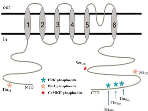
1.4. Kv4.2 channel
Across the neuron membrane, a variety of channels can be found, each contributing to different ionic currents. The largest and most diverse group of these ion channels is the potassium channel family, that has an important role in controlling neuronal excitability and plasticity56. As mentioned above, the
IA current plays an important role in the enhancement of dendritic excitability associated with LTP. The
main mediator of this current in hippocampal neurons is the Kv4.2 channel57.
Kv4.2 belongs to the superfamily of potassium channels with six hydrophobic transmembrane domains (Figure 1.5)58 and is mostly expressed in the distal dendrites of neurons59,60. In response to a
depolarization, the channel pore opens due to a conformational change triggered by the voltage sensor found in the fourth TM domain61.
Channel properties and function can be regulated by post-translational phosphorylation. There are a few highly conserved sites that can be phosphorylated by ERK (extracellular signal-regulated kinases) and other kinases of the MAPK (mitogen-activated protein kinases) family like the threonine 602 residue (Thr602) and threonine 607 (Thr607)62,63. When this happens, IA decreases, and internalization of the
channel occurs63. ERK family can also catalyze the phosphorylation of Ser
616, which suppresses the
channel’s activity64.
PKA also phosphorylates some sites in Kv4.2 channel, such as the threonine 38 residue (Thr38) or
serine 552 residue (Ser552). The consequences of phosphorylation on Thr38 are unknown, but
phosphorylation of Ser552 causes internalization of the channel65.
Another possible phosphorylation site is Ser438, that can be phosphorylated by CaMKII, causing an
7
Figure 1.5. Overview of Kv4.2 channel structure.The channel is composed of six TM domains, with an extracellular P-domain between the fifth and the sixth. It also has three ERK phosphorylation sites (blue stars), Ser616, Thr607 and Thr602; two
PKA phosphorylation sites (orange stars), Thr38 and Ser552; and one CaMKII phosphorylation site (red star), Ser438. CTD:
carboxy-terminal domain; NTD: N-terminal domain. Adapted from 62.
Induction of LTP reduces the levels of Kv4.2 on the membrane of dendrites (Figure 1.6 - B) which is accompanied by a shift in the voltage-dependence of IA. This suggests that control of the trafficking
of the channel, which depends on modifications such as phosphorylation, could underlie a mechanism of LTP expression67,68.
The first evidence that Kv channels could have a role in epilepsy came from the observation that Kv channel blockers induce convulsions in mice69. Studies in chronic TLE models have showed changes
in Kv4.2 expression at several stages of disease progression. In animals with SRSs induced with pilocarpine, an increased excitability of neuronal pyramidal cell dendrites in hippocampal CA1 region was correlated with decreased dendritic availability of Kv4.2 channels and an increase in the phosphorylation of Kv4.2 channel at ERK-sites that is probably mediated by ERK itself70.
8
Figure 1.6. Regulation of dendritic Kv4.2 channels by physiological activity and in epilepsy. Under control conditions (A)
clathrin-mediated internalization, trafficking to the membrane and production of the Kv4.2 channel regulate the density of the channel in the dendritic membrane. The activity of the channel is also controlled by PKA, PKC, and MAPK dependent-signaling. When LTP is induced (B) MAPK dependent-signaling activity and clathrin-mediated internalization are increased. Phosphorylation of the channel is also increased in SE of the pilocarpine model of epilepsy (C) but some of the mechanisms on the internalization of the channel are still unclear. In this situation, there is also a transcriptional downregulation of the channel.57
In another study, in the Li2+-Pilo model of TLE, western-blot and immunohistochemistry studies
have shown an increase in Kv4.2 expression that starts 6 hours after SE and continues to increase until 2 days after the first grade of seizures (latent period). This is followed by a decrease in Kv4.2 expression during the chronic period (50 days after SE). These changes were more prominent in the CA1 and CA3 regions of the hippocampus11.
Besides the changes in phosphorylation and availability/trafficking of the channel on the membrane, a transcriptional down-regulation has also been reported in epilepsy (Figure 1.6 - C)70.
1.5. Pre-synaptic protein: SNAP-25
The synaptosomal-associated protein 25 (SNAP-25) is a component of the trans-SNARE (soluble n-ethylmaleimide-sensitive factor attachment protein receptor) complex, playing an important role at presynaptic vesicle fusion in neurotransmitter release71. Neurotransmitter release is regulated, amongst
other mechanisms, by SNAP-25 palmitoylation. When SNAP-25 is palmitoylated, the protein partitions to the cell membrane allowing the SNARE complex to dissociate during vesicle fusion72. Palmitoylation
is the post-translational modification in which a fatty acid palmitate (or less frequently other fatty acid) is attached covalently to a protein in a cysteine residue (and less frequently to serine and threonine)73.
9
A few observations relate SNAP-25 with synaptic plasticity but no changes have been reported in TLE. Increased mRNA levels of SNAP-25 were associated with the expression of LTP in the granule cells of the DG of the rat hippocampus74. Also, SNAP-25 is phosphorylated by PKC when LTP is
induced with a high-frequency TBS75.
1.6. Post-synaptic density proteins: PSD-95 and gephyrin
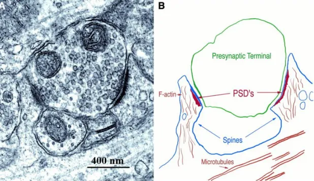
The post-synaptic density (PSD; Figure 1.7) is a complex net of interacting proteins at the synapse that lies just underneath and in close connection with the post-synaptic membrane, opposed to the active zone of the presynaptic neuron. Its composition includes neurotransmitter receptors, effector proteins, enzymes, scaffold proteins and structural proteins29.
Figure 1.7. The PSD, opposing presynaptic terminals forming glutamatergic synapses with two dendritic spines. The
protein complex is visible with electron microscopy as a thickening of approximately 30 nm in the postsynaptic membrane (A). In (B) a tracing of the structures shown in (A), with its most important structures identified, is depicted 76.
The molecules that compose the post-synaptic density have important roles, one of them being the regulation of downstream signaling pathways that adjust the molecular composition of the post-synaptic entities necessary to sustain synaptic plasticity77.
An important protein found in these structures is post-synaptic density protein 95 (PSD-95), which is exclusively found in glutamatergic post-synaptic densities, associating with glutamate receptors and cytoskeletal elements78,79.
PSD-95 has a role in synaptic stabilization and plasticity, through post-translational modifications such as phosphorylation and palmitoylation. PSD-95 palmitoylation on its N-terminus acts as an anchor for the protein in the postsynaptic membrane and is important for protein clustering at the PSD80,81. In
fact, the PSD-95 palmitoylation cycle regulates AMPAR trafficking: high neuronal activity leads to PSD-95 depalmitoylation which induces its dissociation from PSD. Consequently, glutamate receptors are internalized and synaptic strength decreases. With lower neuronal activity, PSD-95 suffers palmitoylation promoting PSD-95 and glutamate receptors trafficking to the postsynaptic membrane80,82.
Expression of PSD-95 lacking palmitoylation sites inhibits the incorporation of AMPARs into synapses during LTP83.
10
The hippocampus of rats with SRS-induced by KA, showed down-regulation of PSD-95 expression 6 weeks post-SE, that correlates with a behavioral deficit. This decrease in PSD-95 expression, alongside other factors, likely contributes to behavioral impairments such as spatial learning memory deficit, anxiety and increased locomotor activity10.
Like PSD-95 in excitatory synapses, gephyrin is found exclusively in inhibitory glycinergic and GABAergic synapses. This 93 kDa protein anchors glycine receptors and type A GABA (GABAA)
receptors, to the cytoskeleton84–86, ensuring the stability that guarantees that the appropriate number of
these receptors are localized at the post-synaptic membrane. Despite this stability, gephyrin scaffold sustains several forms of synaptic plasticity due to rapid changes in its composition77.
Although not as well studied as PSD-95 regarding synaptic plasticity, gephyrin is believed to sustain it through the regulation of GABAA receptor internalization and function, i.e. by regulating the
strength of GABAergic transmission. This regulation depends on changes in gephyrin through post-translational modifications, such as phosphorylation and/or palmitoylation, and interaction with signaling molecules that modulate downstream signaling cascades related to synaptic plasticity in GABAergic synapses87.
Patients with TLE showed aberrant alternative splicing in the gephyrin gene (GPHN), which lead to a gephyrin variant that clusters differently with GABAA receptors88. Also, a decrease in gephyrin
expression was detected in the hippocampus of rats from 24 hours to 2 months after the first grade of seizures induced with Li2+-Pilo89.
1.7. Summary of the changes found in TLE regarding the proteins mentioned
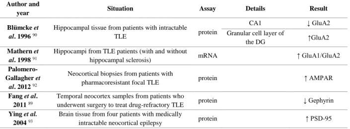
Several studies have been conducted to study what molecular changes happen in TLE, both in human tissue and in rodent models of TLE. Some of the changes in proteins involved in synaptic plasticity studied in the present work, can be found in human tissue (Table 1.1). Changes in these proteins observed in different rodent models of TLE are summarized in Table 1.2. Results for human tissue are very similar to the ones obtained for rat models. Interestingly, for the rat models, the results not only vary between brain regions or even hippocampus areas, but also along different periods of the disease.
Table 1.1. Protein changes found in patients with TLE. The diseased tissues were compared with non-seizure autopsies.
Note that for some studies not all differences found in each study are described, only the ones that are relevant for the present work. ↑ represents an increase; ↓ represents a decrease.
Author and
year Situation Assay Details Result
Blümcke et
al. 1996 90
Hippocampal tissue from patients with intractable
TLE protein
CA1 ↓ GluA2
Granular cell layer of
the DG ↑GluA2
Mathern et
al. 1998 91
Hippocampi from TLE patients (with and without
hippocampal sclerosis) mRNA ↑ GluA1/GluA2
Palomero-Gallagher et
al. 2012 92
Neocortical biopsies from patients with
pharmacoresistant focal TLE protein ↑ AMPAR
Fang et al. 2011 89
Temporal neocortex samples from patients who
underwent surgery to treat drug-refractory TLE protein ↓ Gephyrin
Ying et al. 2004 93
Brain tissue from four patients with medically
11
Table 1.2. Protein changes found in rodent models of TLE. Note that for some studies not all differences found in each
study are described, only the ones that are relevant for the present work. ↑ represents an increase; ↓ represents a decrease; - represents no changes.
Author and
Year Model Assay
Region of
the brain Period Result
Condorelli et
al. 1994 94 Li
2+-Pilo mRNA
Hippocampus SE: 24 hours after treatment with pilocarpine
↓ GluA1 - GluA2 and GluA3 Hippocampus
DG
SE: 12 hours after treatment with pilocarpine
↓ GluA1 ↑GluA3 Hippocampus
CA1
SE: 12-24 hours after treatment with pilocarpine
↓ GluA1 ↓ GluA3
Mathern et
al. 1998 45 Pilocarpine mRNA
Hippocampus
DG Chronic period: 85 days after SE ↑GluA2 Hippocampus CA1 ↓GluA2 Porter et al. 2006 46 Li
2+-Pilo mRNA Hippocampus
DG 2 weeks after SE
↑GluA2 ↓GluA3
Rajasekaran
et al. 2012 95 Pilocarpine protein Hippocampus
SE: 10 or 60 min after the
first seizures ↓GluA2 surface expression
Malkin et al. 2016 3 Li
2+-Pilo mRNA
Hippocampus Latent period: 1 day after treatment with pilocarpine
↓ GluA1 ↓GluA2
mPFC
Latent period: 3 days after
pilocarpine treatment ↑GluA1
1st to the 7th day after
treatment with pilocarpine ↓GluA2
Latent period: 3rd day after
treatment with pilocarpine ↑GluA1/GluA2
Lorgen et al.
2017 96 KA protein Hippocampus 8 weeks after SE ↓ GluA2 Russo et al.
2013 97 Pilocarpine protein Hippocampus 3 hours after SE
↑GluA2 ↓ GluA1 ↓ GluA1PSer831 Lopes et al. 2013 55 Pilocarpine protein Cortex
1, 3 and 12 hours after SE ↑GluA1PSer845 Hippocampus
↑GluA1PSer831 Chronic period: 50 days
after SE ↓GluA1PSer845
Cortex Latent period: 5 days after
SE ↓ GluA1
Hippocampus Chronic period: 50 days
after SE - GluA1 and GluA1PSer831
Lopes et al.
2015 8 Pilocarpine protein
Dorsal
hippocampus Chronic period: 60 days after treatment with
pilocarpine ↓GluA1PSer845 Ventral hippocampus ↓GluA1PSer831 Su et al. 2008 11 Li
2+-Pilo protein Hippocampus
Latent period: 2 days after
SE ↑ Kv4.2 channel
Chronic period: 50 days
after SE ↓ Kv4.2 channel
Sun et al.
2009 10 KA protein Hippocampus
Chronic period: 4 and 6
weeks after SE ↓ PSD-95
Fang et al. 2011 89 Li
2+-Pilo protein Temporal
lobe
from 24 hours to 2 months
12 1.8. Lipid rafts
Since the fluid mosaic model of membrane structure was proposed by Singer and Nicolson in 197298, several new aspects of membrane organization have been uncovered (Figure 1.8), such as the
transmembrane asymmetry of glycerophospholipids, but also the lateral heterogeneity (i.e., non-randomness) in lipid and protein distribution (see 99 for review). In fact, the membrane is currently
viewed as being composed by different functional domains which are distinct not only in their lipid and protein composition, but also in their biophysical properties, such as membrane lateral diffusion coefficient and thickness, and in the fact that they operate in a large range of time and length scales100.
This lateral organization is highly relevant for the neuronal membrane due to all the roles associated to it, some of them already mentioned above, and especially, but not only, in synapses. The existence of lateral interactions between the high diversity of neural membrane components, with different degrees of affinity between them, drives and stabilizes the existence of different domains101. These domains are
involved in the regulation of signaling pathways and other neurophysiological processes. Therefore, disturbance of this dynamic network in the membrane of neurons is associated with several pathologies, especially neurodegenerative diseases102.
Figure 1.8. Schematic representation of the fluid mosaic membrane proposed by Singer and Nicolson modified to highlight the diversity of lipid components and their heterogeneous transversal and lateral distribution. Two types of
domains are illustrated: those that are formed due to the segregation of different lipid components, and lipid annulus surrounding transmembrane proteins. Different colors correspond to different lipid species. The pink bulks represent proteins, and the black branches carbohydrates.103
The term lipid raft was first used by Simons and Ikonen in 1997 to describe the existence of membrane domains that were more rigid (liquid-ordered; lo) formed by the dynamic aggregation of
sphingolipids and cholesterol, that move along the liquid-disordered (ld) lipid bilayer, in a way similar
to a raft on water104. Many studies have been conducted since then with the purpose of better
understanding these domains. Regarding lipid raft composition in mammalian cells, it was found that besides cholesterol, these domains are also rich in a specific sphingolipid, sphingomyelin, and in glycosylphosphatidylinositol (GPI)-anchored proteins. From the Keystone Symposium on lipid rafts and
13
presently accepted. In addition to the already mentioned characteristics, this definition also includes the possibility of small rafts to be stabilized in order “to form larger platforms through protein-protein and protein-lipid interactions”105. The importance of these domains is evidenced by the functions that have
been attributed to them so far, that include organization of signaling molecules for the assembly of signaling platforms or trafficking, and regulation of membrane fluidity.
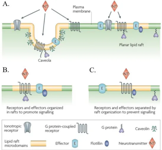
Figure 1.9. Lipids raft organization and possible roles in neurotransmitter signaling. Caveolae and planar lipid rafts (A)
are two types of ordered microdomains found in the CNS. These domains are believed to spatially organize different membrane molecules to promote (B) or prevent (C) signal transduction106.
In the CNS, lipid rafts have been associated with modulation of synaptic signaling by regulating neurotransmission and receptor trafficking107. The lipid raft signaling hypothesis proposes that these
domains organize the spatial distribution of signaling molecules at the membrane in order to promote kinetically favorable interactions that are necessary for signal transduction (Figure 1.9 - B). The opposite might also happen, i.e. the inhibition of interactions by separation of the signaling molecules which results in a dampening of the signaling response (Figure 1.9 - C)106. Two different types of lipid rafts
have been associated with these functions: planar lipid rafts and caveolae (Figure 1.9 - A)106. Caveolae
are flask-shaped membrane invaginations that have caveolins as protein markers. In a similar way, flotillins are marker proteins found in planar lipid rafts. Both proteins are thought to promote stability of these domains. Localization within these microdomains affects the binding affinity of neurotransmitters to ionotropic receptors, such as AMPARs108–110. However, it is still unclear exactly
how location within lipid rafts affects AMPAR properties.
Palmitoylation is one of the mechanisms responsible for proteins recruitment to lipid rafts104.
PSD-95, along other proteins important for synaptic signaling, can be found in these domains in a palmitoylation-dependent fashion. During rat postnatal development an increase in PSD-95 palmitoylation and lipid-raft constituent lipids were observed, which likely facilitate the formation of
14
lipid rafts81. In fact, remodeling of lipid raft composition in the CNS occurs during development and is
believed to contribute to altered cognition and synaptic plasticity observed upon ageing and neurodegenerative diseases106,111. However, it is still unknown if such changes contribute to the cognitive
decline and altered synaptic plasticity observed in TLE, and more studies are needed to better understand this issue.
1.8.1. Membrane model systems
The use of membrane model systems, such as Giant unilamellar vesicles (GUVs), is a good approach to study in detail a limited number of molecular interactions, to determine, amongst the overwhelming complexity surrounding biomembranes, which of those interactions are involved in a given biological process, and if they are altered in pathologies such as TLE. As stated above, cellular membranes can have different domains with different biophysical properties, such as fluidity. These properties vary according to the environment and composition of the membrane itself and are reminiscent of the lipid phases found in membrane model systems100. The existing lamellar phases, i.e., those in which the lipids
are organized into a lipid bilayer, as in biomembranes, under close to physiological conditions are ld, lo
and solid-ordered (so), here listed from more fluid to more ordered and compact112. It is usually
considered that biological membrane domains, are in a fluid state, and therefore are considered either ld
–like or lo –like domains, although evidence in the past decade has emerged for the existence also of so–
like domains in living cells under physiological conditions, at least in yeast113. Lipid phases and domains
have been studied by a variety of spectroscopic and microscopic techniques112. One of those has been
fluorescence spectroscopy and microscopy, which relies on the use of sensitive molecular probes that have a certain preference in partitioning to specific domains, e.g. N-(lyssamine Rhodamine B sulfonyl)-1,2-dioleoyl-sn-glycero-3-phosphoethanolamine (Rhod-DOPE) prefers to partition into ld over lo and is
largely excluded from so domains114.
As membrane model systems, GUVs are liposomes with around 20 to 100 μm of diameter used to study, among others, how lipid composition affects shape and stability of domains. By using a fluorescent probe, domains with sizes on the μm range can be visualized by fluorescence microscopy, preferably using a laser-scanning confocal microscope115,116.
From phase diagrams such as the one present in Figure 1.10, it is possible to estimate the percentage of each phase in the vesicle for each mixture, by using the tie-line which contains the proportion of components used in the experiment and the lever rule112. To obtain tie-lines in ternary systems usually
it is necessary to physically separate the phases in coexistence and to determine the composition of each phase. To obtain the tie-lines in ternary lipid systems, namely from GUV imaging, information regarding the composition of each domain observed in the vesicles studied, especially the percentage of each component, would be needed117. This information can be indirectly estimated from spectroscopic
measurements in which the spectra or quantitative parameters obtained can be separated in to the individual contribution of each phase, such as fluorescence lifetime components of fluorescent probes or Nuclear magnetic resonance (NMR), namely 2H-NMR of a lipid component with perdeuterated acyl
chain versus 1H-NMR of the other lipid. Moreover, there are thermodynamic rules that restrict the
different possible directions for the tie-lines. All together these theoretical and experimental approaches allowed to obtain ternary phase diagrams and several tie-lines, particularly in the ld/lo coexistence region with a high accuracy, with uncertainty in the order of 2 mol% or less. These phase diagrams are now a well-established and fundamental framework in membrane biophysical studies concerning membrane domains and lipid rafts.114
15
Figure 1.10. Miscibility phase diagram for DPPC, DOPC and cholesterol at 24°C. Regions of different phase coexistence are defined by red boundaries as shown: the ld + lo, ld + so and lo + so areas indicate the respectively ld/lo, ld/so, lo/so phase