Activation of Dorsal Raphe Serotonergic Neurons
Promotes Waiting but Is Not Reinforcing
Highlights
d
Optogenetic activation of serotonin neurons promotes
waiting for delayed rewards
d
The effect of serotonin neuron activation is not general to all
motor behaviors
d
Serotonin neuron activation shows no appetitive or aversive
effects
Authors
Madalena S. Fonseca, Masayoshi
Murakami, Zachary F. Mainen
Correspondence
zmainen@neuro.fchampalimaud.org
In Brief
Fonseca et al. show that optogenetic
activation of serotonergic neurons in the
dorsal raphe nucleus prolongs waiting for
delayed rewards but has no aversive or
appetitive effects in several different
behavioral assays. These results
establish a link between serotonin and
impulse control that is independent of
reinforcing effects.
Fonseca et al., 2015, Current Biology25, 306–315 February 2, 2015ª2015 Elsevier Ltd All rights reserved http://dx.doi.org/10.1016/j.cub.2014.12.002
Current Biology25, 306–315, February 2, 2015 ª2015 Elsevier Ltd All rights reserved http://dx.doi.org/10.1016/j.cub.2014.12.002
Article
Activation of Dorsal Raphe
Serotonergic Neurons Promotes
Waiting but Is Not Reinforcing
Madalena S. Fonseca,1,2Masayoshi Murakami,1,2and Zachary F. Mainen1,*
1Champalimaud Neuroscience Programme, Champalimaud
Centre for the Unknown, Avenida de Brası´lia, 1400-038 Lisbon, Portugal
Summary
Background: The central neuromodulator serotonin (5-HT) has been implicated in a wide range of behaviors and affective disorders, but the principles underlying its function remain elusive. One influential line of research has implicated 5-HT in response inhibition and impulse control. Another has sug-gested a role in affective processing. However, whether and how these effects relate to each other is still unclear. Results: Here, we report that optogenetic activation of 5-HT neurons in the dorsal raphe nucleus (DRN) produces a dose-dependent increase in mice’s ability to withhold premature re-sponding in a task that requires them to wait several seconds for a randomly delayed tone. The 5-HT effect had a rapid onset and was maintained throughout the stimulation period. In addition, movement speed was slowed, but photostimulation did not affect reaction time or time spent at the reward port. Using similar photostimulation protocols in place preference and value-based choice tests, we found no evidence of either appetitive or aversive effects of DRN 5-HT neuron activation. Conclusions: These results provide strong evidence that the efficacy of DRN 5-HT neurons in promoting waiting for delayed reward is independent of appetitive or aversive effects and support the importance of 5-HT in behavioral persistence and impulse control.
Introduction
The central neuromodulator serotonin (5-HT) has been impli-cated in a variety of different sensorimotor, affective, and cognitive behaviors, but the general principles that underlie its diverse effects remain unclear. 5-HT has long been associ-ated with response inhibition and impulse control [1, 2], because reductions in 5-HT levels disinhibit behavior sup-pressed by punishment (reviewed in [1]) and increase prema-ture responding for reward [3–6]. Recently, Miyazaki and colleagues showed that neurons in the dorsal raphe nucleus (DRN), the chief source of serotonin to the forebrain, are active while rats wait for delayed rewards and delayed reward-predic-tive cues [7], that blocking DRN 5-HT activity increases prema-ture responding [8], and that photostimulation of DRN 5-HT neurons decreases it [9]. These findings led to the more specific proposal that 5-HT facilitates ‘‘patience’’ for delayed reward [6]. 5-HT has also been hypothesized to play an important role in reinforcement learning, acting in coordination with dopamine (DA) [10–12]. One line of research has highlighted a role for 5-HT in representing aversive events [13–17]. Other studies
have shown that putative 5-HT neurons in the DRN respond to reward [18–21] and that 5-HT depletion impairs reward pro-cessing [22, 23]. Responses to painful mechanosensory stim-uli are attenuated by pharmacological [24] and optogenetic [25] increases in 5-HT. Recently, optogenetic stimulation of DRN 5-HT neurons was even shown to positively reinforce several behaviors [26]. Although this issue appears to be com-plex (see [27]), this raises the possibility that enhancement of waiting by 5-HT optogenetic stimulation could be an indirect consequence of a reward-like signal, possibly by inducing a state that animals seek to maintain.
To address these issues, in the present report, we per-formed a series of experiments examining the effects of opto-genetic activation of DRN 5-HT neurons in both a waiting task and in a series of tasks that assess value via preference and choice. We found that photostimulation of DRN 5-HT neurons led to a dose-dependent increase in the ability of mice to with-hold premature responding in a waiting task. In contrast, using the same photostimulation parameters, we found no evidence of an appetitive or aversive effect of DRN 5-HT activation in two place preference tests, and a third, extremely sensitive, probabilistic value-based choice task. These results provide direct evidence of sufficiency of DRN 5-HT activation in pro-moting waiting behavior, establishing a definitive link between 5-HT and behavioral changes that is independent of reinforc-ing effects.
Results
A Quantitative Paradigm for Assessing Waiting Behavior To study the effect of DRN 5-HT stimulation on waiting behavior, we trained adult transgenic mice expressing CRE re-combinase under the serotonin transporter promoter (SERT-Cre) and wild-type littermates (WT) in a waiting task (Figure 1A) until their performance was stable (minimum 2 months). The experimenters were blind to the mice’s genotype throughout training, surgery, and testing. The task required mice to nose poke at a ‘‘waiting port’’ until the presentation of a tone, after which they could move to a second port to obtain a water reward. The tone was randomly delayed using an exponential distribution (Figure 1A, inset) with a mean adjusted for each animal so that it successfully waited in just over 50% of the tri-als. This promoted a wide distribution of waiting times (Figures 1B and 1C) [28] and provided a sensitive measure of waiting time. Mice performed around 423.26 122.3 trials per session, mean6 SD, total n = 128 sessions from 11 mice. We classified the trials into ‘‘patient’’ (53.7%6 6.3%, mean 6 SD, n = 11 mice) and ‘‘impatient’’ depending on whether the mouse exited the waiting port before or after the tone, respectively. The me-dian waiting time ranged from 1.0 to 7.5 s across mice, with a mean of 2.86 1.8 s (mean 6 SD). Mice understood the tone-reward association, as shown by the prompt response to the tone (Figure 1D; median reaction time 150 ms for the example mouse, population range: 74–316 ms).
Optical Activation of DRN 5-HT Neurons Prolongs Waiting After training, we expressed the light-sensitive ion channel channelrhodopsin-2 (ChR2) in DRN 5-HT neurons using an 2Co-first author
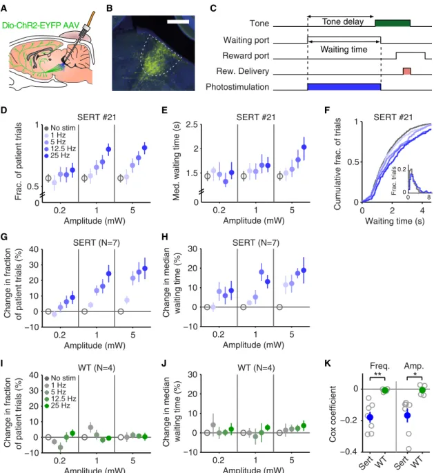
AAV2/1 viral vector (AAV2/1-Dio-ChR2-EYFP) injected into the DRN of SERT-Cre mice (n = 7) or wild-type littermates (WT, n = 4) and implanted an optical fiber in the same location (Figure 2A) (see [25] for more details). Histology performed at the end of testing showed ChR2-YFP expression localized to the DRN in SERT-Cre animals (Figure 2B) and no expression in WT controls (data not shown).
To test the effect of DRN 5-HT activation on waiting behavior, after allowing 3–4 weeks for virus expression and re-training, we delivered photostimulation at different fre-quencies (0, 1, 5, 12.5, and 25 Hz) and amplitudes (0.2, 1, and 5 mW) in randomly interleaved trials, with photostimula-tion beginning when the animal entered the waiting port and lasting until the animal exited it (seeFigure 2C). Photostimula-tion in SERT-Cre mice, but not WT littermates, resulted in an increase in the fraction of patient trials and median waiting time.Figure 2shows waiting task performance for a represen-tative SERT-Cre mouse (Figures 2D–2F) and the population of SERT-Cre mice (Figures 2G and 2H) and WT mice (Figures 2I and 2J). These effects were confirmed with a three-way ANOVA (frequency, amplitude, genotype) on the normalized fraction of patient trials (main effect of genotype, F(1,9) =
10.220, p = 0.011, frequency 3 genotype, F(2.205,19.845) =
3.392, p = 0.002, amplitude3 genotype: F(1.566,14.097)= 3.913,
p = 0.053, no other terms involving genotype were significant) and normalized median waiting time (frequency3 genotype: F(3.000,27.000) = 4.926, p = 0.007, amplitude 3 genotype:
F(2.000,18.000)= 3.565, p = 0.050, no other terms involving
geno-type were significant), followed by separate two-way ANOVAs restricted to SERT-Cre (fraction of patient trials: main effect of frequency, F(1.718,10.307) = 16.439, p = 0.001, main effect of
amplitude: F(1.353,8.118)= 10.633, p = 0.008, n = 7 mice; median
waiting time: main effect of frequency, F(3,18) = 10.094, p <
0.001, main effect of amplitude: F(2,12)= 8.303, p = 0.005, n =
7 mice) and WT (no significant effects in either measure, n = 4 mice).
The dose dependency of photostimulation frequency and amplitude on waiting time were confirmed by a Cox regres-sion. The analysis yielded a negative coefficient for both frequency and amplitude in SERT-Cre animals (Figure 2K; frequency: –0.1796 0.035; amplitude: –0.167 6 0.044, n = 7 mice), that was significantly different from zero (one-sample t test for frequency: p = 0.002, n = 7 mice, and amplitude: p = 0.009, n = 7 mice) and significantly different from WT (two-sample independent t test (SERT versus WT) for fre-quency, p = 0.005; and amplitude, p = 0.0267, NSERT = 7,
NWT= 4), demonstrating a frequency- and
amplitude-depen-dent reduction in the probability of giving up waiting. Effects of DRN 5-HT Photostimulation Are Rapid and Persistent
To characterize the time course of the photostimulation effect on waiting, we estimated the hazard rate of leaving the waiting port (see theSupplemental Experimental Proceduresfor de-tails). DRN 5-HT photostimulation led to a reduction in the hazard rate of leaving in a manner that was both frequency dependent (Figure 3A) and amplitude dependent (data not shown, see alsoFigure 2K for Cox regression coefficients). The hazard rate effect did not decrease or reverse over photo-stimulation time (Figures 3A and 3C). To estimate the earliest detectable effect, we calculated the time at which the cumula-tive hazard rates for trials with and without photostimulation could be distinguished by a permutation test (Figure 3B). Five of the seven SERT-Cre mice had detectable onset times Waiting port
Reward port Tone delay dist.
Tone delay (s) 0.1 0 P ro babi lit y Reward Patient A Waiting time Resp. time Mov. time Poke Wait
Poke out Poke out
0 2 4 6 100 110 120 130 140 150 0 4 8 Waiting time (s) Trial B 0 0.6 0 0.1 0.2 Response time (s) Fraction of trials Tone Impatient Patient 0 0.02 0.04 Fraction of trials 0 2 4 6 8 Waiting time (s) C D Tone Reward Delivery Reward port Waiting port Tone delay Impatient
Figure 1. The Waiting Task and the Behavioral Performance
(A) Schematic diagram of trial events in the waiting task (top). In each trial, a mouse is required to wait for a randomly delayed tone and move to the reward port to obtain a water reward (patient trial). If the mouse fails to wait for the tone, the reward is not available (Impatient trial). An example of the probability distribution of the delays to the tone is shown in the inset. Timeline of the task events and definition of the behavioral parameters (bot-tom). The green rectangle indicates presentation of the tone, and the pink rectangle indicates the water reward.
(B) Snapshot of the waiting behavior. The waiting period in each trial is indicated as a light red or gray bar, representing patient and impa-tient trials, respectively. Green ticks represent the presentation of the tone.
(C) Waiting-time histograms of impatient trials (gray) and patient trials (red) of an example mouse. The histograms show data pooled across sessions. (D) A histogram of reaction time to the tone of an example mouse. The light shaded area indicates the 95% range of reaction time histograms from the shuffled data.
0.2 1 5 −10 0 10 20 30 40 Amplitude (mW)
Change in fraction of patient trials (%)
0.2 1 5 −10 0 10 20 30 40 Amplitude (mW)
Change in fraction of patient trials (%)
0.2 1 5 −10 0 10 20 30 Amplitude (mW)
Change in median waiting time (%)
0.2 1 5 −10 0 10 20 30 Amplitude (mW)
Change in median waiting time (%)
SERT (N=7) WT (N=4) WT (N=4) 0.2 1 5 1.5 2 2.5 Amplitude (mW)
Med. waiting time (s) 0
0.2 1 5
0.5 1
Amplitude (mW)
Frac. of patient trials 0
No stim 1 Hz 5 Hz 12.5 Hz 25 Hz No stim 1 Hz 5 Hz 12.5 Hz 25 Hz D F G H I J K Tone delay Waiting time Dio-ChR2-EYFP AAV A B C SERT (N=7) E −0.4 −0.2 0 Cox coefficient Sert WT Sert WT Freq. Amp.
**
*
0 2 4 0 0.5 1 Waiting time (s)Cumulative frac. of trials
0 8 0 0.2 Frac. trials Photostimulation Rew. Delivery Reward port Waiting port Tone
Figure 2. Photostimulation of DRN 5-HT Neurons Promotes Waiting Behavior
(A) A schematic drawing of the optogenetic approach. DRN neurons are infected with AAV2/1-Dio-ChR2-EYFP. In SERT-Cre mice, 5-HT neurons will express ChR2-YFP (green cells) and can be photoactivated with blue light delivered through an optical fiber implant.
(B) A fluorescence image of a parasagittal section showing localized ChR2-YFP expression in the DRN. YFP in green. DAPI in blue. Scale bar, 500mm. (C) Photostimulation period (blue rectangle) is shown along with the task events. The same format as inFigure 1A.
(D) Dose-dependent increase in the fraction of patient trials with DRN 5-HT photostimulation from an example mouse, SERT #21. Error bars indicate 95% confidence intervals calculated using binomial parameter estimates. Note that all the nonstimulated trials are pooled together and repeatedly plotted across the three amplitudes for visualization purpose, here and elsewhere.
(E) Dose-dependent increase in median waiting times with DRN 5-HT photostimulation in the same example mouse. Error bars indicate a 95 percentile range (2.5–97.5 percentile) of a bootstrap distribution.
(F) Cumulative histograms of waiting times across frequencies (at 5 mW) from the same mouse. Inset shows waiting-time histograms of nonstimulated and stimulated (25 Hz at 5 mW) trials. Patient and impatient trials are pooled together in each histogram.
(G) Dose-dependent increase in the fraction of patient trials for the population of SERT-Cre mice (n = 7). Percent changes in the fraction of patient trials with respect to nonstimulated trials, averaged across mice, are shown. Error bars indicate SEM.
(H) Dose-dependent increase in median waiting times for the population of SERT-Cre mice (n = 7). Percent changes in median waiting time with respect to nonstimulated trials, averaged across mice, are shown. Error bars indicate SEM.
(I) The same as (G) but for the population of wild-type mice (n = 4). (J) The same as (H) but for the population of wild-type mice (n = 4).
(K) Cox regression, demonstrating a dose-dependent increase in waiting performance. Cox regression coefficients for both frequency (left) and amplitude (right) are shown for SERT-Cre mice (blue) and wild-type mice (green). Individual mice in open circles. Averages across mice are shown in filled circles. Error bars represent SEM. **p < 0.01; *p < 0.05 with independent two-sample t test (SERT versus WT). Note that negative coefficients indicate that the higher the frequency (or amplitude), the lower the leaving rate, thus the longer the waiting time.
below 1 s (Figure 3D; range: 0.50–2.14 s; population median 0.66 s).
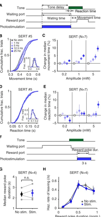
Effects of DRN 5-HT Photostimulation Are Neither Specific to Waiting nor General to All Motor Behavior
Next, we investigated the possibility that the photostimulation effects on waiting reflected general inhibition of behavior or slowing down of motor actions. We first analyzed the move-ment time (time taken from exiting the waiting port to entering the reward port) in the same task and sessions as above (Fig-ure 4A). Median movement time was indeed increased after photostimulation of DRN 5-HT neurons (Figures 4B and 4C). This effect was confirmed with a three-way ANOVA including genotype as a factor (frequency3 genotype: F(3.000,27.000)=
11.280, p < 0.001, amplitude 3 genotype: F(1.778,15.999) =
8.631, p = 0.004) and a follow-up two-way ANOVA restricted to SERT-Cre (frequency, F(2.652,15.911) = 16.851, p < 0.001,
amplitude, F(1.432,8.594)= 17.117, p = 0.002, frequency3
ampli-tude, F(4.194,25.165)= 6.177, p = 0.001, n = 7 mice) and WT (no
significant effects, n = 4 mice) mice.
As another possible indicator of general motoric effects, we examined the tone reaction time, i.e., the time between tone onset and movement onset (Figure 4A). Note that the reaction time period is included in the waiting-time measurement
and occurs when photostimulation is active, whereas the movement time period occurs after the photostimulation has turned off. In contrast to the movement time, tone reaction time was not affected either in the same mouse (Figure 4D) or in the population (Figure 4E) as indicated by a three-way ANOVA (minimum p = 0.175).
To investigate this issue further, we ran a separate experi-ment in which photostimulation was delivered while animals were at the reward port retrieving the water reward (25 Hz at 5 mW, 0–3 s from port entry) (Figure 4F). Although the reward port period did not require animals to wait (water was delivered 200 ms after poke-in, as in the previous experiment), it was otherwise very similar to the waiting period in terms of motor requirements. Yet, photostimulation had no significant effect on the median reward poke duration (Figure 4G, paired t test, p = 0.528, n = 4 mice) or on the hazard rate of leaving the reward port (Figure 4H, Cox coefficient –0.10876 0.1125, one-sample t test, p = 0.405, n = 4 mice). Together, these re-sults show that the effects of DRN 5-HT stimulation are not entirely specific to the action of waiting, because they also increased movement time, but that they did not extend to other motor aspects of the task.
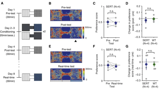
DRN 5-HT Photostimulation Does Not Produce Conditioned or Real-Time Place Preference
To test whether the increase in waiting times observed was related to possible appetitive or aversive reinforcing effects of DRN 5-HT photostimulation [26], we first ran a conditioned place preference (CPP) test, a standard assay for testing the rewarding effects of natural and artificial stimuli (Figure 5A, days 1–4). After a habituation session (pre-test), mice were subjected to 2 days of conditioning (days 2–3). On each day, mice were confined to one side of the box and given either photostimulation (12.5 Hz at 5 mW, parameters that showed robust waiting effects across all mice, for 3 s, every 10 s) or no stimulation. They were then placed in the other side cham-ber and given the alternative treatment. On day 4, mice were retested in the absence of photostimulation (post-test). Occu-pancy plots (time spent in each region of space) for pre- and post-test for an example mouse are shown inFigure 5B. Pref-erence for the chamber paired with photostimulation was as-sessed using a preference score calculated as the difference between the times spent in the stimulated and nonstimulated chamber, divided by the sum of the two. We found no signifi-cant difference in preference score between pre- and post-test (Figure 5C, paired t post-test, p = 0.609, n = 4 mice) or between SERT-Cre and WT controls (Figure 5D, two-sample indepen-dent t test, p = 0.728, n = 4 per group).
We then tested the same mice on a real-time version of the place preference test, 4 days later. In this test, entry to the stim-ulation-assigned chamber (the same side paired with stimula-tion in CPP) triggered optical stimulastimula-tion (12.5 Hz at 5 mW for 3 s, every 10 s, until chamber exit). This version of the place pref-erence procedure is more similar to the waiting-time task in that mice could increase the time of stimulation by staying in one of the chambers. Occupancy plots for an example mouse are shown inFigure 5E. Consistent with the results in the stan-dard CPP assay, there was no significant difference in prefer-ence between the pre-test and the real-time test (Figure 5F; paired t test, p = 0.701, n = 4 mice) or between SERT-Cre and WT animals (Figure 5G; independent two-sample t test, p = 0.987, n = 4 per group). Thus, DRN 5-HT photostimulation showed no reinforcing effects, appetitive or aversive in either standard or real-time place preference tests.
A B
C D
Figure 3. Time Course of the Effect of DRN 5-HT Photostimulation (A) Fast onset and sustained effect of DRN 5-HT photostimulation. Hazard rate of leaving the waiting port is plotted across time for an example mouse, SERT #6. Only data from the 5 mW photostimulation conditions are shown. Error bars indicate a 95 percentile range (2.5–97.5 percentile) of a bootstrap distribution.
(B) Fast onset of the effect of DRN 5-HT photostimulation. Cumulative hazard rate is plotted across time. Data for nonstimulated and stimulated (25 Hz at 5 mW) trials are shown. The red circle indicates the detectable onset time. The inset shows the zoomed-in view near the origin.
(C) Hazard rate of leaving for the population of SERT-Cre mice (n = 7). Aver-ages across mice are shown. Error bars indicate SEM.
(D) Detectable onset times across the population of SERT-Cre mice (n = 7). The black bar indicates the median across mice. Individual mice are shown in open circles.
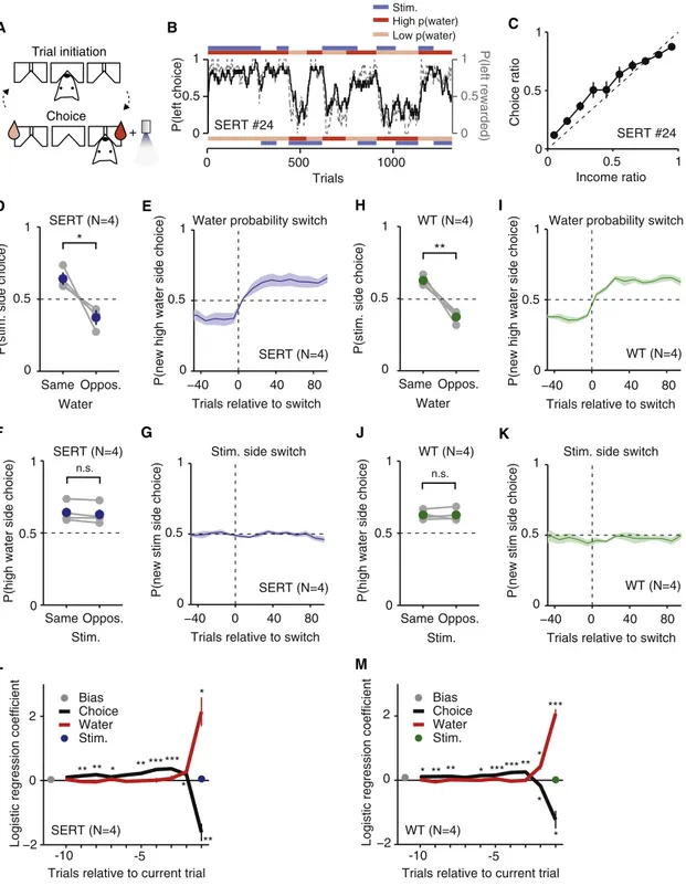
DRN 5-HT Photostimulation Does Not Bias Choices in a Probabilistic Choice Task
To further investigate the possible reinforcing effects of DRN 5-HT photostimulation, we trained and tested the same mice in a probabilistic choice task (Figure 6) [29–31]. This task required mice to initiate a trial with a nose-poke entry and then choose one of two choice ports (Figures 6A and 6B). Each choice port was associated with a specific reward prob-ability and photostimulation probprob-ability. After completion of one block, new reward and photostimulation probabilities were randomly chosen and a new block began.
In this task, mice showed a choice (left/right) ratio that closely matched the reward income ratio (Figure 6C), as ex-pected from Herrnstein’s matching law [29]. Thus, mice’s choices tracked switches in water probabilities (Figure 6E). Furthermore, water significantly affected their overall choice probabilities: the probability of choosing the photostimulation side was significantly higher in blocks where that side was associated with high water probability than in blocks where that side was associated with low water probability (Figure 6D, paired t test, p = 0.027, n = 4 mice). In contrast, we found no sig-nificant difference between the probability of choosing the ‘‘high water probability side’’ in blocks where photostimulation was present on that side or on the opposite side (Figure 6F; paired t test, p = 0.087, n = 4 mice) and no evidence that mice’s choice behavior tracked switches in photostimulation side (Figure 6G;Figures S1A–S1C). Similar results were obtained for WT littermate controls (Figures 6H–6K; water: p = 0.005, photostimulation: p = 0.982, n = 4 mice;Figures S1D–S1F).
We further analyzed the effect of photostimulation by using a logistic regression model [31] to predict the choice behavior using choice history, water reward history, and photostimula-tion history. As shown inFigure 6L, the history of water reward and choices had a strong effect on the choice probability, but there was no significant effect of photostimulation history. This was also true for WT animals (Figure 6M).
Finally, to exclude the possibility that the lack of reinforcing effects of photostimulation was due to insufficient ChR2 acti-vation, we examined the effect of photostimulation on waiting time in the same mice. Because the long training times excluded the possibility of running the mice on the waiting task, we modified the probabilistic choice task (Figure 7A) so that the mice had to wait at the reward port for a variable delay to obtain a water reward (min 0 s, mean 1.0–10.78 s, selected for each session to achieve around 50% waiting success). We expected photostimulation to increase patient waiting but that these effects would be smaller than those during tone waiting [9]. Photostimulation indeed led to a reduction in the hazard rate of leaving the reward port while mice waited for a delayed reward (Figures 7B and 7C). This was confirmed by a significant reduction in the Cox coefficient compared to WT mice (Figure 7D, two sample independent t test, SERT versus WT, p = 0.04). Thus, despite having undergone a long
0.2 1 5 −5 0 5 10 15 Amplitude (mW) Change in median movement time (%)
0.2 1 5 −5 0 5 10 15 Amplitude (mW) Change in median reaction time (%)
SERT (N=7) SERT (N=7) E D A C B Reaction time No stim. Stim. 1 2 3 4 duration (s) n.s.
Reward poke duration (norm.) SERT (N=4) SERT (N=4)
F
G
Median reward poke
0 0.5 1
Haz. rate of leaving (/s)
0 0.4 0.6 0.2 No stim. Stim. H
Cumulative frac. trials
0.05 0.1 0.15 0.2 0
0.5 1
Reaction time (s)
Cumulative frac. trials
Frac. trials 0 0.2 0.1 0 0.1 0.2 0.3 0.4 0.5 0.6 0 0.5 1 Movement time (s) 0.2 0.6 Frac. trials 0 0.2 No stim 1 Hz 5 Hz 12.5 Hz 25 Hz Photostimulation Reward port Waiting port
Tone Tone delay
Wating time Movement time
Photostimulation Reward port Waiting port Tone
Reward poke dur.
3 s
Figure 4. The Effect of DRN 5-HT Photostimulation on Movement Time, Re-action Time, and Time Spent at the Reward Port
(A) Photostimulation period (blue rectangle) and the definition of behavior parameters are shown along with the task events. The same format as in
Figure 1A.
(B) Cumulative histograms of movement times across frequencies (at 5 mW) from an example mouse, SERT #5. Inset shows movement time histograms of nonstimulated and stimulated (25 Hz at 5 mW) trials.
(C) Increase in movement times with DRN 5-HT photostimulation for the population of SERT-Cre mice (n = 7). Percent changes in movement times with respect to nonstimulated trials, averaged across mice, are shown. Error bars indicate SEM.
(D) Cumulative histograms of reaction times across frequencies (at 5 mW) from an example mouse, SERT #5. Inset shows reaction time histograms of nonstimulated and stimulated (25 Hz at 5 mW) trials.
(E) No systematic changes in reaction times with DRN 5-HT photostimula-tion for the populaphotostimula-tion of SERT-Cre mice (n = 7). Percent changes in reacphotostimula-tion times with respect to nonstimulated trials, averaged across mice, are shown. Error bars indicate SEM.
(F) Reward port photostimulation experiment. A subset of the animals used in the waiting-period photostimulation experiment (SERT-Cre, n = 4; WT, n = 2, data not shown). Photostimulation period (blue rectangle) and the defini-tion of the behavior parameter are shown along with the task events. The same format is used as inFigure 1A.
(G) No significant change in reward poke duration with DRN 5-HT photosti-mulation for the population of SERT-Cre mice (n = 4). Individual mice are shown in gray circles. Averages across mice are shown in filled circles. Error bars indicate SEM.
(H) No significant change in hazard rate of leaving the reward port with DRN 5-HT photostimulation, averaged across mice. Error bars indicate SEM. 310
period of expression and testing, photostimulation was still effective in producing waiting-time effects. Furthermore, his-tological examination confirmed that these mice used for the reinforcement assays and the mice used for the main waiting task were similar in terms of the number of infected cells, the position of the infection, and the fiber placement (Figure S2). Based on these results, it is unlikely that the lack of reinforcing effects is explained by insufficient ChR2 activation or other operational differences.
Discussion
Using optogenetic methods, we selectively activated DRN 5-HT neurons while mice performed a waiting task. Our main findings were that activation of DRN 5-HT neurons resulted in an in-crease in mice’s ability to wait in a delayed response task but did not promote conditioned place preference, real-time place preference, or choice bias in a probabilistic choice task. Serotonin Promotes Patient Waiting
Our waiting results are consistent with previous studies showing that reduced levels of 5-HT lead to impulsive re-sponding for reward [3–5, 8, 32–34], that putative 5-HT neurons are active in a situation that involves waiting for a delayed reward or a delayed conditioned cue [7] and that optogenetic DRN 5-HT activation prolongs waiting [9]. We extend these findings by showing that selective activation of DRN 5-HT
neurons is not only causally sufficient to facilitate ‘‘patient’’ waiting, but that this modulation is fully independent of appe-titive or aversive affects, assessed via changes in preference and choice.
We believe these effects are likely to accurately reflect the physiological effects of 5-HT neuron activation because we observed a monotonically increasing, dose-dependent change in waiting performance using 1–25 Hz. This range in-cludes frequencies observed in previous DRN electrophysio-logical studies [7, 18, 19] and is a range over which 5-HT neuron photostimulation produces a monotonic increase in firing rate [25]. Although the effects could have been enhanced by a relatively high degree of synchronization produced by photostimulation, similar results were obtained using a step-function opsin that is likely to produce less synchronization [9]. The effects of photostimulation developed very rapidly (within 0.5 s) and persisted throughout the photostimulation period. Thus, DRN 5-HT photostimulation can affect behavior in a subsecond timescale. This result contradicts the classical view of a slow action of neuromodulatory systems but is consistent with similarly fast DRN neuronal responses to external stimuli [18–21].
Serotonin Does Not Increase Patient Waiting via Reinforcing Effects
Liu et al. [26] recently reported that optogenetic activation of DRN 5-HT neurons can serve as a positive reinforcer. Thus,
0 0.5 1 Preference score SERT (N=4) WT (N=4) −0.2 −0.1 0 0.1 0.2 Pre Real-time 0 0.5 1 SERT (N=4) WT (N=4) −0.2 −0.1 0 0.1 0.2 −0.3 Change in preference
score (post - pre)
n.s. n.s. n.s. n.s. −0.3 Preference score
Change in preference score (real-time - pre)
SERT (N=4) Pre Post Test SERT (N=4) Test C D F G Pre-test Post-test A Day 1 Pre-test x2 (30min)
. . .
Day 2−3 Conditioning (20min/sess.) Day 4 Post-test (30min) Day 8 Real-time (30min) 0 300ms 0 300ms B Pre-test Real-time test EFigure 5. The Effect of DRN 5-HT Photostimulation in the Place Preference Tests
(A) Conditioning and testing schedule for the conditioned place preference (CPP) test (days 1–4) and real-time place preference test (day 8). Photostimu-lation was delivered at 12.5 Hz, 5 mW for 3 s, every 10 s.
(B) Occupancy plots for the pre-test session and post-test session for an example SERT-Cre mouse, SERT #28. The photostimulation paired chamber is indicated by the arrowhead.
(C) No significant change in the preference score after conditioning. Individual mice shown in gray circle. Averages across mice in blue. Error bars indicate SEM. n.s., not significant.
(D) No significant difference in the change in the preference score between SERT-Cre and WT mice for the conditioned place preference test. Individual mice are shown in gray circles. Average across mice shown in blue (SERT-Cre) or green (WT). Error bars indicate SEM.
(E) Occupancy plots for pre-test session and real-time stimulation session for an example SERT-Cre mouse, SERT #28. The blue rectangle indicates the pho-tostimulation-associated chamber. Note that the pre-test session is common for both the conditioned place preference test and real-time preference test. (F) No significant difference in the preference score between the pre-test and real-time session. The same format as shown in (C).
(G) No significant difference in the change in the preference score between SERT-Cre and WT mice for the real-time place preference test. The same format as shown in (D).
Bias Choice Water Stim. Bias Choice Water Stim. P(left choice) 0 0.5 1 0 0.5 1 Income ratio Choice ratio −40 0 40 80 0 0.5 1
P(new high water side choice)
Trials relative to switch
−40 0 40 80 Trials relative to switch 0
0.5 1
P(new stim side choice)
Same 0 0.5 1
P(stim. side choice)
Water probability switch
Stim. side switch Water * 0 0.5 1 Same Oppos. Stim. Oppos.
P(high water side choice)
n.s. SERT (N=4) SERT (N=4) Same 0 0.5 1
P(stim. side choice)
Water Oppos. 0 0.5 1 Same Oppos. Stim.
P(high water side choice)
WT (N=4) WT (N=4) −40 0 40 80 0 0.5 1
P(new high water side choice)
Trials relative to switch
−40 0 40 80 Trials relative to switch 0
0.5 1
P(new stim side choice)
Choice Trial initiation
+
Water probability switch
Stim. side switch
n.s. D E A B C F G J K SERT (N=4) SERT (N=4) WT (N=4) WT (N=4) H I L M
Logistic regression coefficient
-5 -10
Trials relative to current trial
−2 0 2
Logistic regression coefficient
-5 -10
Trials relative to current trial
* ** * *** *** ** ** * ** *** * * * ** *** *** * ** ** * WT (N=4) SERT (N=4) −2 0 2 ** 0 500 1000 Trials Stim. High p(water) Low p(water) 0 0.5 1 0 0.5 1 P(left rewarded)
Figure 6. The Effect of DRN 5-HT Photostimulation in the Probabilistic Choice Task
(A) Schematic diagram of trial events in the probabilistic choice task. In each trial, a mouse is required to enter the center port and then move to the reward port to obtain a water reward delivered in a probabilistic manner. The pink and red water drops indicate the side port associated with the lower and higher probability of water reward, respectively. The blue light indicates the side port associated with the photostimulation (12.5 Hz, 5 mW for 1 s).
(B) The block schedule and mouse choice behavior from example sessions. Probability of choosing the left port (black solid line) overlaid with prob-ability of obtaining reward at the left port (gray dashed line) (moving average of past 20 trials) are shown across trials for an example mouse, SERT #24. Two example sessions are concatenated. The top red/pink bar indicates the probability of water reward associated with the left port in a block of trials. The top blue bar indicates blocks in which the left port was associated with photostimulation. The bottom bars represent the same but for the right port.
(C) Matching behavior. The choice ratio (probability of choosing left) is plotted as a function of income ratio (fraction of obtained reward at the left port in the past 20 trials) for the same mouse. The dashed line indicates the unity line. Error bars represent 95% confidence intervals calculated using binomial param-eter estimates.
(legend continued on next page) 312
the association of waiting behavior and 5-HT activation could potentially be explained as a consequence of a primary affec-tive role. For example, if 5-HT stimulation is hedonically pleasant, increased waiting could result from mice attempting to prolong the effects of 5-HT activation.
Here, we present three experiments arguing against this interpretation. First, DRN 5-HT photostimulation did not induce conditioned place preference, a commonly used behavioral measure of reinforcement, which is sensitive to op-togenetic stimulation of the dopaminergic system [27, 35, 36]. Second, DRN 5-HT photostimulation did not induce ‘‘real-time’’ place preference, a reinforcement measure with similar-ities to self-stimulation protocols [37] in that mice have the opportunity to increase the amount of exposure to stimulation. Third, DRN 5-HT photostimulation failed to bias mice’s choices in a probabilistic choice task that has high statistical power to detect weak appetitive or aversive effects. Similarly, Miyazaki et al. found that DRN 5-HT photostimulation did not reinforce spontaneous nose poking or enhance the ability of a natural reinforcer to foster waiting [9].
The inconsistency between ours and Miyazaki et al.’s results and those of Liu et al. might reflect differences in the population of DRN neurons stimulated. Reinforcement from DRN stimulation results from activation of a subset of neurons within the DRN that activate the dopaminergic sys-tem via a glutamatergic synapse [26, 27]. It is not clear exactly how much overlap there is between this population and the population of 5-HT-containing neurons [26, 27, 38]. Differential recruitment of DRN populations by optogenetic stimulation could reflect differences in the subtype of AAV (AVV2/1 versus AAV2/9) used, the precise targeting of ste-reotaxic injections, or a combination of these factors with light intensity and/or Cre line. Additional differences in the temporal pattern of photostimulation (phasic versus tonic) [9, 39] are less likely to be important, because we used a similar pulsatile photostimulation protocol method as Liu et al. [26]. Regardless of the cause, our study clearly demon-strates that the waiting enhancing effects of DRN 5-HT release can be produced independently of reinforcing ef-fects, consistent with the interpretation of Miyazaki and
(D) Strong effect of water probability on choice. Probability of choosing the photostimulation associated side is plotted separately for the blocks in which the higher water probability was assigned to the same side as the photostimulation side and for the blocks in which the higher water probability was assigned to the opposite side. Individual SERT-Cre mice are shown in gray. Average across mice shown in blue. Error bars indicate SEM. *p < 0.05 with paired t test. (E) Prompt switch in the choice behavior in response to the change in the water probability. Probability of choosing the higher water probability side after the block switch (lower water probability side before the switch) is plotted across trials (bin size = 10 trials) aligned on the block switch, for SERT-Cre mice (n = 4). Only the block switches in which the water probability changed without a change in photostimulation side are included. The shaded area indicates SEM. (F) No significant effect of DRN 5-HT photostimulation on choice. Probability of choosing the higher water probability side is plotted separately for the blocks in which the photostimulation was assigned to the same side as the higher water probability side and for the block when the photostimulation was assigned to the opposite side. The same format as in (D). n.s., not significant.
(G) No apparent shift in choice behavior in response to the change in the photostimulation side. Probability of choosing the photostimulation side after the block switch (no stimulation side before the switch) is plotted across trials aligned on the block switch for SERT-Cre mice (n = 4). Only block switches in which the photostimulation side changed without a change in the water probability are included. The shaded area indicates SEM.
(H–K) The same as (D)–(G) but for wild-type littermates injected, implanted, and tested in the same manner as SERT-Cre mice (WT, n = 4).
(L and M) No significant effect of photostimulation on choice with a logistic regression analysis. (L) Logistic regression coefficients are plotted for the trial history variables up to the tenth trial back for SERT-Cre mice. Filled circles and thick lines indicate population mean. Error bars indicate SEM. In some cases, the error bars are too small to be visible. ***p < 0.001, **p < 0.01, *p < 0.05 (one-sample t test, SERT versus 0, n = 4 mice). (M) The same as (L), but for WT littermates (n = 4 mice).
See alsoFigure S1. Choice Trial initiation Center port Reward port Reward Delivery Photostimulation 0 5 10 0 0.2 0.4
Reward waiting time (s)
Haz. rate of leaving (/s)
0 0.5 1
0 0.2 0.4
Haz. rate of leaving (/s) −0.4 −0.2 0 Cox coefficient SERT WT
*
SERT (N=4) No stim. Stim. A B C DReward waiting time (norm.) Rew. waiting time
Figure 7. Effect of DRN 5-HT Photostimulation on Waiting for Reward in the Modified Probabi-listic Choice Task
(A) Schematic diagram of trial events (left) in the modified probabilistic choice task. In each trial, a mouse is required to enter the center port then move to the reward port, where it has to wait for a variable delay (min 0 s, mean selected to ensure around 50% waiting success) in order to obtain a water reward. Water drops indicate water reward probabilities associated with each side port, as inFigure 6A. Timeline of an example rewarded trial (right). Photostimulation (12.5 Hz, 5 mW) was delivered from reward port entry to reward delivery (rewarded trials) or port exit (non-rewarded trials).
(B) Hazard rate of leaving the reward port is plotted across time for an example mouse, SERT #24. Note that data from both of the reward ports (left and right) were combined. Error bars indicate the 95 percentile range (2.5–97.5 percen-tile) of a bootstrap distribution.
(C) Hazard rate of leaving the reward port for the population of SERT-Cre mice (n = 4). Averages across mice are shown. Error bars indicate SEM. (D) Cox regression coefficients for the photostimulation term are shown for SERT-Cre mice (blue) and wild-type mice (green). Individual mice shown in open circles. Averages across mice shown in filled circles. Error bars indicate SEM. *p < 0.05 with independent two-sample t test (SERT versus WT). See alsoFigure S2.
colleagues [6–9]. The present study also reinforces the finding of McDevitt et al. [27] that reinforcing effects of DRN stimulation are not serotonergic.
How Does 5-HT Promote Patient Waiting?
How does 5-HT neuronal activation lead to increases in wait-ing time? Miyazaki et al. [9], based on waitwait-ing-time effects similar to ours, proposed that 5-HT increases patience for de-layed rewards. An important question is whether 5-HT stimu-lation is inhibiting the impulsive action of leaving for the reward port or is promoting the patient action of remaining in the waiting port (or both). Although our data cannot distin-guish these possibilities, one possible clue is that, in addition to increases in waiting time, we observed a robust slowing of the speed of movement from the waiting port to the reward port. This effect was not observed in Miyazaki et al.’s study [9]. Although the two tasks were very similar, possibly rele-vant differences include greater statistical power (1,000s, in our case, versus 100s of trials), shorter movement duration (around 0.5 s versus around 2.5 s) and more training (around 2 months versus 2 weeks).
Suppressing impatient responses and slowing of move-ment time are both consistent with a prominent early theory of 5-HT function suggesting a general function in promoting ‘‘behavioral inhibition’’ [1]. This interpretation is supported by a large set of 5-HT depletion studies showing effects on the five choice serial reaction time task [3, 5], differen-tial-reinforcement-of-low-rate of responding tasks [33], and go/no-go tasks [4]. 5-HT depletion also increases impulsive choice (i.e., the choice of small, immediate reward over larger, delayed reward) in delay-discounting paradigms ([34] but see [5]) and promotes perseverative responding in re-versal tasks [40].
However, the generality of behavioral inhibition is contra-dicted by the finding that DRN 5-HT photostimulation slowed some, but not all, behaviors we tested. Although it prolonged waiting time and slowed movement times, it did not change the reaction time to the tone (see also [9]) or the time mice spent retrieving the reward. Likewise, 5-HT depletion does not affect the stop-signal reaction time task [32, 41, 42]. The selectivity of effects among similar motor acts suggests that they depend on ‘‘why’’ the behavior is being performed. One alternative to the behavioral inhibition hypothesis is that the DRN 5-HT neu-rons broadcast signals about the availability of delayed benefits that are ‘‘read out’’ by target circuits. Thus, 5-HT would modulate target structures to increase the persistent engagement of behaviors associated with delayed positive outcomes.
Experimental Procedures
All procedures were carried out in accordance with the European Union Directive 86/609/EEC and approved by Direcc¸a˜o-Geral de Veterina´ria of Portugal. Adult C57BL/6 mice were used in all experiments. The SERT-Cre mouse line [43] was used to express channelrhodopsin-2 selectively in serotonergic cells. Optogenetic methods followed methods described previously [25]. Data are presented as mean6 SEM, unless stated other-wise. See theSupplemental Experimental Proceduresfor more details on the methods and procedures.
Supplemental Information
Supplemental Information includes Supplemental Experimental Procedures and two figures and can be found with this article online athttp://dx.doi.org/
10.1016/j.cub.2014.12.002.
Author Contributions
M.S.F., M.M., and Z.F.M. designed the experiments and analyses and wrote the manuscript. M.S.F. conducted the experiments with assistance from M.M. M.S.F. and M.M. analyzed the data.
Acknowledgments
We thank the Z.F.M. laboratory, in particular Eran Lottem, for many discus-sions; Leo Madruga, Ana Santos, Susana Dias, Tatiana Vassilevskaia, and Enrica Audero for technical assistance; Joe Paton, Rui Azevedo, and Marina Fridman for help setting up the probabilistic choice task; and Ri-cardo Ribeiro for help with mouse tracking. We also thank Bassam Atallah, Eric DeWitt, Eran Lottem, Dhruba Banerjee, and Joe Paton for helpful com-ments on the manuscript. This work was supported by Fundac¸a˜o para a Cieˆncia e a Tecnologia (SFRH/BD/52446/2013, M.S.F., SFRH/BPD/46314/ 2008, M.M.), European Research Council Advanced Investigator Grant (250334, Z.F.M.), and Champalimaud Foundation (Z.F.M.).
Received: October 1, 2014 Revised: November 20, 2014 Accepted: December 1, 2014 Published: January 15, 2015 References
1. Soubrie´, P. (1986). Reconciling the role of central serotonin neurons in human and animal behavior. Behav. Brain Sci.9, 319–335.
2. Evenden, J.L. (1999). Varieties of impulsivity. Psychopharmacology (Berl.)146, 348–361.
3. Harrison, A.A., Everitt, B.J., and Robbins, T.W. (1997). Central 5-HT depletion enhances impulsive responding without affecting the accu-racy of attentional performance: interactions with dopaminergic mech-anisms. Psychopharmacology (Berl.)133, 329–342.
4. Harrison, A.A., Everitt, B.J., and Robbins, T.W. (1999). Central serotonin depletion impairs both the acquisition and performance of a symmetri-cally reinforced go/no-go conditional visual discrimination. Behav. Brain Res.100, 99–112.
5. Winstanley, C.A., Dalley, J.W., Theobald, D.E.H., and Robbins, T.W. (2004). Fractionating impulsivity: contrasting effects of central 5-HT depletion on different measures of impulsive behavior. Neuropsychopharmacology29, 1331–1343.
6. Miyazaki, K., Miyazaki, K.W., and Doya, K. (2012). The role of serotonin in the regulation of patience and impulsivity. Mol. Neurobiol. 45, 213–224.
7. Miyazaki, K., Miyazaki, K.W., and Doya, K. (2011). Activation of dorsal raphe serotonin neurons underlies waiting for delayed rewards. J. Neurosci.31, 469–479.
8. Miyazaki, K.W., Miyazaki, K., and Doya, K. (2012). Activation of dorsal raphe serotonin neurons is necessary for waiting for delayed rewards. J. Neurosci.32, 10451–10457.
9. Miyazaki, K.W., Miyazaki, K., Tanaka, K.F., Yamanaka, A., Takahashi, A., Tabuchi, S., and Doya, K. (2014). Optogenetic activation of dorsal raphe serotonin neurons enhances patience for future rewards. Curr. Biol.24, 2033–2040.
10. Daw, N.D., Kakade, S., and Dayan, P. (2002). Opponent interactions be-tween serotonin and dopamine. Neural Netw.15, 603–616.
11. Boureau, Y.-L., and Dayan, P. (2011). Opponency revisited: competition and cooperation between dopamine and serotonin. Neuropsychopharmacology36, 74–97.
12. Cools, R., Nakamura, K., and Daw, N.D. (2011). Serotonin and dopa-mine: unifying affective, activational, and decision functions. Neuropsychopharmacology36, 98–113.
13. Cools, R., Roberts, A.C., and Robbins, T.W. (2008). Serotoninergic regu-lation of emotional and behavioural control processes. Trends Cogn. Sci.12, 31–40.
14. Schweimer, J.V., and Ungless, M.A. (2010). Phasic responses in dorsal raphe serotonin neurons to noxious stimuli. Neuroscience171, 1209– 1215.
15. Takase, L.F., Nogueira, M.I., Baratta, M., Bland, S.T., Watkins, L.R., Maier, S.F., Fornal, C.A., and Jacobs, B.L. (2004). Inescapable shock ac-tivates serotonergic neurons in all raphe nuclei of rat. Behav. Brain Res. 153, 233–239.
16. Dayan, P., and Huys, Q.J.M. (2009). Serotonin in affective control. Annu. Rev. Neurosci.32, 95–126.
17. Deakin, J.F., and Graeff, F.G. (1991). 5-HT and mechanisms of defence. J. Psychopharmacol. (Oxford)5, 305–315.
18. Nakamura, K., Matsumoto, M., and Hikosaka, O. (2008). Reward-depen-dent modulation of neuronal activity in the primate dorsal raphe nu-cleus. J. Neurosci.28, 5331–5343.
19. Ranade, S.P., and Mainen, Z.F. (2009). Transient firing of dorsal raphe neurons encodes diverse and specific sensory, motor, and reward events. J. Neurophysiol.102, 3026–3037.
20. Bromberg-Martin, E.S., Hikosaka, O., and Nakamura, K. (2010). Coding of task reward value in the dorsal raphe nucleus. J. Neurosci.30, 6262– 6272.
21. Inaba, K., Mizuhiki, T., Setogawa, T., Toda, K., Richmond, B.J., and Shidara, M. (2013). Neurons in monkey dorsal raphe nucleus code beginning and progress of step-by-step schedule, reward expectation, and amount of reward outcome in the reward schedule task. J. Neurosci.33, 3477–3491.
22. Rogers, R.D., Tunbridge, E.M., Bhagwagar, Z., Drevets, W.C., Sahakian, B.J., and Carter, C.S. (2003). Tryptophan depletion alters the decision-making of healthy volunteers through altered processing of reward cues. Neuropsychopharmacology28, 153–162.
23. Seymour, B., Daw, N.D., Roiser, J.P., Dayan, P., and Dolan, R. (2012). Serotonin selectively modulates reward value in human decision-mak-ing. J. Neurosci.32, 5833–5842.
24. Reyes-Vazquez, C., Qiao, J.T., and Dafny, N. (1989). Nociceptive re-sponses in nucleus parafascicularis thalami are modulated by dorsal raphe stimulation and microiontophoretic application of morphine and serotonin. Brain Res. Bull.23, 405–411.
25. Dugue´, G.P., Lo¨rincz, M.L., Lottem, E., Audero, E., Matias, S., Correia, P.A., Le´na, C., and Mainen, Z.F. (2014). Optogenetic recruitment of dorsal raphe serotonergic neurons acutely decreases mechanosensory responsivity in behaving mice. PLoS ONE9, e105941.
26. Liu, Z., Zhou, J., Li, Y., Hu, F., Lu, Y., Ma, M., Feng, Q., Zhang, J.-E., Wang, D., Zeng, J., et al. (2014). Dorsal raphe neurons signal reward through 5-HT and glutamate. Neuron81, 1360–1374.
27. McDevitt, R.A., Tiran-Cappello, A., Shen, H., Balderas, I., Britt, J.P., Marino, R.A.M., Chung, S.L., Richie, C.T., Harvey, B.K., and Bonci, A. (2014). Serotonergic versus nonserotonergic dorsal raphe projection neurons: differential participation in reward circuitry. Cell Reports8, 1857–1869.
28. Murakami, M., Vicente, M.I., Costa, G.M., and Mainen, Z.F. (2014). Neural antecedents of self-initiated actions in secondary motor cortex. Nat. Neurosci.17, 1574–1582.
29. Herrnstein, R.J. (1961). Relative and absolute strength of response as a function of frequency of reinforcement. J. Exp. Anal. Behav.4, 267–272. 30. Corrado, G.S., Sugrue, L.P., Seung, H.S., and Newsome, W.T. (2005). Linear-Nonlinear-Poisson models of primate choice dynamics. J. Exp. Anal. Behav.84, 581–617.
31. Lau, B., and Glimcher, P.W. (2005). Dynamic response-by-response models of matching behavior in rhesus monkeys. J. Exp. Anal. Behav. 84, 555–579.
32. Eagle, D.M., Lehmann, O., Theobald, D.E.H., Pena, Y., Zakaria, R., Ghosh, R., Dalley, J.W., and Robbins, T.W. (2009). Serotonin depletion impairs waiting but not stop-signal reaction time in rats: implications for theories of the role of 5-HT in behavioral inhibition. Neuropsychopharmacology34, 1311–1321.
33. Fletcher, P.J. (1995). Effects of combined or separate 5,7-dihydroxy-tryptamine lesions of the dorsal and median raphe nuclei on responding maintained by a DRL 20s schedule of food reinforcement. Brain Res. 675, 45–54.
34. Mobini, S., Chiang, T.J., Ho, M.Y., Bradshaw, C.M., and Szabadi, E. (2000). Effects of central 5-hydroxytryptamine depletion on sensitivity to delayed and probabilistic reinforcement. Psychopharmacology (Berl.)152, 390–397.
35. Tsai, H.-C., Zhang, F., Adamantidis, A., Stuber, G.D., Bonci, A., de Lecea, L., and Deisseroth, K. (2009). Phasic firing in dopaminergic neu-rons is sufficient for behavioral conditioning. Science324, 1080–1084. 36. Lammel, S., Lim, B.K., Ran, C., Huang, K.W., Betley, M.J., Tye, K.M.,
Deisseroth, K., and Malenka, R.C. (2012). Input-specific control of reward and aversion in the ventral tegmental area. Nature491, 212–217. 37. Olds, J., and Milner, P. (1954). Positive reinforcement produced by elec-trical stimulation of septal area and other regions of rat brain. J. Comp. Physiol. Psychol.47, 419–427.
38. Qi, J., Zhang, S., Wang, H.-L., Wang, H., de Jesus Aceves Buendia, J., Hoffman, A.F., Lupica, C.R., Seal, R.P., and Morales, M. (2014). A gluta-matergic reward input from the dorsal raphe to ventral tegmental area dopamine neurons. Nat. Commun.5, 5390.
39. Ranade, S., Pi, H.-J., and Kepecs, A. (2014). Neuroscience: waiting for serotonin. Curr. Biol.24, R803–R805.
40. Clarke, H.F., Dalley, J.W., Crofts, H.S., Robbins, T.W., and Roberts, A.C. (2004). Cognitive inflexibility after prefrontal serotonin depletion. Science304, 878–880.
41. Clark, L., Roiser, J.P., Cools, R., Rubinsztein, D.C., Sahakian, B.J., and Robbins, T.W. (2005). Stop signal response inhibition is not modulated by tryptophan depletion or the serotonin transporter polymorphism in healthy volunteers: implications for the 5-HT theory of impulsivity. Psychopharmacology (Berl.)182, 570–578.
42. Eagle, D.M., Bari, A., and Robbins, T.W. (2008). The neuropsychophar-macology of action inhibition: cross-species translation of the stop-signal and go/no-go tasks. Psychopharmacology (Berl.)199, 439–456. 43. Gong, S., Doughty, M., Harbaugh, C.R., Cummins, A., Hatten, M.E., Heintz, N., and Gerfen, C.R. (2007). Targeting Cre recombinase to spe-cific neuron populations with bacterial artificial chromosome con-structs. J. Neurosci.27, 9817–9823.