As opiniões expressas são da exclusiva responsabilidade do autor
A impressão desta dissertação foi aprovada pela Comissão Coordenadora do Conselho Científico da Faculdade de Medicina de Lisboa em reunião de 17 de Julho de 2008
v
TABLE OF CONTENTS
Pag. Acknowledgements ... xi Summary ... xv Resumo ...ixx Abbreviations ... xxiii 1. GENERAL INTRODUCTION ... 11.1. Relevance of fundamental research to autoimmune diseases ... 1
1.2. Pathogenesis of autoimmune diseases ... 5
1.3. Development of a regulatory T cell concept ... 7
1.4. Origin and mechanism of action of regulatory T cells ... 9
1.5. Regulatory T cells in animal models of autoimmune diseases ... 11
1.6. Regulatory T cells in humans and in human autoimmune diseases ... 13
1.7. Regulatory T cell immunotherapy ... 20
1.8. Aims and scope of the thesis ... 22
2. EFFECT OF CORTICOSTEROID TREATMENT ON THE NUMBER AND FUNCTION OF REGULATORY T CELLS IN MICE... 25
2.1. Introduction ... 26
2.1.1. Regulatory T cell phenotype markers ... 27
2.1.2. T lymphocyte homeostasis ... 28
2.1.3. Triggers of autoimmune diseases... 30
2.1.3.1. Irradiation ... 30
2.1.3.2. Non-steroidal immunosuppressants ... 30
2.1.3.3. Pertussis toxin ... 31
2.1.3.4. Anti-CD25 monoclonal antibody ... 33
2.1.4. Immunosuppression mediated by glucocorticoids ... 34
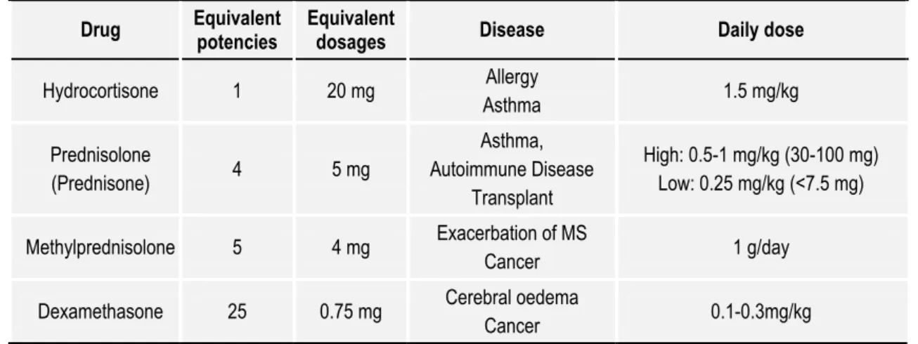
2.1.4.1. Preparations in clinical use ... 35
2.1.4.2. Effect on the immune system ... 36
2.1.4.3. Effect on regulatory T cells ... 38
Table of contents
vi
2.2. Material and methods ... 40
2.2.1. Experimental model ... 40 2.2.2. Mice ... 41 2.2.3. Experimental agents ... 42 2.2.3.1. Hydrocortisone ... 42 2.2.3.2. Cyclophosphamide ... 44 2.2.3.3. Pertussis toxin ... 44
2.2.3.4. Anti-CD25 monoclonal antibody ... 46
2.2.3.5. Irradiation ... 47
2.2.4. Cell recovery, antibodies and flow cytometric analysis ... 47
2.2.5. Cell purification and transfer ... 48
2.2.6. Cell cultures and suppression assay ... 48
2.2.7. Histological evaluation ... 48 2.2.8. Statistical methods ... 49 2.3. Results ... 49 2.3.1. Titrations ... 49 2.3.1.1. Hydrocortisone ... 49 2.3.1.2. Pertussis toxin ... 56 2.3.1.3. Irradiation ... 59
2.3.2. Comparison of immunosuppressive effect on T lymphocyte subpopulations ... 62
2.3.2.1. Hydrocortisone ... 62
2.3.2.2. Cyclophosphamide alone or in combination with hydrocortisone ... 67
2.3.2.3. Pertussis alone or in combination with hydrocortisone ... 82
2.3.2.4. Irradiation alone or in combination with hydrocortisone ... 85
2.3.3 In vivo effect of immunosuppressants on regulation ... 88
2.3.3.1. Regulatory T cell mediated protection of TR¯ mice ... 88
2.3.3.2. Induction of encephalomyelitis in TR+ mice ... 93
2.3.3.2.1. Hydrocortisone ... 93
2.3.3.2.2. Cyclophosphamide alone or in combination with hydrocortisone ... 95
2.3.3.2.3. Pertussis toxin ... 97
2.3.3.2.4. CD25 depletion ... 97
2.3.3.2.5. Irradiation ... 98
2.3.3.3. Modulation of pertussis toxin-induced encephalomyelitis in TR+ mice ... 98
Modulation of regulatory T cells in autoimmune disease
vii
2.3.3.3.2. Hydrocortisone ... 98
2.3.3.3.3. Irradiation ... 100
2.3.4. The effect of hydrocortisone on regulatory T cell expansion ... 100
2.3.5. The effect of hydrocortisone on regulatory T cell function in vitro ... 108
2.4. Discussion ... 112
2.4.1. A short course of steroids, as opposed to cyclophosphamide, did not selectively deplete regulatory T cells ... 112
2.4.2. In the transgenic mouse, lymphopenia only triggered auto-immune disease when regulatory T cells were selectively decreased ... 114
2.4.3. A short course of steroids did not affect regulatory T cell function or expansion upon transfer ... 116
3. RESPONSE OF REGULATORY T CELLS TO INFLAMMATION ... 117
3.1. Introduction ... 117
3.1.1. The aetiology of inflammatory bowel disease ... 117
3.1.2. Influence of bacterial luminal content on the outcome of colitis ... 119
3.1.3. The protection afforded by regulatory T cells in the T cell induced colitis model ... 120
3.1.4. The dynamics of regulatory T cells during acute inflammation ... 121
3.1.5. Objectives ... 122
3.2. Material and methods ... 123
3.2.1. Mice ... 123
3.2.2. Antibodies ... 123
3.2.3. Cell purification and transfer ... 124
3.2.4. Cell recovery and flow cytometric analysis ... 124
3.2.5. Histological evaluation ... 124
3.2.6. Helicobacter PCR ... 124
3.3. Results ... 125
3.3.1. Effect of recipient Helicobacter status upon the rapidity of onset and severity of T cell induced colitis ... 125
3.3.2. The specificity of the immune response to Helicobacter ... 130
3.3.3. Effect of ongoing colitis on regulatory T cells ... 132
3.3.4. Recipient analysis after Foxp3¯ T cell transfer ... 133
3.4. Discussion ... 135
3.4.1. The Helicobacter status of the recipient determines the rapidity of onset and the severity of T cell induced colitis ... 135
Table of contents
viii
3.4.2. The immune response appears to be specific to Helicobacter in
colonized recipients with T cell induced colitis ...136
3.4.3. Regulatory T Cells accumulate selectively in T cell induced colitis ...137
3.4.4. Conversion from Foxp3¯ to Foxp3+ T cells in ongoing T cell induced colitis ...137
3.4.5. Concluding remarks and implications for clinical practice ...138
4. T CELL INDUCED COLITIS ON THE REPRESSION OF ENCEPHALOMYELITIS IN TRANSGENIC MICE ... 141
4.1. Introduction ... 142
4.1.1. Immune mechanisms in immuno-inflammatory diseases ... 142
4.1.1.1. Experimental auto-immune encephalomyelitis ... 143
4.1.1.1.1. Experimental models ... 143
4.1.1.1.2. The blood brain barrier ... 144
4.1.1.1.3. Effector lymphocyte activation ... 145
4.1.1.1.4. Role of cytokines ... 146
4.1.1.1.5. Role of regulatory T cells ... 150
4.1.1.2. T cell induced colitis ... 152
4.1.1.3. Chemical colitis ... 153
4.1.2. The hygiene hypothesis ... 154
4.1.3. Regulatory T cell homeostasis ... 156
4.1.4. Objectives ... 156
4.2. Material and methods ... 157
4.2.1. Mice ... 157
4.2.2. Antibodies and reagents ... 157
4.2.3. Cell purification and transfer ... 157
4.2.4. Cell recovery and flow cytometric analysis ... 157
4.2.5. Cell culture and proliferation assay ... 157
4.2.6. Histological evaluation ... 158
4.2.7. Data analysis ... 158
4.3. Results ... 158
4.3.1. Effect of T cell induced colitis upon spontaneous EAE ... 158
4.3.2. Effect of chemical colitis upon spontaneous EAE ... 163
4.3.3. Effect of donor T cell depletion in TR– mice with T cell induced colitis ... 164
Modulation of regulatory T cells in autoimmune disease
ix 4.3.5. Outcome of co-transfer of different T cell subsets from recipients with
colitis together with transgenic T cells ... 167
4.3.6. The encephalitogenic potential of tg T lymphocytes in TR– mice protected from EAE by T cell induced colitis ... 172
4.3.7 Cytokine production by colitogenic T cells ... 174
4.3.8. Migration of tg T cells in T cell induced colitis ... 175
4.4. Discussion ... 176
4.4.1. T cell induced colitis inhibits spontaneous EAE ... 176
4.4.2. Donor T lymphocytes mediate EAE protection ... 176
4.4.3. Regulatory T cells accumulate selectively in inflammation ... 176
4.4.4. The EAE protective population is CD25 negative ... 178
4.4.5. Effector tg T cells in colitis lose encephalitogenic potential ... 179
4.4.6. Effector tg T cells in colitis are attracted to the inflamed colon ... 181
5. CONCLUSIONS ... 183
5.1. Modulation of regulatory T cells after immunosuppressive therapy ... 183
5.2. Modulation of regulatory T cells during inflammation ... 184
6. CLINICAL RELEVANCE AND PERSPECTIVES ... 187
6.1 Consequences of immunosuppressive therapies ... 187
6.2 Considerations for regulatory T cell immunotherapy ... 188
6.3 Perspectives ... 190
7. REFERENCES ... 193
APPENDIX ... 227
Zelenay, S., Lopes-Carvalho, T., Caramalho, I., Moraes-Fontes, M. F., Rebelo, M., and Demengeot, J. (2005). Foxp3+ CD25- CD4 T cells constitute a reservoir of committed regulatory cells that
regain CD25 expression upon homeostatic expansion. Proc Natl Acad Sci U S A 102 (11): 4091-4096.
Demengeot, J., Zelenay, S., Moraes-Fontes, M. F., Caramalho, I., and Coutinho, A. (2006). Regulatory T cells in microbial infection. Springer Semin Immunopathol 28 (1): 41-50.
xi ACKNOWLEDGEMENTS
I often wonder how an adventure in the world of science, a sense of fulfilment from the experience of adult learning and an enormous personal gain could result from a simple conversation with a colleague during a busy intake in the emergency room, in the summer of 2001. Thank you Carlos Meneses de Oliveira, for telling me that the IGC existed and that perhaps they would take me under their wing. An interview with António Coutinho, an attempt at project writing and a brief meeting with Jocelyne Demengeot changed the course of my life and, in
October 2001 I started to work at the IGC, relieved of ward duties and granted a bursary from “Ministério da Saúde”, for 2 years. I continued emergency duty but in October 2003 realized I needed more time in the laboratory, asked for unpaid leave and supported myself with private clinical commitments, finding a way to spend most of my time at the IGC. I thank my colleagues in the Hospital, José Pimenta da Graça (Director: “Serviço de Medicina II”) and the Clinical Administration of “Hospital de Egas Moniz” for encouragement and granting the necessary permits. In July 2004, I registered as a PhD student at the “Faculdade de Medicina, Universidade de Lisboa”.
I gratefully acknowledge the teaching, supervision and unfailing support of Jocelyne Demengeot. After a period of time where I was allowed to decide on the research topic, she gave guidance and methodology teaching, always encouraging independence, creativity and innovation in practical work and dissertations, allowing me to become progressively more confident and at ease with the work and the scientific method. She imparted both the careful and strict planning required in each individual experiment and the thought processes involved in problem solving and always kept the lines of communication open so that I was never afraid to ask questions. She motivated me to participate in the journal club, regularly present results in structured group meetings, attend seminars and conferences at the IGC and stimulated me to attend courses in core subjects of modern scientific training inherent to the PhD programs in place at the IGC. She encouraged my involvement in the directions of “Sociedade Portuguesa de
Acknowledgements
xii
Imunologia” and the “Núcleo de Estudos de Doenças Autoimunes” of the “Sociedade Portuguesa de Medicina Interna”, and in other research-led activities such as participation in several projects with clinicians and other research groups at the IGC and other scientific institutions, namely on the effect of symbiotics and the effect of IVIG on regulatory T cells. She provided me with the opportunity to review and co-publish scientific papers, organize conferences and teaching courses for medical doctors and to present the work of this thesis at several national and international conferences. It was a great learning experience in a very dynamic laboratory in a very competitive field. I pay tribute to her intelligence, professionalism and honesty and thank her for giving me much more than what is expected from a PhD supervisor.
António Coutinho provided the enthusiasm and encouragement for the initiation of this work, accompanying my progress in the laboratory, forever teaching and bringing resourceful ideas to the project. He gave me the opportunity to present the work in this thesis while representing the IGC at several national meetings, introducing me to other scientists who provided invaluable partnerships and to witness and aid his work in the direction board of the “Sociedade de Ciências Médicas de Lisboa”. He provided the framework which allowed for the development of research projects in patients with Behçet´s disease and with systemic lupus erythematosus. He made me realize that there is much more than practical skills in modern research providing a true example of an academic and of a man of free spirit, who believes that “collaboration is always more effective than competitiveness” and who created a spirit of organizational excellence at the IGC. I believe these principles, which rule the IGC, also apply to the workings of health care institutions and can honestly say that António Coutinho provided me with a new working philosophy based on a simple doctrine of unselfishness, universality and the practice of good. He has influenced my life so what better tribute to him than to state he is a true exemplary figure in my life.
None of this work would have been possible without the help of all the members, past and present, of my group, especially Íris Caramalho, Manuel Rebelo, Santiago Zelenay, Marie Louise Bergman, João Duarte, Leonor Morais Sarmento, Ana Água Doce, Lurdes Duarte, Alexis Perez, Catarina Martins and Paulo Bettencourt. I want to make a special reference to Íris, to thank her very much for her unselfish capacity for constant teaching, for helping me with the practical work while she was a senior PhD student and I was just starting in the lab, for ongoing support and for providing me with a lush example of the multiple domains, which a true scientist must possess. Manel, Santi and Lisa were my main work partners helping me enormously with the practical work and its interpretation and most logical continuation but everyone else helped
Modulation of regulatory T cells in autoimmune disease
xiii too. All became my friends, constituting the type of tight knit support group I required for achieving completion.
My gratitude also goes to Carlos Penha Gonçalves, Jorge Carneiro, Betty Padovan, Rosa Maria, Margarida Vigário, Mafalda Lopes da Silva, Rui Gardner, Teresa Faria Pais, Dinis Calado, Ângelo Chora, Nuno Moreno, Isabel Gordo, Ricardo Ferreira, Greta Martins and Paulo Fontoura at the IGC and Pedro Oliveira (Instituto Portugês de Oncologia Francisco Gentil). I thank Sofia Oliveira and Jorge Crespo for help with the Behçet´s disease project, Constantin Fesel for my involvement in the Lupus project and Carlos Lima (Hospital de Egas Moniz) for the many histology sessions and fruitful discussions. Thank you, Werner Haas, for your initial support, teaching, friendship and many spaghetti dinners.
I would have never completed this thesis without the loving constant and solid support of my uncle Luís Filipe Gusmão who patiently revised this thesis and repeatedly, ever since I know myself, has helped me to think in a logical fashion. I also thank my cousins Leonor Gusmão for revision and Estela Gusmão for edition. I deeply thank my husband and mother for their understanding and support and promise them compensation for all the stolen hours.
xv
SUMMARY
Foxp3+ regulatory T cells (Treg) are a specialized group of lymphocytes that constitute the basis
of control of physiological auto-reactivity. These cells prevent autoimmune disease (AID) by inhibiting the activation of potentially pathogenic effector cells and, in mice and humans, their absence causes a lethal form of autoimmunity. In order to contribute to the study of Treg in AID three different experimental layouts were designed to appraise: (i) the impact of immunosuppression (IS) on the number and function of Treg and on immune regulation; (ii) the response of Treg to inflammation and; (iii) the effect of an inflammatory immune response during the course of an AID.
It was hypothesized (because of their generalized effect on the immune system and inability to cure AID) that steroids (the most commonly used form of non-specific IS) may damage Treg. To determine the impact of IS on Treg number and function, a 5 day course of hydrocortisone (HC) was administered to the anti-myelin basic protein (anti-MBP) T cell receptor (TCR) transgenic (Tg) mouse (TR+ mice) which are naturally protected by natural Treg from
developing experimental autoimmune encephalomyelitis (EAE). A transfer of Treg prevents the EAE which develops when these Tg mice are set in a RAG–/– background (TR– mice). We
concluded that IS caused by a short course of steroids was not deleterious to Treg number or function; however, higher doses, formulations of different potencies and/or longer therapies remain to be formally tested.
To assess the effect of IS on immune regulation we evaluated the impact of several conditions of lymphopenia on EAE onset and severity using the anti-MBP TCR Tg system. The effect of HC alone was compared to irradiation, cyclophosphamide (Cyp), pertussis toxin (Ptx) and CD25 depletion, each alone or in combination therapy with HC. Generalized lymphopenia (steroids or irradiation) was compared to selective Treg depletion alone (mAb mediated CD25 depletion) or to lymphopenia associated with Treg depletion (Cyp). Our results indicate that lymphopenia is a trigger of EAE in TR+ mice only when associated with selective Treg depletion.
Summary
xvi
number and function and the development of AID. In this respect, the combination of HC and Cyp, often used in the clinic, was particularly harmful.
Following a symmetrical reasoning, we next tested whether ongoing immune activity promotes immune regulation by stimulating an increase in Treg number. We utilized an experimental layout designed to assess the response of regulatory T cells to inflammation by testing the possibility of conversion of naïve donor T cells to Foxp3+CD4+ T cells. T-cell induced
colitis (a mouse system of T cell lymphopenia and severe Treg number reduction that commonly serves as a model of human inflammatory bowel disease) provided the means to study the dynamics of inoculated Treg expansion. We reveal that in ongoing severe colitis induced by the transfer of naïve T cells containing a small Treg contaminant, Treg of donor origin accumulate and reduce the severity of colitis. Transfer experiments in which the Treg contaminant was excluded from the donor cell population unequivocally demonstrated naïve T cell conversion to Treg. These findings support the notion that inflammation and immune responses favour Treg accumulation, through expansion and conversion and allow for Treg function.
Bearing in mind that Helicobacter spp. (H) is a recognized facilitator of T cell-induced colitis, it has not been formally established whether T-cell induced colitis is an AID targeted at intestinal tissues or an immunopathology caused by ongoing immune responses specific to Helicobacter. In the course of these experiments we showed that while T cells isolated from H+
mice presenting with severe colitis were not colitogenic upon adoptive transfer in H– mice, they
induce fulminant colitis in H+ recipient mice. Even though a Helicobacter requirement for self Ag
presentation by intestinal dendritic cells (DC) was not excluded, these results suggest that colitogenic T cells in this system are Helicobacter specific and not tissue specific, in favour of an infectious rather than autoimmune aetiology of T cell-induced colitis.
Based on the evidence that T-cell induced colitis results from an exacerbated immune response to enteric bacteria we directly tested whether such an inflammatory immune response would affect the course of a bona fide AID. Colitis was induced in TR– mice by transfer of naïve T
cells. T cell-induced colitis was shown to completely prevent EAE development but was unable to suppress established EAE. Prevention of EAE only occurred with rapid colitis induction in the presence of Helicobacter colonization of recipient mice. Even though protection occurred within a time window which corresponds to that expected from a Treg transfer, these results indicated that the protective immune response that follows the acute phase of T cell-induced colitis is mediated by donor CD4+CD25– T cells.
This work has contributed to further awareness of the mechanisms of lymphopenia as a trigger of AID and has advanced knowledge on the biologic behaviour of Treg in response to an
Modulation of regulatory T cells in autoimmune disease
xvii ongoing inflammatory immune response. Technical advances in the methodology to define and better identify Treg, in conjunction with the recent burst of information describing the number and function of Treg and polymorphisms of genes implicated are expected to strengthen the knowledge about the association of Treg to the pathogenesis and genetic susceptibility of human AID.
Key words: Autoimmune disease; Regulatory T cells; Steroids; T cell-induced colitis;
xix
RESUMO
As células T reguladoras (Treg) são um grupo de linfócitos especializados que constitui a base do controlo da auto-reactividade fisiológica. Estas células previnem a doença auto-imune através da inibição da activação de outros linfócitos T potencialmente patogénicos e, tanto em humanos como no modelo experimental, a sua ausência causa uma forma letal de auto-imunidade. De modo a contribuir para o estudo das Treg na doença auto-imune, foram utilizadas diferentes abordagens experimentais concebidas para avaliar especificamente: (i) o impacte da utilização de imunossupressores (IS) sobre o número e a função das Treg e sobre a regulação do sistema imunitário, (ii) a resposta do organismo à inflamação, no que se refere às Treg e (iii) o efeito de uma resposta inflamatória imune sobre a potencial evolução de uma doença auto-imune.
Foi colocada a hipótese da utilização de corticóides (GC) - a forma de imunossupressão mais frequentemente utilizada - poder ter um efeito negativo sobre as Treg, dado o facto dos IS exercerem um efeito generalizado sobre o sistema imunitário e a sua utilização clínica não levar à cura das doenças auto-imunes. Com o objectivo de determinar o impacte da utilização de IS sobre o número e função das Treg, foi administrada hidrocortisona (HC), diariamente, durante 5 dias, a ratinhos transgénicos. Estes são caracterizados por possuir linfócitos que, na sua maioria, têm um receptor específico para a proteína básica da mielina (ratinhos TR+) e estão
naturalmente protegidos, pelas Treg, contra o desenvolvimento da encefalomielite experimental auto-imune (EAE). Quando não possuem Treg (ratinhos TR–), a transferência deste grupo de
linfócitos previne o desenvolvimento de EAE. O presente estudo permite concluir que a IS causada por HC de curta duração não afecta negativamente o número ou a função das Treg, tornando-se necessário estudar o efeito de uma terapêutica mais prolongada.
Para estudo do efeito da utilização deIS sobre a regulação do sistema imunitário, foram induzidas diferentes condições de linfopenia e foi avaliado o seu impacte na capacidade de indução e gravidade da EAE no ratinho TR+. O efeito da HC foi comparado ao da radiação gama,
da ciclofosfamida (Cyp), da toxina do pertussis (Ptx) e da depleção de CD25, isoladamente ou em conjunto com a HC. A linfopenia generalizada (por efeito da HC, de radiação gama ou
Resumo
xx
combinação dos dois) foi comparada à depleção selectiva de Treg, induzida pelo anticorpo monoclonal anti-CD25, e à linfopenia associada à depleção selectiva de Treg, induzida pela Cyp. Os resultados indicam que a linfopenia apenas constitui um factor desencadeante da EAE nos ratinhos TR+ quando em associação à depleção selectiva de Treg. A utilização de Cyp e Ptx
demonstra a correlação do seu efeito nocivo sobre o número e a função das Treg e o desenvolvimento de doença auto-imune, salientando-se o efeito mais prejudicial da HC quando combinada com a Cyp - uma combinação frequentemente utilizada na prática médica.
Seguindo um raciocínio inverso, a pergunta seguinte refere-se à possibilidade de uma resposta imunitária em curso poder fomentar a regulação imunitária, ao promover um aumento das Treg. O método experimental foi concebido para avaliar a resposta das Treg ao estímulo da inflamação e testar a possibilidade de existir conversão de linfócitos T naïve para células T Foxp3+CD4+. A colite induzida por linfócitos T, um modelo murino de doença inflamatória do
intestino, é caracterizada por linfopenia das células T e redução acentuada de Treg, permitindo estudar in vivo a dinâmica da expansão das células inoculadas. Os resultados revelam que, durante o processo de colite induzida pela transferência de linfócitos T naïve contendo um pequeno contaminante de Treg, estas Treg se acumulam e, após transferência, reduzem a gravidade da colite induzida. Através de um tipo de transferência de células naïve, que permite excluir a presença de um contaminante de Treg, foi comprovada a conversão de células naïve para Treg. Estes resultados corroboram a convicção de que a inflamação e consequente resposta imunitária favorece a acumulação de células Treg e, para além disso, permite que estas exerçam a sua função imunossupressora.
Tendo em conta que a infecção por Helicobacter (H) é facilitadora da colite induzida por linfócitos T, pretendeu-se esclarecer se tal tipo de colite era uma doença auto-imune dirigida para os tecidos intestinais ou uma forma de imunopatologia causada por uma resposta imunitária específica para o Helicobacter. O presente estudo demonstra que linfócitos isolados de recipientes H+ com colite induzida por linfócitos T não são colitogénicos quando transferidos para
recipientes H–, contrastando com a colite grave que induzem em recipientes H+. Estes resultados
demonstram que os linfócitos colitogénicos neste sistema são específicos para o Helicobacter e não reconhecem tecido intestinal, excluindo-se deste modo a hipótese de auto-imunidade.
Finalmente, foi testada a hipótese da colite induzida por linfócitos T (caracterizada por uma resposta imunitária inflamatória a bactérias entéricas) poder exercer influência sobre uma doença auto-imune. Se por um lado ficou demonstrado o papel supressor da EAE através da indução de colite em ratinhos TR–, por outro verificou-se não existir inibição se a EAE já estiver
Modulation of regulatory T cells in autoimmune disease
xxi verificou a prevenção da EAE quando a ocorrência da colite foi mais tardia e menos grave. O aparecimento de EAE apenas foi impedido quando a colite foi induzida precocemente, em recipientes portadores de Helicobacter e quando a transferência de linfócitos indutores de colite foi realizada até às quatro semanas, tal como na protecção conferida aos ratinhos TR– por Treg.
Os resultados do presente estudo indicam contudo que esta protecção é mediada por linfócitos T CD4+CD25– provenientes do dador e não por Treg.
O presente trabalho contribuiu para o conhecimento das características da linfopenia que podem ser desencadeantes de doença auto-imune e do comportamento biológico das Treg face a uma resposta imunitária inflamatória. Avanços técnicos na metodologia para melhor definir e identificar as Treg, em conjunto com a explosão de informação sobre a caracterização do número e função das Treg e polimorfismos de genes implicados, deverão contribuir para um melhor esclarecimento da associação de Treg com a patogénese e a susceptibilidade genética das doença auto-imunes.
Palavras chave: Doença auto-imune; células T reguladoras; glucocorticóides; colite induzida por
xxiii
ABBREVIATIONS
–/– Knock out
Ab Antibody
Ag Antigen
AID Auto-Immune Disease
AIRE Auto-Immune REgulator
ALPS Auto-Immune LymphoProliferative Syndrome APC Antigen-Presenting Cell
APECED Autoimmune PolyEndocrinopathy Candidiasis Ectodermal Dystrophy
APh AlloPhycocyanin
APS 1 Autoimmune Polyendocrinopathy Syndrome type 1 BBB Blood Brain Barrier
CD Crohn’s Disease
CFA Complete Freund Agent CNS Central Nervous System
CsA Cyclosporin A
CS CorticoSteroid
CSF CerebroSpinal Fluid
CTLA4 Cytotoxic T-Lymphocyte Antigen 4
Cyp Cyclophosphamide
D Day
D0 Up to 24 hours post last injection
D1 to D10 First to 10th day after D0
D-5 to D-1 Fifth to first Day before D0
DC Dendritic Cell
DEX DEXamethasone
Abbreviations
xxiv
DP Double Positive (thymocyte) DSS Dextran Sodium Sulphate
EAE Experimental Autoimmune Encephalomyelitis
FCS Fetal Calf Serum
FDA Food and Drug Administration
GC GlucoCorticoid
GF Germ Free
GFP Green Fluorescent Protein H&E Haematoxylin & Eosin
H+ Colonized with Helicobacter spp
HC HydroCortisone HC0.5 HC 0.5 mg/kg HC5 HC 5 mg/kg HC50 HC 50 mg/kg HC5x5 HC 5 mg/kg/day x 5 days HCt Healthy Control
HPF High Power Field
i.p. intra-peritoneal
i.v. intra-venous
IBD Inflammatory Bowel Disease IEL IntraEpithelial Lymphocytes
IFN InterFeroN
IgG Immunoglobulin G
IL InterLeukin
IL-12R IL-12 Receptor
IPEX Immune dysfunction, Polyendocrinopathy, Enteropathy, and X-linked inheritance Irrad. Irradiation
IS ImmunoSuppressant
iTreg induced Treg
IVIG IntraVenous ImmunoGlobulin
LFB Luxol Fast Blue
LIP Lymphopenia Induced Proliferation
Ln Lymph node
Modulation of regulatory T cells in autoimmune disease
xxv
MACS Magnetic-Activating Cell Sorting MBP Myelin Basic Protein
MC MineraloCorticoids
MHC Major Histocompatibility Complex
mLn mesenteric Ln
MOG Myelin Oligodendrocyte Glycoprotein
MP MethylPrednisolone
MS Multiple Sclerosis
n number of mice
NFAT Nuclear Factor of Activated T cells
NK Natural Killer
nmLn non-mesenteric Ln
NOD Non Obese Diabetic
nTreg natural Treg
PB Peripheral Blood
PBS Phosphate Buffer Solution PD-1 Programmed Death receptor-1
PE PhycoErythrin
PLP ProteoLipid Protein
PRED PREDnisolone
Ptx Pertussis toxin RA Rheumatoid Arthritis
RAG Recombination Activating Gene RTE Recent Thymic Emigrants
SCID Severe Combined ImmunoDeficiency
SF Synovial Fluid
SLE Systemic Lupus Erythematosus SP SinglePositive (thymocyte)
Sp Spleen
SPCD4+ SinglePositive CD4+ thymocyte SPF Specific Pathogen Free
T1DM Type 1 Diabetes Mellitus
TCR T Cell Receptor
Abbreviations
xxvi
Tg transgenic
TGF-β Transforming Growth Factor-β
Th T helper
Thy Thymus
TLI Total Lymphoid Irradiation TLR Toll-Like Receptors TNF- Tumor Necrosis Factor-
TR– Anti-MBP TCR Tg on a RAG deficient background
TR+ Anti-MBP TCR Tg on a RAG sufficient background
Treg regulatory T cell UC Ulcerative Colitis
1
1. GENERAL INTRODUCTION
Diabetes mellitus, Graves' disease, multiple sclerosis, rheumatoid arthritis and systemic lupus erythematosus are among a group of diseases of unknown aetiology that affect very different organs or organ systems but share a universal pathogenesis. Common to all is an immune response directed to self, referred to as autoimmunity, the aetiology of a broad spectrum of human illnesses collectively known as autoimmune diseases (AID). This thesis aimed to study: (i) the effect of steroid treatment upon the number and function of regulatory T cells (Treg) in mice; (ii) the response of Treg to inflammation and; (iii) the effect of T cell-induced colitis on the repression of encephalomyelitis in transgenic mice. Even though the study of these three aspects required different experimental layouts - and was therefore included in separate chapters - an effort was made to coherently consolidate them within the common objective namely the understanding of the effects of modulation of Treg in AID.
1.1. Relevance of fundamental research to autoimmune diseases
Some of the most prevalent auto-immune diseases (AID) in the USA are rheumatoid arthritis (RA) with a rate of 0.6%, systemic lupus erythematosus (SLE), with a prevalence of 0.1% in Caucasian and 0.4% in afro-American women (Helmick et al., 2008; Lawrence et al., 2008) and multiple sclerosis (MS) rating 0.04 to 0.17% (Jacobson et al., 1997). In industrialized nations overall prevalence is increasing (Bach, 2002) and this fact is amply illustrated by diabetes mellitus (DM), estimated to rise from a global incidence of 2.8% in the year 2000 to 4.4% in 2030 (Wild et al., 2004). Some AID are actually considered Orphan Diseases, which by definition: (i) affect less than 0.05% of a population, (ii) affect less than 200,000 people in the USA or (iii) for which treatment is not envisaged as profitable for pharmaceutical development.
AID were traditionally taught to have “a mysterious aetiology” or to be designated as a fascinating but poorly understood group of diseases (Davidson and Diamond, 2001) and, even in present times, the inference is often made that when the origin of a disease is obscure, it
Relevance of fundamental research to autoimmune diseases
2
suggests an autoimmune aetiology. The classification of a disease as autoimmune has traditionally been based on the detection of an auto-antibody (auto-Ab) that could be visualized reacting with an affected tissue or cell. Two factors, namely, the rarity of naïve T cells specific for any one “peptide - major histocompatibility complex (MHC)” complex, due to the tremendous diversity of the T cell repertoire, and the much greater technical ease with which auto-Ab are identified in comparison to auto-reactive T lymphocytes, actually constitute an important historical bias in the clinical characterization of AID. The development of microscopes that allow for more sensitive detection of cell surface–bound auto-Ab, the capacity to use the auto-Ab to precipitate the auto-antigens (auto-Ag) and the availability of more sophisticated techniques that allow for the exclusion of infections (such as PCR based assays) are technological advances that resulted in a proliferation of newly recognized autoimmune disorders. Auto-Ag sequencing, purification and studies of uptake and processing, and, conversely, Ab isolation, sequencing and isotype determination, allow for a more certain assessment of the possibility that a specific auto-Ag is the driver of the immune response and auto-Ag that are commercially available can be used for identification of auto-Ab for diagnostic means.
Pathogenic antibodies are directly responsible for the immunopathology that derives from Ag recognition and their presence is usually demonstrated experimentally in vivo or in vitro. In a disease such as lupus, pathology is most likely due to deposition of immune complexes irrespective of their reactivity. Lernmark (2001) presents a review of many auto-Ag which are commercially available as recombinant proteins and can be used for specific autoantibody assays in the diagnosis of AID.
The recognition of specific Ag or Ab is not equivalent to the identification of the defect that may underlie pathological autoimmunity. Indeed, with the exception of a few AID associated with immune-deficiencies (Carneiro-Sampaio and Coutinho 2007), and despite powerful genetic studies, no single gene mutation or aberrant gene expression has been identified as a causative factor in any of these diseases (Spurkland and Sollid, 2006). Ongoing work suggests that instead, polymorphisms of normal genes of the immune system (including MHC), that are usually present in the general human population, contribute to the susceptibility for AID development. These polymorphisms may constitute the basis of (i) heterogeneity in clinical presentations, (ii) clinical overlap between AID and (iii) familial clustering of AID. Some of the identified genes are involved in lymphocyte differentiation while others code for proteins of an unknown function, the precise role of these polymorphisms in disease pathogenesis remaining to be understood (Maier and Hafler, 2008). The possibility also remains that genes involved in organ development, protection or repair are implicated. Risk factors cannot be identified in clinical practice and consequently, at
General introduction
3 the present time, preventative measures cannot be implemented. AID are incurable and remain responsible for considerable morbidity and mortality, justifying need for ongoing research.
Careful phenotyping of cells and lesions using cytometry, advanced molecular biology and imaging techniques (in order to map pathways controlling lymphocyte physiology) should accompany genetic studies providing important clues. In this regard, humans are very poor study material. Because of obvious ethical constraints, the only cells which are generally available for research are from the peripheral blood (PB) and, rarely, from accessible tissues, and these may not necessarily reflect a pathological process in the lymphoid or target organs. In addition, by the time an AID appears in humans, it is very likely that the initial event or the primary immune abnormality have been diluted or masked by the presence of an immune response. In addition, autoimmunity leads to inflammation which in turn stimulates the immune response, further masking the distinction between primary and secondary events.
Rodent models are preferred because they are affordable, allow for mass production, represent almost all the major AID, can be genetically manipulated to study susceptibility and enable the study of early events, disease severity and organ damage, allowing for therapeutic screening. In an ideal model for a specific AID, the animal develops the disease spontaneously and not through immunization, in an effort to mimic natural mechanisms operating in the human disease. The major drawback for the use of rodent animals is the very limited number of mouse strains, each representing one specific MHC haplotype and in practical terms equivalent to one human individual. There are over 100 alleles at several loci within the MHC, the most polymorphic gene within the human genome. As antigen presentation by the MHC and its recognition by the immune system is a central question, MHC variability may contribute to clinical sub-phenotypes of human disease, differentiated by factors such as the group of target organs affected, the frequency of relapses and disease severity. As an example, the programmed death receptor-1 (PD-1) knock out (–/–) mouse presents with a dilated cardiomyopathy in a Balb/c background and
with a glomerulonephritis in a B6 background, remaining to be seen if this is due to a change in the MHC or due to another strain-specific gene that confers a completely different phenotype for the same genetic abnormality (Nishimura et al., 1999; Nishimura et al., 2001). In human AID, an environmental factor is brought into the equation, because concordance disease rates in monozygotic twins are far from 100% (Ebers et al., 1986; Silman et al., 1993; Wandstrat and Wakeland, 2001). In this respect, experimental models do not allow for a re-creation of environmental agents or pressures to which human are subjected in their daily life but, on the other hand, facilitate the study of the presence or absence of specific environmental agents in a
Relevance of fundamental research to autoimmune diseases
4
very controlled fashion, having been helped, in this regard, by the development of germ free facilities.
There has been a major effort for experimental models to phenotypically resemble human disease as completely as possible and animal models, such as that of MS, have originated thousands of reports (Steinman and Zamvil, 2006). Ideally, knowledge of disease pathogenesis gained from experimental models should result in the design of rational therapy and consequent pre-clinical studies and clinical trials, going back and forth to experimental models to check new beneficial observations, critical clues from patient studies, unexpected side-effects and combination therapies, in a very dynamic interaction between the laboratory and the clinic. In practice however, advances in treatment of AID in the past 10 to 15 years are not always a direct consequence of experimental research but rather due to a combination of careful marketing and anecdotal evidence of disease improvement of which chronic interferon- (IFN-) treatment for MS is illustrative, with only a 30% reduction in relapse rate and disability progression. In contrast, on the basis of a trial with a similar effect to IFN (Johnson et al., 1995), glatiramer acetate - a polymer of tyrosine, glutamate, alanine and lysine, believed to bind to MHC molecules and block T cell recognition of myelin basic protein (MBP) - carefully designed and first tested in animal models of MS in the 1970’s (Teitelbaum et al., 1971), took 25 years to be approved by the Food and Drug Administration (FDA) for MS treatment The fact that the use of both IFN-and glatiramer acetate has been recently challenged in systematic meta-analysis (Filippini et al., 2003; Munari and Filippini, 2004) only accentuates the urgent need for ongoing research into the pathogenesis of AID.
There are some recent examples as to why differences between man and mouse may explain why a specific therapy hailed as promising in mice produces unexpected side-effects in humans. Based on the knowledge that T cell help - mediated by the interaction between CD40L (CD154) on T lymphocytes and CD40 on the cognate B cell - is critical for the production of pathogenic auto-Ab in lupus nephritis, it was predicted that anti-CD40 ligand antibody would lead to a reduction in the severity of nephritis and prolonged survival in experimental models (Kalled et al., 1998). Despite favourable results in phase I human studies (Davis et al., 2001), a phase II trial was terminated prematurely because of thromboembolic events (Boumpas et al., 2003), related to the fact that CD40L is also expressed by platelets, both in monkeys and humans but not in mice (Kawai et al., 2000). Another example of unpredictable collateral damage occurred with the use of natalizumab, an anti-integrin (41) monoclonal antibody. This integrin is an adhesion molecule present on lymphocytes and responsible for their adhesion to endothelium, found to be effective in MS and Crohn´s disease. This drug was actually associated with three deaths from progressive multifocal leucoencephalopathy, which occurs due to a viral infection that could not
General introduction
5 have possibly been detected in murine models, since its causative agent - the JC virus - does not infect mice or rats (Van Assche et al., 2005; Kleinschmidt-DeMasters and Tyler, 2005). The recent disastrous result of a phase I clinical trial with TGN1412, a novel superagonist anti-CD28 monoclonal Ab reported experimentally to expand Treg (Beyersdorf et al., 2005), nearly resulted in the death of 6 healthy volunteers due to a cytokine storm (Suntharalingam et al., 2006). CD28 is a co-stimulatory molecule involved in the activation of all T lymphocytes so that in theory this was a possible complication, independently of the results of pre-clinical studies.
Rather than to separate, we need to join “Science Based Medicine” with what we already know from “Evidence Based Medicine”. Progress in the field depends entirely on the very strictest collaboration between fundamental and clinical science, through the constant exchange of questions and clinical translation of research findings to prevention and therapy.
1.2. Pathogenesis of autoimmune diseases
After strong headed dispute in the beginning of the 20th century, when Paul Ehrlich denied the
existence of auto-Ab - reviewed in Coutinho (2005) - the concept of autoimmunity as the actual cause of human disease was established in the 1950’s (Witebsky et al., 1957) and modelled on Koch's postulates, the latter originally intended to establish a causal relationship between microbes and disease. At that time, the pathogenesis of AID focused on B lymphocytes due to a recognized increase in immunoglobulins and the presence of rheumatoid factor, immune complexes and plasma cells. In the mid seventies, it was realized that T lymphocytes predominated in RA infiltrates (Bankhurst et al., 1976) and that removal of thoracic duct lymphocytes (mostly T cells) improved RA (Paulus et al., 1977), as a result of which T lymphocytes became centre point for AID. The reduced auto-Ab production in MRL-lpr/lpr chimeras submitted to Ab mediated T cell depletion (Sobel et al., 1993) and milder renal disease in TCR−/− MRL-lpr/lpr mice (Peng et al., 1998) demonstrated the dependence of auto-Ab
production on T cell help in the MRL-Faslpr model of lupus, where both the Fas mutation and MRL background genes contribute to disease. In addition, allo-Ab production has been shown to be reduced after CD4 T lymphocyte depletion in a model of skin graft rejection (Steele et al., 1996) and, in SLE patients, T cell activation has been correlated with anti-dsDNA production (Spronk et al., 1996). On the other hand, B lymphocytes are indispensable for the development of lupus in Fas sufficient but B cell deficient MRL mice (Chan et al., 1999) and for collagen-induced arthritis (Svensson et al., 1998) and taken together, these observations support a combined role of both B and T lymphocytes in the pathogenesis of AID.
Pathogenesis of autoimmune diseases
6
Evidence that a T lymphocyte transfer alone reproduces AID was shown for EAE (Ben-Nun and Cohen, 1982), thyroiditis (Maron et al., 1983) and arthritis (van Eden et al., 1985). Myelin reactive T lymphocytes have been found in patients with MS (Ota et al., 1990) and, with the help of MHC class II tetramers (i) GAD 65-specific T cells have been identified in pre-diabetic NOD mice (Trudeau et al., 2003) and in pre-diabetic patients (Reijonen et al., 2000; Oling et al., 2005) and (ii) cartilage-specific T cells have been recognized in patients with RA (Kotzin et al., 2000). Importantly, this shift in focus led to T lymphocyte directed therapeutic advances with the use of CTLA-4 (Emery, 2003; Genovese et al., 2005) alongside promising B cell targeted monoclonal Ab treatment (Edwards et al., 2002; Edwards et al., 2004). A well-designed set of experiments involving the transfer of OVA-specific DO11 CD4+ T cells into mice that produce
secreted OVA as an endogenous self-protein reveals different outcomes in the absence of specific cellular subsets, namely: (i) severe systemic autoimmune reaction in the absence of T and B cells; (ii) mild disease when T cells are absent but B cells are present and; (iii) no disease in the presence of T cells, suggestive that T cells prevent expansion and maintain homeostasis and B cells limit subsequent effector responses of auto-reactive CD4+ T cells (Knoechel et al.,
2005).
Criteria to define a disease as autoimmune correctly included the notion that B and T lymphocytes are involved in disease pathogenesis and were established at three different levels of animal experimentation, namely direct, indirect or circumstantial (Rose and Bona, 1993). Transmissibility of the characteristic lesions of the disease by auto-Ab or pathogenic T cells was considered mandatory for direct evidence of an autoimmune mechanism. Indirect evidence required re-creation of the human disease in an animal either spontaneously or through immunization with an analogous Ag to the putative auto-Ag of the human disease. Circumstantial evidence concerned the finding of T lymphocytes or Ab in the affected organ, with improvement of symptoms after the use of immunosuppressive drugs. A working definition of an AID is therefore a disease caused by reactivity of any component of the immune system to self Ag with subsequent inflammatory damage to one or more organs, in the absence of an ongoing infection or other discernable cause (adapted from Davidson and Diamond 2001). It usually involves T lymphocyte activation and/or Ab/immune complex deposition. Even though at the present time AID are still viewed by most clinicians in the framework of diseases of the adaptive immune system, there is a large group of disorders of the innate immune system in humans, – reviewed in Nathan (2002) - of which auto-inflammatory syndromes characterized by mutations in the apoptotic pathways (Padeh and Berkun, 2007) and inflammatory bowel disease linked to polymorphisms of macrophage receptors (Henckaerts et al., 2007) are examples. Disorders of
General introduction
7 innate immunity are characterized by spontaneous inflammation and subsequent activation of the adaptive immune response and will probably be included in the group of AID in future definitions.
1.3. Development of a regulatory T cell concept
It is estimated that 1x10⁶ T lymphocytes, each with its own distinct T cell receptor (TCR)
develops in the thymus every day in a stochastic manner (Tonegawa, 1983). The potential diversity of TCR recombination is of such magnitude that if every T cell was allowed to respond to MHC:peptide complexes without discriminating self from non-self, this would quickly lead to self-destruction. Negative selection in the thymus is a mechanism for preventing attack of healthy self tissues. The development of autoimmune manifestations in recipients of thymic grafts (Thornton et al., 1998) and of a CD25negative thymocyte population (Itoh et al., 1999), transferred from healthy donors, led to the important realization that, under normal circumstances, negative selection is not complete and furthermore, that physiological auto-reactivity must be controlled in the healthy donor.
Physiological tolerance, a property of a living beings, is the absence of immune responses to self and to commensals that are, by definition not pathogenic. As proposed by Coutinho (2005), the only way to conjugate (i) the absence of AID with (ii) the natural occurrence of antibodies and autoreactive T lymphocytes that, despite negative selection, are continuously produced throughout the life of an individual and (iii) the establishment of tolerance solely at the stage of embryonic and perinatal development is, to hypothesize, that a group of cells in the immune system develop in this restricted time window and are responsible for controlling physiological autoreactivity, imparting this memory to cells that develop in later periods of development. Tolerance was shown to be effected by lymphocytes selected in the thymus and was called dominant, on the basis that it could be transferred from one living organism to another by a T lymphocyte transfer (Coutinho et al., 1993; Modigliani et al., 1995; Le Douarin et al., 1996; Modigliani et al., 1996). The T lymphocytes responsible for tolerance were named regulatory T cells (Treg).It should be mentioned that rejection of a transplant performed in the perinatal period is only prevented by the co-implantation of a thymic graft from the donor species (Le Douarin et al., 1996) and it remains to be determined if the thymus is active throughout life producing Treg continuously or if there is actually a requirement for ongoing Treg education of other cells. More importantly, the concept of a dominant form of tolerance predominates over recessive, ignorant or anergic models, providing a form of control exerted by a group of cells that can be manipulated and eventually used therapeutically.
Development of a regulatory T cell concept
8
Because we are unable to confidently identify trigger factors for human AID, at the present time it is not possible to predict whether healthy individuals with benign physiological auto-reactivity or sub-clinical autoimmunity in the form of physiological auto-Ab or self-reactive T lymphocytes, will always remain healthy. This effectively rules out any chance of AID prevention and furthermore, the clinical diagnosis is only possible late in the course of disease, corresponding to the presence of significant target organ destruction. A frequently encountered situation in the clinic is illustrative of these difficulties. RA, SLE, systemic sclerosis, polymyositis, dermatomyositis, mixed connective-tissue disease, and Sjögren syndrome can present with similar clinical features, particularly during the first 12 months of symptoms. However, these have defined discrete diagnostic criteria and patients who present with symptoms, positive serology, or physical findings consistent but not fulfilling criteria for one of these established diseases are diagnosed with undifferentiated connective-tissue disease (UCTD), an entity suggested by LeRoy (1980) and recently defined as (i) signs and symptoms suggestive of a connective-tissue disease, (ii) positive anti-nuclear Ab, and (iii) a disease that lasts at least 3 years (Mosca et al., 2004). It has been shown that UCTD within 12 months of onset usually remains undifferentiated with only a few patients progressing to frank disease (Williams et al., 1999). Studies are ongoing to identify factors that contribute to progression to frank AID and a longitudinal study of the immune system in these patients and their relatives could provide important clues. Other examples include the presence of auto-Ab, in SLE (Arbuckle et al., 2003) and diabetic patients (Schatz and Bingley, 2001), years before the diagnosis of symptomatic disease. The situation is similar with circulating organ-specific auto-Ab predictive of disease, detected in asymptomatic relatives of affected patients years before diabetes (Bonifacio et al., 1990; Riley et al., 1990) and in autoimmune cardiomyopathy development (Caforio et al., 2007). Of note, no study has been able to causally associate a specific infection with an AID which is not surprising, in the light of the fact that patients have multiple infections during their lifetime and those same infections affect individuals without AID.
It is customary to classify AID in systemic or organ specific but in fact, with the exception of demyelinating diseases, most are multi-organ or are associated with a clinical diagnosis of another AID, arguing in favour of a common underlying defect of the immune system. AID may result from the complex interaction between a defective immune system, physiological autoimmunity, an environmental precipitating factor and a genetically susceptible host (Fig.1.1). Regulatory T cell defects are prime candidates for causing immune dysfunction but the relative contribution of each of these factors to the overall equation remains unknown. It is probable that they must coincide for disease to develop. Repertoire deviations in T lymphocytes recently emerged from the thymus have not been reported in the human AID such as SLE, RA or MS.
General introduction
9 Significantly, successful autologous stem cell transplantation in MS patients resulted in a thymic output that generated a new and much more diverse TCR repertoire compared to pre-therapy (Muraro et al., 2005). As follows, there is much more evidence that defects in Treg, rather than in repertoire, occur in human AID and contribute to disease pathogenesis.
PHYSIOLOGICAL AUTOREACTIVITY Treg DYSFUNCTION ENVIRONMENTAL FACTORS GENETIC CONTROL of LYMPHOCYTE FUNCTION and TARGET ORGAN
ACTIVATION AND PROLIFERATION OF AUTO-REACTIVE LYMPHOCYTES
AUTOIMMUNE DISEASE
Fig. 1.1
Pathogenesis of Autoimmune Disease
1.4. Origin and mechanism of action of regulatory T cells
The existence of T suppressor cells capable of restricting the development, function and duration of effector T cell responses was first described over 30 years ago (Gershon et al., 1974) and the transfer of such cells described to induce Ag-specific tolerance in naïve animals (Ramshaw et al., 1976). The notion of T cell-mediated suppression was revived in the 1980´s and early 1990´s (Sakaguchi et al., 1982; Sakaguchi et al., 1985; Fowell and Mason, 1993) but little explored due to an absence of specific phenotypic markers. In 1995, Treg were defined by CD25 surface expression, and their suppressive capacity was demonstrated by the spontaneous induction of autoimmunity in mice that were thymectomized in day three of life and, subsequently, inoculated with CD4+CD25¯ T cells (Sakaguchi et al., 1995; Asano et al., 1996). However, cell surface
Origin and mechanism of action of regulatory T cells
10
expression of CD25 is nonspecific since it is also up-regulated on other CD4+ T cell populations
after activation. Treg express the X chromosome encoded transcription factor “Foxp3” shown to be indispensable for natural Treg development and function (Fontenot et al., 2003; Hori et al., 2003). At present, Foxp3 is the best known unique marker of this group of cells. Natural Treg are CD4+ T cells selected in the thymus through high avidity TCR-cognate self antigen interactions
(Modigliani et al., 1996). Another major population of Foxp3 expressing Treg has recently been described, peripherally generated from naïve CD4+CD25¯ T cells, further reviewed in point 3.1.4.
of the present work.
The fundamental property that defines a Treg is its ability to transfer immunological unresponsiveness from one animal to another or one cell culture to another. Unresponsiveness in vitro is usually measured by the inhibition of proliferation while in vivo measurements include the inhibition of autoimmune disease, graft rejection, allergic reactions or other immune responses. Naturally occurring regulatory T cells have been identified in non-manipulated rodents and humans, and comprise cells of the adaptive immune system (CD4+CD25+Foxp3+ T cells). The
precise mechanisms of Treg mediated suppression of potentially pathogenic T cells are still unclear, with divergent conclusions regarding the importance of cellular interactions, release of soluble mediators and functional modification or killing of APC as summarized in Figure 1.2, reproduced from Mottet and Golshayan (2007) and recently reviewed (Sakaguchi et al., 2008). These discrepancies might be explained by differences in the experimental systems (in vitro versus in vivo), the various disease models studied, the pathogenic effector mechanisms and target organs involved, and the contribution of the genetic background of the mouse strains used.
Fig. 1.2
General introduction
11
1.5. Regulatory T cells in animal models of autoimmune diseases
As shown in Table 1.I, disease development in the presence of a numerical or functional Treg defect provides proof of concept for their fundamental role in effector control. As expected, there are no Treg in scurfy mice, where the defect is precisely a Foxp3 mutation resulting in fatal autoimmunity. The phenotype of IL-2¯/¯, IL-2R¯/¯ or IL-2R¯/¯ mice demonstrates the role of
IL-2 in the development, survival, and function of Foxp3+ Treg. The discrepancy in the numbers of
Treg between IL-2 R or ¯/¯ mice can be explained by the redundant role IL-15 signaling
through the IL-2Rand the common c complex to promote the development of Foxp3+ Treg.
Apart from scurfy, lupus - in the progeny of a cross between New Zealand Black (NZB) and New Zealand White (NZW) mice- and diabetes - in Non Obese Diabetic (NOD) mice- are among the very few natural spontaneous AID diseases of mice. The study of Treg in both diseases has shown that there is a reduction in Treg frequency and number in lymphoid organs, when compared to non-autoimmune prone age-matched wild type animals. In these studies, Treg suppressor function has not been tested but, of note, prior knowledge of the frequency of CD25+
T cells that are Foxp3+ is required for test interpretation, as this frequency affects the results of in
vitro suppression assays that are necessarily based on sorting of CD25+ T cells.
Table 1.I - Spontaneous models of AID and Treg defects
MURINE MODEL TREG DEFECT CONSEQUENCE REFERENCE
Scurfy Absent Treg due to Foxp3 mutation lymphoproliferation Fatal Brunkow et al. 2001
CD25¯/¯-chain IL-2R) Absence of CD25 expressing cells
Lymphoproliferative syndrome Haemolytic anemia Hyper-reactivity to commensals Papiernik et al., 1998 CD25¯/¯-chain IL-2R) Slightly reduced Foxp3+ Treg Antony et al,; 2006
CD122¯/¯(-chain IL-2R) Profound reduction Foxp3+ Treg Malek et al., 2002
STAT5¯/¯
(which mediates signaling from the IL-2R chain)
Profound reduction Treg Burchill et al., 2007
IL-2¯/¯ Slightly reduced Foxp3+ Treg Antony et al,; 2006
BWF1 and SNF1 Reduced number of CD4CD25T cells
compared to Balb/c and DBA/1 Lupus
Wu and Staines 2004 NZM2410 Reduced number of CD4CD25T cells Lupus Chen et al. 2005 (NZB x NZW) F1 Reduced number of CD4CD25T cells Lupus Hsu et al. 2006 (NZB x NZW) F1 Reduced number of CD4compared to Balb/c CD25T cells Lupus Scalapino et al. 2006
NOD Reduced number of CD4CD25T cells
compared to Balb/c Diabetes Wu et al. 2002 continues
Origin and mechanism of action of regulatory T cells
12
Table 1.I - continued
MURINE MODEL TREG DEFECT CONSEQUENCE REFERENCE
NOD No decline in number of CD4 CD25Foxp3 with aging or onset of
diabetes
Diabetes Mellanby et al. 2007
NOD Intra-pancreatic proliferative potential of Treg declines with age Diabetes Tritt et al. 2008
Examples of AID caused or aggravated by depletion of Treg are summarized in Table 1.II. The initially reported failure to induce AID through CD25 depletion by mAb in wt mice (McHugh and Shevach, 2002) can be attributed to the fact that CD25 depletion resulting from mAb (PC61) administration is quite significant but not complete (Zelenay and Demengeot, 2006). When Treg are completely depleted in wt mice then fulminant lethal autoimmunity ensues (Kim et al., 2007; Lahl et al., 2007). These studies provide a strong link between the deficiency, absence or depletion of Treg and development of AID.
Table 1.II - AID caused or aggravated by Treg depletion or absence
MURINE MODEL Treg MANIPULATION CONSEQUENCE REFERENCE
DBA/1 – collagen + CFA CD25 depletion by mAb Hastens arthritis onset Morgan et al. 2003
BDC2.5 TCR Tg mouse crossed
with scurfy No Treg Accelerated Diabetes Chen et al. 2005 Anti-MBP TCR Tg on RAG–/–
background (TR–) No Treg Spontaneous EAE Lafaille et al. 1994
Anti-MBP TCR Tg (TR+) CD25 depletion by mAb
and pertussis toxin Induces EAE Zelenay et al. 2005
B6 - MOG and B10.S – PLP CD25 depletion by mAb Increases EAE severity McGeachy et al. 2005 Reddy et al. 2004;
SJL/J – PLP CD25 depletion by mAb Increases EAE relapse Gartner et al. 2006 Zhang et al. 2004;
NZM Day 3 thymectomy Increase anti-ds DNA titres Bagavant and Tung 2005
NOD-H2(h4) – iodide CD25 depletion by mAb Exacerbation thyroiditis Nagayama et al. 2007
Diphtheria toxin receptor control of Foxp3 gene locus
Treg depletion by
diphtheria toxin Fulminant AID
Kim et al. 2007; Lahl et al. 2007
General introduction
13
1.6. Regulatory T cells in humans and in human autoimmune diseases
Unlike murine cells, activation of human naïve CD4+ T cells was inconsistently reported to result
in Foxp3 expression and development of suppressor activity (Walker et al., 2003; Yagi et al., 2004; Walker et al., 2005). It seems in fact, that unlike murine cells, some human CD4+ T cells
are capable of de novo Foxp3 induction in vitro but in contrast to humans, this up-regulation is transient and does not promote immunosuppressive function (Gavin et al., 2006; Allan et al., 2007). Foxp3 promoter demethylation allowing for stable Foxp3 expression has been reported to reveal the committed Treg population in humans and for the best possible differentiation of recently activated Foxp3 expressing T cells from Treg (Janson et al., 2008).
As shown in Table 1.III, with few exceptions, AID coupled with immunodeficiency are thought to be associated with a defect in Treg generation of thymic origin, through altered apoptosis, thymic antigen presentation or lack of expression of factors, such as IL-2 and Foxp3, indispensable for Treg generation, expansion or function (Ulmanen et al., 2005; Carneiro-Sampaio and Coutinho, 2007). Foxp3 deficiency in humans results in IPEX (Immune dysfunction, Polyendocrinopathy, Enteropathy, and X-linked inheritance), an invariably fatal multi-organ AID (Bennett et al., 2001), equivalent to the pathology observed in Foxp3 deficient scurfy mice (Clark et al., 1999). IPEX actually represents one of the few known examples of an AID with a known aetiology. CD25 deficiency in humans causes an IPEX like disease (Caudy et al., 2007) where CD4+Foxp3+T cells are present in normal numbers, further substantiating the CD25 requirement
for normal Treg function.
The transcription factor Autoimmune Regulator (AIRE) controls the expression of self Ag in the thymus. Mutations in AIRE have been associated with AID in humans, known as autoimmune polyendocrinopathy candidiasis ectodermal dystrophy (APECED) or autoimmune polyendocrinopathy syndrome type 1 (APS 1) (Nagamine et al., 1997). It is thought that, as a result of failure of negative selection, auto-reactive lymphocytes escape to the periphery and become pathogenic. It is controversial if Treg are also affected but contrary to mice (Anderson et al., 2005) it seems that humans with the AIRE mutation have a Treg defect (Kriegel et al., 2004; Kekalainen et al., 2007). Treg have not been tested in the three forms of autoimmune lymphoproliferative syndrome (ALPS) characterized by defects in apoptosis due to mutations in FasL, caspase 10 and FasR (Ulmanen et al., 2005).


