Prediction of Peptide and Protein Propensity
for Amyloid Formation
Carlos Família1,2, Sarah R. Dennison3,4, Alexandre Quintas2, David A. Phoenix4*
1 School of Forensic and Investigative Science, University of Central Lancashire, Preston, PR1 2HE, United Kingdom, 2 Centro de Investigação Interdisciplinar Egas Moniz, Instituto Superior de Ciências da Saúde Egas Moniz, Campus Universitário, Quinta, Da Granja, Monte de Caparica, 2829–511, Caparica, Portugal, 3 Research and Innovation Office, UCLan Biomedical Research Facility, University of Central Lancashire, Preston, PR1 2HE, United Kingdom, 4 School of Applied Science, London South Bank University, 103 Borough Road, London, SE1 0AA, United Kingdom
Abstract
Understanding which peptides and proteins have the potential to undergo amyloid formation and what driving forces are responsible for amyloid-like fiber formation and stabilization remains limited. This is mainly because proteins that can undergo structural changes, which lead to amyloid formation, are quite diverse and share no obvious sequence or struc-tural homology, despite the strucstruc-tural similarity found in the fibrils. To address these issues, a novel approach based on recursive feature selection and feed-forward neural networks was undertaken to identify key features highly correlated with the self-assembly problem. This approach allowed the identification of seven physicochemical and biochemical proper-ties of the amino acids highly associated with the self-assembly of peptides and proteins into amyloid-like fibrils (normalized frequency ofβ-sheet, normalized frequency of β-sheet from LG, weights forβ-sheet at the window position of 1, isoelectric point, atom-based hydrophobic moment, helix termination parameter at position j+1 andΔG° values for pep-tides extrapolated in 0 M urea). Moreover, these features enabled the development of a new predictor (available athttp://cran.r-project.org/web/packages/appnn/index.html) capa-ble of accurately and reliably predicting the amyloidogenic propensity from the polypeptide sequence alone with a prediction accuracy of 84.9 % against an external validation dataset of sequences with experimentalin vitro, evidence of amyloid formation.
Introduction
Amyloid fiber formation has long been associated with several debilitating diseases and in 2014 there were approximately fifty reported human diseases linked to amyloid [1]. These include localized amyloidosis such as pancreatic amyloidosis, atrial amyloidosis of the heart, Alzhei-mer’s disease, Parkinson’s disease, Huntington’s disease and Creutzfeldt-Jakob’s disease [2,3], as well as systemic diseases such as familial amyloid polyneuropathy or immunoglobulin light-chain amyloidosis [3,4]. Amyloid diseases normally arise due to failures in the dedicated
OPEN ACCESS
Citation: Família C, Dennison SR, Quintas A, Phoenix DA (2015) Prediction of Peptide and Protein Propensity for Amyloid Formation. PLoS ONE 10(8): e0134679. doi:10.1371/journal.pone.0134679 Editor: Eugene A. Permyakov, Russian Academy of Sciences, Institute for Biological Instrumentation, RUSSIAN FEDERATION
Received: May 13, 2015 Accepted: July 13, 2015 Published: August 4, 2015
Copyright: © 2015 Família et al. This is an open access article distributed under the terms of the Creative Commons Attribution License, which permits unrestricted use, distribution, and reproduction in any medium, provided the original author and source are credited.
Data Availability Statement: All relevant data are within the paper and its Supporting Information files. Funding: The authors have no support or funding to report.
Competing Interests: The authors have declared that no competing interests exist.
aggregation prevention quality control systems, which existin vivo. Such systems range from simple protein sequence determinants that evolved over generations, to more complex cellular machinery, such as that involved in heat-shock response, unfolded-protein response, endoplas-mic-reticulum associated degradation and autophagy, among others [5].
Over the last two decades several publications have shown that amyloid could be produced through“controlled” fibrillization and with specific biological functions instead of an off-path-way product of protein folding that leads to disease [6]. Examples include the bacterial pili [7], curly fibrils expressed inEscherichia coli and Salmonella (involved in surface colonization and biofilm formation) [8], human pigment binding templates [9] and regulation of the expression reading-through stop-codon in yeast (Saccharomyces cerevisiae, Sup35p) [6]. Amyloid formation is also involved in providing a storage mechanism for several hormones in secretory granules [10] and structural and protective functions in the eggshell of many fish and insects [11–13].
Protein aggregation and subsequent assembly into amyloid like structures is commonly seen as a major problem in large scale expression of peptides and proteins of potential interest within the field of biotechnology. Usually these proteins are recombinant polypeptides from mammalian and viral heterologous genes, which tend to adopt irregular or incomplete folds when overexpressed in prokaryotic hosts. This frequently results in the accumulation of the protein as insoluble aggregates within inclusion bodies, reducing the yield of extraction and purification, and ultimately, the economic viability of the purification process [14]. In contrast, recent work has exploited protein aggregation into amyloid fibrils and subsequent accumula-tion in inclusion bodies with the purpose of improving protein expression. This is based on the assumption that protein storage in inclusion bodies reduces the protein concentration in the cytoplasm, isolating the proteins from the cytoplasmic content and thus protecting them against proteolysis and other degradation pathways [15].
In the area of material science, amyloid fibrils are seen as an important source of innovation, since they may provide insights into a wide range of properties that could be explored in the design of new nanomaterials. The ability of amyloid to self-assemble or self-replicate into well-defined structures, their nanoscale dimensions, the diversity of associated protein sequences, the ease of production and low cost make them key systems for investigation [5,13,16]. Indeed amyloid fiber formation has already been used as a“bottom-up” approach for the fabrication of a wide range of nanostructured materials, from isolated fibers for the construction of syn-thetic monomolecular wires [12] and biotemplated metal wires to be used in nanoscale electri-cal circuitry [17], to ordered amyloid monolayers for the construction of templates for
mineralization and directed crystal growth, or scaffolds for drug delivery and tissue engineering applications [12,18].
Amyloid fibers are unbranched filamentous protein aggregates with an indefinite length and a diameter that can range from 6 to 12 nm [19]. They are commonly formed by polypep-tide chains arranged in a characteristic cross-β conformation with strands perpendicularly ori-ented to the fiber long axis. This structure results in a series of stackedβ-chains that propagate along the fiber [19–21]. Polypeptides within the amyloid fibers are thus arranged in a highly ordered fashion [22]. Despite this structural similarity [23], proteins that can undergo struc-tural changes that ultimately lead to amyloid formation are quite diverse, sharing no obvious sequential or structural homology [24]. Furthermore, a number of researchers have suggested that the ability to form amyloid fibrils is an intrinsic property of the polypeptide backbone [7,24–28]. In fact, it has been shown that many proteins under the appropriate environmental conditions (concentration, ionic strength, temperature, etc.), can aggregate into highly ordered fibrillar structures [7,24,28] forming the tight packed steric zipper that constitutes the core of the protofilament [29]. Under physiological conditions, however, even at high concentrations, the majority of soluble proteins will remain in solution, while hydrophobic proteins usually
tend to form amorphous aggregates [29] and only a relatively small number of proteins actually undergo amyloid fiber formation [7]. This degree of specificity points towards protein
sequence details and native-state integrity or structural stability as major determinants of how easily proteins are able to adopt an amyloid structure under specific environmental conditions [30–32].
Due to the relevance of amyloid in such diverse areas of study as biochemistry, medicine, microbiology, biotechnology and materials science, the knowledge of which and how peptides and proteins undergo amyloid formation is of paramount importance. Experimental identifica-tion of amyloidogenic proteinsin vitro is extremely laborious and time-consuming. Hence, computational approaches that can accurately and reliably predict the amyloidogenic propen-sity of peptides and assess their amyloidogenic potential based on the sequence information alone are extremely valuable. Additionally, such work can help to elucidate key driving forces responsible for amyloid-like fiber formation and stabilization and provide new insights into the self-assembly problem.
Over the last decade, several computational algorithms have been developed [29,33–38]. These follow two major approaches in order to predict the aggregation propensity of proteins into amyloid fibrils and to identify within the sequence, regions more prone to form fibrils [5,39]. Computational algorithms can thus be classified as: i) empirical or sequence based methods that rely on physicochemical and biochemical properties of the amino acids [39], or ii) structure-based methods that normally combine the sequence based methods with three dimensional structural information gathered from atomistic simulation of the protein seg-ments with the crystallographic structure of short fibril forming peptides [39].
Herein, we report the development of a new phenomenological amyloidogenicity propen-sity predictor based on a machine learning approach through recursive feature selection and feed-forward neural networks, taking advantage of all newly published sequences with experi-mental,in vitro, evidence of amyloid formation. This approach relies on the assumptions that: i) small peptide stretches within an amyloidogenic protein can act as amyloid forming facilita-tors that will eventually direct the refolding of the protein along a path involving the formation of an energetically favourable amyloid conformation [40,41] and ii) the minimum length of these facilitator sequences or hot spots comprises six amino acids, given that in the literature there are a large number of hexapeptides within vitro experimental evidence of amyloid forma-tion. These are reflected in thein silico experimental procedure undertaken, where recursive feature selection and neural network training was performed through a dataset of six amino acid sequences while the external validation of the trained neural network was performed with a dataset of peptides and proteins with lengths greater than six amino acids, although using a sliding window of six amino acids.
Recursive feature selection plays a fundamental role, by relieving the artificial neural net-works learning algorithm from the“curse of dimensionality” [42]. This diminishes the number of features composing the input vectors and thus improves its ability to learn. For our selected predictor feature selection identified seven key physicochemical and biochemical features of the amino acids, which are highly related to the self-assembly of peptides and proteins into amyloid fibers. These included the normalized frequency ofβ-sheet [43], normalized frequency ofβ-sheet from a dataset of 44 sample proteins named LG [44], first order neural network neu-ronal weights forβ-sheet at position 1 of a 13 amino acids length window [45], isoelectric point [46], atom-based hydrophobic moment [47], helix termination parameter or theoretical esti-mate of helix-coil stability parameter for the natural occurring amino acids when found at posi-tion j+1 of the C-terminal region of the helix [48] andΔG° values for peptides extrapolated to 0 M urea from a two-state model derived from urea denaturation curves that correlate the disso-ciation constants of peptides containing one of 20 natural occurring amino acids in a guest
position, with the urea concentration [49]. All of these factors relate to biophysical properties which have been consistently pointed out in the literature as fundamental factors in the molec-ular mechanism of amyloid formation and are proved here to have a high correlation with the ability of a sequence to undergo amyloid formation [7,37,50–53].
The developed predictor based on these physicochemical and biochemical characteristics of the amino acids proved able to accurately and reliably predict amyloidogenic propensity from the polypeptide sequence alone and identify hot spots within these sequences. Comparison with other published amyloidogenic propensity prediction methods (Aggrescan [34],
AMYLPRED [37], AMYLPRED2 [54], FoldAmyloid [38], MetAmyl [55], Pafig [39],Pasta [35], Pasta2 [56], Tango [33], Waltz [29] and Zyggregator [36]) showed a high accuracy value based on the classification of the training dataset (78.0%) and was only outperformed by MetAmyl (79.1%). However, it obtained the highest accuracy value based on the classification of an exter-nal validation sequence dataset (84.9%), outperforming all methods, including MetAmyl (83.4%).
Results
Orthogonal vectors based artificial neural networks
In order to establish an internal reference predictor based on amino acids present within the sequences, and their relative order, several artificial neural networks were trained based on input vectors computed through an orthogonal encoding of the amino acids present. The neu-ral network with the highest accuracy was selected and showed an oveneu-rall prediction accuracy of 82.8% in the classification of the training sequence dataset, with a sensitivity of 83.0%, a specificity of 82.6%, a positive predictive value of 80.0% and a negative predictive value of 85.3%. Classification of the external validation sequences dataset rendered an overall accuracy of 73.1%, a sensitivity of 90.6%, and a specificity of 31.0%, with a positive predictive value of 75.9%, and a negative predictive value of 57.9%.
Physicochemical and biochemical based artificial neural networks
A physicochemical and biochemical description of the polypeptide sequences was created through encoding the sequence based on the APDBase [57] and AAindex [58,59] databases of physicochemical and biochemical properties of the amino acids. The computed features vectors were then submitted to recursive feature selection. This resulted in 96, 10, 13, 548 and 810 selected features for the input vectors computed through the APDBase encoding, and 109, 14, 334, 100 and 969 selected features for the input vectors computed through the AAindex encod-ing, for the internal classifiers of random forests (rf), naïve bayes (nb), support vector machines (svm), shrinkage discriminant analysis (sda) and sparse partial least squares (spls),
respectively.
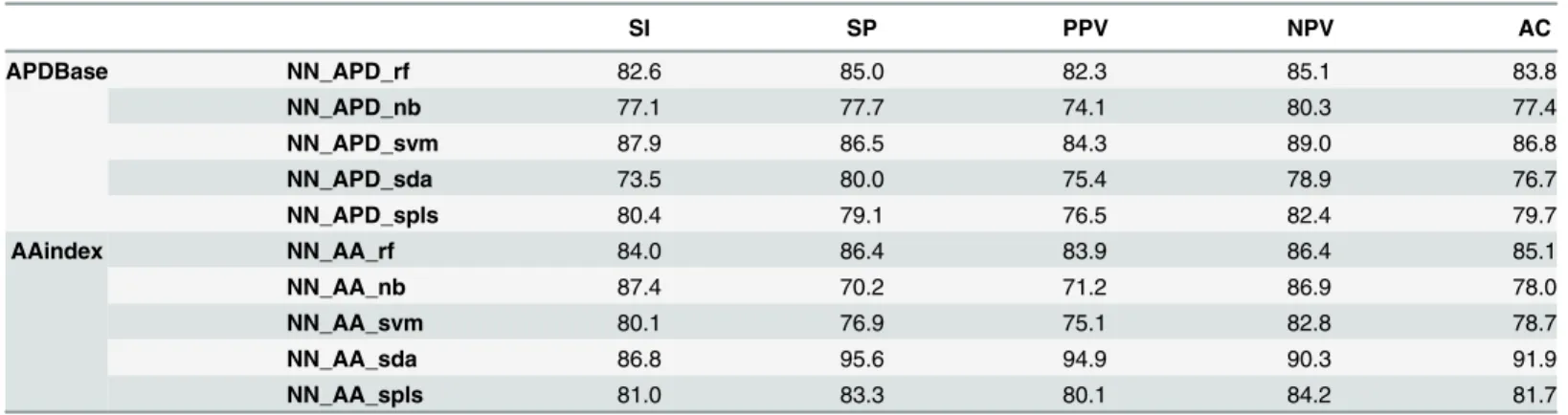
Artificial neural networks were trained using newly computed input vectors based on these selected features. For neural networks trained with feature vectors computed through APDBase encoding, the values obtained for the overall accuracy range between 76.7% (NN_APD_sda) to 86.8% (NN_APD_svm), for sensitivity from 73.5% (NN_APD_sda) to 87.9% (NN_APD_svm) and specificity from 77.7% (NN_APD_nb) to 86.5% (NN_APD_svm) (Table 1). For neural networks trained with feature vectors computed through AAindex encoding, the overall accu-racy values obtained range from 78.0% (NN_AA_rf) to 91.9% (NN_AA_sda), sensitivity from 80.1% (NN_AA_svm) to 87.4% (NN_AA_nb) and specificity from 70.2% (NN_AA_nb) to 95.6% (NN_AA_sda) (Table 1).
Classification of the external validation sequences dataset for neural networks trained with the selected features vectors computed through the APDBase encoding, showed high overall
accuracy for 4 of the 5 neural networks selected. The most effective neural network had an accuracy of 83.0% (NN_APD_rf), a sensitivity of 89.4% and a specificity of 67.8%. While for neural networks trained with the selected features vectors computed through the AAindex encoding, only 2 of the 5 neural networks selected showed high overall accuracy. The best accu-racy shown was 84.9% (NN_AA_nb) with a sensitivity of 87.4% and a specificity of 78.9%, and the second best was 82.2% (NN_AA_rf), with a sensitivity of 90.8% and a specificity of 62.6% (Table 2).
Analysis of the amyloidogenicity propensity prediction selected artificial
neural network
The artificial neural network based on the description of the polypeptide sequences through the physicochemical and biochemical properties of the amino acids that showed the highest overall accuracy in the classification of the external validation sequences dataset (NN_AA_nb) was selected for further analysis, and will hereafter be referred as APPNN, standing for Amy-loidogenicity Propensity Prediction Neural Network. This neural network was trained with a small subset of 14 features selected from the input vectors computed through the AAindex database of physicochemical and biochemical properties of the amino acids, by the Naïve
Table 1. Training sequences dataset classification results (%) for the selected neural networks obtained through APDBase or AAindex encoding, after feature selection with one of the internal classifiers, rf, nb, svm, sda and spls. Where SI is the sensitivity, SP the specificity, PPV the positive pre-dictive value, NPV the negative prepre-dictive value and AC the overall accuracy, averaged after 10-fold stratified resampling.
SI SP PPV NPV AC APDBase NN_APD_rf 82.6 85.0 82.3 85.1 83.8 NN_APD_nb 77.1 77.7 74.1 80.3 77.4 NN_APD_svm 87.9 86.5 84.3 89.0 86.8 NN_APD_sda 73.5 80.0 75.4 78.9 76.7 NN_APD_spls 80.4 79.1 76.5 82.4 79.7 AAindex NN_AA_rf 84.0 86.4 83.9 86.4 85.1 NN_AA_nb 87.4 70.2 71.2 86.9 78.0 NN_AA_svm 80.1 76.9 75.1 82.8 78.7 NN_AA_sda 86.8 95.6 94.9 90.3 91.9 NN_AA_spls 81.0 83.3 80.1 84.2 81.7 doi:10.1371/journal.pone.0134679.t001
Table 2. External validation sequences dataset classification results (%) for the selected neural networks obtained through APDBase and AAindex encoding, after feature selection with one of the internal classifiers, rf, nb, svm, sda and spls. Where SI is the sensitivity, SP the specificity, PPV the positive predictive value, NPV the negative predictive value and AC the overall accuracy, averaged after 10-fold stratified resampling.
SI SP PPV NPV AC APDBase NN_APD_rf 89.4 67.8 86.8 73.5 83.0 NN_APD_nb 78.7 82.5 91.3 62.4 79.9 NN_APD_svm 91.8 40.1 78.7 68.2 76.6 NN_APD_sda 91.9 16.3 72.7 46.2 69.8 NN_APD_spls 94.1 17.9 73.1 57.6 71.4 AAindex NN_AA_rf 90.8 62.6 85.3 73.4 82.2 NN_AA_nb 87.4 78.9 90.8 72.3 84.9 NN_AA_svm 95.6 12.6 72.4 51.0 71.2 NN_AA_sda 93.5 15.7 72.6 52.1 70.4 NN_AA_spls 93.9 12.5 72.1 48.9 70.0 doi:10.1371/journal.pone.0134679.t002
Bayes classifier embedded within the recursive feature selection algorithm. Identification of these features revealed that three consisted of summation properties of the amino acid of the Normalized frequency ofβ-sheet [43], the Normalized frequency ofβ-sheet from LG [44] and the Weights forβ-sheet at the window position of 1 [45]. Another two consisted of the values of standard deviation and range of the Isoelectric Point [46], while a further nine consisted of the standard deviation, range and mean absolute deviation of the Atom-based hydrophobic moment [47], the Helix termination parameter at position j+1 [48] and theΔG° values for the peptides extrapolated to 0 M urea [49].
A comparative analysis between the APPNN and several published prediction methods (Aggrescan, AMYLPRED, AMYLPRED2, FoldAmyloid, MetAmyl, Pafig, Pasta, Pasta2, Tango, Waltz and Zyggregator), was undertaken for the classification of both training and external val-idation sequences datasets to assess if differences existed and if so if they were statistically sig-nificant. Prediction results were used, after bootstrapping, to compute the values of sensitivity, specificity, positive predictive value, negative predictive value and accuracy, with correspond-ing 95% confidence intervals; and after 10-fold stratified resamplcorrespond-ing, to determine if the accu-racy values obtained for all predictors were sampled from populations with identical
distributions (case in which all differences between groups are due to random sampling) through Friedman’s test.
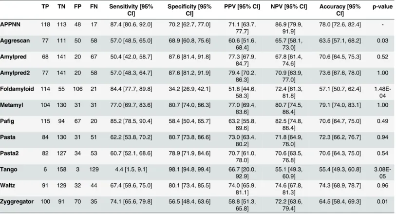
The results obtained for the classification of the training sequences dataset (Table 3), shown for APPNN, high values of specificity (70.2%) and positive prediction value (71.1%), although these were surpassed by several other methods. In contrast, APPNN showed the highest values for sensitivity (87.4%) and negative predictive value (86.9%). APPNN also shown a high accu-racy value (78.0%), only outperformed by the Metamyl predictor (79.1%). Friedman’s test showed differences between predictors’ accuracy to be statistically significant
(chi-squared = 62.7812, df = 11, p = 2.81E-09) with pairwise comparisons identifying key differ-ences between the APPNN and the methods Aggrescan, Foldamyloid and Tango and Zyggre-gator (p< 0.05).
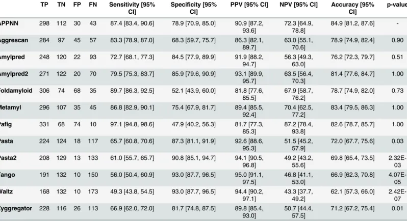
The results obtained for classification of the external validation sequences dataset (Table 4), shown APPNN had high values of sensitivity (87.4%), specificity (78.9%), positive predictive value (90.9%) and negative predictive value (72.3%). However it was only for the accuracy value (84.9%) that APPNN was able to outperform all other prediction methods. Friedman’s test again showed that the differences between predictors’ accuracy was statistically significant (chi-squared = 78.1777, df = 11, p-value = 3.318E-12) with pairwise comparison confirming superior performance of APPNN compared to Pasta, Pasta2, Tango, Waltz and Zyggregator (p< 0.05).
Discussion
Orthogonal based artificial neural networks
The orthogonal encoding of the amino acids, due to its simplicity, has been used in several sec-ondary structure prediction algorithms [45,60,61], even if only to establish an internal refer-ence to compare to the developed predictor and determine if the features included are more informative than merely, the type of amino acids present within the sequence and their relative position [62,63]. With this in mind, several artificial neural networks have been trained, based on input vectors computed through the orthogonal encoding of amino acids, from which, the neural network with the best overall accuracy and lowest accuracy differences between training, testing and validation sub-dataset classifications was selected, as described in the methods sec-tion. This neural network showed relatively high values of sensitivity (83.0%), specificity (82.6%), positive and negative predictive values (80.0% and 85.3% respectively) and overall
accuracy (82.8%) in the classification of the sequences present in the training sequences data-set. However, the results obtained for the classification of the sequences present in the external validation sequences dataset were, except for sensitivity (90.6%), considerably lower for speci-ficity (31.0%), positive and negative predictive values (75.9% and 57.9% respectively) and over-all accuracy (73.1%). These results could be an indication that the rules developed by the selected neural network are not easily generalized across more diverse sequences or that the relationships established between the features present in the input vectors and expected out-comes do not entirely describe the amyloid forming propensity problem when transposed to peptide and protein sequences with lengths greater than six amino acids. It is interesting how-ever, that this encoding method allowed amyloidogenicity propensity prediction with relatively high accuracy (73.1%), considering the simplicity of the information provided to the learning algorithm.
Physicochemical and biochemical based neural networks
The physicochemical and biochemical description of the polypeptide sequences was obtained through the encoding sequence based on the APDBase and AAindex databases of physicochemical and biochemical properties of the amino acids. After which, feature selec-tion was performed with the purpose of reducing the dimensionality of the computed input vectors, enabling the smallest subset of features to be chosen while providing the highest
Table 3. Results obtained for the classification of the training sequences dataset for each predictor, where TP corresponds to the number of true positives, TN to the number of true negatives, FP to the number of false positives and FN to the number of false negatives. The values of sensitivity, specificity, positive predictive value (PPV), negative predictive value (NPV) and accuracy, with corresponding 95% confidence intervals, were obtained using bootstrap replicates. The p-value corresponds to the p-value obtained for the comparison of the accuracy values between the APPNN and each given other predictors using the Wilcoxon-Nemenyi-McDonald-Thompson post-hoc test performed after 10-fold stratified resampling.
TP TN FP FN Sensitivity [95% CI]
Specificity [95% CI]
PPV [95% CI] NPV [95% CI] Accuracy [95% CI] p-value APPNN 118 113 48 17 87.4 [80.6, 92.0] 70.2 [62.7, 77.0] 71.1 [63.7, 77.7] 86.9 [79.9, 91.9] 78.0 [72.6, 82.4] -Aggrescan 77 111 50 58 57.0 [48.5, 65.0] 68.9 [60.8, 75.6] 60.6 [51.6, 68.4] 65.7 [58.1, 73.0] 63.5 [57.1, 68.2] 0.03 Amylpred 68 141 20 67 50.4 [42.0, 58.7] 87.6 [81.4, 91.8] 77.3 [67.9, 84.7] 67.8 [61.4, 74.6] 70.6 [64.5, 75.3] 0.52 Amylpred2 77 141 20 58 57.0 [48.3, 64.7] 87.6 [81.2, 91.9] 79.4 [70.2, 86.3] 70.9 [63.9, 77.0] 73.6 [67.6, 78.0] 1.00 Foldamyloid 114 55 106 21 84.4 [77.7, 89.8] 34.2 [26.9, 42.1] 51.8 [44.6, 58.3] 72.4 [61.3, 81.8] 57.1 [50.7, 62.4] 1.48E-04 Metamyl 104 130 31 31 77.0 [69.7, 83.6] 80.7 [74.0, 86.3] 77.0 [69.4, 83.6] 80.7 [74.5, 86.4] 79.1 [74.0, 83.1] 1.00 Pafig 115 94 67 20 85.2 [78.5, 90.4] 58.4 [50.4, 65.7] 63.2 [55.8, 69.6] 82.5 [74.8, 88.4] 70.6 [64.7, 75.0] 0.49 Pasta 84 130 31 51 62.2 [53.8, 70.2] 80.7 [73.8, 86.6] 73.0 [63.4, 80.2] 71.8 [64.9, 78.0] 72.3 [66.2, 76.7] 0.94 Pasta2 82 127 34 53 60.7 [52.1, 68.6] 78.9 [71.9, 84.6] 70.7 [61.0, 78.0] 70.6 [63.5, 76.8] 70.6 [64.3, 75.0] 0.54 Tango 6 158 3 129 4.4 [1.5, 9.1] 98.1 [94.8, 99.4] 66.7 [20.0, 92.9] 55.1 [49.3, 60.9] 55.4 [49.3, 60.8] 3.08E-05 Waltz 91 129 32 44 67.4 [59.6, 75.0] 80.1 [73.4, 85.5] 74.0 [65.9, 81.1] 74.6 [67.8, 81.3] 74.3 [68.9, 78.7] 0.96 Zyggregator 100 91 70 35 74.1 [65.6, 79.8] 56.5 [48.4, 63.6] 58.8 [51.3, 65.8] 72.2 [63.6, 79.4] 64.5 [58.4, 69.3] 0.01 doi:10.1371/journal.pone.0134679.t003
possible generalization, so improving the neural networks’ learning performance. In the present study, feature selection was performed using three wrapper methods of recursive fea-ture selection, caret, Boruta, and penalizedSVM with five different classifiers (spls, sda, nb for caret, rf for Boruta and svm for penalizedSVM). This resulted in the selection of 96, 10, 13, 548 and 810 features for the input vectors computed through the APDBase encoding database and 109, 14, 334, 100 and 969 features for the input vectors computed through the AAindex encoding dataset for rf, nb, svm, sda and spls, respectively. For each subset of selected features, new input vectors were created and used in the training of several neural networks from which 5 neural networks per encoding dataset were selected. The selected neural networks showed high values of sensitivity (73.5% to 87.9%), specificity (70.2% to 95.6%), positive and negative predictive values (71.2% to 94.9% and 78.9% to 90.3% respec-tively) and overall accuracy (76.7% to 91.9%) in the classification of the sequences present in the training sequences dataset. However, in the classification of the sequences present in the external validation sequences dataset, only three of the selected neural networks
(NN_APD_rf, NN_AA_rf and NN_AA_nb inTable 2) for both encoding schemes, showed high values of sensitivity (89.4%, 90.8% and 87.4%), specificity (67.8%, 62.6% and 78.9%), positive (86.8%, 85.3% and 90.8%) and negative (73.5%, 73.4% and 72.3%) predictive values and overall accuracy (82.2% and 84.9%), suggesting that svm, sda and spls were not the most appropriate classifier methods for this specific type of classification problem in contrast to rf and nb classifiers.
Table 4. Results obtained for the classification of the external validation sequence dataset for each predictor, where TP corresponds to the num-ber of true positives, TN to the numnum-ber of true negatives, FP to the numnum-ber of false positives and FN to the numnum-ber of false negatives. The values of sensitivity, specificity, positive predictive value (PPV), negative predictive value (NPV) and accuracy, with corresponding 95% confidence intervals, were obtained using bootstrap replicates. The p-value corresponds to the p-value obtained for the comparison of the accuracy values between the APPNN and each given predictor using the Wilcoxon-Nemenyi-McDonald-Thompson post-hoc test performed after 10-fold stratified resampling.
TP TN FP FN Sensitivity [95% CI]
Specificity [95% CI]
PPV [95% CI] NPV [95% CI] Accuracy [95% CI] p-value APPNN 298 112 30 43 87.4 [83.4, 90.6] 78.9 [70.9, 85.0] 90.9 [87.2, 93.6] 72.3 [64.9, 78.8] 84.9 [81.2, 87.6] -Aggrescan 284 97 45 57 83.3 [78.9, 87.0] 68.3 [59.7, 75.7] 86.3 [82.1, 89.7] 63.0 [55.1, 70.6] 78.9 [74.9, 82.4] 0.90 Amylpred 248 120 22 93 72.7 [68.1, 77.3] 84.5 [77.9, 89.9] 91.9 [88.2, 94.7] 56.3 [49.3, 63.0] 76.2 [72.3, 79.7] 0.51 Amylpred2 271 122 20 70 79.5 [75.3, 83.7] 85.9 [79.6, 90.9] 93.1 [89.9, 95.7] 63.5 [56.4, 70.3] 81.4 [77.6, 84.7] 1.00 Foldamyloid 306 74 68 35 89.7 [86.3, 92.5] 52.1 [43.9, 60.0] 81.8 [77.6, 85.5] 67.9 [58.7, 76.2] 78.7 [74.9, 82.0] 0.73 Metamyl 296 107 35 45 86.8 [82.9, 90.1] 75.4 [67.9, 81.7] 89.4 [85.5, 92.4] 70.4 [62.5, 77.2] 83.4 [79.5, 86.3] 1.00 Pafig 331 68 74 10 97.1 [94.8, 98.6] 47.9 [40.2, 56.3] 81.7 [77.3, 85.3] 87.2 [78.4, 93.8] 82.6 [78.7, 85.7] 1.00 Pasta 224 124 18 117 65.7 [60.8, 70.6] 87.3 [81.1, 91.9] 92.6 [88.6, 95.3] 51.5 [45.2, 57.9] 72.0 [67.7, 75.6] 0.03 Pasta2 208 129 13 133 61.0 [55.7, 65.7] 90.8 [85.1, 94.7] 94.1 [90.5, 96.8] 49.2 [43.2, 55.6] 69.8 [65.4, 73.5] 2.32E-03 Tango 191 132 10 150 56.0 [50.4, 60.9] 93.0 [87.7, 96.5] 95.0 [91.1, 97.5] 46.8 [41.1, 53.0] 66.9 [62.3, 70.8] 4.07E-05 Waltz 168 132 10 173 49.3 [43.8, 54.5] 93.0 [87.7, 96.5] 94.4 [90.2, 97.1] 43.3 [37.7, 49.2] 62.1 [57.3, 66.0] 2.42E-07 Zyggregator 228 116 26 113 66.9 [62.0, 72.0] 81.7 [74.8, 87.5] 89.8 [85.4, 93.0] 50.7 [44.4, 57.5] 71.2 [67.2, 75.4] 0.01 doi:10.1371/journal.pone.0134679.t004
Analysis of the amyloidogenicity propensity prediction of the selected
artificial neural network
The artificial neural network based on the description of the polypeptide sequences through the physicochemical and biochemical properties of the amino acids that showed the highest overall accuracy, and therefore, the most successful predictor developed, was selected for fur-ther study (NN_AA_nb). This neural network, henceforth referred as APPNN, showed values of sensitivity, specificity, positive predictive value, negative predictive value and overall accu-racy of 87.4%, 70.2%, 71.1%, 86.9% and 78.0%, respectively, when classifying the sequences present in the training sequences dataset and 87.4%, 78.9%, 90.9%, 72.3% and 84.9%, respec-tively, when classifying sequences present in the external validation sequences dataset.
This newly developed predictor was generated from training with input vectors computed through the AAindex database of physicochemical and biochemical properties of amino acids, after recursive feature selection with the internal classifier Naïve Bayes, where a subset of 14 features was selected. These were the summation of the values of the Normalized frequency of β-sheet [43], the Normalized frequency ofβ-sheet from a dataset of 44 sample proteins named LG [44], the weights of a first order neural network neuronal forβ-sheet at the window position of 1 [45], the standard deviation of the isoelectric point [46], and standard deviation, range and mean absolute deviation of the atom-based hydrophobic moment [47], the helix termination parameter or theoretical estimate of helix-coil stability parameter for the natural occurring amino acids when found at position j+1 of the C-terminal region of the helix [48], and theΔG° values, which provides a measure of structural stability, for the peptides extrapolated to 0 M of urea from a two-state model derived from urea denaturation curves that correlated the dissoci-ation constants of the peptides containing one of the 20 natural occurring amino acids in a guest position, with the urea concentration [49]. Interestingly the amino acids’ propensity to formβ-sheet and α-helices and the hydrophobic moment have been consistently pointed out in the literature as fundamental factors in the molecular mechanism of amyloid formation and are proved here to have a high correlation with the ability of a sequence to form amyloid [7,37,50–53]. Moreover, the isoelectric point of the amino acids in a peptide or protein sequence can affect the intrinsic propensity of a sequence to undergo conformational changes into amyloid fibrils as a result of charge variations caused by pH changes in the environment. TheΔG° values for peptides extrapolated to 0 M of urea is a quantitative measure of the effect of the amino acid point mutations on the conformational stability of peptides. This can be directly correlated with the propensity for a peptide or protein to undergo conformational changes into amyloid intermediates and consequently amyloid fibers, as polypeptides generally need to partially fold (intrinsically unfolded polypeptides) or partially unfold (globular pro-teins) in order to achieve theβ-rich amyloidogenic intermediates [64]. Interestingly, these fea-tures fit into the three major groups of feafea-tures identified by Maurer-Stroh and co-workers in the development of the Waltz algorithm (α-helical, β-sheet and solvation-related hydrophobic-ity propensities).
A more detailed analysis of the APPNN classification results obtained using training and external validation sequences datasets, showed some level of disagreement in terms of sensitiv-ity, specificsensitiv-ity, positive and negative predictive values and accuracy. This was found to be even more marked for the neural networks that had been developed using feature selection based on other methodologies. This could be due to the low number of training sequences, which allied to the low variability of these sequences and may have led to an small over fitting of the neural network and thus to a lower generalization capability when classifying never seen sequences. Moreover, the assumption that for a sequence to be considered amyloidogenic there needs to be at least one amyloidogenic six amino acid stretch within the sequence (hard coded into the
algorithm), does not take into account the fact that only one stretch may not be enough to effectively produce the destabilization of the entire peptide or protein structure required for the transition from its native state into amyloid fibers. Additionally, it does not take into account possible interactions between stretches, amyloidogenic or non-amyloidogenic, which could enhance or even inhibit amyloid formation. For these reasons it is thus expected that the length of the input sequences may play a major role in prediction results, where the overall accuracy of our predictor should be higher for smaller sequences. In order to mitigate this problem, the algorithm developed here provides a per amino acid prediction score, which could be used for further analysis.
A comparative analysis between the accuracy of APPNN and several others published pre-diction methods (Aggrescan, AMYLPRED, AMYLPRED2, FoldAmyloid, MetAmyl, Pafig, Pasta, Pasta2, Tango, Waltz and Zyggregator), was undertaken for the classification of sequence datasets assembled from the literature (training sequences and external validation sequences datasets). The results show that APPNN has high overall accuracies for the classifi-cation of the training (78.0%) and the external validation (84.9%) sequence datasets. MetAmyl outperformed APPNN in the classification of the training sequences dataset with an accuracy of 79.1%, although APPNN outperformed MetAmyl in the classification of the external valida-tion sequences dataset, where MetAmyl showed an accuracy value of 83.4%. Analysis showed that the differences between the accuracy values obtained for APPNN and the other predictions was statistically significant hence confirming that the APPNN was able to provide enhanced propensity prediction compared to Pasta (p-value of 0.03), Pasta2 (p-value of 2.32E-03) Tango (p-value of 4.07E-05), Waltz (p-value of 2.42E-07) and Zyggregator (p-value of 0.01).
In this study we have thus developed a highly accurate and effective method for the predic-tion of amyloid propensity based on the polypeptide amino acid sequence alone. This was achieved using a very small subset of highly relevant physicochemical and biochemical amino acid properties. Overall, this study not only provides a new amyloidogenicity propensity pre-diction method but also gives new insights into the key driving forces underpinning the self-assembly of peptides and proteins into amyloid-like fibers.
Methods
Sequence datasets
A dataset of polypeptide sequences with experimentalin vitro evidence of amyloid formation was assembled from the combination of sequences present in published datasets used in several amyloidogenicity propensity prediction studies [28,29,33,34,54,55,65–68]. In addition, several sequences meeting this requirement were also added (seeS1andS2files for references). This resulted in the construction of two distinct datasets (training sequences dataset and external validation sequences dataset) in which each sequence is associated with a binary target value, representing its ability to form amyloid. The training sequences dataset (S1 File) is exclusively formed by peptides of six amino acids in length, with a total of 296 sequences, from which 161 have been reported negatively and 125 positively for amyloid formation. The external valida-tion sequences dataset (S2 File) is a more general dataset comprising a total of 483 peptide and protein sequences with lengths greater than six amino acids, from which 142 have been reported negatively and 341 positively for amyloid formation.
Sequence encoding
In order to convert sequence information into numerical vectors that could identify each sequence uniquely and be used to train the artificial neural networks, three encoding schemes were prepared in MATLAB [69]. These schemes utilized a simple orthogonal encoding for the
twenty naturally occurring amino acids, two datasets of amino acid physicochemical and bio-chemical properties, the Amino Acid Index Database version 9.1 (AAindex) [58,59] and the Amino Acid Physicochemical Properties Database (APDBase) [57].
AAindex is a database of numerical indices that represent the physicochemical and bio-chemical properties of individual amino acids, from which only the first dataset (aaindex1) was used. This dataset contains a total of 544 characteristics, 531 of which, had no missing values for any of the twenty natural occurring amino acids and thus were used in the calculations [58,59].
APDBase is a smaller database containing a total of 242 physicochemical and biochemical properties for all twenty naturally occurring amino acids. APDBase was derived from two other databases, AAindex and ProtScale by Mathura and Kolippakkam, based on properties they felt were most relevant to the study of protein sequence, structure, and function [57].
Sequences present in both training and external validation sequence datasets were encoded throughin house built scripts in MATLAB [69] programing language. Feature vectors based on the orthogonal descriptors of the amino acids, were created by the linear combination of the respective individual amino acids orthogonal vectors (S1 Table). Feature vectors, based on the physicochemical and biochemical properties of the amino acids were obtained by the concate-nation of several smaller vectors containing the single characteristics of the amino acids, the cumulative summation of these characteristics and some basic mathematical and statistical measures of these characteristics (summation, mean, harmonic mean, median, mode, standard deviation, interquartile range, mean absolute deviation, range, kurtosis and skewness).
Feature vectors pre-processing
Pre-processing was performed for all generated feature vectors, prior to training and external validation of the neural networks, through data normalization by mapping the mean and stan-dard deviation to 0 and 1 respectively (except for feature vectors based on the orthogonal encoding), through removal of features with no variation across samples and removal of dupli-cated features. This pre-processing was performed to circumvent scale effects of some features over others and to improve feature selection performance.
Feature selection
Feature selection was performed for the characteristics vectors computed through both physi-cochemical and biochemical properties of the amino acids encoding datasets (AAindex and APDBase). Feature selection was performed utilizing three recursive feature selection wrapper methods, from the caret package v.5.15–48 [70], Boruta package v.1.6 [71] and penalizedSVM package v.1.1 [72] for R v.2.15.3 [73] with four different internal classifiers (sparse partial least squares (spls), shrinkage discriminant analysis (sda), both linear, and naïve bayes (nb) for caret, and random forests (rf) for Boruta and support vector machines (svm) for penali-zedSVM). After this the input vectors were trimmed to match each set of selected features and subsequently used in the training of 5 neural networks per encoding dataset. The generated neural networks were posteriorly validated through the classification of the external validation sequences dataset as described below.
Artificial neural networks
MATLAB’s [69] Neural Networks Toolbox [74] was used to create feed forward fully con-nected neural networks. The weights and biases of the neural networks were initialized with the Nguyen-Widrow layer initialization function (initializing weights and biases randomly but evenly across each layer’s input space). The activation function selected for the hidden layer
was the symmetric sigmoid function and for the output layer was the linear function. The learning algorithm used was the scaled conjugate gradient backpropagation (backward propa-gation of errors) and the performance measure used to stop training was the mean absolute error. The number of neurons present in the input layer was set to match the dimensions of the different feature vectors. The number of neurons present in the hidden layer was computed based on the number of dimensions of the feature vectors (n/3) for the orthogonal based vectors
and(n/2 +1)for the physicochemical and biochemical properties of the amino acid based
vec-tors). The number of neurons in the output layer was one.
For each of the computed input vectors, neural networks were trained after random division of the input sequences dataset (the hexapeptides dataset) into three distinct subsets, the train-ing, test and validation subsets, comprising 70%, 15% and 15% of the overall training samples, respectively. The best neural network was selected for the feature vectors computed through the orthogonal encoding scheme from a total of 5000 trained networks, and for feature vectors computed through the physicochemical and biochemical encoding scheme from a total of 1000 trained networks. This selection was based on the values of accuracy and standard deviation obtained for the training, test, validation subsets and overall dataset, where the neural network with the highest average accuracy was selected, provided that the standard deviation was below 7.5%. The selected neural networks were posteriorly validated by the classification of the sequences present in the external validation dataset. This was performed by the submission of the pre-processed individual input vectors, generated by a sliding window of six amino acids that was run through the polypeptide sequence, to the corresponding neural network. A sequence was considered amyloidogenic if at least one of these six amino acid windows was classified amyloidogenic.
Comparison with other prediction algorithms
A careful comparison of the best neural network (APPNN) and other published methods for amyloid propensity prediction (Aggrescan, AMYLPRED, AMYLPRED2, FoldAmyloid, MetAmyl, Pafig, Pasta, Tango, Waltz and Zyggregator) was undertaken. This comparison took into account that both training sequences and external sequences validation datasets have been produced from the literature specifically for this work. Thus the amyloidogenic propensities of the sequences present in both datasets were evaluated by all these methods through the use ofin house built Python version 2.7.5 [75] scripts that allowed sequence sub-mission and results retrieval (input parameters and considerations made to classify a sequence as amyloidogenic from the provided outputs, can be found inS2 Table, organized by prediction method).
Prediction results for both training sequences and external validation sequence datasets were summarized for each predictor in confusion matrices containing the obtained values for sensitivity, specificity, positive predictive value, negative predictive value and accuracy, with corresponding 95% confidence intervals. These values were calculated with the R v.2.15.1 [73] package boot v.1.2–10 [76,77] through bootstrapping performed with 2000 replicates. More-over, after 10-fold stratified resampling of the data with the package caret v.5.15–48 [70], the Friedman’s test provided in the package coin v.1.0–23 [78,79] was used to determine if the accuracy values obtained for all predictors were sampled from populations with identical distri-butions, which was followed by the Wilcoxon-Nemenyi-McDonald-Thompson post-hoc test [80] from the package multcmp v.1.3–2 [81], for pairwise comparison between APPNN and every other classifier.
Supporting Information
S1 File. Training sequences dataset. (DOCX)
S2 File. External validation sequences dataset. (DOCX)
S1 Table. Orthogonal encoding of the amino acids. (DOCX)
S2 Table. Input parameters and considerations made to classify a sequence as amyloido-genic from the provided outputs, organized by prediction method.
(DOCX)
Acknowledgments
The first author would like to thank Ana Santos, Vitor Família, Branca Proença for all their encouragement and support. The authors also thank Professor Silvio Tosatto for all the clarifi-cations provided about the Pasta prediction method, and Professor Sebastian Maurer-Stroh for the availability and help regarding interpretation of the results provided by the Waltz predic-tion method.
Author Contributions
Conceived and designed the experiments: CF SRD AQ DAP. Performed the experiments: CF. Analyzed the data: CF. Contributed reagents/materials/analysis tools: CF SRD AQ DAP. Wrote the paper: CF SRD AQ DAP.
References
1. Knowles TPJ, Vendruscolo M, Dobson CM. The amyloid state and its association with protein misfold-ing diseases. Nat Rev Mol Cell Biol 2014; 15:384–96. doi:10.1038/nrm3810PMID:24854788 2. Stefani M, Dobson CM. Protein aggregation and aggregate toxicity: new insights into protein folding,
misfolding diseases and biological evolution. J Mol Med Berlin Ger 2003; 81:678–99.
3. Chiti F, Dobson CM. Protein misfolding, functional amyloid, and human disease. Annu Rev Biochem 2006; 75:333–66. doi:10.1146/annurev.biochem.75.101304.123901PMID:16756495
4. Selkoe DJ. Folding proteins in fatal ways. Nature 2003; 426:900–4. doi:10.1038/nature02264PMID: 14685251
5. Belli M, Ramazzotti M, Chiti F. Prediction of amyloid aggregation in vivo. EMBO Rep 2011; 12:657–63. doi:10.1038/embor.2011.116PMID:21681200
6. Fowler DM, Koulov AV, Balch WE, Kelly JW. Functional amyloid—from bacteria to humans. Trends Biochem Sci 2007; 32:217–24. doi:10.1016/j.tibs.2007.03.003PMID:17412596
7. Nerelius C, Fitzen M, Johansson J. Amino acid sequence determinants and molecular chaperones in amyloid fibril formation. Biochem Biophys Res Commun 2010; 396:2–6. doi:10.1016/j.bbrc.2010.02. 105PMID:20494101
8. DePas WH, Chapman MR. Microbial manipulation of the amyloid fold. Res Microbiol 2012; 163:592– 606. doi:10.1016/j.resmic.2012.10.009PMID:23108148
9. Kelly JW, Balch WE. Amyloid as a natural product. J Cell Biol 2003; 161:461–2. doi:10.1083/jcb. 200304074PMID:12743097
10. Maji SK, Perrin MH, Sawaya MR, Jessberger S, Vadodaria K, Rissman RA, et al. Functional amyloids as natural storage of peptide hormones in pituitary secretory granules. Science 2009; 325:328–32. doi: 10.1126/science.1173155PMID:19541956
11. Baskakov IV. Thermodynamics and Protein Folding. In: Sipe JD, editor. Amyloid Proteins, Weinheim, Germany: Wiley-VCH Verlag GmbH; 2005, p. 65–80. doi:10.1002/9783527619344
12. Harrison RS, Sharpe PC, Singh Y, Fairlie DP. Amyloid peptides and proteins in review. Rev Physiol Biochem Pharmacol 2007; 159:1–77. doi:10.1007/112_2007_0701PMID:17846922
13. Cherny I, Gazit E. Amyloids: not only pathological agents but also ordered nanomaterials. Angew Chem Int Ed Engl 2008; 47:4062–9. doi:10.1002/anie.200703133PMID:18412209
14. Ventura S, Villaverde A. Protein quality in bacterial inclusion bodies. Trends Biotechnol 2006; 24:179– 85. doi:10.1016/j.tibtech.2006.02.007PMID:16503059
15. Idicula-thomas S, Balaji PV. Protein aggregation: A perspective from amyloid and inclusion-body for-mation. Curr Sci 2007; 92:758–67.
16. Pastor MT, Esteras-Chopo A, López de la Paz M. Design of model systems for amyloid formation: les-sons for prediction and inhibition. Curr Opin Struct Biol 2005; 15:57–63. doi:10.1016/j.sbi.2005.01.004 PMID:15718134
17. Scheibel T, Parthasarathy R, Sawicki G, Lin X, Jaeger H, Lindquist SL. Conducting nanowires built by controlled self-assembly of amyloid fibers and selective metal deposition. Proc Natl Acad Sci U S A 2003; 100:4527–32. doi:10.1073/pnas.0431081100PMID:12672964
18. Rajagopal K, Schneider JP. Self-assembling peptides and proteins for nanotechnological applications. Curr Opin Struct Biol 2004; 14:480–6. doi:10.1016/j.sbi.2004.06.006PMID:15313243
19. Sunde M, Blake C. The structure of amyloid fibrils by electron microscopy and X-ray diffraction. Adv Protein Chem 1997; 50:123–59. PMID:9338080
20. Kelly JW. Alternative conformations of amyloidogenic proteins govern their behavior. Curr Opin Struct Biol 1996; 6:11–7. PMID:8696966
21. Sipe JD. The Beta-pleated Sheet Conformation and Protein Folding: A Brief History. In: Sipe JD, editor. Amyloid Proteins, Weinheim, Germany: Wiley-VCH Verlag GmbH; 2005, p. 49–61. doi:10.1002/ 9783527619344
22. Astbury WT, Dickinson S, Bailey K. The X-ray interpretation of denaturation and the structure of the seed globulins. Biochem J 1935; 29:2351–60.1. PMID:16745914
23. Sunde M, Serpell LC, Bartlam M, Fraser PE, Pepys MB, Blake CC. Common core structure of amyloid fibrils by synchrotron X-ray diffraction. J Mol Biol 1997; 273:729–39. doi:10.1006/jmbi.1997.1348 PMID:9356260
24. Hamodrakas SJ. Protein aggregation and amyloid fibril formation prediction software from primary sequence: towards controlling the formation of bacterial inclusion bodies. FEBS J 2011; 278:2428–35. doi:10.1111/j.1742-4658.2011.08164.xPMID:21569208
25. Dobson CM. The structural basis of protein folding and its links with human disease. Philos Trans R Soc Lond B Biol Sci 2001; 356:133–45. doi:10.1098/rstb.2000.0758PMID:11260793
26. EvaŽ. Amyloid-fibril formation. Eur J Biochem 2002; 269:3362–71. doi:10.1046/j.1432-1033.2002. 03024.xPMID:12135474
27. Chiti F, Stefani M, Taddei N, Ramponi G, Dobson CM. Rationalization of the effects of mutations on peptide and protein aggregation rates. Nature 2003; 424:805–8. doi:10.1038/nature01891PMID: 12917692
28. Goldschmidt L, Teng PK, Riek R, Eisenberg D. Identifying the amylome, proteins capable of forming amyloid-like fibrils. Proc Natl Acad Sci U S A 2010; 107:3487–892. doi:10.1073/pnas.0915166107 PMID:20133726
29. Maurer-Stroh S, Debulpaep M, Kuemmerer N, Lopez de la Paz M, Martins IC, Reumers J, et al. Explor-ing the sequence determinants of amyloid structure usExplor-ing position-specific scorExplor-ing matrices. Nat Meth-ods 2010; 7:237–42. doi:10.1038/nmeth.1432PMID:20154676
30. Quintas A. The Tetrameric Protein Transthyretin Dissociates to a Non-native Monomer in Solution. A novel model for amyloidogenesis. J Biol Chem 1999; 274:32943–9. doi:10.1074/jbc.274.46.32943 PMID:10551861
31. Trovato A, Chiti F, Maritan A, Seno F. Insight into the structure of amyloid fibrils from the analysis of globular proteins. PLoS Comput Biol 2006; 2:e170. doi:10.1371/journal.pcbi.0020170PMID: 17173479
32. Oliveberg M. Waltz, an exciting new move in amyloid prediction. Nat Methods 2010; 7:187–8. doi:10. 1038/nmeth0310-187PMID:20195250
33. Fernandez-Escamilla AM, Rousseau F, Schymkowitz JWH, Serrano L. Prediction of sequence-depen-dent and mutational effects on the aggregation of peptides and proteins. Nat Biotechnol 2004; 22:1302–6. doi:10.1038/nbt1012PMID:15361882
34. Conchillo-Solé O, de Groot NS, Avilés FX, Vendrell J, Daura X, Ventura S. AGGRESCAN: a server for the prediction and evaluation of“hot spots” of aggregation in polypeptides. BMC Bioinformatics 2007; 8:1–17. doi:10.1186/1471-2105-8-65PMID:17199892
35. Trovato A, Seno F, Tosatto SCE. The PASTA server for protein aggregation prediction. Protein Eng Des Sel 2007; 20:521–3. doi:10.1093/protein/gzm042PMID:17720750
36. Tartaglia GG, Vendruscolo M. The Zyggregator method for predicting protein aggregation propensities. Chem Soc Rev 2008; 37:1395–401. doi:10.1039/b706784bPMID:18568165
37. Frousios KK, Iconomidou VA, Karletidi CM, Hamodrakas SJ. Amyloidogenic determinants are usually not buried. BMC Struct Biol 2009; 9:44. doi:10.1186/1472-6807-9-44PMID:19589171
38. Garbuzynskiy SO, Lobanov MY, Galzitskaya OV. FoldAmyloid: a method of prediction of amyloido-genic regions from protein sequence. Bioinformatics 2010; 26:326–32. doi:10.1093/bioinformatics/ btp691PMID:20019059
39. Tian J, Wu N, Guo J, Fan Y. Prediction of amyloid fibril-forming segments based on a support vector machine. BMC Bioinformatics 2009; 10 Suppl 1:1–8. doi:10.1186/1471-2105-10-S1-S45
40. López de la Paz M, Serrano L. Sequence determinants of amyloid fibril formation. Proc Natl Acad Sci U S A 2004; 101:87–92. doi:10.1073/pnas.2634884100PMID:14691246
41. Teng PK, Eisenberg D. Short protein segments can drive a non-fibrillizing protein into the amyloid state. Protein Eng Des Sel 2009; 22:531–6. doi:10.1093/protein/gzp037PMID:19602569
42. Jimenez LO, Landgrebe DA. Supervised classification in high-dimensional space: geometrical, statisti-cal, and asymptotical properties of multivariate data. IEEE Trans Syst Man Cybern Part C (Applications Rev) 1998; 28:39–54. doi:10.1109/5326.661089
43. Chou PY, Fasman GD. Prediction of the secondary structure of proteins from their amino acid sequence. Adv Enzymol Relat Areas Mol Biol 1978; 47:45–148. doi:10.1016/0968-0004(77)90440-6 PMID:364941
44. Palau J, Argos P, Puigdomenech P. Protein secondary structure. Studies on the limits of prediction accuracy. Int J Pept Protein Res 1982; 19:394–401. doi:10.1111/j.1399-3011.1982.tb02620.xPMID: 7118409
45. Qian N, Sejnowski TJ. Predicting the secondary structure of globular proteins using neural network models. J Mol Biol 1988; 202:865–84. PMID:3172241
46. Zimmerman JM, Eliezer N, Simha R. The characterization of amino acid sequences in proteins by sta-tistical methods. J Theor Biol 1968; 21:170–201. PMID:5700434
47. Eisenberg D, McLachlan AD. Solvation energy in protein folding and binding. Nature 1986; 319:199– 203. doi:10.1038/319199a0PMID:3945310
48. Finkelstein AV, Badretdinov AY, Ptitsyn OB. Physical reasons for secondary structure stability: alpha-helices in short peptides. Proteins 1991; 10:287–99. doi:10.1002/prot.340100403PMID:1946339 49. O’Neil KT, DeGrado WF. A thermodynamic scale for the helix-forming tendencies of the commonly
occurring amino acids. Science 1990; 250:646–51. PMID:2237415
50. Hamodrakas SJ, Liappa C, Iconomidou VA. Consensus prediction of amyloidogenic determinants in amyloid fibril-forming proteins. Int J Biol Macromol 2007; 41:295–300. doi:10.1016/j.ijbiomac.2007.03. 008PMID:17477968
51. Kallberg Y, Gustafsson M, Persson B, Thyberg J, Johansson J. Prediction of amyloid fibril-forming pro-teins. J Biol Chem 2001; 276:12945–50. doi:10.1074/jbc.M010402200PMID:11134035
52. Pawar AP, Dubay KF, Zurdo J, Chiti F, Vendruscolo M, Dobson CM. Prediction of“aggregation-prone” and“aggregation-susceptible” regions in proteins associated with neurodegenerative diseases. J Mol Biol 2005; 350:379–92. doi:10.1016/j.jmb.2005.04.016PMID:15925383
53. Yoon S, Welsh WJ. Rapid assessment of contact-dependent secondary structure propensity: rele-vance to amyloidogenic sequences. Proteins 2005; 60:110–7. doi:10.1002/prot.20477PMID: 15849755
54. Tsolis AC, Papandreou NC, Iconomidou VA, Hamodrakas SJ. A consensus method for the prediction of“aggregation-prone” peptides in globular proteins. PLoS One 2013; 8:e54175. doi:10.1371/journal. pone.0054175PMID:23326595
55. Emily M, Talvas A, Delamarche C. MetAmyl: a METa-predictor for AMYLoid proteins. PLoS One 2013; 8:e79722. doi:10.1371/journal.pone.0079722PMID:24260292
56. Walsh I, Seno F, Tosatto SCE, Trovato A. PASTA 2.0: an improved server for protein aggregation pre-diction. Nucleic Acids Res 2014; 42:W301–7. doi:10.1093/nar/gku399PMID:24848016
57. Mathura VS, Kolippakkam D. APDbase: Amino acid Physico-chemical properties Database. Bioinfor-mation 2005; 1:2–4. PMID:17597840
58. Kawashima S, Ogata H, Kanehisa M. AAindex: Amino Acid Index Database. Nucleic Acids Res 1999; 27:368–9. PMID:9847231
59. Kawashima S, Pokarowski P, Pokarowska M, Kolinski A, Katayama T, Kanehisa M. AAindex: amino acid index database, progress report 2008. Nucleic Acids Res 2008; 36:202–5. doi:10.1093/nar/ gkm998
60. Rost B, Sander C. Prediction of protein secondary structure at better than 70% accuracy. J Mol Biol 1993; 232:584–99. doi:10.1006/jmbi.1993.1413PMID:8345525
61. Sasagawa F, Tajima K. Prediction of protein secondary structures by a neural network. Comput Appl Biosci 1993; 9:147–52. PMID:8481816
62. Hu H, Pan Y, Harrison R, Tai PC. Improved protein secondary structure prediction using support vector machine with a new encoding scheme and an advanced tertiary classifier. IEEE Trans Nanobioscience 2004; 3:265–71. PMID:15631138
63. Zamani M, Kremer SC. Protein secondary structure prediction using support vector machines and a codon encoding scheme. 2012 IEEE Int Conf Bioinforma Biomed Work 2012:22–7. doi:10.1109/ BIBMW.2012.6470326
64. Rochet JC, Lansbury PT. Amyloid fibrillogenesis: themes and variations. Curr Opin Struct Biol 2000; 10:60–8. doi:10.1016/S0959-440X(99)00049-4PMID:10679462
65. DuBay KF, Pawar AP, Chiti F, Zurdo J, Dobson CM, Vendruscolo M. Prediction of the absolute aggre-gation rates of amyloidogenic polypeptide chains. J Mol Biol 2004; 341:1317–26. doi:10.1016/j.jmb. 2004.06.043PMID:15302561
66. Yoon S, Welsh WJ. Detecting hidden sequence propensity for amyloid fibril formation. Protein Sci 2004; 13:2149–60. doi:10.1110/ps.04790604PMID:15273309
67. Tartaglia GG, Cavalli A, Pellarin R, Caflisch A. Prediction of Aggregation Rate and Aggregation-Prone Segments in Polypeptide Sequences. Fakultat der Universitat Zurich, 2005. doi:10.1110/ps. 051471205
68. Thompson MJ, Sievers SA, Karanicolas J, Ivanova MI, Baker D, Eisenberg D. The 3D profile method for identifying fibril-forming segments of proteins. Proc. Natl. Acad. Sci. U. S. A., vol. 103, 2006, p. 4074–8. doi:10.1073/pnas.0511295103
69. The MathWorks I. MATLAB 2011.
70. Kuhn M. Building Predictive Models in R Using the caret Package. J Stat Softw 2008; 28:1–26. 71. Kursa MB, Rudnicki WR. Feature Selection with the Boruta Package. J Stat Softw 2010; 36:1–13. 72. Becker N, Werft W, Toedt G, Lichter P, Benner A. penalizedSVM: a R-package for feature selection
SVM classification. Bioinformatics 2009; 25:1711–2. doi:10.1093/bioinformatics/btp286PMID: 19398451
73. R Development Core Team. R: A language and environment for statistical computing (Version 2.15.3). 2008.
74. The MathWorks I. Neural Networks Toolbox for MATLAB 2011. 75. Python Software Foundation. Python, version 2.7.5 2013.
76. Davison AC, Hinkley DV. Bootstrap Methods and Their Applications. Cambridge, UK: Cambridge Uni-versity Press; 1997.
77. Canty A, Ripley B. boot: Bootstrap R (S-Plus) Functions 2014.
78. Hothorn T, Hornik K, van de Wiel MA, Zeileis A. A Lego System for Conditional Inference. Am Stat 2006; 60:257–63. doi:10.1198/000313006X118430
79. Hothorn T, Hornik K, van de Wiel MA, Zeileis A. Implementing a Class of Permutation Tests: The coin Package. J Stat Softw 2008; 28:1–23.
80. Hollander M, Wolfe DA. Nonparametric Statistical Methods. 2nd ed. New York: John Wiley & Sons; 1999.
81. Hothorn T, Bretz F, Westfall P. Simultaneous Inference in General Parametric Models. Biometrical J 2008; 50:346–63.