DOI: http://dx.doi.org/10.18363/rbo.v77.2020.e1884 Short Communication / Oral Medicine
The Contribution of Dentistry on The Sturge-Weber
Syndrome Diagnosis and Management -
Report of Two Cases
Bruno Teixeira Gonçalves Rodrigues,1 Lucas Alberto dos Santos Nunes,1 Flávia Souza Pereira de Jesus Almeida,1 Nathália de Almeida Freire,1 Mônica Simões Israel1
1Department of Diagnosis and Therapeutics, State University of Rio de Janeiro, Rio de Janeiro, RJ, Brazil
• Conflicts of interest: none declared.
A
bstrActObjective: The aim of this study was to report two cases of female patients diagnosed with Sturge-Weber syndrome (SWS), its main oral and maxillofacial clinicals
manifestations and dental management. Case report: Case 1. A 30-year-old female patient, referred to the Oral Medicine Clinic of State University of Rio de Janeiro for evaluation of a diffuse reddish lesion over her face and mouth. During the clinical examination, port-wine stains (PWS) were detected on the right side of her face and intraoral discoloration were suggestive of SWS. As the patient referred no symptoms, she was referred to a neurology team to investigate alterations in the central nervous system (CNS). Case 2. A 19-year-old woman was referred for evaluation of a 5-month lasting gingival bleeding in the right mandibular and maxillary alveolar mucosa. Extraoral exam showed the presence of PWS on the right side of her face. Intraorally, the gingival hyperplasia caused malocclusion, plaque deposition and severe bleeding. The SWS diagnosis was established due to these findings. Patient was referred for periodontal treatment. Conclusion: Thus, it is essential that the dentist has knowledge of this condition clinical manifestations in order to do the correct diagnosis and purpose the correct treatment.
Keywords: Sturge-Weber Syndrome; Port-Wine Stain; Gingival Hyperplasia.
Introduction
S
turge-Weber syndrome (SWS) - also called as
encephalotrigeminal angiomatosis - is a rare
developmental non-hereditary congenital condition
characterized by facial cutaneous vascular nevus (nevus
flammeus or port-wine stain) in association with venous
angiomas in leptomeninges over the cerebral-cortex, usually
unilateral - which often follows the outline distribution of
trigeminal nerve.
1,2SWS often shows morphological and histological
alterations in the periodontal tissues - frequently gingival
hyperplasia involving the maxilla, mouth floor, lips, buccal
mucosa, palate or tongue is observed, ipsilateral to the
port-wine stain on the face. Therefore, it is important for
the clinician to know this syndrome oral manifestations
and the complications that may occur - such as increased
risk of hemorrhage during surgical procedures - in order
to purpose the correct treatment amongst others medical
specialties.
1,2,3Herein, we report two cases of female patients diagnosed
with SWS presenting extra and intraoral manifestations.
Case Report
Case 1. A 30-year-old white female patient was referred
from a dental clinic to the Oral medicine clinic, State
lesions on her face and mouth - that kept growing, but
were present since younger ages. Family history was
non-contributory, and the patient did not report any previous
surgeries or neurological disturbs. On extraoral examination
(Figure. 1.a, 1.b), a right-sided hemihypertrophy of the face
with port-wine stains was observed, extending along the
second division of the trigeminal nerve unilaterally. Her
mouth was deviated toward the inferior-right side of her face.
The upper lip and alar base were swollen, edematous, and
incompetent. Also, there was increased malar prominence
and facial asymmetry. Intraoral exam revealed the presence
of a diffuse reddish to purple discoloration over the hard
and soft palate on the right side, not exceeding the midline
(Figure 1.c). Patient referred to no intraoral symptoms or
periodontal alterations. The clinical findings confirmed
the Sturge-Weber syndrome diagnosis. However, despite
the oral manifestations, as the patient complained about
no symptoms, she was oriented regarding the syndrome
and referred to a neurology service in order to investigate
possible alterations.
Case 2. A 19-year-old white female patient was referred
for evaluation of a 5-month lasting gingival bleeding in the
right mandibular and maxillary alveolar mucosa. Medical
history was non-contributory, and patient denied any
addictions regarding alcohol or tobacco abuse. Extraoral
of the face with port-wine stains following the outline
distribution of trigeminal nerve on the face crossing the
midline. A significant facial asymmetry could be observed
with mouth deviation toward the left side and both
upper and lower lips presented swollen, edematous and
incompetent (Figure 2a, 2b). Intraoral clinical examination
revealed the presence of a diffuse gingival enlargement in
both right maxilla and mandible along with reddish-areas
throughout the right hard palate, buccal mucosa, tongue and
mouth floor - that did not cross the midline. Blanching on
pressure could be noted in the enlarged gingiva, suggesting
angiomatous lesion. Both right maxillary and mandibular
teeth were misplaced, and significant plaque and calculus
deposition was presented due to lack of oral hygiene (Figure
2c). Due to the clinical findings the Sturge-Weber syndrome
diagnosis was established. Patient was oriented regarding
the syndrome and its complications and referred to the
Periodontology clinic to start periodontal treatment - Plaque
control and gingival excision - and to a neurology service to
investigate alterations in the central nervous system.
Discussion
Sturge-Weber syndrome is a very uncommon, non-familial,
congenital condition of unknown etiology. SWS presents
varied clinical features that may affect the central nervous
system (CNS), eyes, skin (specially the face) and the oral
cavity - which the dental clinician should be aware of when
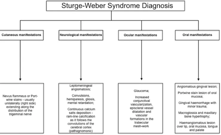
performing the routine stomatologic exam. Its diagnosis
can be established by the presence of port-wine stain on the
face followed by other signs, such as glaucoma, epilepsy, and
mental retardation (Figure 3).
1,2,3Table 1 highlights the extra
and intraoral features reported on previously SWS cases as
well as its dental management, thus highlighting the role of
the dentist on its diagnosis and treatment.
1,2,4-17The Roach Scale is used as a classification of the
SWS manifestations:
18Type 1 presents with facial and
leptomeningeal angiomas and glaucoma may occur; Type
2 involves facial angioma alone - no CNS alteration is
observed - and may have glaucoma and finally, type III shows
isolated leptomeningeal angiomas and usually glaucoma.
Furthermore, when the CNS and facial angiomas are present,
SWS is referred to as complete, whereas it is incomplete
when only one of these areas is affected.
12Both of our cases
presented as the incomplete type II form of Sturge-Weber
syndrome - with the presence of facial angiomas and no CNS
alteration or glaucoma.
Figure 1. A - Frontal View - a right-sided hemihypertrophy of the face
with port-wine stains following the trigeminal nerve second division is observed. B - Profile View. C - Intraoral features - Notice that the reddish discoloration on the right side do not exceed the midline and it is located ipsilateral to the PSW on the face.
Figure 2. A - Frontal View - a right-sided hemihypertrophy of the face
with port-wine stains. B - Profile View. C - Severe periodontal alterations may be observed on this patient presented with SWS in both right maxilla and mandible. D - Gingival hypertrophy on the right maxilla.
Figure 3. Sturge-Weber syndrome diagnostic features. Table1. Sturge-Weber syndrome clinical findings and treatment
Author Sex Age (years) Extraoral features Intraoral features Dental manegement
Gyarmati et al.4 Female 10 Bilaterally PWS; slight
swelling of the upper lip; 1.5 cm gingival growth NS el-Mostehy
et al.5 Female 14 PWS on the left face
Gingival swelling on the left face; mass of hypertrophied, deep red, easily bleeding angiomatous tissue on the left mandibular vestibular; periodontal pockets; teeth mobility
Gingival growth excision; debrided of tissue tags and calculus; root surfaces
Yukna et al.6 Male 25
PWS on the left face, extending from the forehead to the neck and from the midline to the ear
Dusky-red lesion on the left side involving the buccal mucosa, floor of the mouth, pharynx, palate, uvula, and the gingiva of both the maxilla and the mandible. Slight to moderate gingival hyperplasia, coupled with a marginal gingivitis; carious lesions associated with the left posterior teeth
Plaque-control instructions; seaIing and root planning; extractions; pocket elimination by flap surgery
Perez et al. 7 Female 6 Bilaterally PWS, bilateral
congenital glaucoma
Red swelling involving the upper anterior gingiva in the region of the central incisors and the mucosa of the upper lip; carious lesions; malocclusion
Plaque control, carious teeth restore; forward to the Orthodontic clinic for evaluation and treatment.
Yamashiro
et al.8 Male 35 Bilaterally PWS; Glaucoma Gingival enlargement and periodontitis.
Extractions and gingivectomy. De Benedittis
et al.9 Male 25
Bilaterally PWS, hemiplegia, glaucoma, blind in the right eye,
Macroglossia secondary to the hemangioma; gross angiomatous mass
Gingivectomy using Nd:YAG laser
Bhansali et al.1 Male 15
Hemiplegia on the right side along with PWS; right-sided hemihypertrophy of the face; upper and lower
Gingival enlargement involving all four quadrants of the oral cavity; periodontal pockets; purplish-red discoloration over the soft palate, the floor of the mouth bilaterally, and the buccal mucosa on the
Scaling and root planing; extraction; gingivectomy
Conceição et
al.2 Male 29 Lower lip hemangioma
Hemangioma in retromolar trigone and in the lower surface of the tongue seen as purplish red spots without symptomatology
None
Pagin et al.10 Male 43 PWS on the right side of
the face
Stain in the entire hard and soft palates, and the alveolar ridge and buccal mucosa on the right side. Poor oral hygiene, calcified masses in both supragingival and subgingival sites, with swelling and generalized inflammation throughout the gingiva
Technical hygiene instruction; successive sessions to remove dental calculus by ultrasound.
Manivannan
et al.11 Female 20
PWS on the left side of the face; facial asymmetry and macrochelia
Prominent reddish purple gingival enlargement posteriorly on the left side; teeth on the lower arch were malpositioned; poor oral hygiene, extensive amounts of plaque and calculus.
Oral prophylaxis, surgical excision of the lesion under general anesthesia and replacement of the worn out bridge in the upper anterior region.
Babaji et al.12 Female 8 PWS on the right side of
the face
Maxilla on the right side revealed reddish discoloration of gingiva extending from labial frenum to the first molar region with osseous enlargement and drifting of teeth; gingival hyperplasia
Plaque control, oral prophylaxis at regular interval, oral hygiene instructions. Mobile deciduous were extracted Kalakonda
et al.13 Male 23
PWS on the left side of the
face Severe gingival overgrowth; loss of knife edge contour and bulbous papillae
Gingivectomy with diode laser (Picasa 810 nm 1.8 W continuous mode) Kalakonda
et al.13 Male 32
PWS on the left side of the face; facial asymmetry; swelling of the left side of the upper lip
Gingival overgrowth Nonsurgical periodontal therapy under strict aseptic conditions. Kalakonda
et al.13 Female 12
PWS on the right side of the face; reddish discoloration of the sclera of the right eye
Discrete, sessile, reddish-pink gingival
overgrowth; bleeding Gingivectomy;
Tripathi et al.14 Female 15 PWS on the right side of
the face
Unilateral hyperplastic lesions on the right side of the maxilla
Oral plaque control; prophylaxis
Pontes et al.15 Female 23
PWS on the right side of the face; swelling of the superior lip
Large overgrowth in the right maxilla extending from teeth 11 to 18 and a minor growth in the right mandible encompassed the area between teeth 42 and 48
Surgical excision with an electric scalpel; flap surgery, gingivectomy, osteotomy, osteoplasty, and extractions Shaikh et al.16 Male 11 PWS on the right side of
the face; Initial glaucoma
Inflamed and hypertrophied gingiva of the right upper and lower quadrants; carious lesions; calculus; erythematous reddish pink patches on the mucosa
Plaque control; extraction
Neerupakam
et al. 17 Female 18
PWS on the right side of the face; glaucoma
Hyperplasia of right side gingiva, including interdental, marginal and attached gingiva; palatal ecchymosis
Curettage of right maxillary region; gingivectomy using the dioode 980 nm laser Present case 1 Female 30
Right-sided
hemihypertrophy of the face with PWS; facial asymmetry
Diffuse reddish to purple discoloration over the hard and soft palate on the right side, not exceeding the midline None
Present case 2 Female 19
Right-sided
hemihypertrophy of the face with PWS; facial asymmetry
Diffuse gingival enlargement in both right maxilla and mandible along with reddish-areas
Referred to the Periodontology clinic for gingival excision and plaque control
The SWS cases reported in table 1
1,2,4-17showed a slight
preference for women (55%), most of them diagnosed in
the second decade of life with a mean age of 20,65 - when
perceptive signs of the syndrome manifests such as the
port-wine stains which 50% occurred unilaterally on the right
side and 25% on the left side; 25% bilaterally and only 5% no
PWS were present. The case 1 reported in our study has an
interesting fact due to the late diagnosis - age 30 - even though
the patient presented with a right-sided PWS since younger
ages. Both of our cases presented with unilaterally PWS on
the right side - which is most commonly reported.
Sturge-Weber syndrome is clinically important to the
dentist because of the periodontal alterations often present
and their higher risk to develop excessive bleeding. The most
frequent oral manifestations include gingival hyperplasia
2- usually the maxillary - and diffuse reddish to purple
discolorations throughout the oral anatomic sites usually
ipsilateral to the PWS on the face.
1,2,4-17Also, macroglossia
may be presented secondary to an angioma located in the
tongue.
9Furthermore, a periodontal sounding examination
must take place because patients with SWS - due to the
gingival growth - may develop periodontal pockets
1,5,10and
periodontitis.
8Carious lesions and calculus or even teeth
mobility and malocclusion secondary to poor oral hygiene
are also clinical features observed within patients with
this condition,
5,6,7,10,11,12thus enhancing the importance to
rigorously follow-up these patients with oral prophylactic
measures. Both right maxillary and mandibular teeth were
misplaced and significant plaque and calculus deposition
secondary to the gingival growth were observed in case 2
of this study.
Sturge-Weber syndrome treatment and its prognosis
will depend on the severity of its clinical manifestations -
depending on the type.
18CNS alterations are treated by a
medical team and it includes non-invasive methods such
as anticonvulsants for seizures, medications for controlling
the intraocular pressure in glaucoma and symptomatic
treatment for manifestations like headache to even surgeries -
including glaucoma surgery, lobectomy or hemispherectomy
- and PWS can be treated by laser therapy.
19,20However,
the dentist must integrate the multidisciplinary team that
will aid a patient with SWS as periodontal alterations are
often observed. Large hyperplastic gingival growth can be
surgically excised with an scalpel
5,6,13electric scalpel
15(less
risk of bleeding) or even by high intensity laser therapy.
13,17Mild to moderate periodontal alterations could be treated
by a non-surgical periodontal treatment like oral plaque
control; prophylaxis, calculus removal and sealing and root
planing.
5,6,10,11,12,13,14,16Also, the restoration of the carious
lesions and orthodontic treatment for malocclusions must be
indicated on some cases.
7Even though both of our cases did
not present any CNS alteration at the moment of diagnosis,
they were referred to a medical team to investigate possible
alterations. Case 1 did not present any significant periodontal
alterations, so no treatment was recommended at that
time. However, case 2 was referred to initiate a periodontal
treatment at the Periodontology clinic of State University of
Rio de Janeiro.
Periodontitis is a condition of the periodontal tissues
that results in attachment loss and destruction of alveolar
bone - which involves a complex variety of immune system
cells and inflammatory cytokines.
21An increasing number
of reports have shown associations between the presence of
HLA antigens and the periodontal disease - the upregulation
of HLA-DR antigens on epithelial cells at all sites in the oral
cavity in patients presenting periodontitis is observed.
22Therefore, we wonder whether this association could be
studied on the periodontal manifestations often observed in
the SWS and if it plays an important role in its development
and severity.
Conclusion
Sturge-Weber syndrome (SWS) is a rare developmental
non-hereditary congenital condition characterized by
facial cutaneous vascular nevus in association with venous
angiomas in leptomeninges that often manifest periodontal
alterations, usually the gingival hyperplasia. The syndrome
manifestations may vary and are better treated by a
multidisciplinary team - in which the dentist must integrate
in order to provide oral health and quality-of-life to these
patients.
Acknowledgment
We would like to thank Professor Carlos Marcelo da Silva
Figueredo - Department of Integrated Clinical Procedures
- Periodontics of State University of Rio de Janeiro - for the
reception and treatment of the patients presented in this
study after diagnosis.
4. Gyarmati I. Oral change in Sturge-Weber’s disease. Oral Surg Oral Med Oral Pathol. 1960;13:795-801.
5. el-Mostehy MR, Stallard RE. The Sturge-Weber Syndrome: its periodontal signifi-cance. J Periodontol. 1969;40(4):243-246.
6. Yukna RA, Cassingham RJ, Carr RF. Periodontal manifestations and treatment in a case of Sturge-Weber syndrome. Oral Surg Oral Med Oral
References
1. Bhansali RS, Yeltiwar RK, Agrawal AA. Periodontal management of gingival enlarge-ment associated with Sturge-Weber syndrome. J Periodontol. 2008;79(3):549-55.
2. Conceição JG, Santos LF, Bahia TP, Silva VD, Ramos ME, Israel M. Sturge-Weber syndrome: a case report. RSBO (Online). 201;8(4):469-72.
Sturge-Submitted: 08/03/2020 / Accepted for publication: 10/08/2020
Corresponding author Mônica Simões Israel
E-mail: monica.israel@uerj.br
Mini Curriculum and Author’s Contribution
1. Bruno Teixeira Gonçalves Rodrigues – DDS. Contribution: Effective scientific and intellectual participation for the study; data acquisition, data interpretation; Design of the study; preparation and draft of the manuscript; manuscript writing; critical review and final approval. ORCID: 0000-0001-7678-2588
2. Lucas Alberto dos Santos Nunes – DDS. Contribution: effective scientific and intellec-tual participation for the study; data acquisition, data interpretation; prepa-ration and draft of the manuscript; manuscript writing. ORCID: 0000-0002-1095-5332
3. Flávia Souza Pereira de Jesus Almeida- DDS; MSc. Contribution: Technical proce-dures, writing of case report. ORCID: 0000-0002-1332-1431 4. Nathália de Almeida Freire - DDS; MSc. Contribution: Manuscript review. ORCID: 0000-0002-3053-0665
5. Mônica Simões Israel DDS; PhD. Contribution: Guiding professor, responsible for technical procedures; preparation and draft of the manuscript; critical review and final approval. ORCID: 0000-0002-9234-7903
a 6-year-old girl. Int J Paediatr Dent. 2005;15(2):131-135.
8. Yamashiro M, Furuya H. Anesthetic management of a patient with Sturge-Weber syndrome undergoing oral surgery. Anesth Prog. 2006;53(1):17-19. 9. De Benedittis M, Petruzzi M, Pastore L, Inchingolo F, Serpico R. Nd:YAG laser for gingivectomy in Sturge-Weber syndrome. J Oral Maxillofac Surg. 2007;65(2):314-316.
10. Pagin O, Del Neri NB, Battisti Mde P, Capelozza AL, Santos PS. Periodontal manifes-tations and ambulatorial management in a patient with Sturge-Weber syndrome. J Craniofac Surg. 2012;23(6):1809-1811.
11. Manivannan N, Gokulanathan S, Ahathya RS, Gubernath, Daniel R, Shanmu-gasundaram. Sturge-Weber syndrome. J Pharm Bioallied Sci. 2012;4(2):S349-S352.
12. Babaji P, Bansal A, Choudhury GK, et al. Sturge-weber syndrome with osteohyper-trophy of maxilla. Case Rep Pediatr. 2013;2013:964596.
13. Kalakonda B, Pradeep K, Mishra A, et al. Periodontal management of sturge-weber syndrome. Case Rep Dent. 2013;2013:517145.
14. Tripathi AK, Kumar V, Dwivedi R, Saimbi CS. Sturge-Weber syndrome: oral and ex-tra-oral manifestations. BMJ Case Rep. 2015;2015:bcr2014207663. 15. Pontes FS, Conte Neto N, da Costa RM, Loureiro AM, do Nascimento LS, Pontes HA. Periodontal growth in areas of vascular malformation in patients
with Sturge-Weber syndrome: a management protocol. J Craniofac Surg. 2014;25(1):e1-e3.
16. Shaikh SM, Goswami M, Singh S, Singh D. Sturge-Weber syndrome - A case report. J Oral Biol Craniofac Res. 2015;5(1):53-56.
17. Neerupakam M, Reddy PS, Babu BA, Krishna GV. Sturge Weber Syndrome: A Case Study. J Clin Diagn Res. 2017;11(5):ZD12-ZD14.
18. Roach ES. Neurocutaneous syndromes. Pediatr Clin North Am. 1992;39(4):591-620
19. Siddeswari R, Manohar S, Abhilash T. Sturge Weber syndrome. J Med Allied Sci. 2014;4(2):88–90.
20. Caiazzo A, Mehra P, Papageorge MB. The use of preoperative percutaneous transcatheter vascular occlusive therapy in the management of Sturge-Weber syn-drome: report of a case. J Oral Maxillofac Surg. 1998;56(6):775-78. 21. Löe H, Anerud A, Boysen H, Morrison E. Natural history of periodontal disease in man. Rapid, moderate and no loss of attachment in Sri Lankan laborers 14 to 46 years of age. J Clin Periodontol. 1986;13(5):431-445. 22. Bisson-Boutelliez C, Miller N, Demarch D, Bene MC. CD9 and HLA-DR expression by crevicular epithelial cells and polymorphonuclear neutrophils in periodontal disease. J Clin Periodontol 2001; 28: 650–656.