Effects of supervised exercise intervention on cardiorespiratory
fitness and health-related quality of life in breast cancer patients
during treatment
Dissertação apresentada às Provas de Doutoramento em Ciências do Desporto (Decreto-Lei no 74/2006, de 24 de Março), com vista ao grau de Doutor em Ciências do Desporto, sob a orientação do Professor José Manuel da Costa Soares.
Dissertation presented to the PhD test in Sports Sciences (Decree-Law nº 74/2006 of 24 March), in order to obtaing the PhD degree in Sports Science under the orientation of Prof. Doutor José Manuel da Costa Soares.
Eduardo Nuno Marques da Silva Moitas de Oliveira - 2012 -
Bibliographic Register
Oliveira, E. (2012) Effects of supervised exercise intervention on cardiorespiratory fitness and health-related quality of life in breast cancer patients during treatment. Faculdade de Desporto da Universidade do Porto
Acknowledgements
I would like to extend my deepest thanks to:
Prof. Doutor José Soares, mentor and counsellor of this thesis, for the motivation during all my academic way, an example of professionalism, persistence and ambition. For his friendship, understanding and trust on me, which were the basics for leading me to this task.
Prof. Doutora Maria João Cardoso, determinative during the experimental stage of this thesis. For her character, dynamism, pragmatism, exigency and generosity, which had a deep mark on me during this process. For being always available to help and improve my academic and professional competencies.
Prof. Doutor André Seabra, for his enthusiasm in the statistic handling and for all his availability to help in this work. For all the motivation and incentives during this thesis’s work out.
Dr. Rui Faria, for his help in getting the apropriate equiqment in order to make this work practicable.
Prof. Doutor Carlos Lopes, for his advices at the beginning of this project.
Drª. Sofia Magalhães and Drª. Sara Oliveira, Mama Help’s physiotherapists, for their availability in the experimental stage of this thesis. For clarifying all doubts in the intervention periods and for transmitting their professional experience in working with breast cancer women.
Drª. Diana Carvalho, for the friendship and incentive in dynamizing and organizing the physical exercise at Mama Help.
Manuela Gonçalves, for the organization and professionalism devoted to make the community aware of the physical exercise at Mama Help, whose cooperation was vital for the elaboration of the physical exercise and its practicability.
Drª. Sofia Pinto, for the availability and cooperation in the early stages of the physical exercise’s programme.
Sameiro Braga and Elisa Duval, for helping in my integration and for the joy they daily bring into Mama Help.
Cristina Lopes, for the friendship and incentive to materialize the project of physical exercise at the Mama Help.
Lovely ladies of the physical exercise at Mama Help, for their example of determination, overcoming and hope. For accepting the programme enthusiastically and for all joys and anxieties we shared all the time. Their attitude was a vivid encouragement during this thesis’s elaboration.
Pedro Fraga and Nuno Mendes, for the friendship over the last years and for the constant incentives during the thesis’s elaboration
Rowers of RCFP, for being a harmonious group and for transmitting human values to the professional life. For their competitiveness, overcoming and continuous efforts to overtake adversity, as a team.
Prof. Doutor José Augusto Rodrigues dos Santos, for inculcate a competitive spirit on me, on sports as well as on professional extent. For his friendship and comprehension and for being a reference in surpassable principles.
Prof. Doutor José Magalhães, Prof. Doutor António Ascensão and Prof. Doutor Paulo Santos, for the friendship and unconditional support during all my academic way and for being always available to clarify my doubts.
My family, for allowing me the access to education since my infancy, basing it in the human values of the truth, respect, integrity, honesty and on going knowledge. For the their love and incentive through to my personal and professional upbringing.
Index
Pag
Acknowledgements III
Index V
List of figures VI
List of tables VII
List of Abbreviations VIII
Abstract IX
Resumo XI
1. Introduction 13
2. Review 19
2.1. Cancer Related Fafigue 21
2.1.1.Treatments and toxicity 22
2.1.2. Peripheral and central fatigue 30
2.1.3. Exercise and cancer related fatigue 31
2.2. Cancer, exercise and quality of life 35
3. Objectives 41
4. Material and Methods 45
5. Results 57
6. Discussion 71
7. Conclusions 89
8. References 93
List of figures
Pag
Figure 1. Effect of treatment, disease and detraining in Cancer Related
Fatigue. 33
Figure 2. Flow of breast cancer patients through the study. 48
Figure 3. Percentage of change from baseline to 12 week in VO2max, power output, ventilation, anaerobic threshold, handgrip, sit-stand test and leg extension in breast cancer patients.
63
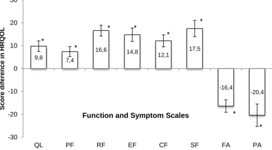
Figure 4. Score change from baseline to 12 week in quality of life, physical function, role function, emotional function cognitive function, fatigue and pain in breast cancer patients.
64
Figure 5. Percent of patients that increased, decreased, or did not change in VO2max, power output, sit-stand and leg extension, from baseline to 12 weeks of supervised exercise.
64
Figure 6. Percent of patients that increased, decreased, or did not change in global QOL, physical function, emotional function, fatigue and pain, from baseline to 12 weeks of supervised exercise.
64
Figure 7. Percentage of change from baseline to 12 week in VO2max, watts, sit-stand test and leg extension from exercise program and control group of breast cancer patients.
67
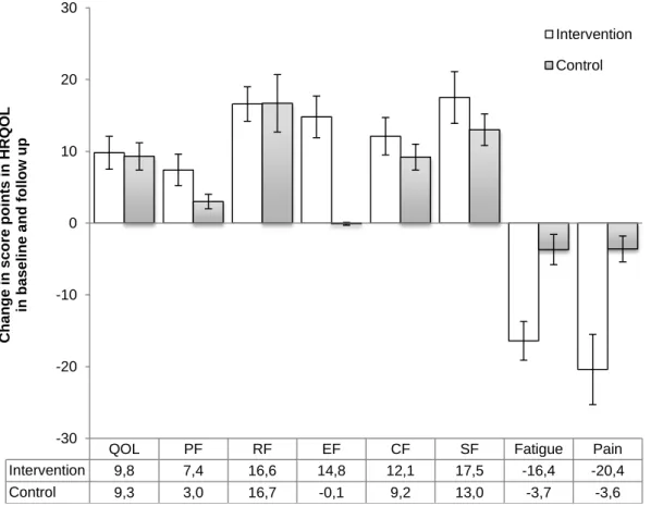
Figure 8. Score change from baseline to 12 week in quality of life, physical function, role function, emotion function, cognitive function, social function, fatigue and pain in breast cancer patients intervention group and control group.
68
Figure 9. Comparison of percent of change decrease in VO2max, Watts, sit stand test and leg extension from baseline to week 12, between healthy group and breast cancer patients in intervention exercise program.
List of Tables
Pag
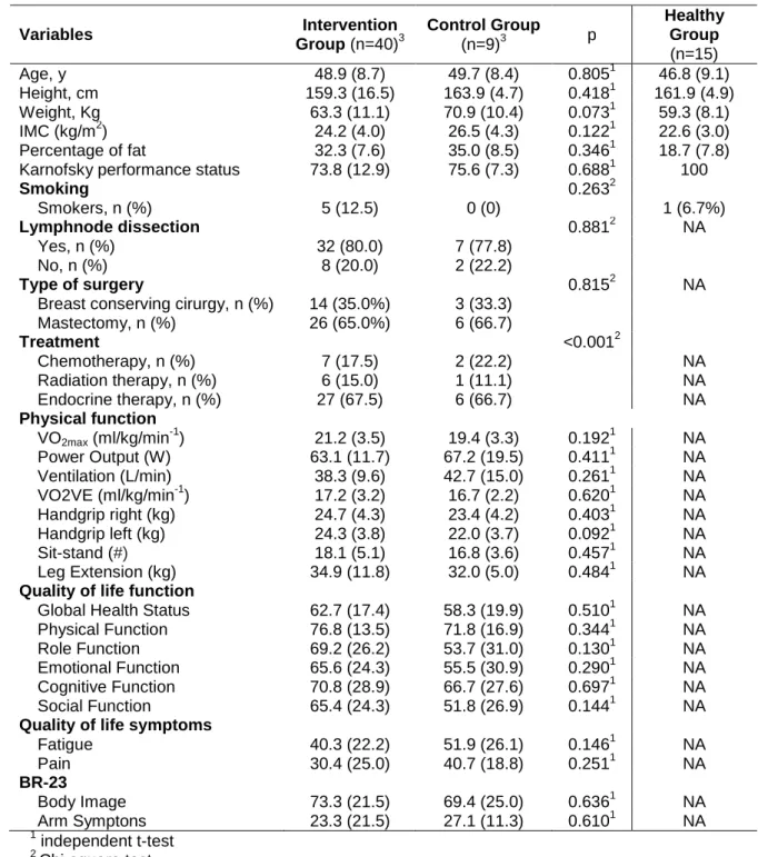
Table 1. Baseline demographic characteristics, diagnosis and treatment
according to group. 60
Table 2. Baseline and follow-up muscle strength and cardiorespiratory fitness components of breast cancer patients in supervised exercise program.
61
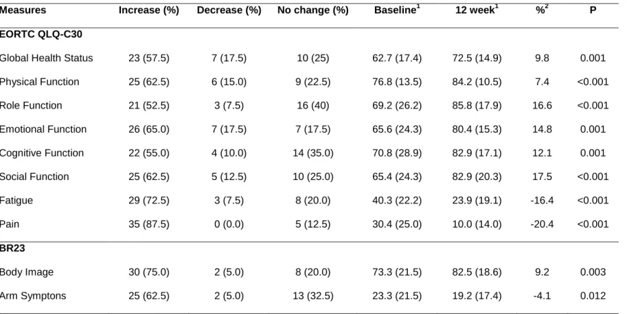
Table 3. Baseline and follow-up health-related quality of life subscales (EORTC QLQ-C30/BR23) of breast cancer patients in the control group.
62
Table 4. Baseline and 12-week muscle strength and cardiorespiratory
fitness components in intervention group and control group. 65
Table 5. Baseline and 12-week health-related quality of life subscales (EORTC QLQ-C30/BR23) in intervention group and control group.
66
Table 6. Baseline and 12-week muscle strength and cardiorespiratory
List of Abbreviations
VO2max Maximal Aerobic Power
EORTC European Organization Research Treatment of Cancer HRQOL Health Related Quality of Life
QOL Quality of Life
ACSM American College Sports Medicine
VO2VE Oxygen Consumption at Ventilatory Anaerobic Threshold
FACT Functional Assessement of Cancer Therapy CRF Cancer Related Fatigue
QL General Quality of Life PF Physical Functioning RF Role Functioning CF Cognitive Functioning SF Social Functioning FA Fatigue PA Pain
BRBI Body Imagem BRAS Arm Symptoms
Abstract
BACKGROUND. With 1.3 million new breast cancer cases reported every year and improved survival, it is important to develop interventions to maintain health related quality of life (HRQOL) during and after cancer treatment. Aerobic and strength training is an intervention that can enhance HRQOL and physical components during treatment.
OBJECTIVES. To examine the effect of 12 weeks of supervised exercise in physical capacity measurement and HRQOL in breast cancer patients during treatment.
METHODS. Breast cancer patients were allocated into an exercise intervention program (EI, n=40) and to a control group (CG, n= 8) that received standard care. The intervention comprised cardiovascular training at 70-85% of VO2max in cycle ergometer, resistance training and specific rehabilitation arm exercises. Patients were trained during 60 minutes, two times per week, during 12 weeks, with progression in time and intensity during the intervention period, with the main goal to achieve, at week 12, thirty minutes at 85%VO2max. Physical capacity and health related quality of life EORTC QLQ-C30 were assessed pre and post intervention. General linear model for repeated measures was used to compare (baseline and 12-week follow-up) with group assignment and time x group interaction included as fixed effects (p.05 for significance).
RESULTS. Highly significant increases were achieved with an increase of 17.0% vs. 7.7% in VO2max, and 46,6% vs. 4.9% in power output, respectively, in the intervention group compared to the control group (p.05). Gains in strength occurred only in the intervention group with and increase in leg extension by 13.4% (p.05). Intervention and control groups improved global health status (10.2 vs. 9.3 points) but emotional status (16.6 vs. 11.0 points) and role function (14.9 vs. -0.1 points) were significantly higher in the intervention group (p.05). The fatigue decreased by 16.4 points in the intervention group compared with only 2.7 points in the control group (p<.001). A significant decrease in pain was obtained for the intervention group with a reduction by 20.4 points vs. 2.6 points for the control group (p<.001).
CONCLUSIONS. A supervised and individualized prescriptive exercise program, results in an improvement in functional ability and HRQOL functions in women with breast cancer.
Resumo
INTRODUÇÃO. Com 1.3 milhões de novos casos de cancro todos os anos e com o aumento da taxa de sobrevivência, é importante desenvolver intervenções relacionadas com a qualidade de vida (QOL) durante e depois do tratamento do cancro. O exercício físico pode ser uma estratégia não farmacológica para melhorar a QOL e a capacidade física durante o tratamento da doença.
OBJETIVOS. Avaliar o efeito de um programa de exercício físico supervisionado durante 12 semanas nas alterações físicas, funcionais e a QOL, durante o tratamento, em mulheres com cancro da mama.
MÉTODOS. As mulheres com cancro da mama foram incluídas em dois grupos distintos, sendo que 40 participaram num programa de exercício físico (EF, n=40), enquanto que as restantes integraram o grupo de controlo (GC, n=8). O programa de exercício físico consitiu em treino cardiovascular no ciclo-ergómetro a 70-85% VO2max, treino de resistência de força com bandas terapêuticas e exercícios específicos de reabilitação para os membros superiores. O programa de exercício físico teve a duração de 60 minutos, duas vezes por semana durante 12 semanas. Durante o período de aplicação do programa de exercício físico houve uma progressão no tempo e intensidade com o objetivo de realizar na 12ª semana 30 minutos a 85% VO2max. A capacidade física e a QOL (EORTC QLQ-C30) foram avaliadas antes e depois do programa de intervenção. Para comparar o efeito do programa de exercício físico antes e após a intervenção utilizou-se a Anova de medidas repetidas com a análise da interação tempo x grupo (nível de significância p.05).
RESULTADOS. Verificaram-se aumentos significativos de 17.0% vs 7.7% no VO2max e 46,6% vs 4.9% na potência ao VO2max, respectivamente no EF comparado com GC (p.05). O aumento da força na leg extension ocorreu apenas no EF 13.4% (p.05). Ambos os grupos aumentaram a qualidade de vida geral (10.2 vs 9.3) mas a função emocional (16.6 vs 11.0) e role função (14.9 vs -0.1) foram significativamente mais altos no grupo EF (p.05). A fadiga diminuiu cerca de 16.4 pontos no grupo EF comparativamente com 2.7 pontos no GC (p<.001). Foi demonstrado um decréscimo significativo da dor apresentado apenas pelo grupo de intervenção com uma redução de 20.4 vs 2.6 pontos para o grupo de controlo (p<.001).
CONCLUSÕES. Um programa de exercício físico supervisionado, individualizado e prescritivo durante 12 semanas induziu numa melhoria significativa das capacidades funcionais e QOL em mulheres com cancro da mama.
1
In the western world, cardiovascular and oncologic diseases are the main causes of premature death (Pedersen & Saltin, 2006). Cancer is the leading cause of death in economically developed countries and the second leading cause of death in developing countries. The burden of cancer is increasing in economically developing countries as a result population aging and growth as well as, increasingly, an adoption of cancer-associated lifestyle choices including smoking, physical inactivity, and fast food (Jemal et al., 2011). Breast cancer in female and lung cancer in males are the most frequently diagnosed cancers and the leading cause of cancer death for each sex in both economically developed and developing countries except lung cancer is preceded by prostate cancer as the most frequent cancer among males in economically developed countries (Jemal et al., 2011).
Worldwide, 1.3 milion new breast cancer cases were reported every year accounting for 23% of the total new cases and 14% (458.000) of the total cancer deaths in 2008 (Jemal et al., 2011). More recent studies in the United States estimate that the 5-year survival rate for all cancers is roughly 62% and represents an approximate total of 9 million cancer survivors. The annual percentage change in the incidence of invasive breast cancers decreased modestly among older women but increased among younger (<40 years) and it has been reported that approximately 25% of breast cancers occur in women less than 50 years of age (e.g. premenopausal) in Europe and in US (Brinton et al., 2008). Early detection and advanced treatment option have led to a steady improvement in year relative survival rate in developed countries where the 5-year relative survival rate is currently 87% (Eheman et al., 2012). Data from 2003 show that 5-year relative survival exceeded 90% for patients with breast cancer and reached levels of about two thirds for patients with colorectal cancer and kidney cancer and patients with non-Hodgkin's lymphoma (Brenner et al., 2007).
This trend has resulted in a growing population of breast cancer survivors who are potentially faced with a number of long-term side effects of cancer and its
treatment (Campbell et al., 2011). Some of these include decrease aerobic capacity and strength, weight gain, fatigue and reduced quality of life.
Treatment is focused on curing the disease and preventing relapse due to metastic disease (Hoving et al., 2009). This fact accentuates the importance of investigation regarding the development of strategies to improve patients’ quality of life, reduce the recurrence risk, the probability of contracting other diseases and prolonging the population’s survival (Courneya, 2003).
The quality of life in oncological patients is often affected by the fatigue that they feel. In fact, approximately 70% of the cancerous population reports sensations of fatigue and asthenia during radiotherapy, chemotherapy or after surgery. Furthermore, approximately 30% of the population that survives cancer continues to manifest signs of persistent fatigue (Dimeo, 2001). Therefore, in all the types of cancer, the cancer related fatigue (CRF) severely influences several components of the quality of life since in aggravating symptoms like pain, nausea and dyspnoea, it causes physical, economic and social consequences that are devastating to the patients (Winningham, 2001). Cancer related fatigue (CRF) as a profound, negative impact on daily activities, social activities and overall quality of life (Cella et al., 2002).
Devoogdt et al. (2010) studied the evolution of the total physical activity levels occupational, sport and household activity on breast cancer patients. Physical activity levels are still significantly lower 12 months after treatment for breast cancer than at the pre-operative level. A greater decrease in occupational activities was associated with having a spouse, lymph node stage pN2 or pN3 (versus stage pN0 or pN1) complete axilary dissection (versus sentinel node biopsy), more than 20 lymph nodes dissected (versus less than 10) or undergoing chemotherapy. Moreover, older patients and those smoking before surgery had a higher risk of having a decreased sport activity level 12 months after surgery.
In this study, 1 year after the surgery breast cancer patients showed a 85% reduction from baseline in occupational activities (Devoogdt et al., 2010).
So, cancer patients, and in particular those at risk for a decreases physical activity level and affected by fatigue, should be identified, and encourage to increase their activities with supervised exercise programs. In fact, CRF and decreased physical activity may leed to difficulties in return to work.
Being unable to return to work after cancer treatment, frequent or prolonged work absenteeism, or problems with work performance may impose a significant economic impact on the survivor and her family (Chirikos et al., 2002). Furthermore, it is stated that the longer people are absent from their jobs, the lower the likelihood is that they will ever return to work. Moreover, Whereas loss of occupational identity can be a source of significant anxiety and depression, continuing or returning to the workplace allows many patients to maintain a sense of normalcy or control (Peteet, 2000). Most of the interventions with patients receiving adjuvant treatment show some improvement on quality of life or an other physical and psychological outcomes, such CRF, but do not pay attention to the aspect of work which is considered to be an important contributor to quality of life (Hoving et al., 2009). Some studies investigated return to work following breast cancer surgery. In a systematic review, Hoving et al. (2009) concluded the lack of methodology sound intervention studies on breast cancer survivors with outcome return to work indicating that returning to work programs and vocational rehabilitation for breast cancer survivors should be further developed and evaluated.
At 3 months pos-operative surgery, only 19% of patients working pre-operatively has returned to work and after 12 months the proportion had increased to 60% with patients working in a part-time employment than before surgery (Devoogdt et al., 2010). In other study elaborated in Canada, showed at 12 months, 88% of the breast cancer patients had returned to work (Drolet et al., 2005). There is a high variability among studies about working rates after breast cancer surgery being a possible explanation the difference in health care systems between US, Canada and European countries. In US, after 3 months, 60% of breast cancer patients were working but assistance with transportation, limitation in upper-body strength, and employment in jobs requiring physical activiy were needed (Satariano & DeLorenze, 1996).
In light of this information, it is the general purpose of this study is analyse and describe the advantages of the individualized effect of training and exercise, with a clinic purpose, in the alteration of the aerobic/ muscular functions and quality of life outcomes in breast cancer patients during treatment.
2
2.1 Cancer Related Fatigue
Every day people experience fatigue and this symptom is commonly associated with diseases such as depression, multiple sclerosis, arthritis and renal disease, as well as different medical and pharmacological treatments (Schwartz et al., 2001). Patients treated for cancer disease experience a different and more disruptive fatigue. In fact, cancer related fatigue (CRF) is the most prevalent and disturbing side effect of treatment for the majority of cancer patients. CRF is described as being more intense and overwhelming than fatigue before treatment, and exercise is an intervention proposed to reduce fatigue (Adamsen et al., 2004; Dimeo et al., 1999; Lucia et al., 2003; Oldervoll et al., 2003; Watson & Mock, 2004).
Cancer induces an abnormal inflammatory response with a high level of pro-inflammatory cytokines that are responsible for the progression and clinical deterioration of the disease (Winkelman, 2004). Cancer treatment itself contributes to muscular atrophy, through the use of high doses of immunosuppressant drugs such as cyclosporine, cyclosfosfamide or glucocorticoids that increase the protein catabolism. The muscular atrophy is essentially the result of the production of tumour necrosis factor-alpha (TNFa) that accentuates the inflammatory response in the musculoskeletal structure (Winkelman, 2004).
Most investigators consider that the origin of CRF is multifactor and not just a result of a single factor (Lucia et al., 2003; Ryan et al., 2007; Stasi et al., 2003; Watson & Mock, 2004); on top of that, the interaction between the various aetiological mechanisms is complex. In fact, fatigue represents just a part of the problem that is the physical incapacity experienced by cancer patients. Fatigue is usually defined as a sensation of lack of physical strength, weakness or loss of energy. However, fatigue becomes a pathological condition when it persists in ordinary daily activities, perseveres during a long period of time, does not improve with rest, or becomes severe enough so that it reduces daily physical activity (Dimeo, 2000). Although surviving cancer is a growing reality, medical intervention is almost always necessary.
2.1.1 Treatments and Toxicity
The most common treatments for cancer are surgery, radiotherapy and chemotherapy. Despite these medical interventions, the patients’ quality of life may suffer consequences. Since in 60% of cases of cancer survivors surgery is used, depending on the location and the extent of the operation, a significant morbidity may occur (complications with wounds, infections, loss of function, diminished movement amplitude, diarrhoea, dyspnoea, pain, drowsiness and lymphatic oedema) (Courneya, 2003).
Radiotherapy is used at a certain stage of the treatment in approximately 50% of cancer survivors. Generally, radiotherapy is administered in small fractions repeated over a period of 5 to 8 weeks to maximize the cellular death of the cancerous cells and minimize the damage to normal cells (Courneya, 2003). However, it can induce toxicity in normal cells and depends on the location of the radiation (pain, blistering, reduced elasticity, diminished movement amplitude, nausea, fatigue, pulmonary fibrosis, dry mouth, and cardiomyopathy).
The tumour suppressor protein p-53 is required for most DNA-damage-induced apoptosis. Unfortunately, mutations in the p-53 gene are observed in upwards of 70% of human cancer cases, thus reducing the efficacy of some therapies (63-14). After surgery to remove a tumour (lumpectomy or mastectomy), high levels of ionizing radiation are used to destroy any remaining breast cancer cells in the breast, chest wall, or axillary area. Cell death from radiation therapy occurs primarily during mitosis, when extensive DNA damage makes duplication of the genome impossible (mitotic cell death). P-53 Dependent apoptosis occurs at the G1 checkpoint of the cell cycle and ensures that DNA carrying genomic defects is not replicated. However, in the absence of p53, or when p53 is not functional, the cells become insensitive to low doses of radiation. A high dose of radiation induces necrosis and cytoplasmic constituents being released into the surrounding tissue, which serves as potent inflammatory signals (Bower et al., 2009). Recent data suggest that the fatigue experienced with radiation therapy is associated with the activations of
Systemic therapy is prescribed in several types of cancer. This treatment may be chemotherapy, hormone therapy and immunotherapy. Chemotherapy is administered intravenously or orally, in repeated cycles during 2-4 weeks from 3 to 6 months. Chemotherapy may cause some adverse effects such as fatigue, anorexia, nausea, neutropenia, peripheral neuropathies, ataxia and cardio toxicity. Furthermore, approximately 55% of patients are submitted to adjuvant chemotherapy during the intervention.
Chemotherapy drugs can disrupt the cell cycle at several critical steps and thereby induce apoptosis. Taxane drugs work to bind and stabilize cell microtubules to prevent them from separating chromosomes during cell division (anaphase of mitosis), thereby arresting cell cycle; doxorubicin (adriamycin) prevents DNA replication by inhibiting the enzymes, topoisomerases, that cut on strand of the DNA to unwind it during replication; and 5-fluorouracil (5FU, adrucil) inhibits thymidylate synthetase that functions to incorporate the thymine nucleotide into DNA during replication. Some of the more rapidly dividing normal, healthy cells are damaged (e.g. hair follicles, blood cells), subsequent mitosis from resident stem cells can soon replace those lost to chemotherapy or radiation (Clarkson & Kaufman, 2010). Over the past years, different studies have shown that fluorouracil, doxorubicin and cyclophosphamide (FAC) regimen as an adjuvant therapy for early stage of breast cancer is an effect treatment. Moreover, the doxetaxel, doxorubicin and cyclophosphamide regimen (TAC) has recently shown significant improvements in the rates of disease-free and overall survival even more than FAC (Lee et al., 2009). Some studies showed that TAC was more toxic than FAC with respect to neutropenic fever events and many extra hematological side effects such as asthenia, stomatitis, diarrhea and myalgia (Martin et al., 2005). Moreover, breast cancer patients who received TAC as treatment had less QOL scores, compared with FAC after the end of chemotherapy. Nevertheless, these differences gradually disappeared in different scales of QOL lasting for months after finishing chemotherapy (Bastani & Ahmad Kiadaliri, 2011). So, during follow-up period, patients in the TAC group experienced a higher improvement than FAC group. Hormone therapy is administered orally and can have several side effects such as weight gain, loss of muscular mass, accumulation of fat mass in the torso
and face, osteoporosis, fatigue and increased susceptibility to infections (Demark-Wahnefried et al., 2012). Lastly, immunotherapy is a recent form of treatment that maximizes the effect of other drugs as well as the defence mechanisms against cancerous cells. However, currently, in several clinical cases a combination of various types of treatment is being applied. In this situation, the timing and the treatment sequence depends on the staging and type of cancer. Consequently, it is possible that some cancer survivors may be treated on several occasions with different forms of treatment (Courneya, 2003). The supply of oxygen to the mitochondrion is a critical factor in the regulation of energy production. An adequate supply of energy to the cells requires the integrity of all the stages in the capture cascade, transport and use of oxygen. Anatomical and functional alterations due to the treatment of cancer can affect cells’ oxygen supply. On the other hand, the treatment itself, in inducing cardiovascular toxicity, can cause a dysfunction of the left ventricle (indicated by diminished systolic volume), ventricular contractility alterations and consequently diminished cardiac debt. Furthermore, pulmonary toxicity resulting from the treatment may cause a reduction of the lung’s full capacity, diminished vital capacity, a reduction of the inspiratory and diffusion capacities which, in its turn, compromise O2 and CO2 gas exchanges (Schneider et al., 2007).
Moreover, diminished pulmonary volume due to illness (pulmonary metastases and pleural effusion) or as a sequel of the treatment (lobotomy, pulmonary fibrosis after radiotherapy) may alter the ventilation-perfusion ratio, causing difficulty to the saturation of haemoglobin (Lucia et al., 2003).
Therefore, the treatment of cancer can reduce O2 transport capacity through the
aerobic path by the different path of physiological mechanisms (Dimeo, 2000; Lucia et al., 2003).
This toxicity is often observed in women with breast cancer during and after treatment. However, these negative effects can be attenuated through the intervention of an exercise programme. By means of an exercise intervention of 3 weekly training sessions over a period of 6 months, between 45-75% HRmax,
Schneider et al. (2007) observed, in breast cancer survivors, improved systolic arterial pressure (-2.6%), diastolic arterial pressure (-3.4%), and cardiac
improved (+2.8%), as did maximum oxygen consumption (+15.1%). In the specific case of chemotherapy, the marrow may be affected, which consequently diminishes the production of red blood cells. Cardiotoxic cytostatic agents, such as anthracycline and cyclosfosfamide, can reduce blood flow to the musculo-skeleton (Dimeo, 2000).
The loss of muscular function induced by a prolonged period of bed rest can result in a substantial loss of muscular mass, plasmatic volume and reduced cardiac debt that impairs energetic efficiency (Levine et al., 1997). This clinical status is very similar to being bedridden, since 6 weeks in this condition was enough to induce diminished left ventricular mass in 8.0+2.2% and is connected to reduced final diastolic volume on the second week in 14+1.7% (Perhonen et al., 2001). Furthermore, treatment with high doses of corticosteroids results in diminished muscular mass. Inducing immunosuppression using cyclosporine (CsA) may result in mitochondrial myopathy, loss of capillary density and, consequently, loss of physical capacity (Zbreski et al., 2006). CsA is an immunosuppressant commonly used in organ transplants. This drug acts as a cytosolic phosphatase protein inhibitor in T-Killer lymphocytes. In the Zbreski et al. (2006) study, the administration of CsA resulted in muscular atrophy, inducing a 22% decrease in fiber cross-sectional area. These last occurrences are clinically indistinguishable from alterations caused by detraining or by reducing physical activity.
The majority of breast cancers are estrogen receptor-positive, although estrogen receptor negative disease is more frequent in premenopausal (37%) than postmenopausal women (21%) (Anderson et al., 2002). Breast cancers that occur in younger women are of concern because these cancers are often hormone receptor negative (estrogen receptor ER- and progesterone receptor PR-), are high grade, and are diagnosed at advanced stages (Klauber-DeMore, 2005). Current treatment guidelines recommend that premenopausal women with hormone receptor-negative disease receive adjuvant chemotherapy, and that those with hormone-positive disease receive adjuvant endocrine therapy (tamoxifen +- ovarian function suppression) with or without adjuvant chemotherapy based on the biology and extend of the primary tumor within the breast and regional lymph nodes (Burstein et al., 2010). Among the
adjuvant therapy options for premenopausal patients with breast cancer, both endocrine therapy (ovarian suppression or tamoxifen) and chemotherapy can result in substantial bone loss from suppression of estrogen levels (Headley et al., 1998; Nikander et al., 2012).
Women who have premature menopause as a result of chemotherapy due to breast cancer are at an increased risk of bone loss and may be at risk for early development of osteoporosis (Headley et al., 1998). Moreover, women who became permanently amenorrheic as a result of chemotherapy had 14% lower bone mass density than women who maintained menses after chemotherapy (Headley et al., 1998). Rates of bone loss occurring with cancer therapy can be up to tenfold higher than normal (Guise, 2006).
Nikander et al. (2012) examined the impact of aerobic exercise training (3x/week) in bone structural strength and observed a positive training effects on bone structural traits (1.2%), physical performance (~3-4%) and body composition (~1-3%) in breast cancer patients. However, resistance training is more effective in maintaining bone density (Schwartz et al., 2007). Knobf et al. (2008) conducted 12-24 week supervised weight-loaded aerobic intervention with women with breast cancer who were 3 year from completing breast cancer treatment. Bone remodeling was measured by serum biomarkers and no significant changes in serum osteocalcin, or lean muscle mass were observed. However, women at high risk for weight gain and bone loss maintained their weight and bone mass. A home-based strength training intervention, in postmenopausal breast cancer survivors with ostopenia or osteoporosis, resulted in improvements on bone mineral density with a significant increase in muscle strength for hip flexion, hip extension, and knee flexion, and BMD of the spine and hip (Waltman et al., 2003). A controlled pilot trial was carried out to assess the feasibility and efficacy of an aerobic exercise in enhancing physical performance of breast cancer patients after adjuvant treatments. Aerobic and step training during 12 weeks and the magnitude of the load during training sessions, which seemed to be sufficiently high to increase bone mass in different patients, suggest that this kind of vigorous training regimen is feasible among breast cancer survivors and could have a potential to prevent bone loss
aerobic exercise appeared to be well-tolerated and an effective training mode among breast cancer survivors who had recently completed heavy adjuvant treatments such as chemotherapy (Nikander et al., 2007).
As cancer treatment regimens evolve, patients live longer but, sometimes, with the unwanted complication of anaemia. Anemia is a common haematologic manifestation in cancer and can increase the disease’s aggressiveness, causes fatigue and reduces quality of life (QOL) and can also limit the treatment (Straus et al., 2006).
One of the many causes of CRF is anemia which comes with the disease and its treatment. Although it is not the only cause of CRF, anemia is the easiest to document using standard complete blood count haemoglobin (Hgb) levels for mild (10.00-11.99 g/dL), moderate (8.00-9.90 g/dL), and severe (<8.00 g/dL) anemia. These clinical definitions allow us to establish criterion of anemic cancer patients who can be tested for levels of self-reported fatigue and its impact on function (Cella et al., 2002).
Moreover, it has been demonstrated that a state of anemia is an indicator of a disease’s independent prognosis (Ludwig & Strasser, 2001). It has also been demonstrated in cancer patients that a state of mild or moderate anemia (Hb values 10-12g/dL and 8-10g/dL, respectively) is significantly associated with a low survival rate (Ludwig & Strasser, 2001). In cancer patients, the incidence of chronic anemia depends on the type of malignancy, the staging and duration, and also type and intensity of treatment (Ludwig & Strasser, 2001).
A multinational study, comprising Europe, carried out by the European Cancer Anaemia Survey (ECAS) documented the prevalence, incidence and severity of anemia in a numerous and representative sample of the European cancerous population (Ludwig et al., 2004). In this study it was demonstrated that anemia was more frequent in patients with gynaecologic cancers (81,4%), lung cancer (77,0%) and lymphoma/myeloma (72,9%). Some authors report that the prevalence of anemia is particularly high in patients with lung carcinoma (50%-60% with Hb <11 g/dL) (Kosmidis & Krzakowski, 2005; Ludwig et al., 2004), especially when compared to less prevalence in colorectal and breast carcinoma (approximately 10-20%); the total incidence of anemia was 53,7%. In this case, patients that had been submitted to chemotherapy had a higher
incidence of anemia (63,7%) compared to concomitant chemo-radiotherapy (41,9%) or radiation therapy (19,5%) (Ludwig et al., 2004).
In the case of lung cancer, cytotoxic chemotherapy is one of the main factors that contribute to the probability of developing or aggravating pre-existing anemia (Dowlati et al., 1997). Therefore, the incidence of anemia depends on the chemotherapy treatment that is used. The combination of cysplastin and
etoposide, commonly used in the treatment of small cell lung carcinoma (SCLG)
is associated with severe anemia in 16-55% of patients (Itri, 2000). In advanced non-small lung cell carcinoma (NSCLC), one of the firstly used treatments is paclitaxel and platinum therapies. However, the combination of these two drugs can induce anemia in approximately 78% of patients and, approximately 33% of patients can develop severe anemia (Langer et al., 1995). In these cases, the mechanism involved in chemotherapy-induced anemia includes direct myelosuppression and, in some cases, loss of renal function. These data are reinforced by the ECAS study since chemotherapy treatment induced mild to moderate anemia in 8-100% of patients suffering from NSCLC and 0-87% in those with SCLC (Ludwig et al., 2004). Moreover, severe anemia (Hb < 8 g/dL) and life-threatening anemia (Hb < 6.5 g/dL) occurs in 0-40% of patients with NSCLC and 0-55% in those suffering from SCLC (Groopman & Itri, 1999). Because of this, approximately 43% of patients with lung carcinoma were given blood transfusions, while patients suffering from breast carcinoma receiving blood transfusions did not exceeded 19% (Kosmidis & Krzakowski, 2005). Therefore, we can conclude that platinum compounds (like cysplatin or carboplatin) seem to particularly induce myelosuppression.
Generally speaking, the hemoglobin values in the ECAS study are categorized between 10,0 and 11,9 g/dL. Nevertheless, the longer chemotherapy is used the higher is the risk of developing anemia. This fact was confirmed by progressive proportion of patients with low levels of hemoglobin (i.e. <10,0 g/dL) as the cycles increased (Ludwig et al., 2004). In fact, anemia was observed in 19,5% of patients in the first cycle with an increased incidence to 56,7% on the fifth cycle of chemotherapy. If baseline hemoglobin level is approximately 15 g/dL and subsequently these values are reduced by 2 g/dL during the first 3/4
transfusion to treat anemia at a certain stage during treatment. On the other hand, if a similar Hb decrease from a 11 g/dL baseline level occurs during the first cycle of treatment, practically all patients will need a transfusion (Langer et al., 2002). Therefore, it is necessary to determine the optimal cut-off value for the treatment of anemia, which is a critical criterion to maximize quality of life and possibly other outcomes in cancer patients. The ASH/ASCO guidelines recommend that treatment using erythropoietin starts when hemoglobin values decrease to <10,0 g/dL (Rizzo et al., 2002). Besides, when patients are symptomatic due to anemia and hemoglobin values are between 10 g/dL and 12 g/dL, the decision of implementing the start of the treatment is at the doctor’s discretion (Ludwig et al., 2004).
Furthermore, the ECAS study revealed that patients with lower hemoglobin values also had lower WHO Performance Scores when compared to those with higher hemoglobin values. 50,7% of patients with <8,0 g/dL hemoglobin had 2-4 WHO scores, as did 40% of those with 8.0-9.9 g/dL hemoglobin. On top of which, almost a quarter (24,8%) of patients with 10,0-11,9 g/dL hemoglobin had 2-4 WHO scores (Ludwing et al., 2004). These results can lead to a better understanding of anemia when associated with cancer therapy, thus optimizing the patient’s treatment and improving quality of life during that period. These results are reinforced by the Cella study that analyzed the use of Functional Assessment of Cancer Therapy (FACT) to assess the impact of fatigue or anemia in cancer patients’ QOL. Patients with Hb > 12 g/dL (defined as non anemic) felt less fatigue, better wellbeing and higher general levels of QOL, when compared to those who were not anemic (Cella, 1997).
Moderate physical exercise can be a strategy to increase hemoglobin. In the specific case of breast cancer, women who undergo radiation therapy are prone to diminished erythrocyte levels during the months after treatment. As previously analyzed, this erythrocyte decrease appears to be associated to complications that include fatigue, anemia, depression and reduced physical functioning (Lucia et al., 2003). Furthermore, diminished erythrocyte levels seem to be connected to a decrease in the survival rate in some types of cancer. Drouin et al. (2006) studied the influence of moderately intense aerobic exercise and its positive effect in hemoglobin (HB), haematocrit (HCT) and red
blood cells (RBC) in women with breast cancer in stages 0 to 4 (Tis, N0, M0; to T0-4, N3-M0) subjected to a 7-week radiation therapy regimen (Drouin et al., 2006). The exercise intervention consisted of walking 3 to 4 times a week, at an intensity between 50% and 70% CFmax. Through this research, investigators
observed that moderately intense aerobic exercise during radiation therapy in breast cancer prevented the reduction of erythrocyte levels when compared to control group. Moreover, the authors verified a positive correlation between the peak of aerobic capacity and the final erythrocyte evaluation, corroborating the concept of the connection between erythrocyte level and improved physical capacity (Drouin et al., 2006).
2.1.2 Peripheral Fatigue and Central Fatigue
The main mechanisms of fatigue have been categorized in two principal components: peripheral fatigue and central fatigue. Peripheral fatigue is a result of the neuromuscular system’s inability to carry out a task in response to central stimulation. Two recent studies have analysed the effect of radiotherapy in neuromuscular fatigue. Monga et al. (1997) observed in prostate cancer patients diminished neuromuscular efficiency (a muscular function parameter) in the anterior skeletal muscle tibia’s fast fibres. The patients were submitted to a radiation treatment protocol with 68 to 70Gy in 34 to 38 fractions during a period of 7 to 8 weeks. However, when the effect of radiotherapy was studied, using certain psychometric scales, to ascertain the psychological and emotional state, and sleep parameters, no association was made between radiotherapy and clinical fatigue (Monga et al., 1997). Moreover, the same patients did not have diminished VO2max, assessed by modified Bruce protocol, which led to the
conclusion that neuromuscular instead of cardiorespiratory factors contribute mostly to increased muscular fatigue. Therefore, 68 to 70Gy of ionizing radiation in the pelvic area of patients who are undergoing treatment for prostate cancer, emphasizes neuromuscular fatigue. In the majority of patients, the fatigue persisted 5 to 6 weeks after radiotherapy had ended, which may indicate that the nature of the fatigue presumably resulted from subtle lesions in the muscular structure. Authors speculate that ionizing radiation may have
mitochondrial membranes (Monga et al., 1997). This alteration causes a disruption in the strength producing mechanisms and/or a calcium abduction by the sarcoplasma and by the Ca-ATPase, which entails a deficiency in the excitation-contraction process (Bigland-Ritchie et al., 1995). Furthermore, this alteration in the cytoskeleton can cause an electrolyte gradients’ imbalance in the cellular membranes, thus inducing a K+ accumulation in the extracellular medium (plasma). In the Agroyannis et al. (1992) study, it was observed that in 21 patients with malignant tumours (lung, breast, endometrium and prostate), 36 to 60Gy of radiation during 20 to 30 fractions of radiotherapy caused a significant increase in the release of seric transferrin and TNF-.
On the other hand, central fatigue affects the central nervous system and is the result of a progressive failure in the transmission of stimuli by the motoneuron. A study on patients with chronic fatigue syndrome demonstrated that a plasmatic free tryptophan increase can potentially lead to a high level of cerebral serotonin (5-HT) (Castell et al., 1999).The tryptophan and the branched-chain amino acids (BCAAs) compete to enter the brain through the same transport – albumin (Ryan et al., 2007). Several protocols that use exercise as a way to induce fatigue reveal that the increase of cerebral 5-HT levels during exercise is the result of a reduction of BCAAs, which allows the tryptophan to enter the brain through the hematoencephalic barrier. On top of which, exercise increases the concentration of plasmatic free fatty acids that bind with the albumin, causing an increase of free tryptophan in the plasma. Supporting this, research on animal models has demonstrated that concentrations of cerebral 5-HT increase during exercise, reaching the highest concentration at the point of fatigue (Ryan et al., 2007).
2.1.3. Exercise and cancer related fatigue
More recently, exercise has been tested in the treatment/reduction of cancer-related fatigue (Dimeo, 2001; Winningham, 2001). Schwartz et al. (2001) showed that fatigue was significantly reduced on exercise days compared with non-exercise days. Furthermore, the amount of exercise, measured by the number of minutes of actual practice, was significantly associated with fatigue
levels. There is growing evidence that aerobic exercise programs can reduce fatigue in cancer patients receiving chemotherapy (Adamsen et al., 2004; Adamsen et al., 2006; Dimeo et al., 2003; Dimeo et al., 1999; Quist et al., 2006; Schwartz et al., 2001; Winningham et al., 1989).
Mock et al. (2001) studied the effect of home-based exercise at least 90 min per week for three or more days in women with breast cancer who reported significantly less fatigue and emotional distress as well as higher functional ability and QOL than the women who were less active during treatment.
The literature describes exercise as having potential importance in the quantitative reduction of fatigue. The qualitative data in this study highlight the experience of cancer patients having a comfortable sense of fatigue while simultaneously improving their physical form and increasing their strength. Data from Adamsen et al. (2004) describe different forms of fatigue in cancer patients. The sensation of fatigue which results from exercising (i. e. exercise induced fatigue) is experienced as a positive and natural tiredness and is furthermore associated with an after-effect of improved physical well-being, tranquillity, release, relaxation, and, for some, typically improved sleep (Cella et al., 2002). It is a sense of fatigue that is desirable because the after-effects are familiar as they were experienced during sports activities prior to their illness (Watson & Mock, 2004). This contrasts with fatigue induced by chemotherapy which can be characterized as negative because of its connection with the experience of physical discomfort. It is not a sense of fatigue that is desired but rather is imposed on the body from the outside through medication, and usually leads to a passive physical state. Adamsen et al. (2004) showed that cancer patients can transform the nature of fatigue – from a negative chemotherapy induced fatigue into a positive exercise induced fatigue.
The impact of exercise on fatigue was significant and reflects the effectiveness of low to moderate intensity regular exercise in maintaining functional ability and reducing fatigue in patients with breast cancer. Exercise was consistently associated with reducing fatigue the day of exercise and one day afterward over the first two cycles of chemotherapy in breast cancer patients (Schwartz et al., 2001). However, current level of fatigue increased when exercise exceeded 60
Treatment Disease Detraining
F
Faattiigguuee
Attenuated fatigue Physical Activity / exercise Sedentary habits
State of “chronic” fatigue
minutes, probably due to changes in nutrition and hydration states (Schwartz et al., 2001).
In short, physical inactivity induces an accentuated loss of muscular mass and loss of cardiorespiratory capacity. Therefore, lack of physical activity causes a tendentiously perpetual condition of diminished activity leading to fatigue and vice-versa (Figure 1). This mechanism may explain persistent fatigue and physical performance impairment in several patients, even years after ceasing of treatment (Dimeo, 2000; Lucia et al., 2003).
Image 1: Effect of treatment, disease and detraining in Cancer Related Fatigue (Adapted from
Rundle, (Rundle, 2005).
In a cross sectional study, 3 years after breast cancer treatment, persistent pain and sensory disturbances remain clinically significant problems among 47% breast cancer patients who received surgery (Gartner et al., 2009). In fact, fatigue and pain overlap, and both seem to be additionally associated with mental health (Gartner et al., 2009).
The cause of pain in cancer patients and in general, remains unclear. A 4 year follow-up showed an increase in pain symptoms in those women taking tamoxifen at baseline; in those reporting depression or stressful life events during the first 12 months after enrolment. Nevertheless, exercise at baseline had a beneficial effect on pain recovery (Rief et al., 2011). So, pain should be a major outcome in evaluations of cancer treatment and rehabilitation programs.
In 2009, with support from Siteman Cancer Centre and the Oncology Nursing Society, the ACSM convened an expert roundtable to review the literature on exercise in cancer survivors and issue guidelines for activity, along with recommendations on exercise testing and prescription (Schmitz et al., 2010). The ACSM guidelines for cancer survivors emphasis on returning to normal daily activities as quickly as possible after surgery and continuing these activities as much as possible during any adjuvant treatments (Wolin et al., 2012).
Low to moderate exercise interventions of variable duration seem to be the standards applied across existing studies. Predominantly, those studies have examined the effects of a single activity, e.g. cardiovascular training on stationary bicycles, rather than resistance to the exercise as the one of modality (Galvao & Newton, 2005). More studies are needed to provide evidence of whether specific patient groups, at different stages of the disease and/or with different diagnoses and treatments, can benefit from exercise. So, due to the lack of evidence, no recommendations can be issued at present (Lucia et al., 2003).
A multidimensional 6 weeks high intensity exercise training design intervention in cancer patients with different types of solid tumours and haematological malignancies undergoing chemotherapy, increased VO2max in 16% and dynamic
strength of 40%. Furthermore, it was observed a significant reduction in treatment-related symptoms and significantly improved their general well-being and quality of life specifically with a reduction of fatigue and pain (Adamsen et al., 2006). In accordance with these results, Kolden et al. (2002) verified through a supervised 16 week program involving cardiovascular, resistance and flexibility training, 1h three times a week in 40 sedentary women with breast cancer, an increase in strength of 36% and in cardiovascular capacity of 15%. These results suggest that cancer patients undergoing chemotherapy may benefit from a multidimensional exercise interventions.
Knutsen et al. (2006) reported any adverse effects such as unintentional physical reactions, cardiac, or respiratory arrest, or hypotension during VO2max
showed that patients felt significantly safer in performing the maximum physical capacity tests and in using their bodies in a 6 week high intensity training.
2.2. Cancer, exercise and quality of life
Investigation regarding exercise in cancer survivors began in the early 80’s but published articles became consistent in the late 90’s only. The Alberta University in Edmonton, Canada, by means of investigator Kerry Courneya, has been a driving force in clinical trials in this area (Courneya, 2003; Courneya et al., 2008; Courneya & Friedenreich, 2011).
The positive effect and the advantage of exercise in cancer patients were described for the first time by (Winningham et al., 1989).
Physical exercise is one of the foremost areas of research in cancer control with potential to improve the lives of cancer; the context “cancer survivor” indicates any person who has or had cancer, from the time of diagnosis onward (Ingram & Visovsky, 2007). The psychological and physical symptoms resulting from cancer and its treatments (e.g., pain, fatigue, nausea, vomiting, depression) will contribute to excessive patterns of rest and also contribute to immobility. Bed rest will precipitate muscle weakness, atrophy, and functional impairment (Ingram & Visovsky, 2007).
Evidence of the benefits of exercise for cancer survivors has mounted steadily over the past in the areas of psychology and quality of life outcomes. Recently, improvements in physical functioning, body weight and composition, muscle strength and endurance (Quist et al., 2006), and immune function have been reported.
The exercise interventions are low to moderately intense (exercise intensity between 55% and 85% HRmax) with very similar protocols throughout the
various studies (Courneya & Friedenreich, 1999; Dimeo et al., 1999). These rules are in accordance with ACSM guidelines, that state that 6 to 7 weeks of training are enough to obtain significant muscular hypertrophy gains. As to the frequency of training, two to three weekly training sessions are sufficient to promote the desired adaptations (Garber et al., 2011).
Several clinical trials have studied the relationship between clinical exercise, quality of life and psychosocial parameters in cancer survivors. The exercise programs in the trials consisted mainly of 3 weekly sessions of moderate and vigorous exercise, with a duration that increased progressively up to 45` over a period of 3 to 4 months. These studies demonstrated that exercise reduced anxiety, depression, improved self-esteem and diminished fatigue-related symptoms (Herrero et al., 2006; Quist et al., 2006).
One of the best direct indicators of cardio respiratory capacity is VO2max. This
variable is an excellent indicator of a person’s health condition and an independent mortality predictor in healthy or unhealthy individuals (Garber et al., 2011). Initially, some authors alerted to the possibility that vigorous exercise, including maximal physical evaluation tests, could impair the cancer patient’s health during chemotherapy due to the administered drugs’ adverse effects. However, contrary to what was expected, the study carried out by Knutsen et al. did not observe adverse effects such as hypotension, cardiac or respiratory arrests, during the assessment of aerobic and muscular functions following maximum protocols (Knutsen et al., 2006).
Obesity is a risk factor for some of the most common cancers in the western world, such as post menopausal breast cancer and colorectal cancer. This is one of the reasons why cancer patients are overweight or obese at the time of diagnosis (Brown et al., 2003). Furthermore, there is strong evidence that excessive weight increases the recurrence risk in several cancers, as well as increased mortality risk (Eheman et al., 2012). At present, the International Agency for Research on Cancer estimates that 25% of cancer cases are caused by excessive weight, obesity and a sedentary life (Ahlberg et al., 2003). The current tendency of increasing number of adults and children that are overweight or obese and, simultaneously, have low levels of physical activity makes the connection of their lifestyle with cancer a matter of public health (Campbell & McTiernan, 2007). Therefore, exercise has been used as a mean to reduce obesity and to promote a healthy lifestyle.
Empirically, strength training is generally inappropriate for people with an illness as well as for cancer survivors, and for this reason there is still little investigation
observed improved quality of life in cancer patients even when the exercise program is discontinued for a period of time due to a specific reason (anemia, infections and demotivation). An 8-week exercise program of aerobic training combined with strength exercises, with 3 weekly 90 minute sessions, in women with breast cancer, significantly increased muscular mass and reduced fat mass and these improvements were linked to an improved quality of life (EORTC QLQ-C30) (Herrero et al., 2007). On top of that, in breast cancer survivors, even after an 8-week detraining period QOL was not diminished, as were not dynamic force and specific functional tasks of muscular capacity. However, the same period of detraining reduced cardio respiratory capacity by ~6ml.Kg.min-1 (VO2peak) (Herrero et al., 2007). Therefore, cardiorespiratory adaptations are
quickly lost. Nevertheless, strength training should be integrated as a component in a training program since it attenuates muscular atrophy induced by treatment and sedentary habits in cancer survivors (Inui, 2002). In this case, ACSM guidelines recommend that 6 to 7 weeks of training are enough to obtain significant muscular hypertrophy gains. As far as frequency is concerned, 2 to 3 weekly training sessions are sufficient for both men and women (Schmitz et al., 2010). In fact, aerobic and strength training improves quality of life in a way that is practically unparalleled in patients with various types of cancer, that is to say, an increase of almost 43% in dynamic muscular force accompanied by a 21% increase in the improvement of QOL after 18 weeks of aerobic and strength training (De Backer, Van Breda, et al., 2007).
In another recent study, during the hospitalization of patients submitted to high-dose chemotherapy, an exercise program of aerobic resistance that consisted of pedaling for 30 minutes on a reclined cycle ergometer, reduced fatigue and loss of physical performance. On the other hand, chemotherapy-related complications, namely aplasia (quantity of neutrophils < 500/mL and platelets < 20,000/mL) and duration of hospitalization, were significantly inferior in the trained group, when compared to the control group (Dimeo et al., 1999).
In the specific case of prostate cancer treatment, the deprivation of androgens can cause an adverse effect – bone mineral density loss, progressing to osteoporosis. It is described that this bone mineral density loss, of 3 to 5% in the first years of treatment, results in a higher incidence of fractures
(Friedenreich, 2001). Therefore, not only has exercise an important role in preventing prostate cancer but it also prevents the adverse effects that can occur during therapy (Campbell & McTiernan, 2007).
Since approximately 60% of cancer survivors are 65 years old or more, there has been an interest in studying the potentials of exercise in these ages. In fact, since aging is connected to a decline in neuromuscular function and diminished capacity to synthesize proteins, the scientific community questions whether the elderly are subject to physical exercise’s positive adaptations. Besides, studies in the last decades have demonstrated that the elderly revealed improved physiologic function and positive adaptations such as those that were observed in younger adults (Drouin, 2004). Exercise, in this type of population, requires some precautions in several situations due to some contraindications.
Psychological factors also have an important function in the aetiology of cancer-related fatigue’s genesis. In this particular case, depression is considered a contributing factor to fatigue in cancer patients. The effect of physical activity is not limited to improved muscular and cardiovascular functions. In fact, improved physical performance can increase a patient’s sensation of control, independence and self-esteem; this improved self-esteem can result in better social interaction, reduced anxiety and fear (Dimeo, 2000).
Segal et al. (2001) did not observe any effect of structured exercise on aerobic fitness and in HRQOL in breast cancer patients during treatment. These results are, to some extend, in line with (Thorsen et al., 2005) who did not observed any beneficial effect on fatigue, mental distress and HRQOL parameters of the 14-week supervised home-based training program intervention shortly after chemotherapy. The fact that patients performed multiple supervised activities by themselves could represent additional stress for them. However, VO2max
increased by 6.4 ml/kg/min-1 in the patients in the interventions group and only 3.1 ml/Kg/min-1 in the patients in the control group corresponding to 23% and 10%, respectively) showing a beneficial effect on CRF in young and middle-aged cancer patients. Opposite to this findings Segal et al. (2003) demonstrated a positive effect of a 12-week supervised strength-training program on fatigue and overall HRQOL in men receiving androgen deprivation for prostate cancer.
Dimeo, Fetscher, et al. (1997) observed a positive effect of an interval exercise, using a cycle ergometer in bed, on fatigue and psychological distress in cancer patients during hospitalization.
Courneya et al. (2003) reported a positive effect on fatigue and in overall HRQOL in breast cancer patients several months after treatment.
Burnham and Wilcox (Burnham & Wilcox, 2002) observed a significantly higher overall HRQOL improvement in the exercise group compared with the control group but mental distress between groups was the same.
Mock et al. (1997) demonstrated a positive effect of a 6-week individualized self-paced progressive walking program on fatigue and anxiety in breast cancer patients during radiotherapy. However, there were no significant differences between the exercise group and the control group in depression.
The magnitude of changes in mental distress, fatigue, and HRQOL parameters in trials in this area are not consistent. It is not clear enough whether these differences of outcome are the result of differences in the patients characteristics and/or the type and the timing of the intervention or if these variations are explained by different methods of assessment (Thorsen et al., 2005).
The application of the principles of exercise training (specificity, overload, progression, initial values, reversibility and diminishing returns) in a design of an exercise prescription ensures the most appropriate type and dose of exercise to promote de desired outcome (Campbell et al., 2011).
De Backer et al. (2009) conducted a systematic review to summarize the research of previous studies that used resistance training with cancer patients. The duration of the resistance training programs ranged from 3 to 24 weeks, with an average of 12 weeks and most of them prescribed two or three sessions per week. Furthermore, most of the studies applied resistance with aerobic training. Few studies described the number of sets and repetitions, and among 10 of them, intensities ranged from 30-60 to 60-85% of 1RM. With regard to aerobic exercises, treadmill walking and stationary cycling were the most frequently prescribed sessions. Intensities of aerobic exercises varied from 40% to 90% of heart rate maximum. Studies incorporating resistance training prescribed either exercises using machines or resistance bands (Spence et al.,
2010). As outcomes measures, most of the studies assessed muscle strength (1RM), endurance tests (maximal number of repetitions at 60-75% of 1RM), hand grip tests. The results demonstrate that increases in muscular strength occur in women with breast cancer who receive resistance training (Loprinzi & Cardinal, 2012).
Evidence suggests that health professionals might encourage breast cancer’s patients to increase physical activity in a consistent way during treatment, because regular practice of exercise, as soon as possible after surgery and during treatment, may reduce secondary effects.
Patients with breast cancer should start to increase gradually their physical activity to a 150 minutes per week within an moderate intensity or to a 75 minutes per week at a vigorous intensity as soon as they finish their chemotherapy or radiotherapy treatments; as well as patients reaching progressively the minimum exercise guidelines, should be persuaded to increase their physical activity level to 180 minutes per week of moderate intensity up to vigorous intensity, because these activity levels are necessary to prevent the recurrence of breast cancer (Holmes et al., 2005).
3
The main objective of the study was to examine the effect of 12 weeks of supervised exercise in physical capacity, lower and upper body strength and QOL, in breast cancer patients, during treatment.
Primary outcome was measured through the change in cardiovascular fitness and strength and the evaluation of health related quality of life, between baseline and post intervention.
A secondary objective of the study was to assess the feasibility of supervised exercise program and respective effects upon cancer related fatigue, in breast cancer patients, during treatment.
Secondary outcome was assessed through the change VO2max, power
output and in pain and fatigue symptoms between baseline and post intervention program. Results were compared with a control group.
Another second objective of this study was to examine aerobic and muscle functional changes, induced by a 12 weeks combined aerobic and endurance exercise program, in breast cancer patients and compares it with healthy women.
This outcome was assessed through the changes in physical components between baseline and post intervention program, using health results as reference.