A comparative evaluation of efficacy of a reciprocating
system for gutta-percha removal with or without a solvent
Nancy Kudsi Carvalho,1 Gustavo Ribeiro Alvares,1 Luciana Moura Sassone,1 Renato Liess Krebs,1 Tauby de Souza Coutinho-Filho,1 Emmanuel João Nogueira Leal da Silva1
1Department of Endodontics, Rio de Janeiro State University (UERJ), Rio de Janeiro, RJ, Brazil
• Conflicts of interest: none declared. AbstrAct
Objective: to evaluate the efficacy of Reciproc System, with or without solvent for endodontic filling removal from oval shaped root canals. The time required for gutta-percha removal and the instrument fracture were also evaluated. Material and Methods: forty straight single-rooted premolars were prepared up to a size 30 and filled with gutta-percha and sealer, then randomly assigned to two retreatment groups (n = 20), treated with Reciproc System with or without chloroform (RS+C and RS). For all roots, procedural errors, time of retreatment and apically extruded material were recorded. After retreatment, roots were split longitudinally and then photographed. All images were evaluated with Image Tool software to calculate the percentage of residual material. Kolmogorov Smirnov and t tests were used to determine significant differences at P < .05. Results: no treatment completely removed the root filling material from the root canals. Analysis of the total area did not reveal statistical differences between the two groups (P >.05). Both groups also showed similar results in all tested thirds, with no statistical differences (P >.05). RS without solvent was faster in filling material removal than RS+C (P <.05). For both groups, none of the instruments showed intracanal fracture or visible signs of plastic deformation. Conclusion: taken together, our results demonstrated that Reciproc System used without solvent required less time to remove the same amount of root filling material than Reciproc System with solvent. However, regardless of solvent use, the same cleaning quality was obtained.
Keywords: Endodontic retreatment; Reciprocating; Instrumentation; Solvents.
Introduction
W
hen nonsurgical retreatment is indicated, effi-cient removal of the filling material from the root canal system is essential to ensure a favor-able outcome.1,2Numerous techniques have been proposedfor removing root filling materials, including stainless steel hand files,3-5 use of adjunctive solvents and/or ultrasonics,6-8
and nickel-titanium (NiTi) rotary systems.3,6,9,10In addition,
it is recommended to use a solvent to facilitate the remov-al of gutta-percha by softening it.2 Chloroform is a widely
used solvent, because of its effective action in dissolving sealing material. Its use has had some restrictions because of its possible carcinogenic potential. Nonetheless, chloro-form is considered safe if used carefully and in a clinically controlled manner.11 Another limitation is that chloroform
leaves a thin layer or a soft gutta-percha film.12
Recently a new technique using reciprocating motion was proposed for endodontic treatment.13 This method
re-lieves stress on the instrument by special counter-clockwise (cutting action) and clockwise (release of the instrument) movements and, therefore, extends the durability of a NiTi rotary instrument and increase its resistance to fatigue com-pared to continuous rotation motion.14,15 Even though there
is accumulating evidence of safety and shaping effectiveness of the reciprocating motion,13-16 knowledge of the potential
benefits of using this system in retreatment cases is still lacking.17-19 Nevertheless, there is no agreement about which
methods should be used for root canal filling removal using reciprocating motion.
The aim of this study was to evaluate the efficacy of
Re-ciproc system, with or without solvent for endodontic filling removal from oval shaped root canals. The time required for gutta-percha removal and the instrument fracture were also evaluated. The null hypothesis tested was that there was no significant difference in the efficacy of the Reciproc system regardless of the use of gutta-percha solvent.
Material and Methods
Sample Selection
This study was approved by the Ethics Committee of the Piracicaba Dental School, State University of Campinas – UNICAMP, Piracicaba, SP, Brazil. Forty maxillary premo-lars extracted with a single oval-shaped canal were selected from a random collection of teeth freshly extracted due to periodontal or prosthetic reasons. Bucco-lingual and me-sio-distal radiographic projections were used to select teeth with single oval-shaped canal, that is, a long:short diame-ter ratio of the canal ≥2.5:1 at 5 mm from the apex.20 The
teeth with straight roots, mature root apex and similar an-atomical characteristics were selected for this study. After access cavity, the canal patency was established with a size 10 K-type file (Dentsply Maillefer, Ballaigues, Switzerland). Root canals that were patent to greater than International Standards Organization (ISO) size 20 and/or with curvature >15 degrees were discarded.
Root Canal Preparation and Filling
A size 15 K-file (Dentsply Maillefer) was placed into the canal until it was visible at the apical foramen. The work-ing length was established 1 mm short of this length. Root
canals were prepared using the Protaper Universal instru-ments (Dentsply Maillefer) used according to manufactur-er’s instructions. The following sequence was used: SX file [1/2 of the working length (WL)], S1 file (2/3 of the WL), S2 file (2/3 of the WL), and F1, F2, and F3 files (full WL). Root canals were irrigated with 2 mL of 5.25% sodium hypochlo-rite (NaOCl) at each change of file. Then, the smear layer was removed with 5 mL of 17% ethylenediaminetetraacetic acid (EDTA) for 3 min and flushed with 10 mL of distilled water.
Before the obturation, root canals were dried with paper points and filled with a .06 taper gutta-percha point size 35 coated with AH Plus sealer (Dentsply De Trey, Konstanz, Germany) using the continuous wave of condensation tech-nique (System B; Analytic Technology, Redmond, WA, USA). The access openings were sealed with a temporary filling material (Coltosol F, Còltene/Whaledent AG, Alstat-ten, Switzerland). Teeth were radiographed at different an-gles to verify the quality of filling procedure and the pres-ence of bubbles. Samples displaying radiographic void inside the gutta-percha were discarded and replaced. The samples were then placed in 100% humidity for 14 days to ensure complete setting of the sealer. After this period, all sam-ples were pair-matched. Pair selection was based on visual and radiographic similarity in shape and working length size. One tooth from each pair was randomly distributed (http://www.random.org) into 2 experimental groups (n=20).
Root Canal Retreatment
Each 20-specimen group was treated according to the fol-lowing gutta-percha removal techniques:
• Reciproc group (RS) – Root canal filling material was removed using a R40 Reciproc (VDW, Munchen, Germany) file having a size 40 at the tip and taper of .06 over the first 3mm, used in a reciprocating, slow in-and-out pecking mo-tion with an amplitude of approximately 3 mm and brushing action with lateral pressing movements. After three pecking motions, the instrument was removed from the canal and cleaned with sterile gauze. The instruments were used with a VDW electric motor using Reciproc motion program, ac-cording to the manufacturer’s instructions.
• Reciproc with Chloroform group (RS+C) – Before the instrument introduction, chloroform (0.5 mL) was placed in the pulp chamber to soften the gutta-percha for 30 seconds. This time was not counted as instrumentation time. Solvent was delivered all at once. Then, the R40 instrument was in-troduced and used as described for RS group.
For both groups, root canals were irrigated with 5.25% NaOCl after each removal of the file. Retreatment was judged to be complete when no gutta-percha or sealer was detected on the file surfaces or inside the root canal or dentinal walls
after evaluation under dental operating microscope (DFV, Valença, RJ, Brazil). After retreatment, smear layer was re-moved with 5 mL of 17% EDTA for 3 minutes and flushed with 10 mL of distilled water. Supported by previous study,21
Reciproc files were used safety in 2 samples and discarded after instrumentation.
Gutta-percha Removal Assessment
After the filling removal, samples were grooved longitu-dinally with a low-speed saw under water cooling and split into halves with a chisel. Halves were viewed through a stereomicroscope (Zeiss Stemi SV6, Carl Zeiss, Jena, Ger-many). Pictures were taken with a camera (Axio Cam, Carl Zeiss) at a magnification of 10x. The images were transferred to image analysis software (UTHSCSA Image Tool 3.0, San Antonio, TX, USA) to measure the areas of residual filling material and root canal walls. One blinded observer was used to assess remnants of filling materials. Areas contain-ing remaincontain-ing sealcontain-ing material (gutta-percha and sealer) in each part of the root canal were identified, analyzed and measured. The amount of remaining filling materials and total root canal area observed from the three thirds were added. The calculation of area percentages of residual filling material were obtained by dividing the area with residual material by the corresponding root canal area and multiply-ing by 100. The averages of the values obtained from each half were used.
Time Required for Gutta-percha Removal
The total time needed to complete the procedure was recorded for each sample. Time measurements were per-formed by the same operator. Time included irrigation pro-tocol during the retreatment.
Procedural Errors
The number and type of fractured and deformed instru-ments were visually analyzed using loups with 3x magnifi-cation.
Statistical Analysis
Data were subjected to Kolmogorov Smirnov and Stu-dent’s t-test with the significance set at the 95% confidence level.
Results
All samples had filling material remnants in the canal, except 2, in the RS group. Table 1 shows the averages of the area of residual filling material to the area of root ca-nals after instrumentation for each group. Analysis of the total area revealed no statistical differences between the two groups (P>.05). Both of them also showed similar results in all tested thirds, without any statistical differences (P>.05)
Regarding the mean time of retreatment, the use of Recip-roc files with no solvent was significantly faster than with the use of solvent (P<0.05) (Table 2). For both groups, no separated and no permanently deformed files were noted.
Discussion
Conventional endodontic retreatment is the treatment of choice for the endodontic failures cases.22-24 The removal of
the filling material as much as possible is essential to obtain the elimination of remains of necrotic tissues or bacteria that may be causing inflammation and periapical lesions. 12
However, the complete removal of root filling material from root canal irregularities, such as oval extensions, isthmus and apical deltas, represents a challenge due to the anatom-ical complexity and consequently inaccessibility of instru-ments and irrigants.5
Residual root filling obturation materials on root canal can block irrigating solutions and intracanal medicaments from contacting the surface of the underlying dentin, pre-venting disinfection.25 The permanency of bacteria, or their
products and infected pulp tissue remnants which may be hidden beneath the root canal filling material may reduce the cleaning and disinfecting capacities of the mechanical and chemical procedures.26 Hence, the unremoved organic
content are major causes of maintenance the persistent peri-apical infection.27,28
Conventionally, removal of gutta-percha using hand-files with or without solvent29 can be a tedious and
time-con-suming process, especially when the root canal filling ma-terial is well condensed.12 A recent study showed that
recip-rocating technique was effective in endodontic retreatment, requiring less time than rotary and hand files.19,27,3 The
pres-ent study was designed to evaluate the ability of Reciproc System to remove root canal filling material from the canal with or without solvent. Despite the undesirable properties of chloroform, such as the local toxicity in contact with per-iradicular tissues, hepatotoxicity and nephrotoxicity,31,32 it is
the most efficient gutta-percha solvent and one of the most used in clinics.31-33 Based on these informations, chloroform
was the chosen solvent for this study.
Neither Reciproc System with nor Reciproc System with-out solvent rendered the canals free of root filling material. Indeed, both retreatment methods left substantial amounts of material on the canal walls. This could be attributed to the internal anatomy of mandibular premolars, which have some recesses that hinder complete removal of filling ma-terial, regardless of the kind of file, motion or even the gut-ta-percha solvent used. These aspects increase the difficulty of the root canal cleaning and shaping that may require ad-ditional procedures in root canal retreatment as quoted in previous studies.5,34,35 The oval-shaped root canals were
se-lected for this evaluation to provide additional challenges in endodontic retreatment due to their noncircular anatomy.36
Several methods have been applied to evaluate the qual-ity of root filling removal after various endodontic retreat-ment techniques.37-39 Previous studies showed that SEM can
provide direct topographical and morphological data on the root filling materials.40 Recently, Micro-CT can supply 3D
and accurate quantitatively and qualitatively data of the root canal and its content, representing a reproducible and non-invasive method that allows a detailed assessment of mor-phological characteristics of the object under analysis36,41,42
and reduces potential operator bias in the interpretation of the results.43
Nevertheless, the present study used stereomicroscope magnification to assess the effectiveness of a solvent in end-odontic retreatment associated to reciprocating instrumen-tation. Although this is a recognized methodology, a disad-vantage of the method applied is that only a small portion of the root canal can be evaluated and the principal limitation of the method is that part of the specimen may be lost in the sectioning procedure.2,39,44 Compared to the methods of
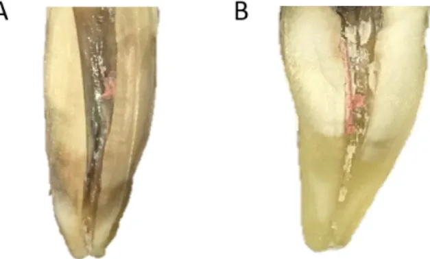
Figure 1. Representative slices of (A) RS and (B) RS + C groups
Group Cervical Middle Apical Total
RS 2.5% (1.9)Aa 4.2% (2.2)Aa 14.7% (13.1)Ab 6.2% (4.9)A RS+C (2.4)3.1%Aa 7.4% (4.3%)Aa 17.4% (11.3)Ab 8.7% (5.4)A
Table 1. Mean ± SD of the area of residual filling material by canal region after instrumentation, as well as statistical significance*
*For each column, values followed by same capital letters are statisti-cally similar (P >.05). For each row different lowercases represent sig-nificant differences between the different root canal thirds in the same group (P >.05).
Table 2. Mean ± SD of the time necessary for retreatment and number (percentage) of cases that had file fracture recorded for the different groups, as well as statistical significance*
Group Time (s) Fracture (%)
RS 172.3 ± 41.3A 0 (0%)
RS+C 245.2 ± 62.9B 0 (0%)
*Different capital letters represent significant differences between the tested groups (P >.05).
analysis that use sectioning, Micro-CT maintains the integ-rity of the sample that can be reused.38,45,46
Previous studies showed a shorter retreatment time when files were used associated with solvent.6,12 Conversely, the
use of Reciproc System without solvent speeded up retreat-ment time in this study. Similar results were also reported by Takahashi et al.,3 using ProTaper Universal Retreatment
Files (URF). They showed that while Protaper URF without solvent completed the procedure within 5.7 min, their use with chloroform took 8.8 min for complete removal of the old root canal filling.
Solvents should be used only in cases of extreme difficulty in removing the gutta-percha, as they create a thin layer of softened gutta-percha that adheres to the root canal wall.
Such a layer can be easily forced into the complex root canal anatomy (isthmuses, cul de sacs, lateral canals and irregu-larities) that are not touched by the instruments.12,47 Once
created, accentuates the challenges of filling removal and requires more time to be removed. The use of an operating microscope can improve visualization of this layer and aid in its removal.
Conclusion
Taken together, our results demonstrated that Reciproc System used without solvent required less time to remove the same amount of root filling material than Reciproc Sys-tem with solvent. However, regardless of solvent use, the same cleaning quality was obtained.
operative pain after endodontic retreatment using rotary or reciprocating instru-ments: a randomized clinical trial. J Endod. 2017;43:1084-8.
18. Silva EJNL, Ferreira VM, Silva CC, Herrera DR, De-Deus G, Gomes BP. In-fluence of apical enlargement and complementary canal preparation with the Self-Adjusting File on endotoxin reduction in retreatment cases. Int Endod J. 2017;50:646-51.
19. Silva EJ, Brito ME, Ferreira VD, Belladonna FG, Neves AA, Senna PM, De-Deus G. Cytotoxic effects of the debris apically extruded during three differ-ent retreatmdiffer-ent procedures. J Oral Sci. 2016;58:211-7.
20. De-Deus G, Reis C, Beznos D, de Abranches AM, Coutinho-Filho T, Pacio-rnik S. Limited ability of three commonly used thermoplasticized gutta-percha techniques in filling oval-shaped canals. J Endod 2008;34:140-5.
21. Bueno CSP, Oliveira DP, Pelegrine RA, Fontana CE, Rocha DGP, Bueno CEDS. Fracture Incidence of WaveOne and Reciproc Files during Root Canal Preparation of up to 3 Posterior Teeth: A Prospective Clinical Study. J Endod. 2017;43:705-8.
22. Moiseiwitsch JR, Trope M. Nonsurgical root canal therapy treatment with apparent indications for root-end surgery. Oral Surg Oral Med Oral Pathol Oral Radiol Endod. 1998;86:335-40.
23. Del Fabbro M, Taschieri S. A systematic review on the outcome of surgical vs non-surgical procedure for the retreatment of periapical lesions. Minerva Stomatol. 2007;56:621-32.
24. Kvist T. Endodontic retreatment. Aspects of decision making and clinical outcome. Swed Dent J Suppl. 2001; 144:1-57
25. Rôças IN, Siqueira JF Jr. Characterization of microbiota of root canal-treated teeth with posttreatment disease. J Clin Microbiol. 2012;50:1721-4.
26. Fabricius L, Dahle´n G, Sundqvist G, Happonen RP, Möller AJ. Influence of residual bacteria on periapical tissue healing after chemomechanical treat-ment and root filling of experitreat-mentally infected monkey teeth. Eur J Oral Sci. 2006;114:278-85.
27. Silva EJ, Orlowsky NB, Herrera DR, Machado R, Krebs RL, Coutinho–Filho Tde S. Effectiveness of rotatory and reciprocating movements in root canal filling material removal. Braz Oral Res. 2015;29:1-6.
28. Ricucci D, Siqueira JF Jr, Bate AL, Pitt Ford TR. Histologic investigation of root canaltreated teeth with apical periodontitis: a retrospective study from twenty-four patients. J Endod. 2009;35:493-502.
29. Hulsmann M, Stotz S. Efficiency, cleaning ability and safety of different devic-es for gutta-percha in root canal retreatment. Int Endod J. 1997;30:227-33. 30. Zuolo AS, Zuolo ML, da Silveira Bueno CE, Chu R, Cunha RS. Evaluation of the efficacy of TRUShape and Reciproc file systems in the removal of root filling material: an ex vivo micro-computed tomographic study. J Endod. 2016;42:315-9. 31. Wennberg A, Orstavik D. Evaluation of alternatives to chloroform in end-odontic practice. Endod Dent Traumatol. 1989;5:234-7.
32. Zakariasen KL, Brayton SM, Collinson DM. Efficient and effective root canal retreatment without chloroform. J Can Dent Assoc. 1990;56:509-12.
33. Wilcox LR. Endodontic retreatment with halothane versus chloroform sol-vent. J Endod. 1995;21:305-7.
34. Ma J, Al-Ashaw AJ, Shen Y, Gao Y, Yang Y, Zhang C, et al. Efficacy of ProTaper References
1. Mounce R. Endodontic retreatment possiblities: evaluation, limitations and considerations. Compend Contin Educ Dent. 2004;25:364-8.
2. Tasdemir T, Yildirim T, Celik D. Comparative study of removal of current endodontic fillings. J Endod. 2008;34:326-9.
3. Takahashi CM, Cunha RS, de Martin AS, Fontana CE, Silveira CFM, da Sil-veira Bueno CE. In vitro evaluation of the effectiveness of ProTaper Universal rotary retreatment system for gutta-percha removal with or without a solvent. J Endod. 2009;35:1580-3.
4. Bramante CM, Fidelis NS, Assumpção TS, Bernardineli N, Garcia RB, Bra-mante AS, et al. Heat release, time required, and cleaning ability of Mtwo R and ProTaper Universal retreatment systems in the removal of filling material. J En-dod. 2010;36:1870-3.
5. Zmener O, Pameijer CH, Banegas G. Retreatment efficacy of hand versus au-tomated instrumentation in oval-shaped root canals: an ex vivo study. Int Endod J. 2006;39:521-6.
6. Giuliani V, Cocchetti R, Pagavino G. Efficacy of ProTaper Universal retreat-ment files in removing filling materials during root canal retreatretreat-ment. J Endod. 2008;34:1381-4.
7. Unal GC, Kaya BU, Tac¸ AG, Keçeci AD. A comparison of the efficacy of con-ventional and new retreatment instruments to remove gutta-percha in curved root canals: an ex vivo study. Int Endod J. 2009;42:344-50.
8. Bernardes RA, Duarte MA, Vivan RR, Alcalde MP, Vasconcelos BC, Bramante CM. Comparison of three retreatment techniques with ultrasonic activation in flattened canals using micro-computed tomography and scanning electron mi-croscopy. Int Endod J. 2016;49:890-7
9. Baratto-Filho F, Ferreira EL, Fariniuk LF. Efficiency of the 0.04 taper Pro-File during the re-treatment of guttapercha- filled root canals. Int Endod J. 2002;35:651-4.
10. Gergi R, Sabbagh C. Effectiveness of two nickel-titanium rotary instruments and a hand file for removing gutta-percha in severely curved root canals during retreatment: An ex vivo study. Int Endod J. 2007;40:532-7.
11. McDonald MN, Vire DE. Chloroform in the endodontic operatory. J Endod. 1992;18:301-3.
12. Sae-Lim V, Rajamanickam I, Lim BK, Lee HL. Effectiveness of Profile .04 taper rotary instruments in endodontic retreatment. J Endod. 2000;26:100-4. 13. Yared G. Canal preparation using only one Ni-Ti rotary instrument: prelimi-nary observations. Int Endod J. 2008;41:339-44.
14. De-Deus G, Brandão MC, Barino B, Di Giorgi K, Fidel RA, Luna AS. As-sessment of apically extruded debris produced by the single-file ProTaper F2 technique under reciprocating movement. Oral Surg Oral Med Oral Pathol Oral Radiol Endod. 2010;110:390-4.
15. Varela-Patiño P, Ibañez-Párraga A, Rivas-Mundiña B, Cantatore G, Otero XL, Martin-Biedma B. Alternating versus continuous rotation: a comparative study of the effect on instrument life. J Endod. 2010;36:157-9.
16. Arias A, Perez-Higueras JJ, de la Macorra JC. Differences in cyclic fatigue re-sistance at apical and coronal levels of Reciproc and WaveOne new files. J Endod. 2012;38:1244-8.
Post-Submitted: 08/08/2017 / Accepted for publication: 08/27/2017 Corresponding Author
Emmanuel João Nogueira Leal Silva E-mail: nogueiraemmanuel@hotmail.com
Mini Curriculum and Author’s Contribution
1. Nancy Kudsi Carvalho - DDS, MSc. Contribution: bibliographical research, experimental procedures, manuscript writing. 2. Gustavo Ribeiro Alvares - DDS, MSc, PhD. Contribution: bibliographical research, experimental procedures, manuscript writing. 3. Luciana Moura Sassone - DDS, MSc, PhD. Contribution: manuscript writing, manuscript review, image editing.
4. Renato Lies Krebs - DDS, MSc, PhD. Contribution: manuscript writing, manuscript review.
5. Tauby de Souza Coutinho-Filho - DDS, MSc, PhD. Contribution: manuscript writing, manuscript review.
6. Emmanuel João Nogueira Leal da Silva - DDS, MSc, PhD. Contribution: manuscript review, statistical analysis, image editing, work supervisor and paper submission. Universal Rotary Retreatment system for gutta-percha removal from oval root
canals: a micro-computed tomography study. J Endod. 2012;38:1516-20. 35. Rechenberg DK, Paque F. Impact of cross-sectional root canal shape on filled canal volume and remaining root filling material after retreatment. Int Endod J. 2013;46:547-55.
36. Versiani MA, Pecora JD, de Sousa-Neto MD. Flat-oval root canal preparation with self-adjusting file instrument: a micro-computed tomography study. J En-dod. 2011;37:1002-7.
37. Mello Junior JE, Cunha RS, Bueno CE, Zuolo ML. Retreatment efficacy of guttapercha removal using a clinical microscope and ultrasonic instruments: part I - an ex vivo study. Oral Surg Oral Med Oral Pathol Oral Radiol Endod. 2009;108:59-62.
38. Zuolo AS, Mello JE, Cunha RS, Zuolo ML, Bueno CE. Efficacy of reciprocat-ing and rotary techniques for removreciprocat-ing fillreciprocat-ing material durreciprocat-ing root canal retreat-ment. Int Endod J. 2013;46:947-53.
39. Saad AY, Al-Hadlaq SM, Al-Katheeri NH. Efficacy of two rotary NiTi instru-nents in the removal of gutta-percha during root canal retreatment. J Endod. 2007;3:38-41.
40. Upadhyay V, Upadhyay M, Panday RK, Chturvedi TP, Bajpai U. A SEM eval-uation of dentinal adaptation of root canal obturation with GuttaFlow and con-ventional obturating material. Indian J Dent Res. 2011;22:881.
41. Asheibi F, Qualtrough AJ, Mellor A, Withers PJ, Lowe T. Micro-CT evalua-tion of the effectiveness of the combined use of rotary and hand instrumentaevalua-tion in removal of Resilon. Dent Mater J. 2014;33:1-6.
42. Paque F, Peters OA. Micro-computed tomography evaluation of the prepara-tion of long oval root canals in mandibular molars with the self-adjusting file. J Endod. 2011;37:517-21.
43. Solomonov M, Paque F, Kaya S, Adigüzel O, Kfir A, Yiğit-Özer S. Self-adjust-ing files in retreatment: a highresolution micro-computed tomography study. J Endod. 2012;38:1283-7.
44. Kfir A, Tsesis I, Yakirevich E, Matalon S, Abramovitz I. The efficacy of five retreatment techniques: microscopic vs. radiographic evaluation. Int Endod J. 2012;45:35-41.
45. Rios MA, Villela AM, Cunha RS, Velasco RC, De Martin AS, Kato AS, et al. Efficacy of 2 reciprocating systems compared with a rotary retreatment system for gutta-percha removal. J Endod. 2014;40:543-6.
46. Ozy € €urek T, Demiry€urek EO. Efficacy of different nickel-titanium in-struments in € removing gutta-percha during root canal retreatment. J Endod. 2016;42:646-9.
47. Gu LS, Ling JQ, Wei X, Huang XY. Efficacy of ProTaper Universal rotary re-treatment for gutta-percha removal from root canals. Int Endod J. 2008;41:288-95.