i
Idálio de Jesus Contreiras Viegas
Licenciado em Biotecnologia
N6-methyladenosine, a new modification in T. brucei
epitranscriptome
Dissertação para obtenção do Grau de Mestre em Genética Molecular e Biomedicina
Orientador: Luísa Miranda Figueiredo, PhD, Universidade do Porto
Júri:
Presidente: Doutora Paula Maria Theriaga Mendes Bernardo Gonçalves Arguente: Doutor Diogo Pinto da Cruz Sampaio e Castro
Vogal: Doutora Luísa Miranda Figueiredo
iii
Idálio de Jesus Contreiras Viegas
Licenciado em Biotecnologia
N6-methyladenosine, a new modification in T. brucei
epitranscriptome
Dissertação para obtenção do Grau de Mestre em Genética Molecular e Biomedicina
Orientador: Luísa Miranda Figueiredo, PhD, Universidade do Porto
iv
N6-methyladenosine, a new modification in T. brucei epitranscriptome
Copyright Idálio Viegas, FCT/UNL, UNL
A Faculdade de Ciências e Tecnologia e a Universidade Nova de Lisboa têm o direito, perpétuo e sem limites geográficos, de arquivar e publicar esta dissertação através de exemplares impressos reproduzidos em papel ou de forma digital, ou por qualquer outro meio conhecido ou que venha a ser inventado, e de a divulgar através de repositórios científicos e de admitir a sua cópia e distribuição com objectivos educacionais ou de investigação, não comerciais, desde que seja dado crédito ao autor e editor.
v
Agradecimentos
“Um dia sem rir é um dia desperdiçado”
Charles Chaplin
Muito obrigado Luísa. Muito obrigado por todo o apoio, toda a dedicação, paciência e por me acompanhares em todos os momentos desde que cheguei ao teu grupo. Agradeço-te pela chefe que és, e acima de tudo, pela pessoa que és, sabes ensinar e motivar, estimulas que os teus alunos tenham opinião, sejam criativos, críticos e cresçam como cientistas e pessoas. Foi um privilégio aprender contigo.
Obrigado Francisco (Xico), foste o coorientador deste trabalho, sendo incansável na dedicação e esforço. Obrigado pela dedicação, pela disponibilidade, empenho, e acima de tudo pela paciência para ensinar e ajudar, estiveste sempre presente ao longo do projeto tornando-o possível.
Ao longo do ano, dia após dia de convivência, os elementos da UPAR estiveram sempre disponíveis para ajudar, sempre disponíveis para apoiar, sempre disponíveis para ensinar. Mais que convivência profissional estiveram disponíveis para realmente conviver, falar e sorrir, passaram de colegas a amigos. Por tudo isto, o meu muito obrigado a todos, agradeço á Pena, ao Daniel, á Margarida, ao Fabien, à Filipa, á Sandra, á Leonor, á Mafalda, á Helena e ao novo reforço o Fábio.
Como sempre faço, agradeço a todos os meus amigos, não sendo necessário estar listar nomes, eles sabem quem são. Obrigado.
Um obrigado a toda a minha família, que me acompanha e apoia desde sempre. Um obrigado muito especial aos meus pais, a quem devo tudo, mais que tudo, que com grande esforço e sacrifício possibilitaram que eu crescesse e seja a pessoa que sou. Obrigado. Um obrigado ao meu irmão, o miúdo mais importante á face da Terra.
Fui breve nas palavras, mas acreditem, quando digo obrigado estou mesmo grato!
Por fim, quero dedicar o todo meu esforço e dedicação a quem já vou tarde para agradecer, gostava de puder agradecer, estou muito grato. Diz a nossa cultura que e função do padrinho é estar presente e ajudar nos momentos mais difíceis, o meu padrinho esteve presente desde que nasci e quando eu precisei ajudou-me sem hesitar. Sem essa ajuda não estaria aqui neste momento, a escrever os agradecimentos deste trabalho, não estaria a perseguir um sonho de miúdo. Obrigado padrinho.
vii
Resumo
A doença do sono em humanos é causada pelo Trypanosoma brucei, um parasita eucariota unicelular. Neste parasita, quase todos os genes são transcritos constitutivamente e, portanto, a sua regulação é sobretudo por mecanismos pós-transcricionais. A N6-metiladenosina (m6A) é uma
modificação presente no RNA que tem sido associada à regulação da expressão génica ao nível pós-transcricional em vários eucariotas. Com base nestas observações propus que esta modificação existe no transcriptoma de T. brucei e é um mecanismo de regulação da expressão génica ao nível pós-transcricional. Neste trabalho, pela primeira vez, detetou-se esta modificação no RNA. Também encontrei esta modificação no DNA, sendo esta a primeira descrição de um organismo em que esta modificação existe nos dois tipos de ácidos nucleicos. A modificação m6A no RNA parece ser
dinâmica: verifiquei que os níveis variam em diferentes condições biológicas, nomeadamente aumentam durante a diferenciação entres dois estadios do ciclo de vida e quando os parasitas são colocados em condições de stress provocadas por alta densidade celular. Bioinformaticamente foi procurado no genoma deste parasita, genes candidatos que codificam possíveis enzimas que catalisam a formação e a remoção desta modificação. Encontrou-se uma possível metiltransferase e seis possíveis demetilases. Para testar a sua possível função, foram geradas linhas celulares knockouts da possível metiltransferase (Tb927.7.6620) e de duas possíveis demetilases (Tb927.4.460 denominada TbALKBH1 e Tb927.5.980 denominada TbALKBH2). A quantificação dos níveis de m6A no RNA
dos knockouts da metiltransferase e de uma demetilase (TbALKBH1) não revelou evidência que suportasse a possível função proposta. No entanto, um aumento nos níveis de m6A no RNA do
knockout da possível demetilase TbALKBH2 indica que poderá ser uma demetilase de m6A no RNA.
A evidência apresentada nesta tese levanta a possibilidade de um novo mecanismo de regulação pós transcricional em T. brucei através desta modificação no epitranscriptoma do parasita.
ix
Abstract
Trypanosoma brucei is a unicellular eukaryote parasite that causes human sleeping sickness. In this parasite, transcription is mainly constitutive and gene expression regulation occurs essentially at post-transcriptional level. N6-methyladenosine (m6A) is an RNA modification associated
with post-transcriptional gene regulation in eukaryotes. These observations led to the proposal that this modification occurs in T. brucei transcriptome and is involved in post-transcriptional gene regulation. In this thesis, m6A was detected for the first time in T. brucei RNA and additionally in DNA, from
bloodstream and procyclic life stages. As far as I know, this is the first description of an organism in which has m6A is found in both type of nucleic acids. In RNA, I observed that the levels are regulated
in different biological circumstances, namely, it increases during differentiation from bloodstream to procyclic life-cycle stages and it also increases when parasites are stressed by being placed at high cell density. T. brucei genome was searched with bioinformatics tools to find enzymes that catalyse the formation and the removal the of m6A modification in RNA. One putative RNA m6A
methyltransferase and six putative demethylases were found. Knockout cell lines of the putative methyltransferase (Tb927.7.6620) and of two putative demethylases (Tb927.4.460 named TbALKBH1 and Tb927.5.980 named TbALKBH2) were generated to test their putative functions. Quantification of m6A levels in RNA from the knockout cell lines did not reveal evidence that support the putative
function of the methyltransferase and one demethylase (TbALKBH1). However, knockout of TbALKBH2 resulted in a slight increase in m6A levels, suggesting that this candidate could be an
RNA m6A demethylase. The evidence presented in this thesis raises the possibility of
post-transcriptional gene regulation mediated by the presence of m6A modification in T. brucei
epitranscriptome.
xi
Contents
Agradecimentos ... v
Resumo ... vii
Abstract ... ix
Index of figures ... xiii
Index of tables ... xv
Abbreviations ... xvi
1.Introduction ... 1
1.1 Trypanosoma brucei ... 1
1.1.1. Human African trypanosomiasis and human infective subspecies ... 1
1.1.2. T. brucei life cycle ... 2
1.1.3. Genome Organization in T. brucei ... 3
1.1.4. Gene expression in T. brucei ... 4
1.2. N6-methyladenosine (m6A) in RNA ... 8
1.2.1. RNA modifications ... 8
1.2.2. N6-methyladenosine ... 11
1.2.3. Chemical reactions ... 11
1.2.4. Enzymes and Reversibility ... 12
1.2.5. Biological functions ... 14
1.2.6. Molecular mechanisms and targets ... 15
1.3. Objectives ... 17 2. Methods ... 19 2.1. Parasite culture ... 19 2.2. Differentiation ... 19 2.3. RNA extraction ... 20 2.4. DNA extraction ... 20 2.5. Cloning ... 20 2.6. Transfections ... 21 2.7. Immunoblot ... 22 2.8. EpiQuick m6A Quantification ... 23 2.9. Bioinformatics ... 23 3. Results ... 25
3.1. Immunoblot detection of m6A in T. brucei RNA ... 25
3.2. Detection and quantification of m6A in T. brucei (in bloodstream and procyclic forms) ... 28
xii
3.4. Levels of m6A in density stress condition ... 32
3.5. Characterization of putative m6A methyltransferase and demethylases enzymes ... 33
3.5.1. Identification of putative RNA m6A methyltransferase... 33
3.5.2. Identification of putative RNA m6A demethylases ... 35
3.5.3. Generation of knockout cell lines of putative enzymes ... 38
3.5.4. Measurement of m6A levels in knockout cell lines ... 45
4. Discussion ... 47
4.1. N6-methyladenosine (m6A) in RNA of T. brucei ... 47
4.2. N6-methyladenosine (m6A) in DNA of T. brucei ... 48
4.3. Levels of RNA m6A during differentiation ... 49
4.4. m6A RNA modification is sensitive to cell density ... 50
4.5. Putative RNA m6A methyltransferase ... 51
4.6. Putative RNA m6A demethylases... 52
5. Conclusion ... 55
6. References ... 56
xiii
Index of figures
Figure 1.1: Human African trypanosomiasis distribution.……….……..2
Figure 1.2: T. brucei life cycle ……….3
Figure 1.3: Ribonucleoside modifications found in RNA ………...………10
Figure 1.4: N6-methyladenosine structure………..…………11
Figure 1.5: RNA m6A methylation/demethylation pathway……….….…13
Figure 1.6: Molecular mechanisms and functions of m6A ………….…….….………..……..… 16
Figure 3.1: Immunoblot to detect m6A ……….……….…………..………… 26
Figure 3.2: Levels of m6A in total RNA from T. brucei ………...………..….…… 30
Figure 3.3: Flow cytometry of differentiated cells ………...………… 31
Figure 3.4: Levels of m6A in total RNA during in vitro differentiation ………..…………31
Figure 3.5: Levels of m6A of T. brucei from high density culture ……….….……..32
Figure 3.6: MSA of sequences of the Probable N6-adenine methyltransferases ………...…………... 35
Figure 3.7: MSA of AlkB, ALKBH and TbALKBH proteins…….…..……… 37
Figure 3.8: KO cell lines generation strategy ………...……… 38
Figure 3.9: Agarose gel of the inserts to clone pIV vectors…………..……….……...………… 40
Figure 3.10: Agarose gel of the vectors digestion ……… 40
Figure 3.11: Agarose gel of resistance genes integration in Tb.927.5.980 locus ……….…… 41
Figure 3.12: Agarose gel of resistance genes integration in Tb.927.4.460 locus……….……… 42
Figure.3.13: Agarose gel of resistance genes integration in Tb.927.7.6620 locus ………….……..… 42
Figure 3.14: Agarose gel of KO cell lines locus ……….……… 43
Figure 3.15: Growth curve of KO cell lines ………..…………..………44
xv
Index of tables
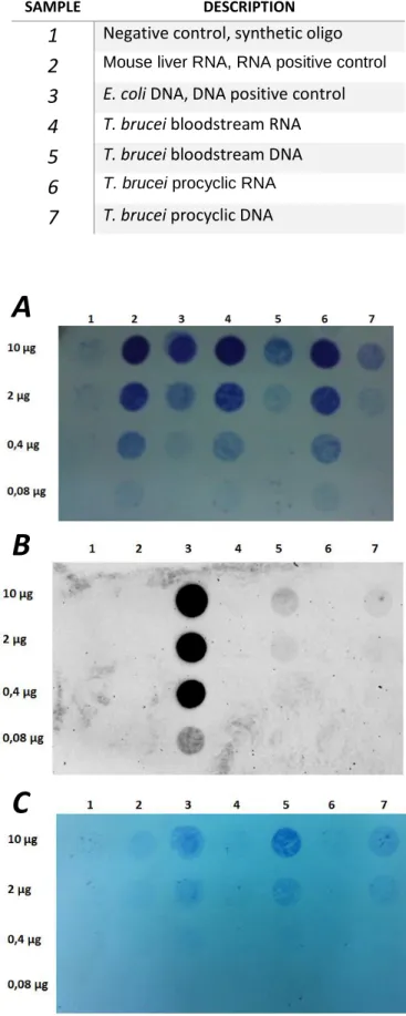
Table 3.1: Samples spotted in immunoblot membrane……….….26
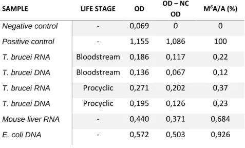
Table 3.2: Detection of m6A in RNA and DNA samples………..……….….. 29
Table 3.3: T. brucei proteins with 2OG-Fe(II) oxygenase domain ………….………..……….…….. 36
Table 3.4: Vectors designed to KO the candidate genes ……….……..…………39
Table 3.5: Inserts to generate the pIVs vectors ………..………….….. 39
Table 3.6: Restriction digestions to confirm the cloned plasmids ……….………..…..40
Table 3.7: Amplifications of resistance genes integration in Tb927.5.980 locus ………..…….. .41
Table 3.8: Amplifications of resistance genes integration in Tb927.4.460 locus ………...….. 42
Table 3.9: Amplifications of resistance genes integration in Tb927.7.6620 locus ………... 42
xvi
Abbreviations
AAT: Animal African trypanosomiasis ALBA: Acetylation lowers binding affinity ALKBH: AlkB homologue
BLAST: Basic Local Alignment Search Tool Bp: Base pair
BSF: Bloodstream form CNS: Central nervous system
DALYs: Disability-adjusted life years DNA: Deoxyribonucleic Acid
DNase I: Deoxyribonuclease I ECL: Enhanced chemiluminescence EP: EP procyclins
E-value: Expect value
FTO: Fat and mass associated protein GPEETS: GPEET procyclins
HAT: Human African trypanosomiasis HMM: Hidden Markov Model
IME4: Inducer of Meiosis 4 KO: Knockout
lncRNA: long non-coding RNA m5C: 5-methylcytosine
m6A: N6-methyladenosine
m7G: 7-methylguanosine
Me-RIP: m6A-specific methylated RNA
immunoprecipitation
METTL14: Methyltransferase like 14 METTL3: Methyltransferase like 3 miRNA: Micro RNA
mRNA: Messenger Ribonucleic Acid
MSA: Multiple sequence alignment MTA: mRNA adenosine methylase
ncRNA: Non-coding RNA NEB: New England Biolabs NPC: Nuclear pore complex ORF: Open reading frame PC: Procyclic form
PCR: Polymerase Chain Reaction Poll I: RNA polymerase I
Poll II: RNA polymerase II RBP: RNA binding protein RNA: Ribonucleic Acid RNase A: Ribonuclease A RRM: RNA recognition motif rRNA: Ribosomal Ribonucleic Acid SAM: S-adenosylmethionine
SL: Spliced leader
SN2: Nucleophilic Substitution bi-molecular
TDB: Trypanosome dilution buffer tRNA: Transfer Ribonucleic Acid TSS: Transcription start site TTS: Transcription termination site UTR: Untranslated region
VSG: Variable surface glycoprotein
WTAP: Wilms’ tumor 1-associating protein YLL: Years of life lost
1
1.Introduction
1.1 Trypanosoma brucei
1.1.1. Human African trypanosomiasis and human infective subspecies
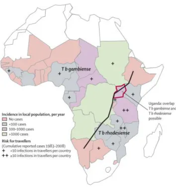
Trypanosoma brucei (T. brucei) is a unicellular protozoan parasite. This parasite causes the Human African trypanosomiasis (HAT), also known as sleeping sickness (Brun et al., 2010). This disease occurs in 36 sub-Saharan countries, where 6314 new cases were reported in the year of 2013 (Franco et al., 2014). The impact caused by HAT in the population was estimated in 1.6 million disability-adjusted life years (DALYs) per year and 27 years of life lost (YLL) per death (WHO, 2012) . Clinically, HAT presents two stages: first the haemolymphatic stage, characterized by the presence of parasites in the blood and in the interstitial space of diverse tissues. Second the meningoencephalitic stage, characterized by infiltration of parasites in the central nervous system (CNS) (Brun et al., 2010; Kennedy, 2004). During the first stage the main symptoms are fever, pruritus, lymphadenopathy and hepatosplenomegaly. Sleep disturbances that result from dysregulation of the circadian rhythm is the major symptom of the second stage. These sleep disturbances are responsible for the attribution of the name sleeping sickness. If untreated, HAT ultimately leads to coma and death (Brun et al., 2010; Kennedy, 2004).
Two subspecies of T. brucei can cause HAT, T. brucei rhodesiense (East Africa) and T. brucei gambiense (West Africa). The distribution is represented in figure 1.1. The infection caused by T. brucei rhodesiense is acute, leading to death in weeks or months. As for T. brucei gambiense, the disease has a progressive course with a chronic infection and death occurs after several years (3-7 years) (Brun et al., 2010; Kennedy, 2004). Parasites are transmitted to humans by the tsetse, a fly from the Glossina species, during its blood meal (Brun et al., 2010; Dyer et al., 2013). Besides HAT, this parasite also causes Animal African trypanosomiasis (AAT, or nagana) in other mammals like cattle (Brun et al., 2010; Steverding, 2008).
2
Figure 1.1. HAT distribution with incidence and risk for travellers indicated. Black line separate the T.b. gambiense and T.
b. rhodesiense distribution. Adapted from (Brun et al., 2010).
1.1.2. T. brucei life cycle
The life cycle of T. brucei is divided between the mammalian host and the tsetse vector (Figure 1.2.) (Fenn and Matthews, 2007). In the blood of the mammalian host, parasites proliferate as bloodstream slender forms. When the parasite population increases, cells are able to differentiate into non-dividing stumpy forms, using a quorum sensing mechanism (Fenn and Matthews, 2007; Matthews et al., 2004). Stumpy cells are competent to complete the life cycle if uptaken by the tsetse. When stumpy cells enter the midgut of the transmission vector, the progression of differentiation into procyclic forms takes place. Procyclic cells migrate to proventriculus and differentiate into epimastigote forms. After this event, some epimastigote cells migrate to the salivary glands of the fly, where they differentiate into non-dividing metacyclic forms (Dyer et al., 2013; Fenn and Matthews, 2007). It was proposed that in the salivary glands of the tsetse, T. brucei could pass through a meiotic stage in the life cycle, besides the mitotic cell division (Peacock et al., 2011). When the tsetse takes another blood meal from a mammal, metacyclic forms are injected into its bloodstream, where the parasites differentiate into bloodstream slender cells closing the cycle (Matthews et al., 2004).
3
Figure 1.2. T. brucei life cycle (courtesy of Daniel Pinto-Neves.
Cell differentiation throughout life cycle implies that parasites can adapt to different environments (Fenn and Matthews, 2007; Matthews, 2005). This adaptation is defined by alterations of gene expression. Comparison of the transcriptome of different life-cycle stages revealed that around 28-40% of genes are differentially expressed between bloodstream slender forms and procyclic forms (Nilsson et al., 2010; Veitch et al., 2010). Consistent with these observations, recent studies comparing protein expression between bloodstream slender forms and procyclic forms revealed that around 33-48% of the proteome is different (Butter et al., 2013; Gunasekera et al., 2012).
Also during differentiation, some genes are co-regulated establishing multiple post-transcriptional regulons. These co-regulated clusters are formed by transcripts from genes involved in diverse functions and some regulons are composed of genes involved in the same biochemical pathway. For example, genes involved in cell division and macromolecular biosynthesis are co-regulated during differentiation, like ribosomal and flagellar proteins encoding genes (Queiroz et al., 2009).
1.1.3. Genome Organization in T. brucei
T. brucei is a diploid organism whose 26 Mb genome is organized in different classes of chromosomes: eleven pairs of megabase chromosomes (around 1 Mb - 6 Mb), one to six intermediate chromosomes (200 Kb – 900 Kb) and around one hundred minichromosomes (50 Kb - 150 Kb) (El-Sayed et al., 2000). The megabase chromosomes have been sequenced and shown to harbour 9 068 genes (Berriman et al., 2005). Recent studies allowed the identification of 1 114 new non-annotated genes (Kolev et al., 2010). Genes are organized in polycistronic units, similar to bacterial operons, but unlike these, genes in the same polycistronic unit do not seem be involved in the same pathway
4
(Berriman et al., 2005). Unlike megabase chromosomes, which are diploid, the intermediate chromosomes and minichromosomes seem to be haploid (Ersfeld, 2011).
20% of the genes of T. brucei encode for variant surface glycoproteins (VSGs) (Cross et al., 2014), which form a dense layer at the surface of parasite. VSGs are transcribed in a monoallelic fashion (Rudenko, 2010; Taylor and Rudenko, 2006), so that only one VSG coat is exposed to the immune system at a time. By a mechanism known as antigenic variation, some cells of the parasite population periodically change the expressed VSG, allowing these cells to escape the immune system and ensure the infection persists (Rudenko, 2010; Taylor and Rudenko, 2006).
1.1.4. Gene expression in T. brucei
Transcription in trypanosomes shows clear differences from “canonical” eukaryotes. Due to the polycistronic organization of the genes, they are transcribed as a polycistronic transcript by RNA polymerase II (Pol II), without any apparent transcriptional control (Clayton, 2002; Palenchar and Bellofatto, 2006). The polycistronic transcript is processed into individual mRNAs through a process called trans-splicing. In this process, a 39 nucleotide RNA cap sequence, termed spliced leader, is added to the 5’ end of the newly transcribed gene, while a poly-A tail is added at the 3’ end of upstream transcribed gene (Liang et al., 2003; Palenchar and Bellofatto, 2006). RNA polymerase I (Pol I) transcribes not only rRNA (18S, 5.8S and 28S), but also life-cycle stage specific proteins, including VSGs (in bloodstream forms) and procyclins (EPs and GPEETs, in procyclic forms) (Palenchar and Bellofatto, 2006). Unlike Pol II , Pol I is regulated at transcriptional level (Rudenko, 2010).
1.1.4.1. Transcriptional control in T. brucei
In eukaryotes, gene expression is controlled by an interconnected net of diverse mechanisms. These mechanisms involve genetic elements like promoters (Juven-Gershon and Kadonaga, 2010), transcription factors assembly (Lemon, 2000), enhancers and silencers (Kolovos et al., 2012; Ong and Corces, 2011). Beyond genetic elements, gene expression is regulated by molecular mechanisms that do not involve changes in DNA sequence and that are called epigenetics (Goldberg et al., 2007; Jaenisch and Bird, 2003). Epigenetic mechanisms include DNA methylation (Bird, 2002; Jones, 2012), non-coding RNAs (Kaikkonen et al., 2011; Mercer and Mattick, 2013), alterations in chromatin structure mediated by histone modifications (Bannister and Kouzarides, 2011; Kouzarides, 2007),
5
chromatin remodelling (Clapier and Cairns, 2009; Saha et al., 2006) and organization of chromatin in the nucleus (Fedorova and Zink, 2008; Schneider and Grosschedl, 2007).
In T. brucei, Pol II promoters lack well-established genetic elements. The only exception is the promoter of the Spliced Leader gene (Schimanski et al., 2005). Besides the lack of unidentified promoters, very few transcription factors can be found in the genome of this parasite (Iyer et al., 2008). Histone variants are present at transcription start sites (TSS) and transcription termination sites (TTS), suggesting that chromatin may play an important role in defining key functional sites of the chromosomes (Siegel et al., 2009).
A wide variety of chemical modifications has been found in histones of several eukaryotes: acetylation, methylation, phosphorylation, ubiquitylation, sumoylation, ADP ribosylation, deimination, proline isomerization) (Bannister and Kouzarides, 2011). These modifications are dynamic. The enzymes that add the chemical modifications are called “writers” and those that remove are called “erasers” (Jakovcevski and Akbarian, 2012). Histone modifications can function by the recruitment of effectors proteins or complexes. These recruited complexes have histone modifications recognition domains and are called “readers” of the epigenetic code (Kouzarides, 2007; Yun et al., 2011). T. brucei has fewer histone modifications, than most other eukaryotes (Figueiredo et al., 2009). Some modifying enzymes have been characterized, including lysine acetyltransferase (MYST family and EPL3 homologues), deacetylases (HDAC 1-2,HDAC 3-4 and SIR2 related histone deacetylases) and lysine methyltransferases (Disruptor of telomerase silencing DOT1 homologues) (Figueiredo et al., 2009).
In mammalian cells, DNA methylation (5-methylcytosine) is an important epigenetic mark that occurs mainly in CG repetitions (CpG islands) located at transcription start sites and is involved in gene repression (Bird, 2002). DNA methylation can also occur in other transcriptional start sites without CpG islands, in gene bodies, at regulatory elements and at repeat sequences (Jones, 2012). 5-methylcytosine has also been detected in nuclear DNA of T. brucei, both in bloodstream and procyclic forms (Militello et al., 2008). A putative 5-methylcytosine methyltransferase has been found in the genome (Militello et al., 2008), but the activity was not yet empirically tested.
Besides this DNA modification, bloodstream form of T. brucei has another unusual modification in its DNA, the base J (β‑d-glucopyranosyloxymethyluracil) (Gommers-ampt et al., 1993). Base J is found mainly in the telomere repeats, repetitive sequences and transcription termination sites. Two base J binding proteins were found, J-binding protein 1 (JBP1) and J-binding protein 2 (JBP2). These base J binding proteins are required for the synthesis of base J. (Borst and Sabatini, 2008). In Leishmania, base J is required for proper genome wide Pol II transcription termination (van Luenen et al., 2012), however this genome wide function is not conserved in T. brucei, where base J controls transcription termination at specific locations (Reynolds et al., 2014).
6
1.1.4.2 Post-transcriptional control in T. brucei
Diverse post-transcriptional mechanisms influence gene expression, including nuclear transport (Köhler and Hurt, 2007; Strambio-De-Castillia et al., 2010), RNA decay (Garneau et al., 2007; Wilusz and Wilusz, 2004), and translation regulation. These processes can be mediated by the binding or interaction of diverse RNA binding proteins (Glisovic et al., 2008; Lunde et al., 2007) and non-coding RNAs with mRNAs (Kaikkonen et al., 2011).
Nuclear transport
mRNAs need to be transported from the nucleus to the cytoplasm, where they are translated. mRNAs are exported via nuclear pore complexes (NPCs), cylinder structures composed of many proteins that cross the double nuclear membrane. NPCs are also involved in epigenetic control of gene expression through interactions with the chromatin (Rodríguez-Navarro and Hurt, 2011; Strambio-De-Castillia et al., 2010). The structure of the NPC seems to be conserved in T. brucei, suggesting that RNA export in T. brucei could be similar to other eukaryotes (DeGrasse et al., 2009).
RNA stability
RNA stability is one major factor in the regulation of gene expression. RNA stability depends on the interaction between several molecular mechanisms, which include untranslated regions (UTRs) (Mignone et al., 2002), non-coding RNAs (Kaikkonen et al., 2011) and RNA-binding proteins (Glisovic et al., 2008; Lunde et al., 2007). The presence in the UTRs of different motifs, recognition regions and secondary structures influences the interaction of proteins and non-coding RNAs. In general, the regulatory elements present in 5’ UTR are more associated with translation efficiency, while the elements in the 3’ UTR are associated with mRNA stability (Mignone et al., 2002). In T. brucei, diverse putative regulatory elements were found in UTRs, which could potentially regulate gene expression (Mao et al., 2009). Besides the presence of regulatory elements, transcripts that encode for the same protein could have different UTRs as result of heterogeneity in trans-splicing (Nilsson et al., 2010).
RNA binding proteins
In a cell, RNA molecules are associated to RNA binding proteins, which control RNA transport, decay and translation (Glisovic et al., 2008; Lunde et al., 2007). Therefore, RNA binding proteins are important factors in the regulation of gene expression. There are diverse RNA binding proteins, which are characterized by the presence of one or several RNA binding domains, including for example the RNA-binding domain, K-homology domain, RGG box (Glisovic et al., 2008; Lunde et al., 2007). T. brucei has diverse proteins with RNA binding domains, including RNA recognition motif (RRM), “acetylation lowers binding affinity” domains (ALBA), Pumilio domains (RBP) and
7
Zinc-Finger domains (CCCH) (Clayton, 2013; Kolev et al., 2014). RNA binding proteins are involved in diverse biological processes, for example overexpression of one RNA binding protein, RBP6, in procyclics leads to differentiation to epimastigotes and metacyclics forms (Kolev et al., 2012). Another example is the response to heat shock mediated by the RNA binding protein ZC3H11. This protein binds and stabilizes diverse mRNAs encoding heat shock proteins (Droll et al., 2013).
RNA decay
Steady state levels of RNAs in a cell are affected by their rate of decay (Garneau et al., 2007; Wilusz and Wilusz, 2004). The usual mechanisms that lead to RNA decay can be divided in three pathways: the deadenylation-dependent mRNA decay, in which the first step is the removal of poly A tail, followed by the decapping and degradation of the RNA from 5’ to 3’ end (5’3’ decay); alternatively, after the removal the poly A tail the degradation can start at the 3’ end (3’5’ decay); The other pathway is the deadenylation-independent mRNA decay, in which the decay starts with mRNA decapping, followed by degradation from the 5’ end; The endonuclease-mediated mRNA decay is the third pathway that starts with an internal cleavage of RNA by an endonuclease, followed by the degradation of the RNA fragments (Garneau et al., 2007; Wilusz and Wilusz, 2004). In T. brucei the majority of RNAs are probably degraded by a deadenylation-dependent mRNA pathway. (Clayton, 2014) T. brucei has deadenylation enzymes (NOT complex) (Färber et al., 2013) and enzymes involved in 5’3’ decay (XRNA) (Manful et al., 2011). However, no enzymes have been identified that could be responsible for mRNA decapping.
Regulation by non-coding RNAs
Diverse small non-coding RNAs regulate gene expression at the post-transcriptional level (Ghildiyal and Zamore, 2009). The most studied small RNAs are the micro RNAs (miRNA) and small interfering RNAs (siRNA), which are very similar in their biochemical properties and pathways of action (He and Hannon, 2004). T. brucei has an intrinsic interference RNA pathway that leads to RNA degradation (Ngô et al., 1998). Several components of the pathway have been identified: two DICER proteins (TbDCL1 and TbDCL2) (Patrick et al., 2009) and the argonaute protein (AGO1) (Shi et al., 2004). The small RNAs of T. brucei have around 23-26 nt and originate from diverse genomic sources, including natural antisense transcripts, tRNAs, rRNAs and transposable elements. (Zheng et al., 2013b). Bioinformatic analysis have proposed a group of putative miRNAs that could target VSGs (Mallick et al., 2008).
Long non-coding RNAs (lncRNAs) can also regulate gene expression (Kung et al., 2013; Wilusz et al., 2009). These are RNAs which are typically longer than 200 nt and do not encode for proteins (Kung et al., 2013). lncRNAs can be classified based on the genomic localization from which they are transcribed, including long intronic ncRNAs (from introns), natural antisense transcripts (transcribed in the complementary strand of the ORF) and stand-alone ncRNAs (transcribed from
8
transcription units independent of ORFs) (Kung et al., 2013). The functions of lncRNAs are diverse, ranging from regulation of transcription, chromatin structure and nuclear organization (Wilusz et al., 2009). At the post-transcriptional level, lncRNAs typically interact with mRNAs and this interaction modulates their processing, stability and translation (Kung et al., 2013).
Cytoplasmic storage of mRNAs
Another means of regulating gene expression is by storing and/or degrading RNA in cytoplasmic structures (Balagopal and Parker, 2009; Eulalio et al., 2007). RNAs can be stored without degradation, delaying translation, a process called translational repression. RNAs can be stored in constitutive structures, called P-bodies, or in temporary stress granules (Balagopal and Parker, 2009; Eulalio et al., 2007). T. brucei has P-bodies in the cytoplasm where RNAs are stored as part of the RNA processing pathway (Cassola, 2011). Stress granules are formed under heat shock and they are associated with the storage of transcripts. When normal grow conditions are established, stored mRNAs are released again to the translating pool. Starvation conditions in T. brucei lead to the combination of P-bodies with several ribonucleic complexes forming granules called mRNA granules, where transcripts are stored (Cassola, 2011).
Translation regulation
Protein translation is also subject to regulation. This can happen globally at the level of translation initiation. Translation of specific mRNAs is also dependent on the binding of proteins that recognize regulatory elements in the UTRs (Gebauer and Hentze, 2004). Translation efficiency is also important in T. brucei, varying among transcripts, in a range around 117 fold in the procyclic forms and around 64 fold in bloodstream form (Vasquez et al., 2014).
1.2. N6-methyladenosine (m
6A) in RNA
1.2.1. RNA modifications
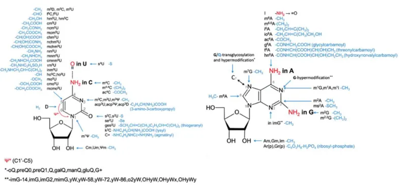
More than one hundred different post-transcriptional chemical modifications have been found in RNA molecules, either in the four nitrogen bases or in the RNA backbone (Cantara et al., 2011; Machnicka et al., 2013). These chemical alterations could be the ligation of a chemical group (small or bulky) or an isomerization, as summarized in figure 1.3., and the same nucleotide could have more than one modification at the same time. Different RNA types including mRNAs, tRNAs, rRNAs, snRNAs and miRNAs are modified and RNA modifications are present in the three domains of life (Cantara et al., 2011; Machnicka et al., 2013). Potentially these modifications could modulate the
9
function, stability and information content of the RNA molecule, however the role of these modifications are, in general, not understood (Helm and Alfonzo, 2014; Li and Mason, 2014). In some cases the function of specific modifications starts to be revealed, including that of 7-methylguanosine, pseudouridine and 5-methylcytosine.
7-methylguanosine (m7G, guanosine methylated in N7 position) is present in tRNA and rRNA
in bacteria and, additionally to these RNAs types, in eukaryotes is also present in mRNA (Cantara et al., 2011; Machnicka et al., 2013). In eukaryotes mRNA, m7G is added to the 5’ end of the primary
transcript through a triphosphate bridge forming a structure called cap (Cowling, 2010). These reactions occurs co-transcriptionally and are catalysed by capping enzymes. First, two enzymes (RNA 5’ triphosphatase and guanylyltransferase) promote the ligation of a guanosine cap, that is methylated by another enzyme, the RNMT (RNA guanine-7- methyltransferase) forming the m7G cap (Cowling,
2010). Presence of cap in 5’ end of mRNA is essential to mRNA processing, and is involved in diverse steps including transcription, polyadenilation, splicing, transport, stability and translation. For example, in splicing 5’ cap is bound by a protein complex, (cap binding complex) which interacts and recruits splicing complex components. To be translated, the majority of mRNAs requires the presence of m7G cap structure. The process occurs by the binding of eIF4F (eukaryotic initiation factor 4) to cap
structure promoting the recruitment of ribosomal components and initiator tRNA (Cowling, 2010). Pseudouridine (ψ; 5-ribosyluracil) results from isomerization of uridine. The process involves breaking the N1-C1’covalent bound between the nitrogenous base and the ribose, followed by a 180º rotation of the nitrogenous base and formation of one C5-C1’ ligation between the ribose and the nitrogenous base (Ge and Yu, 2013). This alteration generates an additional hydrogen bond donor. Isomerization reaction is catalysed in specific places by box H/ACA RNAs, with some rare cases where it is catalysed by protein pseudouridylases (Ge and Yu, 2013). Pseudouridine is found in tRNAs, rRNAs, snRNAs and mRNAs, usually located in functional regions of the molecules, for example the peptidyl transferase center (PTC), the decoding center, the A-site finger, and subunits interaction sites in rRNAs (Ge and Yu, 2013). Presence of pseudouridine increases the affinity of rRNAs and tRNAs promoting efficient translation. In mRNAs this modification affects the codon specificity and, therefore, the coding potential. The three stop codons (UAA, UAG and UGA) present in the standard genetic code, have uridine. When the uridine is modified to pseudouridine, translation does not stop, an aminoacylated tRNAs binds the modified codon and translation continues, process denominated nonsense suppression. Presence of pseudouridine in tRNAs anticodons promotes the recognition of alternate codons, and potentially the pseudouridines in codons could have a similar effect (Ge and Yu, 2013).
10
5-methylcytosine is found in bacterial rRNA and eukaryotic tRNA, rRNA and mRNA (Cantara et al., 2011; Machnicka et al., 2013). RNA m5C methyltransferases form a large protein
family composed of sub-families that include the RsmB family, RsmF/YebU family, Dnmt2 family, RlmI family, and Ynl022 family. Only some members identified in these families were empirically verified to 5-methylcytosine methyltransferase activity, like for example the E. coli RsmB and yeast Trm4 (Motorin et al., 2010). E. coli RsmB catalyse the methylation in naked 16S rRNA, but not in assembled 30S subunits. Yeast Trm4 catalyse the methylation in specific position of tRNA, namely positions 34, 40, 48 and 49. Positions 34 and 40 are only methylated in the precursors of the tRNA that carry the amino acids leucine and phenylalanine (Motorin et al., 2010). Biological function of m5C is
not completely understood, although, in tRNAs, the methylated cytosine appears in specific positions and seems to be involved in structural conformation and stability. Degradation of tRNA is apparently increased in molecules lacking some methylated positions. In rRNA and mRNAs the function of m5C
is not yet understood (Motorin et al., 2010).
Figure 1.3. Representation of the ribonucleoside modifications found in RNA, the nomenclature is according MODOMICs database. Adapted from (Machnicka et al., 2013).
11
1.2.2. N6-methyladenosine
One RNA modification is N6-methyladenosine (m6A), which differs from canonical adenosine
by the presence of a methyl group (CH3) in the nitrogen atom linked to carbon six (N6) (Figure 1.4.).
The presence of m6A on RNA molecules was detected in the 1970’s, in the polyadenylated RNA
fraction. (Desrosiers et al., 1974; Perry and Kelley, 1974) and was measured to be around 0.1 to 0.4% of total adenosines (Darnellt, 1975; Perry and Kelley, 1975). The methylated adenosine occurs mainly in the consensus sequence [G/A/U]-[G/A]-A-C-[U/A/C], (where the underlined A correspond to the m6A) in a non-stoichiometric ratio. (Carroll SM, Narayan P, Rottman FM, 1990; Csepany et al., 1990;
Narayan et al., 1994). In bacteria, this modification is present in rRNA, tRNA (Cantara et al., 2011) and adenines in DNA can also be methylated in N6 position (N6-methyladenine) (Wion and Casadesús, 2006).
1.2.3. Chemical reactions
This modified nucleoside is formed by the transfer of a methyl group from S-adenosylmethionine (AdoMet or SAM) to adenosine. This reaction is catalysed by RNA m6A
methyltransferases (discussed below) (Bokar et al., 1997; Liu et al., 2014). These enzymes are very similar to the DNA m6A methyltransferases (Bujnicki et al., 2002), therefore it is likely that the
catalytic mechanism is preserved. Based on the crystal structure of M.TaqI, (DNA m6A
methyltransferase from Thermus aquaticus) the reaction involves the interaction between the N6 of adenine to be methylated with one conserved motif (IV) in these enzymes (Goedecke et al., 2001). This interaction leads to the formation of hydrogen bounds between the hydrogens of amine group and the motif IV amino acids, promoting a hybridization change from sp2 to sp3 in the nitrogen atom. This
leaves a free electron pair, not conjugated to the aromatic system, that attacks the methyl group in SAM, resulting in a nucleophilic substitution (SN2) (Goedecke et al., 2001). The reverse reaction, N6-Fig 1.4. structures of the canonical adenosine (left) and the N6-methyladenosine (right). Adapted from (Jia et al., 2013).
12
methyladenosine to adenosine, is catalysed by RNA m6A demethylases (discussed ahead) (Jia et al.,
2011; Zheng et al., 2013a). The mechanism proposed involves the hydroxylation (ligation of one OH) to the methyl group (N6 position) forming one intermediate, N6-hydroxymethyladenosine. This intermediate can oxidize directly to adenosine (releasing a formaldehyde molecule). Besides direct oxidation of N6-hydroxymethyladenosine to adenosine, this intermediate can be oxidized to a second intermediate, N6-formyladenosine. The adenosine is produced by the oxidation of this second intermediate. These two intermediates are stable at physiological conditions and were detected in mammalian RNA, opening the possibility that they could have additional biological functions (Fu et al., 2013).
1.2.4. Enzymes and Reversibility
From HeLa cells nuclear extracts, it was possible to partially purify a multi-subunit protein complex, that catalyses the RNA adenosine methylation in vitro, which is dependent of SAM (Bokar et al., 1994). One of the subunits of the complex, which binds to SAM, was identified as MT-A70 (or METTL3) (Bokar et al., 1994). Cloning of MT-A70 gene (Bokar et al., 1997) allowed bioinformatics analysis by phylogenetic inference, with similar sequences found in databases being clustered in four subfamilies (A to D). Altogether they form the MT-A70 protein family (Bujnicki et al., 2002). Fold recognition analysis results indicates a structure with a consensus SAM - dependent MTase fold, characterized by α/β/α “sandwich” with seven central β-strands. In this consensus fold, several amino-methyltransferases characteristic motifs were identified. One of the most conserved is motif IV (N/D/S)-P-P-(F/W/Y/H) (Bujnicki et al., 2002). The MT-A70 protein family is very similar to the m6A
DNA Methyltransferase families as they share the SAM - dependent MTase fold and the conserved motifs (Malone et al., 1995).
Besides MT-A70, two other subunits of the m6A RNA methyltransferase complex were
revealed: METTL14 (methyltransferase like 14) and WTAP (Wilms’ tumor 1 (WT1)-associating protein) (Liu et al., 2014; Ping et al., 2014; Wang et al., 2014b). METTL14 is a member of the MT-A70 family, containing the characterized motifs and possesses m6A RNA methyltransferase activity
(Liu et al., 2014; Ping et al., 2014; Wang et al., 2014b). MT-A70 and METTL14 form a heterodimer in a 1:1 ratio and the catalytic activity of the dimer is higher than the subunits alone (Liu et al., 2014; Wang et al., 2014b). Knockdown of MT-A70 or METTL14 leads to a decrease in m6A levels and the
stability of each subunit depends of the presence of the other (Liu et al., 2014; Ping et al., 2014; Wang et al., 2014b). The subunit WTAP does not have any m6A RNA methyltransferase domain (Ping et al.,
2014), and it does not have catalytic activity as an independent subunit either (Liu et al., 2014). However, WTAP interacts with the MT-A70-METTL14 heterodimer and affects its catalytic activity
13
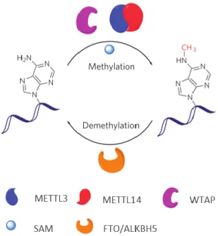
Figure 1.5. RNA m6A methylation/demethylation pathway. Methylation is SAM dependent and catalysed by METTL3-METTL14 heterodimer. WTAP is a regulatory subunit of the methyltransferase dimer. Demethylation is catalysed by FTO or ALKBH5. Adapted from (Liu et al., 2014)
in vivo (Liu et al., 2014; Ping et al., 2014). This suggests that this subunit has a regulatory role in the m6A RNA methyltransferase complex. Besides these subunits, another protein that interacts with the
methyltransferase complex and results in a decrease of methylation after knockdown is KIAA1429 (Schwartz et al., 2014).
Enzymes responsible for removing m6A from RNA have also been recently identified. The
first gene identified was the human FTO gene (which encodes for the fat and mass associated protein) (Jia et al., 2011). This protein demonstrates oxidative demethylation activity in vitro in dsDNA, ssDNA and ssRNA (Fu et al., 2013). Three dimensional crystal structure of FTO revealed that this protein is composed of two domains, a catalytic N-terminal AlkB-like domain and a C-terminal domain (now named FTO C-terminal domain). (Han et al., 2010). The observation that N6-methyladenosine in RNA is a main substrate of FTO (Jia et al., 2011) was a breakthrough, because for the first time it was demonstrated that this RNA modification is reversible, a key feature of regulatory mechanisms. Recently a second RNA m6A demethylase, ALKBH5, was identified in humans (Zheng
et al., 2013a). This protein is a member of the ALKBH family, a family of homologues of the bacterial AlkB. The demethylase catalytic activity of ALKBH5 is comparable to the FTO activity (Zheng et al., 2013a).
The known RNA methylation process is summarized in figure 1.5. The demonstration of reversibility raised the possibility of post-transcriptional gene regulation mediated by m6A and other
RNA modifications and leads to the introduction of the concepts epitranscriptome and RNA epigenetics (He, 2010; Saletore et al., 2012).
14
1.2.5. Biological functions
The relevance of this modification in biological systems can be addressed through the resulting phenotypes when the enzymes are disturbed through knockout, knockdown, mutations or overexpression. In HeLa cells, the lack of the methyltransferase MT-A70 results in apoptosis (Liu et al., 2014). Knockdown of the mouse homologue in embryonic stem cells affects the self-renewal capability (Wang et al., 2014b). In S. cerevisiae, m6A RNA methylation is only found in the sexual life
stage, and the MT-A70 homologue, Ime4, is involved in the induction of meiosis and sporulation. Mutations that result in IME4 loss of function lead to defects in sporulation (Clancy et al., 2002). Besides induction of sporulation, lineage restriction is partially dependent on the activity of this gene (Agarwala et al., 2012). A. thaliana homologue of MT-A70, MTA is highly expressed in dividing tissues and essential to seed development (Zhong et al., 2008). The homologue found in D. melanogaster, DmIme4, is essential and affects oogenesis through notch signalling (Hongay and Orr-Weaver, 2011).
The FTO gene has been associated with metabolic disorders (Wang et al., 2012) and obesity (hence the name, at fat mass and obesity-associated) (Dina et al., 2007). Overexpression of FTO demethylase leads to a food intake increase and obesity in mice (Church et al., 2010) and affect hepatic metabolism in liver cell lines (Bravard et al., 2014). Also, the activity of the dopaminergic signalling in the midbrain is regulated by FTO (Hess et al., 2013). The lack of ALKBH5 demethylase in mice affects fertility due to the occurrence of apoptosis in spermatogenesis (Zheng et al., 2013a).
Besides the interference of the methyltransferases/demethylases, the use of methylation inhibitors allows to understand the biological effects of m6A RNA methylation. Using this strategy, it
was recently demonstrated that the circadian rhythm is affected by this modification (Fustin et al., 2013). Circadian rhythm are biological activities, for example activity/rest behaviour, that follow a cycle of around twenty four hours. This rhythmic behaviour in time is due to an endogenous self-sustained molecular clock that is synchronized with environmental stimulus and the time that the cycle takes is called period (Merrow et al., 2005). The inhibition of methylation as well as MT-A70 knockdown elongates the period, while the opposite effect was observed with MT-A70 overexpression, demonstrating that RNA m6A regulates the speed of the circadian clock (Fustin et al.,
2013)
The fact that the perturbation of RNA methylation/demethylation balance affects diverse biological process in different organisms suggests that this modification has an important role in biological systems influencing a wide range of pathways.
15
1.2.6. Molecular mechanisms and targets
To understand the molecular mechanisms and functions of m6A RNA modification, two
independent groups have identified the RNA molecules harbouring the modification in humans and mice. This was achieved by immunoprecipitation of m6A containing RNAs, followed by high
throughput sequencing (Dominissini et al., 2012; Meyer et al., 2012). In one study, m6A was identified
in 5,768 and 8,843 human (from HEK293T cell line), and mouse brain transcripts, respectively (Meyer et al., 2012). In the other study, m6A was identified in 7,240 and 3,442 transcripts in humans (HepG2
cell line) and mice respectively (Dominissini et al., 2012). These two studies demonstrated that this RNA modification is widely distributed throughout the transcriptome (Dominissini et al., 2012; Meyer et al., 2012). Both studies revealed that m6A is more enriched in the 3’ UTR near the stop codon,
although peaks could also be detected within other regions of the transcripts (Dominissini et al., 2012; Meyer et al., 2012). Moreover, the frequent motifs found in these enriched regions agree with the previous biochemically determined motif (Carroll SM, Narayan P, Rottman FM, 1990; Dominissini et al., 2012; Meyer et al., 2012; Narayan et al., 1994). Besides human and mouse, the methylated transcriptome was analysed in yeast, which identified peaks in 1183 transcripts (Schwartz et al., 2013).
In addition to the identification of the targets, the detection of m6A in several mouse tissues,
and the increase in its levels from embryo to adult development suggest that this modification is widespread and dynamic (Meyer et al., 2012). Perturbation of the RNA methylation through knockdown of RNA methyltransferases (MT-A70, Mettl14), RNA demethylase (ALKBH5) or the methyltransferase regulator subunit (WTAP) followed transcriptome analysis thought RNA-Seq or microarray reveal diverse alterations in gene expression (Dominissini et al., 2012; Ping et al., 2014; Wang et al., 2014b; Zheng et al., 2013a). These observations suggests that RNA m6A methylation
could be an epigenetic modification that regulates gene expression at post-transcriptional level.
What is the molecular mechanism of action of m6A modification? This question does not have
a unique answer. Multiple studies have suggested multiples mode of action:
1. The effect of m6A in gene expression regulation could be mediated by readers of the
epigenetic code, effector proteins that recognize and bind m6A in RNA leading to the consequence in
the transcript (for example alteration in stability). So far, three m6A binding proteins were found in
humans, YTHF1, YTHF2 and YTHF3 (Dominissini et al., 2012; Wang et al., 2014a). The YFTH2 knockdown leads to an increase in the average lifetime of target transcripts and reduced translation efficiency, suggesting that this interaction destabilizes RNAs. In accordance with this observation, this protein co-localizes with markers of P-bodies (complexes involved in mRNA storage and decay) suggesting an involvement in mRNA decay (Wang et al., 2014a). Similar effect of m6A in reducing
16
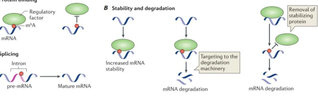
Figure 1.6. Molecular mechanisms and functions of m6A. A) to promote or block RNA–protein interactions; B) to regulate RNA stability through diverse mechanisms. C) to influence splicing efficiency. Adapted from (Meyer and Jaffrey, 2014)
effect is not due to a recruitment of a reader protein, but through blocking the binding of HuR, a protein that stabilizes RNA (Wang et al., 2014b).
2. This modification has also been proposed to be important in splicing. The first evidence is that m6A methyltransferases and demethylases co-localize with nuclear speckles (complexes involved
in pre-mRNA processing and splicing) (Jia et al., 2011; Liu et al., 2014; Zheng et al., 2013a). Second, some introns also contain the m6A modification (Dominissini et al., 2012; Meyer et al., 2012).
3. Knockdown of ALKBH5 demethylase leads to an increase in the rate of mRNA export from the nucleus to the cytoplasm (Zheng et al., 2013a). Together with the observation that methylation inhibitors lead to an increase in the nuclear retention of some mRNAs, namely the circadian RNAs (Fustin et al., 2013), it has been proposed that m6A modification may be involved in the nuclear
transport of RNAs.
4. Localization of m6A in transcripts occurs mainly in 3’ UTR, near the stop codon, therefore
localizes close to the typical binding sites of microRNAs (3’ UTR). This suggest that m6A could
interfere with the action of microRNAs (Meyer et al., 2012).
Although much remains to be studied on the exact role of m6A in different organisms, it is
clear that this RNA modification is involved in regulation of gene expression at a post-transcriptional level (Fu et al., 2014; Meyer and Jaffrey, 2014). Molecular mechanisms and functions proposed that lead the effects in transcriptome are summarized in figure 1.6.
17
1.3. Objectives
In several eukaryotes recent evidence indicates that the presence of N6-methyladenosine (m6A) in RNA is associated to processes of post-transcriptional gene regulation. Because in T. brucei,
gene expression is mainly regulated at the post-transcriptional level, it is reasonable to hypothesise that m6A RNA may be a novel mechanism of gene regulation in this parasite. In this thesis, I had four
main objectives. First, I tested if this modification was present in RNA of trypanosomes. Then I determined if the levels of m6A were regulated in different biological conditions. Third, I searched the
parasite genome to identify putative enzymes that may add or remove this modification. Finally, I used genetic tools to test the role of some of these enzymes.
19
2. Methods
2.1. Parasite culture
T. brucei brucei SMOx (Single Marker from Oxford), a modified version of the pleomorphic strain AnTat1.1E, which contains T7 polymerase and TET repressor, was cultured at 37°C in 5% CO2
in HMI-11 medium (HMI-9 medium (Hirumi, 1989) without serum plus) supplemented with puromycin at 0,1 µg/mL (Invitrogen cat: ant-pr-1). Cell density was maintained below 1 x 10⁶
cells/mL (usually cultures were passed when they reached around 0,5 x 10⁶ cells/mL, unless stated
otherwise). Knockout cell lines were cultured under the same conditions, with the additional drugs supplemented to the medium (G418 at 2,5 µg/mL (Invitrogen cat: ant-gn-5) and hygromycin at 5 µg/mL (Invitrigen cat: ant-hm-1). Procyclic culture was cultured at 27°C in 5% CO2 in DTM medium
(Vassella and Boshart, 1996). Cell density was maintained between 1 – 10 x 106 cells/mL.
2.2. Differentiation
Bloodstream culture at a density around 1-2 x10⁶ cells/mL were centrifuged at 1800 rpm, for
10 minutes at room temperature. Cells were re-suspended in DTM (Vassella and Boshart, 1996), at a cell density of 1-4 x10⁶ cells/mL with freshly prepared cis-aconitate (Sigma-Aldrich cat: A3412) at a
final concentration of 6 mM. Culture was incubated at 27°C and 5% CO2.
The efficiency of differentiation was confirmed by the expression of procyclin through flow cytometry. 0,5 million cells were centrifuged at 2800 rpm for 10 minutes at 4°C. The cells were transferred to an eppendorf tube and centrifuged again at 2800 g for 4 minutes at 4°C. Cell pellet was re-suspended in FITC conjugated anti-procyclin antibody (Cedarlane’s cat: CLP001F) diluted in HMI-11 in a 1:500 dilution and incubated 15 minutes at 4°C. After incubation, the cells were spun at 2800 g for 4 minutes at 4°C and washed three times with HMI-11. After washing, the cells were re-suspended in HMI-11 and analyzed with BD LSR Fortessa (Becton Dickinson cytometers).
20
2.3. RNA extraction
About 50 million parasites were centrifuged at 1800 rpm for 10 minutes at 4°C. Cells were transferred to eppendorf tubes and washed twice with TDB (5 mM KCl, 80 mM NaCl, 1 mM MgSO4,
20 mM Na2HPO4, 2 mM NaH2PO4, 20 mM glucose, pH 7.4), except for procyclic cells that were
washed with PBS (137 mM NaCl, 2.7 mM KCl, 10 mM Na2HPO4, 2 mM KH2PO4, pH 7,4). Cell
pellets were re-suspended in TRIzol (Life Technologies, cat: 15596) and RNA was extracted according to the manufacturer’s instructions. RNA samples were quantified with NanoDrop 2000 Spectrophotometer (Thermo Scientific). 0,5U of DNaseI (New England Biolabs, NEB, cat: M0303S) were added per µg of RNA in DNaseI buffer (NEB cat: B0303S) and incubated at 37°C for 45 minutes. After incubation, EDTA was added to a final concentration of 5 mM and heat inactivated at 75°C for 10 minutes.
2.4. DNA extraction
About 50 million parasites were spun at 1800 rpm for 10 minutes at 4°C. Cells were transferred to eppendorf tubes and washed twice with TDB, except for procyclics which were washed with PBS. Cell pellets were re-suspended in DNAzol (Life Technologies, cat: 10503-027) and DNA was extracted according to the manufacturer’s instructions. DNA samples were quantified with NanoDrop 2000 Spectrophotometer (Thermo Scientific). DNA was treated with RNase A (Carl Roth cat: 7156) (100 µg/mL) at 37°C for 2 hours.
2.5. Cloning
All pIV plasmids possess a common backbone, with an ampicillin resistance gene and an origin of replication for bacteria, which was obtained by digestion of pFAB2 (from Luisa Figueiredo Lab) with NotI-HF (NEB cat: R3189S) and KpnI-HF (NEB cat:R3142S) (CutSmart™ Buffer at 37°C, 5 Units per µg of DNA, for 4 hours). Inserts were amplified with Phusion High-Fidelity DNA Polymerase (Thermo Scientific, cat: F5305) according to the manufacturer’s instructions. Inserts that correspond to genomic recombination sites were amplified from T. brucei genomic DNA. G418 and hygromycin resistance genes were amplified from pLF13 plasmid and p2T7TA, respectively (Luisa Figueiredo Lab). Primer sequences are described in a table in annexes. Digestion and PCR amplification products were purified through gel extraction with QIAquick Gel Extraction Kit (Quiagen cat: 28706) according to the manufacturer’s instructions. After extraction, a sample of each
21
of the fragments was run in agarose gel to confirm its purity. Fragments designed for each plasmid were cloned with the In-Fusion® HD Cloning Kit (Clontec cat: 639649). Briefly, 100 ng of vector backbone was mixed with 100 ng of an equimolar mixture of inserts. The reaction was incubated at 50 °C for 15 minutes. This mixture was then diluted 1:5 with TE buffer (10 mM Tris-HCl, 1 mM EDTA, pH 8.0) and used to transform competent JM109 E. coli bacteria (Promega cat: L2001). 50 µL of E. coli were mixed with 5µL of diluted In-Fusion reaction and incubated at 42°C for 45 seconds and immediately put on ice for 2 minutes (heat shock). Then, 950 µL of SOC medium at room temperature was added and incubated at 37°C for 1 hour, under gentle agitation. Bacteria were centrifuged at 8000 rpm for 2 minutes, re-suspended in 150 µL of SOC medium, plated in LB agar supplemented with ampicillin (100µg/mL) and incubated at 37°C overnight. Colonies obtained were grown in LB liquid medium overnight and the plasmid extracted with Fast-n-Easy Plasmid Mini-Prep Kit (Jena Bioscience cat: PP-204L) according to the manufacturer’s instructions. Isolated plasmids were
digested to confirm if the cloned plasmids correspond to the desired plasmids. All enzymes used to digest the plasmids were from NEB. pIV1 was digested with EcoRI-HF (cat: R3101S) and KpnI-HF (cat: R3142S), pIV2 was digested with BamHI-HF (cat: R3136S) and SacI-HF (cat: R3138S), pIV3 was digested with BsaI-HF (cat: R3535S) and KpnI-HF (cat: R3142S), pIV4 was digested with EcoRI-HF (cat: R3101S) and NotI-HF (cat: R3189S), pIV5, was digested with BamHI-HF (cat: R3136S) and KpnI-HF (cat: R3142S) , and pIV6 was digested with EcoRI-HF (cat: R3101S) and NotI-HF (cat: R3189S). All reactions were performed in CutSmart™ Buffer at 37°C, 5 Units per µg of DNA, for 4 hours. To confirm that the constructs were correctly cloned, the plasmids were sequenced in STAB VIDA (primer table in annexes).
2.6. Transfections
pIV plasmids were digested with NotI-HF (cat: R3189S) and KpnI-HF (R3142S) (from NEB), in CutSmart™ Buffer at 37°C, 5 Units per µg of DNA, for 4 hours (with the exception of pIV1 that was digested with KpnI-HF and NdeI (cat: R01115) in CutSmart™ Buffer in the same conditions). Digestion products were purified by ethanol precipitation. 1/10 volume of sodium acetate (3M, pH 5,2) and 2,5 volumes of ice cold ethanol were added to each digestion reaction and incubated for 1 hour at -80°C. After incubation, DNA was centrifuged at 13 200 rpm for 30 minutes at 4°C. DNA pellets were washed with 70% ethanol and centrifuged at 13 200 rpm for 10 minutes. Supernatants were discarded inside the flow chamber for sterility maintenance. When pellet was dry, DNA was ressuspended in 10 µL of mili-Q water (inside the flow chamber).
Transfections were done by electroporation (Burkard et al., 2007). Briefly, 30 million cells were spun at 1800 rpm for 10 minutes at room temperature and re-suspended in 90 µL of “Roditi
22
buffer” (90mM Na-PO4, 5mM KCL, 50mM HEPES, 0.15mM CaCl2, pH 7,3). Purified DNA sample
(5-10 µg in 10 µL) was added to the cells and the mixtures were transferred to 2mm gap cuvettes (BioRad Gene Pulser/MicroPulser Cuvettes). Electroporations were performed with X-001 program in the Amaxa Nucleofector (Lonza Cologne AG, Germany) and immediately parasites were diluted in HMI-11 at 37°C and plated in three plates of 24 wells, with 10-fold dilution to each plate. Plates were incubated at 37 °C and selection drugs were added to the wells 8-16 hours later. Obtained clones were genotyped through amplification of the locus and the recombination regions, with Taq DNA Polymerase (Thermo Scientific), or Phusion High-Fidelity DNA Polymerase (Thermo Scientific) according to the manufacturer’s instructions (primers’ table in annexes).
2.7. Immunoblot
RNA samples were denatured by incubation at 50°C for 20 minutes and placed immediately on ice. DNA samples were denatured by boiling for 10 minutes and placed immediately on ice. One volume of 20X SSC (3 M NaCl; 0,3 M sodium citrate; pH 7) was added to each sample. Nucleic acid samples were spotted in a positive charged nylon membrane (Amersham Hybond™-N+ ) and fixed by UV crosslink (Stratalinker® UV Crosslinker, autocrosslink mode: 120 mJ/cm2 with 254 nm). Membranes were stained with methylene blue (0,02% in 0,3 M sodium acetate pH 5,5) and washed with milli-Q water to reduce background. Staining was removed by washing with distaining solution (0,2X SSC, 1% SDS) and washed twice with PBS/Tween 0.1% for five minutes each wash. After that, the membrane was blocked in 5% milk in PBS/Tween 0.1% for one hour. Blocked membrane was incubated with anti-m6A antibody (Millipore, rabbit polyclonal cat: ABE572) in a 1:1.000 dilution in
3% milk in PBS/Tween 0.1% overnight at 4°C. Membrane was washed three times with PBS/Tween 0.1% for 5 minutes each wash and incubated 45 minutes with secondary anti-rabbit IgG antibody (HRP-Linked, GE Healthcare) in a 1:10.000 dilution. Membrane was washed three times with PBS/Tween 0.1% for 5 minutes each wash and was developed by incubation for 4 minutes with Plus-ECL reagents (enhanced chemiluminescence, PerkinElmer). Luminescence was captured in ChemiDoc XRS System (Bio-Rad) for 30 minutes.
23
2.8. EpiQuick m
6A Quantification
Detection and quantification of m6A was done with EpiQuik™ m6A RNA Methylation
Quantification Kit (EPIGENTEK cat: P-9005) according to the manufacturer’s instructions. Briefly, nucleic acids were incubated in individual wells with binding solution at 37°C for 90 minutes, washed three times with diluted wash buffer, and incubated with the capture antibody (1:1000 dilution) for one hour at room temperature. The wells were washed three times with diluted wash buffer, and incubated with detection antibody (1:2.000 dilution) for 30 minutes at room temperature. After four washes with diluted wash buffer, wells were incubated with enhancer solution (1:5.000 dilution) for 30 minutes at room temperature. Wells were washed five times as before and developed for 5 minutes, when the reaction stopped. Absorbance was measured at 450 nm in a plate reader (Tecan, model Infinite M200).
2.9. Bioinformatics
Sequences search of putative T. brucei RNA m6A methyltransferases and demethylases was
performed with BLAST in TriTrypDB (Aslett et al., 2010). Putative T. brucei RNA m6A
methyltransferases and demethylases domains search was done in Pfam database (Finn et al., 2014). Sequences from T. brucei putative candidates were obtained from TriTrypDB and the remain sequences (human ALKBH1-8, E. coli AlkB and eukaryotic containing Pfam domain PF10237 sequences) were obtained from UniProt (Apweiler et al., 2004) . Domains from the previous described sequences were identified with Pfam and manually curated. Multiple sequence alignments were done with Clustal Omega (Sievers et al., 2011), manually edited with Jalview (Waterhouse et al., 2009) and colored with Chroma (Goodstadt and Ponting, 2001) with black and white default parameters (table of color and corresponding properties in annexes). Secondary structure was predicted with PsiPred (McGuffin et al., 2000) and Jpred (Cole et al., 2008).