Novel Clostridium thermocellum Type I Cohesin-Dockerin
Complexes Reveal a Single Binding Mode
*
□SReceived for publication, August 3, 2012, and in revised form, October 25, 2012Published, JBC Papers in Press, November 1, 2012, DOI 10.1074/jbc.M112.407700
Joana L. A. Bra´s‡1,2, Victor D. Alves‡1,3, Ana Luísa Carvalho§, Shabir Najmudin‡, Jose´ A. M. Prates‡, Luís M. A. Ferreira‡, David N. Bolam¶, Maria Joa˜o Roma˜o§, Harry J. Gilbert¶, and Carlos M. G. A. Fontes‡4
From the‡Centro de Investigaça˜o Interdisciplinar em Sanidade Animal, Faculdade de Medicina Veterina´ria, Universidade Te´cnica de Lisboa, Po´lo Universita´rio do Alto da Ajuda, Avenida da Universidade Te´cnica, 1300-477 Lisboa, Portugal, the§REQUIMTE/CQFB, Departamento de Química, FCT-UNL, 2829 –516 Caparica, Portugal, and the¶Institute for Cell and Molecular Biosciences,
Newcastle University, The Medical School, Newcastle upon Tyne NE2 4HH, United Kingdom
Background:In general, dockerins present two homologous cohesin-binding interfaces, which confer increased flexibility into cellulosomes.
Results:The structure of two novel Coh-Doc complexes reveals a dockerin single-binding mode. Conclusion:Single-binding mode dockerins bind, preferentially, to cell surface cohesins. Significance:The dual binding mode is a property of cellulosomal dockerins.
Protein-protein interactions play a pivotal role in a large number of biological processes exemplified by the assembly of the cellulosome. Integration of cellulosomal components occurs through the binding of type I cohesin modules located in a non-catalytic molecular scaffold to type I dockerin mod-ules located at the C terminus of cellulosomal enzymes. The majority of type I dockerins display internal symmetry reflected by the presence of two essentially identical cohesin-binding surfaces. Here we report the crystal structures of two novel Clostridium thermocellum type I cohesin-dockerin complexes (CohOlpC-Doc124A and CohOlpA-Doc918). The data revealed that the two dockerins, Doc918 and Doc124A, are unusual because they lack the structural symmetry required to support a dual binding mode. Thus, in both cases, cohesin recognition is dominated by residues located at posi-tions 11, 12, and 19 of one of the dockerin binding surfaces. The alternative binding mode is not possible (Doc918) or highly limited (Doc124A) because residues that assume the critical interacting positions, when dockerins are reoriented by 180°, make steric clashes with the cohesin. In common with a third dockerin (Doc258) that also presents a single binding mode, Doc124A directs the appended cellulase, Cel124A, to the surface of C. thermocellum and not to cellu-losomes because it binds preferentially to type I cohesins located at the cell envelope. Although there are a few excep-tions, such as Doc918 described here, these data suggest that there is considerable selective pressure for the evolution of a
dual binding mode in type I dockerins that direct enzymes into cellulosomes.
Biological nanomachines combining a range of complimen-tary enzyme activities are critical to cellular function. Cellulo-somes are one of nature’s most elaborate and highly efficient multienzyme complexes that deconstruct cellulose and hemicellulose, two of the most abundant polymers on Earth (1–3). Thus, cellulosomes play a major role in carbon recy-cling and provide an opportunity to explore the largely untapped energy provided by plant biomass, by the bioen-ergy and bioprocessing sectors. It is now well established that the complex physical and chemical structure of plant cell walls restrict their access to hydrolytic enzymes. Aerobic microorganisms that utilize plant biomass as a significant nutrient express extensive repertoires of degradative enzymes, primarily glycoside hydrolases but also lyases and esterases, which attack the structural polysaccharides of the plant cell wall. In contrast, microbial anaerobes, due to envi-ronmental selective pressures, have a lower protein produc-ing capacity and organize enzymes into cellulosomes, which enhance enzyme synergy and substrate targeting (see Refs. 1 and 2 for review).
The cellulosome of the thermophilic bacterium Clostridium thermocellumhas been extensively explored (4, 5). It consists of a large non-catalytic multimodular protein, termed CipA, that contains nine tandemly repeated type I cohesins that recognize type I dockerins located in the cellulosomal enzymes (6, 7). Type I cohesins of CipA display a very high level of sequence identity. It was thus suggested that there is little discrimination by the dockerins and their protein receptors presented by the cellulosome scaffold (8). Primary scaffoldins, such as CipA, may also contain a C-terminal divergent type II dockerin that spe-cifically recognizes type II cohesins located on the bacterium’s envelope, thereby providing a mechanism for the cell surface attachment of cellulosomes (9). Thus, different cohesin-dock-*This work was supported by Fundac¸a˜o para a Cieˆncia e a Tecnologia Grants
PEst-C/EQB/LA0006/2011, PTDC/BIA-PRO/69732/2006, PTDC/QUI-BIQ/ 100359/2008, and PTDC/BIA-PRO/103980/2008.
□S This article containssupplemental Table 1S and Figs. 1S–3S.
The atomic coordinates and structure factors (codes4dh2and3ul4) have been deposited in the Protein Data Bank (http://wwpdb.org/).
1Both authors contributed equally to this work.
2Supported by Fundac¸a˜o para a Cieˆncia e a Tecnologia individual fellowship
SFRH/BD/38667/2007.
3To whom correspondence may be addressed. Tel.: 213652876; Fax:
351-213652889; E-mail: [email protected].
4To whom correspondence may be addressed. Tel.: 213652876; Fax:
351-213652889; E-mail: [email protected].
by guest on March 11, 2019
http://www.jbc.org/
erin (Coh-Doc)5 specificities (in C. thermocellum type I and
type II) are responsible for the correct assembly of the multien-zyme complex (type I) and its direct attachment to the orga-nism (type II), respectively.
Structural studies on type I Coh-Doc complexes of C. ther-mocellum(10, 11) and Clostridium cellulolyticum (12), a meso-philic bacterium that produces a cellulosome analogous to the former microorganism, provided insights into the molecular determinants of protein-protein recognition that mediate the assembly of these protein complexes. Dockerins fold into two ␣-helices and EF-hand calcium-binding loop motifs, each cor-responding to one of the two duplicated segments (10, 12). Thus, the structure of the N-terminal␣-helix and EF-hand cal-cium-binding loop can be precisely superimposed over the equivalent structures at the C-terminal end, leading to an inter-nal 2-fold symmetry in the dockerin molecule (11). The impli-cations of this internal symmetry were realized when it was observed that type I dockerins present two cohesin binding sur-faces because they can bind their cognate protein module either through the analogous N- or C-terminal ␣-helices (11). In C. thermocellumtype I dockerins, residues that dominate the hydrogen bond network with cohesins are located at positions 11 and 12 of the calcium binding loop and are usually a Ser-Thr pair (10). When the dockerin is 180° reverse oriented, the equiv-alent residues (Ser45 and Thr46) in the C-terminal dockerin
helix participate in cohesin recognition (11). The Ser-Thr dyad symmetry observed in C. thermocellum dockerins is replaced, in C. cellulolyticum, by hydrophobic residues, which accounts for the lack of affinity between protein partners from different species. The dockerin dual binding mode may reduce the steric constraints that are likely to be imposed by assembling a large number of different catalytic modules into a single cellulosome. In addition, the switching of the binding mode between two conformations may also introduce quaternary flexibility into multienzyme complexes, thus enhancing substrate targeting and the synergistic interactions between some enzymes, partic-ularly exo- and endo-acting cellulases.
Currently, it is unclear whether the dual-binding mode dis-played by C. thermocellum and C. cellulolyticum dockerins is universal to all cellulosomal enzymes. The genome sequence of C. thermocellum ATCC 27405 encodes 72 polypeptides con-taining type I dockerin sequences. Alignment of the 72 dock-erin sequences at the two ligand binding sites revealed a strong conservation of the amino acids that mediate cohesin recogni-tion (particularly Ser11, Thr12, and a Lys-Arg motif at positions
18 and 19). Recently, we described the identification of four dockerins, of proteins Cthe_0435 (Cel124A), Cthe_0918, Cthe_0258, and Cthe_0624 (Cel9D-Cel44A), which deviate from the canonical C. thermocellum motifs at least in one of the cohesin binding interfaces (13). Here we describe the struc-ture of two complexes in which two different type I cohesins are bound to these unusual dockerin modules. The data indicate that a cohort of C. thermocellum type I dockerins display a sin-gle binding mode. The possible biological significance for the single binding mode displayed by these dockerins is discussed.
EXPERIMENTAL PROCEDURES
Cloning and Expression
DNA encoding type I dockerins of Cthe_0435 (Cel124A, res-idues 31–112) and Cthe_0918 (resres-idues 1146 –1209) and type I cohesins of Cthe_0452 (OlpC, residues 108 –258) and Cthe_3080 (OlpA, residues 30 –177) were amplified by PCR from C. thermocellum genomic DNA using the thermostable DNA polymerase NZYDNAChange (NZYTech Ltd.) and prim-ers described in Table 1. Genes encoding the type I dockerin modules of Cthe_0435 and Cthe_0918, here termed Doc124A and Doc918, respectively, were ligated into NdeI_BamHI-di-gested pET3a (Novagen). Genes encoding cohesin modules termed CohOlpC and CohOlpA, which derive from proteins Cthe_0452 and Cthe_3080, respectively, were ligated into NheI_XhoI-restricted pET21a (Novagen). Recombinant co-hesins contained a C-terminal His6tag. To express the
dock-erin and the cohesin genes in the same plasmid, the recombi-nant pET3a derivative was digested with BglII and BamHI, to excise the dockerin gene under the control of the T7 promoter, which was subcloned into the BglII site of recombinant pET21a so that both genes were organized in tandem. Through this approach, it was possible to express both Doc124A and Coh452 and also Doc918 and Coh3080 in the same cells. Doc124A and Doc918 were also subcloned into pET32a vector (Merck) restricted with EcoRI and XhoI. Recombinant dockerins were expressed in fusion with thioredoxin to improve dockerin sol-ubility and stability. OlpA and OlpC cohesins were also cloned into BglII- and EcoRI-digested pRSETa (Invitrogen). Mutant derivatives of both dockerins were synthesized (NZYTech Ltd.) with codon usage optimized for expression in Escherichia coli (Table 2). The synthesized genes contained engineered EcoRI and XhoI recognition sequences at the 5⬘- and 3⬘-ends, respec-tively, which were used for subsequent subcloning into pET-32a (Merck), as described above.
Protein Purification
Cohesin-Dockerin Complexes—The Coh-Doc complexes CohOlpC-Doc124A and CohOlpA-Doc918 were expressed in E. coliTuner cells, grown at 37 °C to A600of 0.5. Recombinant
protein expression was induced by adding isopropyl--D
-thio-galactopyranoside to a final concentration of 0.2 mMand
incu-bation for 16 h at 19 °C. The recombinant proteins were puri-fied by immobilized metal ion affinity chromatography using Sepharose columns charged with nickel (HisTrapTM).
Frac-tions containing the purified Coh-Doc complexes were buffer-exchanged, using PD-10 Sephadex G-25 M gel filtration
col-5The abbreviations used are: Coh, cohesin; Doc, dockerin; ITC, isothermal
titration calorimetry; r.m.s., root mean square; H-bond, hydrogen bond.
TABLE 1
Primers used to obtain the genes encoding the cohesin and the dock-erin derivatives used in the present study
Engineered restriction sites are shown in boldface type.
Clone Sequence (5ⴕ 3 3ⴕ) Direction
Doc124A CTCCATATGTGGAATAAGGCAGTTATT Forward CACGGATCCTTACGAATTGTAAGAGTTC Reverse Doc918 CTCCATATGGTTGTGCTTAATGGTGAC Forward
CACGGATCCCTATATAGTTATAAGTCC Reverse
CohOlpC CTCGCTAGCGTTGTGGCAATTCATG Forward
CACCTCGAGTTTTTCAATTTCCAC Reverse
CohOlpA CTCGCTAGCCAAACAAACACCATTGAA Forward CACCTCGAGTGCCTCCGGAGCGGATGC Reverse
by guest on March 11, 2019
http://www.jbc.org/
umns (Amersham Biosciences), into 20 mMTris-HCl buffer,
pH 8.0, containing 2 mMCaCl2. A further purification step by
anionic exchange chromatography was performed by using a column loaded with Source 30Q matrix and a gradient elution of 0 –1MNaCl (GE Healthcare). Fractions containing the
puri-fied complex were then concentrated with Amicon 10-kDa molecular mass centrifugal membranes and washed three times with 2 mMCaCl2. The final protein concentration was adjusted
to 21 g/liter in 2 mMCaCl2for CohOlpC-Doc124A complex
and was 16 g/liter for CohOlpA-Doc918 complex.
Unbound Cohesins and Dockerins—Dockerins Doc124A, Doc918, and the respective mutant derivatives cloned in pET32a were expressed in E. coli Origami cells. CohOlpC and CohOlpA cloned in pRSETa vector were expressed in E. coli Tuner cells. Growth was performed at 37 °C to midexponential phase (A600⫽ 0.5) in Luria broth. Recombinant protein
expres-sion was induced with 1 mM(Origami) or 0.2 mM(Tuner)
iso-propyl--D-thiogalactopyranoside and incubation for 16 h at
19 °C. The recombinant proteins were purified by immobilized metal ion affinity chromatography as described above and buf-fer-exchanged into 50 mMNa-Hepes buffer, pH 7.5, containing
2 mMCaCl2and then subjected to gel filtration using a HiLoad
16/60 Superdex 75 column (GE Healthcare) at a flow rate of 1 ml/min.
Isothermal Titration Calorimetry (ITC)
ITC experiments were carried out essentially as described previously (11, 12), except that the titrations were at 55 °C, and proteins were in 50 mMNa-HEPES buffer, pH 7.5, containing 2
mMCaCl2. During titration, the dockerin (40M) was stirred at
300 rpm in the reaction cell, which was injected with 28 succes-sive 10-l aliquots of ligand comprising cohesin (180 M) at
200-s intervals. Integrated heat effects, after correction for heats of dilution, were analyzed by non-linear regression using a single site-binding model (Microcal ORIGIN, version 5.0, Microcal Software). The fitted data yielded the association con-stant (Ka) and the enthalpy of binding (⌬H). Other
thermody-namic parameters were calculated by using the standard ther-modynamic equation,⌬RTlnKa⫽ ⌬G ⫽ ⌬H ⫺ T⌬S.
Crystallization and Data Collection
Protein crystals were obtained using the hanging drop, vapor diffusion method. CohOlpC-Doc124A complex crystals grew in 2 M ammonium sulfate, pH 4.6 (condition 32 of Crystal
Screen HR2-110 from Hampton Research) in drops with 7 g/li-ter protein and were harvested afg/li-ter 5–7 weeks at 19 °C. CohOlpA-Doc918 complex crystals grew in 0.2Mlithium
sul-fate, 10% (w/v) PEG 8000⫹, 10% (w/v) PEG 1000, pH 7.5 (con-dition 14 of Clear Strategy Screen I MD1-14 from Molecular Dimensions) in drops with 16 g/liter protein and were har-vested after 3–5 weeks at 19 °C. Crystals were cryocooled with paratone in liquid nitrogen prior to data collection at beamline ID14 –2 at the European Synchrotron Radiation Facility (ESRF, Grenoble, France) at 100 K using an ADSC QUANTUM 4R CCD detector and at a wavelength of 0.9330 Å.
The two data sets were integrated using MOSFLM (14) and scaled with SCALA (15) from the CCP4 suite (16). CohOlpC-Doc124A crystals belong to the P3221 space group, with cell
constants a⫽ b ⫽ 90.76 Å, c ⫽ 135.07 Å and diffracted beyond 1.75 Å resolution. Matthews (17) coefficient calculations sug-gested the presence of two molecules in the asymmetric unit (2.84 Å3/Da and 56.8% of solvent content). CohOlpA-Doc918
crystals belong to I4122 space group, with cell constants a⫽ b ⫽
130.05 Å, c⫽ 70.19 Å and diffracted beyond 1.95 Å resolution. Matthews coefficient calculations suggested the presence of one molecule in the asymmetric unit (3.0 Å3/Da and 59% of
solvent content).
Structure Determination and Refinement
Structure determination of CohOlpC-Doc124A was based on two data sets that were processed in MOSFLM (14) merged and combined with SORTMTZ from the CCP4 suite (16) and scaled in SCALA (15). Phasing was performed by molecular replacement with the program BALBES (18) using a search model based on the Protein Data Bank structures 2ccl, 1aoh, 2vn6, 1nv8, and 1ixh, mainly related to cohesin and dockerin modules from C. thermocellum and C. cellulolyticum (11, 12). Density modification, together with non-crystallographic sym-metry averaging, was done with the DM program from the
TABLE 2
Protein sequences of dockerins Doc124A and Doc918 and respective mutant derivatives
Amino acid changes are shown in boldface type.
Protein sequence (N3 C terminus)
Doc124A WNKAVIGDVNADGVVNISDYVLMKRYILRIIADFPADDDMWVGDVNGDNVINDIDCNYLKRY LLHMIREFPKNSYNSA Doc124A_m1 WNKAVIGDVNADGVVNISDYVLMAAYILRIIADFPADDDMWVGDVNGDNVINDIDCNY LKRYLLHMIREFPKNSYNSA Doc124A_m2 WNKAVIGDVNADGVVNISDYVLMAAYILRIIADFPADDDMWVGDVNGDNVINDIDC NYLAAYLLHMIREFPKNSYNSA Doc124A_m3 WNKAVIGDVNADGVVNDSDYNYMKRYILRIIADFPADDDMWVGDVNGDNV INDIDCNYLKRYLLHMIREFPKNSYNSA Doc124A_m4 WNKAVIGDVNADGVVNDSDYNYMKRYILRIIADFPADDDMWVGDVNGDNVIN IIDCVLLKRYLLHMIREFPKNSYNSA Doc124A_m5 WNKAVIGDVNADGVVNISDYVLMKRYILRIIADFPADDDMWVGDVNGDNVINDIDCNYLKR YQQHMIREFPKNSYNSA Doc124A_m6 WNKAVIGDVNADGVVNISDYVLMKRYQQRIIADFPADDDMWVGDVNGDNVINDIDCNYLK RYQQHMIREFPKNSYNSA Doc918 VVLNGDLNRNGIVNDEDYILLKNYLLRGNKLVIDLNVADVNKDGKVNSTDCLFLKKYILGLITI Doc918_m1 VVLNGDLNRNGIVNDEDYILLKNYLLRGNKLVIDLNVADVNKDGKVNDEDCLFLKNYILGLITI Doc918_m2 VVLNGDLNRNGIVNSTDYILLKKYLLRGNKLVIDLNVADVNKDGKVNDEDCLFLKNYILGLITI Doc918_m3 VVLNGDLNRNGIVNDEDYILLKNYLLRGNKLVIDLNVADVNKDGKVNQQDCLFLKKYILGLITI by guest on March 11, 2019 http://www.jbc.org/ Downloaded from
CCP4 suite (16). ARP/wARP (19) was used to automatically build the protein model. Model completion, editing, and initial validation were carried out in COOT (21). Initial restrained refinement of the molecular model was done using REFMAC version 5.5 (22), and water molecules were added/validated according to the following criteria: a compatible water-shaped peak (Fo⫺ Fc⬎⬃3, 2Fo⫺ Fc⬎⬃1.5), whose center was
within acceptable hydrogen bond distance to the closest pro-tein atoms or other waters (⬃2.4–3.2 Å), and B-factors similar to neighboring atoms but less than 80 Å2. The final cycles of
refinement were done with the program PHENIX.REFINE from the PHENIX suite (23). The two molecules in the asym-metric unit are arranged as a dimer of heterodimers, the later composed of chains A/B or C/D. All atoms in the protein could be properly assigned and refined, apart from a few initial (first 6 and 7 residues from chains A and C, respectively, and the first 6 residues from chains B and D) and final residues (last 6 and 7 residues from chains A and C, respectively, and the last 6 and 8 residues from chains B and D, respectively) in the polypeptide chains. The final model also includes 461 water molecules and four calcium ions. R-work and R-free converged to 18.1 and 21.5%, respectively. Model assessment and validation were car-ried out by PHENIX.POLYGON (24) and MOLPROBITY (25) from the PHENIX suite and PROCHECK (26). According to these programs, the final model contains 99.5% of the residues in most favored and allowed regions of the Ramachandran plot and 0.5% of the residues in generously allowed regions of the plot.
Structure determination of CohOlpA-Doc918 was similarly done by molecular replacement with BALBES using structures
with Protein Data Bank entries 2ccl and 2vn6 as models (11, 12). Density modification with non-crystallographic symmetry was done with the PARROT (27) program from the CCP4 suite. ARP/wARP was used to automatically build the protein model. Model completion, editing, and initial validation were also car-ried out in COOT. The dockerin start model had to be manually rebuilt due to a mistracing error by ARP/wARP, originating from the presence of dockerin’s two duplicated segments that share a striking sequence similarity and strong structural con-servation. Refinement procedures were done as described for CohOlpC-Doc124A. R-work and R-free converged to 17.5 and 20.6%, respectively. Two chains were found in the asymmetric unit, arranged as a dimer (A/B). Protein residues could be prop-erly assigned and refined, apart from the first 5 and last 10 residues from chain A and the first 2 residues from chain B. The final model includes 224 water molecules and two calcium ions. Model assessment and validation using the above-mentioned tools produced a final model with 100% of the residues in the most favored and allowed regions of the Ramachandran plot. Data collection and refinement details data for the two com-plete structures are summarized in Table 3.
RESULTS AND DISCUSSION
Expression and Crystallization of Novel Coh-Doc Complexes
In a previous study (13), C. thermocellum type I dockerins were shown to bind to the nine type I cohesins of CipA and the single cohesin modules of the cell surface protein OlpA or OlpC (Fig. 1). The majority of C. thermocellum dockerins (68 of 72), exemplified by the well characterized dockerin of Xyn10B (10,
TABLE 3
Data collection and refinement statistics
CohOlpC-Doc124A CohOlpA-Doc918
Space group P3221 I4122
Unit cell parameters a⫽ b, c (Å);␣, , ␥ (degrees) 90.76, 135.07; 90, 90, 120 130.05, 70.19; 90, 90, 90
Matthews parameter (Å3/Da) 2.81 3.0
Data collection statistics
X-ray source ESRF, ID14-2 ESRF, ID14-2
Wavelength (Å) 0.933 0.933
No. of unique reflections 66027 22341
Resolution limits (Å) 78.60–1.75 (1.80–1.75)a 91.96–1.95 (2.05–1.95) Completeness (%) 99.9 (99.6) 100 (100) Redundancy 14.9 (8.8) 14.4 (13.8) Average I/(I) 18.7 (2.4) 23.3 (1.8) Rsym(% ) 0.090 (0.887) 0.080 (0.420) Refinement statistics Resolution limits (Å) 39.30–1.80 32.79–1.95 R-work 0.181 0.175 R-free 0.215 0.206
No. of protein residues in the asymmetric unit 456 205
No. of water molecules in the asymmetric unit 461 178
No. of atoms in the asymmetric unit 7844 1824
r.m.s. deviation bond length (Å) 0.014 0.014
r.m.s. deviation bond angles (degrees) 1.296 1.314
Average temperature factor (Å2)
Protein main chain 26.4 28.2
Protein side chain 34.5 33.8
Water molecules 41.9 37.5
Sulfates 65.5 50.6
Calcium 24.5 20.0
Ramachandran plot
Residues in most favored regions (%) 91.1 89.4
Residues in additionally allowed regions (%) 8.4 10.6
Residues in generously allowed regions (%) 0.5 0
Protein Data Bank code 4dh2 3ul4
a
Values in parentheses are for the highest resolution shell.
by guest on March 11, 2019
http://www.jbc.org/
11), display a distinctive internal symmetry that is compatible with a dual binding mode. These dockerins display preferential recognition for OlpA and CipA cohesins. In contrast, two C. thermocellumdockerins, from the protein of unknown func-tion, Cthe_0258, and the recently described cellulase, Cel124A (28), display a 2- and 10-fold preferential binding, respectively, to the cell envelope cohesin of OlpC (13). A third dockerin, of the bifunctional cellulase Cel9D-Cel44A, displays two cohesin-binding interfaces with different specificities; the dockerin can interact with C. cellulolyticum cohesins through the N-termi-nal interface and with C. thermocellum counterparts through the C-terminal binding site (13). A fourth dockerin, from the protein of unknown function Cthe_0918, binds equally well to CipA and OlpA and displays a lower affinity for OlpC. The primary sequences of these four dockerins lack the distinctive symmetry at the binding interfaces, which may explain, at least for dockerins of Cthe_0258, Cel124A and Cel9D-Cel44A, the observed differences in ligand specificity (13). The dockerin dual binding mode does not favor crystallization of protein complexes, and the usual approach used to study the Coh-Doc interaction involves the inactivation of one of the dockerin
cohesin-binding interfaces through site-directed mutagenesis (11, 12). Because the four unusual dockerins described above seem to present a single binding interface, wild-type proteins were used for these structural studies. Here, we have used established strategies for the production and purification of Coh-Doc complexes, which involve the co-expression of both proteins in E. coli cells (29).
Structure of Type I Coh-Doc Complexes
The structures of OlpA type I cohesin bound to the dockerin of the protein Cthe_0918 (CohOlpA-Doc918) and of the OlpC type I cohesin in complex with the dockerin of Cel124A (CohOlpC-Doc124A) were solved to 1.95 and 1.75 Å resolu-tion, respectively (Fig. 2). In C. thermocellum, OlpA and OlpC cohesins are the only two type I cohesins that do not belong to CipA and show significant deviations in the putative residues that participate in dockerin recognition, when compared with the nine highly homologous cohesins of CipA (13) (for details, seesupplemental Fig. 1S).
Structure of OlpA and OlpC Type I Cohesins—The 146-resi-due OlpA cohesin in complex with its cognate dockerin dis-plays an elongated nine-stranded flattened-sandwich struc-ture, defined by two-sheets (A and B) in a classical jelly roll topology (supplemental Fig. 2S) (10).-Sheet A is composed of the-strands (and respective residues) 4 (residues 51–58), 7 (residues 99 –108), 2 (residues 22–30), 1 (residues 9 –17), and 9 (residues 138 –146), whereas-sheet B includes -strands 5 (residues 69 –74), 6 (residues 79 – 85), 3 (residues 37– 45), and 8 (residues 115–129).-Strand 8 is the longest and partly inte-grates both-sheets. -Sheet B forms a distinctive planar pla-teau amid a molecule with a flattened and overall curved cylin-drical shape (supplemental Fig. 2SC). The OlpC cohesin module with 159 amino acid residues exhibits the same type I cohesin architecture, where-sheet A is composed of the same -strands (and respective residues), 4 (residues 60–67), 7 (res-idues 108 –117), 2 (res(res-idues 31–39), 1 (res(res-idues 15–27), and 9 (residues 149 –161), whereas -sheet B includes -strands 5 (residues 78 – 83), 6 (residues 88 –94), 3 (residues 48 –54), and 8 (residues 129 –142), the latter also contributing to both
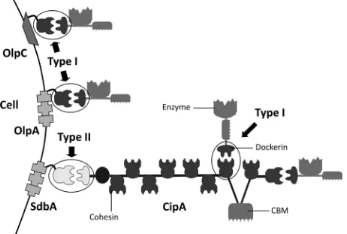
FIGURE 1. C. thermocellum cellulosome. C. thermocellum scaffoldin (CipA) contains nine type I cohesins and thus organizes a multienzyme complex that incorporates nine enzymes. The C-terminal type II dockerin of CipA binds specifically to type II cohesin modules found in cell surface proteins. Individ-ual enzymes may also adhere directly to the bacterium cell envelope by bind-ing the sbind-ingle type I cohesins found in OlpA and OlpC.
FIGURE 2. Structure of the novel type I Coh-Doc complexes, CohOlpA-Doc918 and CohOlpC-Doc124A. The dockerin structure is rainbow-colored (blue, N terminus; red, C terminus). Calcium ions are modeled as magenta spheres. The cohesin surface is depicted as dots, whereas the dockerin surface is solid white with the main contact surface area highlighted in red. CohOlpA-Doc918 shows a C terminus (helix-3)-dominated Coh-Doc interface, whereas CohOlpC-Doc124A reveals an N terminus (helix-1)-dominated Coh-Doc interface.
by guest on March 11, 2019
http://www.jbc.org/
-sheets. Both cohesins contain a highly hydrophobic core (supplemental Fig. 2SB).
Both type I cohesin structures in complex with their respec-tive protein partners reveal a striking similarity (supplemental Fig. 2SA). In comparison with the second cohesin of CipA (CohCipA2), a structure superposition with CohOlpA has an r.m.s. deviation of 1.04 Å (between 136 C␣pairs with a sequence identity of 35.3%) and 1.18 Å with CohOlpC (133 C␣ pairs, 38.3% sequence identity). The two novel cohesins superpose with each other with an r.m.s. deviation of 1.14 Å (139 C␣pairs, 34.5% sequence identity). Noteworthy structural divergences occur between-strands 4 and 5 (which include a small ␣-he-lix), where both CohOlpA and CohOlpC have a shorter loop than CohCipA2, and on the loop between -strands 7 and 8 that, compared with CohCipA2, is slightly longer in CohOlpA and considerably larger in CohOlpC, increasing the main lon-gitudinal axis length of these two proteins by around 2 and 10 Å, respectively (supplemental Fig. 2SA). The-sheet B interface area, evaluated on the basis of its solvent-accessible area when in complex with its cognate dockerin (PDBePISA) (20), was 686 Å3 for CohCipA2, 803 Å3 for CohOlpA, and 729 Å3 for
CohOlpC.
Structure of Type I Dockerins—The structures of dockerins of Cthe_0918 and Cel124A, here termed Doc918 and Doc124A, respectively, are organized in two␣-helices, arranged in an
antiparallel orientation (N-terminal or helix-1 and C-terminal or helix-3) connected through an extended loop displaying a small helix (helix-2) (Fig. 3). In Doc918, helix-1 is composed of residues 15–27, helix-2 extends between residues 35 and 39, and helix-3 extends from residue 48 to 60. In Doc124A, the respective residues are as follows: helix-1 (residues 17–29), helix-2 (residues 39 – 45), and helix-3 (residues 53– 65). Helix-2 connects the other two helices, both of which provide the two putative cohesin binding interfaces. In fact, this region, limited by the distal end of helix-1 and the C terminus of helix-2, con-tains a large amount of the structural variability found among the core C␣ trace of these dockerins. In Doc918, the region connecting the two helices is less structured than in DocXyn10B, presenting a single turn on its␣-helix, similar to a type I C. cellulolyticum dockerin (12). The internal sequence duplication and nearly perfect 2-fold symmetry was quantified by an internal superposition between helix-1 and -3 within each structure. Doc918 shows an r.m.s. deviation of 0.57 Å for 23 C␣ pairs, and in Doc124A, both segments overlap almost as well, with an r.m.s. deviation of 0.66 Å for 26 C␣pairs. Lack of con-servation in the key contacting residues when the two putative binding surfaces are compared should prevent a dual binding mode, which is explored below. Both dockerins contain two Ca2⫹ions coordinated by several residues in the canonical
EF-hand calcium-binding loop. The coordination of the two
cal-FIGURE 3. Structure superposition and important contact residues. A, dockerin superposition between DocXyn10B (blue), Doc918 (yellow), and a 180°-rotated Doc124A (salmon). Residues with a significant contribution to the Coh-Doc contact surface area are shown in stick representations and numbered according to the Protein Data Bank entries 1ohz, 3ul4, and 4dh2. B, cohesin superposition between CohCipA2 (blue), CohOlpA (yellow), and a CohOlpC (salmon). Residues important to the Coh-Doc contact interface are shown above the-sheet B plane, in stick representations and numbered according to the respective Protein Data Bank entries. C, dockerin sequence alignment and interacting residues. Residues with a significant contribution to the Coh-Doc contact surface area are marked with a top variable width small arrow. Residues involved in hydrophobic interactions are shown with a gray background, whereas a box highlights residues with polar interactions. DocXyn10B_180 denotes a 180° binding interface rotation.
by guest on March 11, 2019
http://www.jbc.org/
cium ions is similar to the metal ions observed in the type I dockerins of C. thermocellum and C. cellulolyticum in complex with their cognate protein partners (supplemental Fig. 3SA).
Novel Type I Coh-Doc Complex Interfaces
In contrast with what was previously observed for other type I complexes, the dockerins described here in complex with their protein partners seem to present a single binding mode. Thus, in the CohOlpA-Doc918 complex, binding is dominated by the Doc918 C-terminal helix. In contrast, in the CohOlpC-Doc124A complex, binding is orchestrated by the dockerin N-terminal helix (Fig. 2). In these two novel Coh-Doc struc-tures, the complex interface has a significant hydrophobic nature. Using the solvation free energy gain at complexation, calculated by PDBePISA (⌬iGin kcal/mol (20)), the
CohOlpA-Doc918 interaction is more hydrophobic (⫺10.6 kcal/mol) than that of CohOlpC-Doc124A (⫺7.7 kcal/mol), which in turn exceeds the CohCipA-DocXyn10B value of ⫺6.4 kcal/mol. However, the negative values upon binding are less significant than those of the highly hydrophobic C. cellulolyticum type I complex (Protein Data Bank code 2vn6) with⫺14.9 kcal/mol. These differences reflect the numerous hydrophobic residues, involved in the Coh-Doc complex interface, enumerated in detail insupplemental Table 1Sand highlighted in supplemen-tal Figs. 1S and 3S. Thus, the numbers of cohesin and dockerin hydrophobic residues implicated in the interface of the CohOlpA-Doc918 are greater than in the CohCipA2-DocXyn10B complex. Although the hydrophobic contact net-work of CohOlpC-Doc124A is also extensive, the hydrophobic residues that contribute to the heterodimer interface are con-tributed primarily by Doc124A.
The major hydrophobic contact residues located at the sur-face of cohesins CipA2, OlpA, and OlpC include a completely conserved leucine (Leu83, Leu83, and Leu92, respectively), which
is assisted by upstream hydrophobic residues Val81, Ala81, and Val90and downstream by Ala85, Leu85, and a divergent Asp94in
OlpC, respectively. Other important contributors correspond to Leu129, Met132, and Leu146, respectively. With respect to the
dockerins Xyn10B-␣3/Xyn10B-␣1, 918, and 124A, the major hydrophobic contact residues are Leu22/Leu56, Leu27, and
Leu65, respectively, at position 22 of the less interacting binding
interface. In addition, in position 15 of the dominating inter-face, residues Leu49/Thr15, Leu53, and Val22make a significant contribution to cohesin recognition. The above mentioned conserved leucine located at the surface of the three cohesins is part of an important hydrophobic pocket formed in CohCipA2 by Ala72, Tyr74, Val81, and Leu83, which is occupied by Leu22or
Leu56 from DocXyn10B in the two possible binding modes,
respectively. Using the same relative structural positioning order, for CohOlpA, we find Asn72, Ala81, and Leu83, which
accommodate Leu27from Doc918. As for CohOlpC, residues Asn81, Val90, and Leu92form a hydrophobic pocket that is
occu-pied by the equivalent Doc124A residue, Leu65, found in the
opposite C-terminal interface.
The heterodimer interfaces are assisted by a network of direct and bridged hydrogen bonds and salt bridge interac-tions (described in detail in Table 4). Compared with DocXyn10B(␣3) in a similar C-terminal binding conformation
(10), Doc918 reveals a more imbalanced distribution of polar bonds, favoring helix-3 residues. Although the Ser/Thr dyads of both complexes share an equivalent contribution, the main dif-ference occurs at the Lys56/Lys57pair of Doc918 that contribute with one salt bridge and two direct H-bonds, whereas in DocXyn10B(␣3), the equivalent Ser52makes no polar bonds,
and Arg53establishes a single salt bridge. Again, in comparison
with the N-terminal bound DocXyn10B(␣1), Doc124A reveals some striking differences with respect to the relevant Ser-Thr pair, which is replaced by a divergent Ile18-Ser19 motif. In
Doc124A the N-terminal binding face interacts with CohOlpC, through significant hydrophobic contacts. The only direct polar interactions mediated by helix-1 occur via positions 18 and 19 (Lys25-Arg26), through six direct bonds (two salt bridges
from Lys25and four H-bonds from Arg26) and a couple of water
bridged H-bonds involving Ile18. In contrast, the N-terminal
bound configuration of DocXyn10B reveals a hydrogen bond network around, and dominated by, the conserved Ser-Thr pair and also some involvement of residues 18 and 19. Also in con-trast to DocXyn10B(␣1), Doc124A presents in the opposite interface (␣3) a stronger polar contribution participated in by four residues, Lys61(one salt bridge), Leu64(one H-bond), Leu65
(one H-bond), and His66 (one salt bridge), whereas in
DocXyn10B, Leu56and mainly Arg57make polar contacts with
the CipA cohesin (Fig. 3, A and C; detailed contacts in supple-mental Figs. 1S and 3S). Extending the comparison of Doc124A to C. cellulolyticum type I complex (2vn5/2vn6) in an analogous
TABLE 4
Network of polar interactions in novel type I Coh-Doc complex interfaces
by guest on March 11, 2019
http://www.jbc.org/
binding conformation, the major difference consists of a much subdued polar interaction network found in the latter, espe-cially at the␣3 interface, where only positions 22 and 23 reveal direct contacts (12).
The cohesin-interacting residues can be grouped into three regions corresponding to-strands 3, -strands 5/6, and the loop between-strands 8 and 9 (Fig. 3B; detailed con-tacts in supplemental Fig. 1S). Around the 3 region, the important interactions are quite similar among CohCipA2/ CohOlpA, because equivalent residues Asn37/Ser39and Asp39/
Asp41, respectively, establish relevant polar contacts with the
dockerin Ser/Thr pair. Conversely, the equivalent CohOlpC residue Ser48does not display any polar contacts, and Asn50,
equivalent to CohCipA2 Asp39, establishes a single H-bond
with the dockerin. In the5/6 cohesin region, notable differ-ences between CohCipA2, CohOlpA, and CohOlpC occur, respectively, at Arg77, Asp77, and Asp86residues; Arg77makes
an H-bond with its target dockerin, whereas the equivalent acidic residues of the other two cohesins are not implicated on the interface. In the8-loop-9 region, the corresponding res-idues Asn127, Asn130, and Phe144 in CohCipA2, OlpA, and
OlpC, respectively, reveal some differences in their capacity to recognize the dockerin protein partner. In a helix-3-dominated binding, CohCipA2-Asn127does not exhibit any contacts with
its dockerin, whereas CohOlpA-Asn130 makes two bridged
H-bonds. However, in a helix-1-dominated binding, CohCipA2 uses its Asn127to make two H-bonds with Doc-Arg19, whereas
in CohOlpC, the backbone of Phe144establishes two H-bonds
with Doc-Arg26. In addition, in the Glu131/Glu134/Pro148
posi-tion of the cohesins, both acidic residues from CohCipA2 and
CohOlpA form an H-bond with the critical threonine found at position 12 of the dockerin, whereas CohOlpC-Pro148does not
contribute to dockerin recognition.
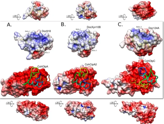
Further analysis of the differences between the canonical type I cohesin and this work on cell-bound cellulosomal cohesins was based on the predicted negative hydrogen bond-accepting regions in an electrostatic surface potential evalua-tion using the Poisson-Boltzmann electrostatics calculaevalua-tion on the PDB2PQR server (30) and visualization of the results in UCSF Chimera (31) (Fig. 4). As reported previously (32), cohesins are strikingly negatively charged in the binding inter-face plateau, whereas dockerins present a suitable complemen-tary positive-to-neutral surface. Compared with CohOlpC and CohCipA2, CohOlpA shows an elongated polar region that extends beyond the binding interface. As described for the type II cohesin of SdbA (32), the opposite cohesin surfaces in CohOlpC and CohOlpA are more positively/neutrally charged, which was suggested to be important to promote a tighter inter-action of cell surface cohesins to the negatively charged pepti-doglycan layer.
Analysis of the type I Coh-Doc interfaces provides significant insights into the previously described tight binding of Doc124A to CohOlpC, in comparison with the lower affinity displayed by this dockerin toward the cohesins of CipA and OlpA (13). The hydrophobic nature of Ile18 at the critical position 11 of the
Doc124A interface, establishes a strong network of apolar con-tacts with CohOlpC, namely with Asn50, Asn140, and Cys142.
These CohOlpC pivotal residues are replaced by an aspartate (position 50) and by small residues, namely glycine or alanine, at the other two positions in CohCipA and CohOlpA cohesins.
FIGURE 4. Electrostatic surface potential for the Coh-Doc molecules. In each panel, the central image shows the top cohesin binding plateau with a licorice model of the bound dockerin in N to C terminus rainbow color ramped style. Above, there is a view of the molecular dockerin binding surface. The top and bottom images are smaller scaled and orthogonal representations, respectively, of the dockerin and cohesin. A, CohOlpA and Doc918. B, CohCipA2 and DocXyn10B. C, CohOlpC and Doc124A. The electrostatic potential is contoured in UCSF Chimera from⫺6 (red) to ⫹6 (blue) (arbitrary Chimera units).
by guest on March 11, 2019
http://www.jbc.org/
The aspartate residue, equivalent to CohOlpC-Asn50, found in
both OlpA and CipA cohesins is also highly relevant for the recognition of typical type I dockerins because, together with Asn37, it establishes conserved hydrogen bonds with the canon-ical serine residues usually found at position 11. In CohOlpC, the latter residues are replaced with Ser48and Asn50,
respec-tively, whose side chains were found more than 4 Å apart and thus presumably unavailable for H-bond formation. In addition, dock-erin position 12 of the binding interface makes some relevant polar contacts with the mentioned residue of CohCipA2-Asn37
and also Glu131. Again, in CohOlpC, the equivalent Ser48side
chain orientation is unsuitable for those contacts, and the Pro148, which substitutes Glu131, is manifestly non-reactive. Probing the Importance of Contact Residues in Dockerins
To identify the dockerin residues that are involved in cohesin recognition, a mutagenesis study informed by the previously
described type I complex structures was implemented. Previ-ous data suggest that the implications of single changes in dock-erin activity may be relatively modest, so the strategy used here involved the change of particular groups of residues that are believed to play a cooperative role in cohesin recognition (10 –13).
CohOlpA-Doc918 Complex—Site-directed mutagenesis and ITC data of the CohOlpA-Doc918 complex (Fig. 5 and Table 5), show that the replication of the relevant residue environment from dockerin helix-1 into the C-terminal helix-3, which dom-inates ligand recognition (mutant Doc918_m1: S49D, T50E, and K57N), precluded any binding, which reinforces a vital role for the Ser-Thr motif, similar to the canonical type I Coh-Doc interaction (10). The drastic decrease in affinity obtained with mutant Doc918_m3 (S49Q and T50Q) also supports a major role for the C-terminal dockerin Ser-Thr motif. The K57N
FIGURE 5. Alignment of Doc918 sequence and its mutant derivatives (A) and representative ITC experiments using the proteins generated in this
study (B). In A, mutated residues are highlighted in gray. In B, the upper parts of each panel show the raw heats of binding, whereas the lower parts show the
integrated heats after correction for heat dilution. The curve represents the best fit to a single-site binding model. TABLE 5
Thermodynamics of the binding between C. thermocellum dockerins and their mutant derivatives and type I cohesins of OlpC and OlpA
Thermodynamic parameters were determined at 328.15 K. ND, values too low to be determined.
Cohesin Dockerin Ka ⌬G ⌬H T⌬S
M⫺1 kcal mol⫺1 kcal mol⫺1 kcal mol⫺1
OlpC Doc124A 3.07E7⫾ 3.57E6 ⫺11.24 ⫾ 0.18 ⫺40.70 ⫾ 0.18 ⫺29.46
Doc124A_m1 6.91E4⫾ 1.10E4 ⫺7.16 ⫾ 0.47 ⫺9.08 ⫾ 0.47 ⫺1.92
Doc124A_m2 ND ND ND ND
Doc124A_m3 7.40E6⫾ 3.43E5 ⫺10.44 ⫾ 0.09 ⫺28.29 ⫾ 0.09 ⫺17.85
Doc124A_m4 6.12E7⫾ 7.20E6 ⫺11.84 ⫾ 0.04 ⫺28.90 ⫾ 0.04 ⫺17.06
Doc124A_m5 1.68E6⫾ 1.50E5 ⫺9.34 ⫾ 0.34 ⫺32.38 ⫾ 0.34 ⫺23.04
Doc124A_m6 1.50E5⫾ 1.39E4 ⫺7.77 ⫾ 2.80 ⫺39.86 ⫾ 2.80 ⫺32.09
OlpA Doc918 8.77E7⫾ 1.31E7 ⫺11.80 ⫾ 0.22 ⫺47.89 ⫾ 0.22 ⫺36.09
Doc918_m1 ND ND ND ND
Doc918_m2 6.07E6⫾ 5.42E5 ⫺10.18 ⫾ 0.22 ⫺30.49 ⫾ 0.22 ⫺20.31
Doc918_m3 2.47E6⫾ 3.52E5 ⫺9.62 ⫾ 0.39 ⫺27.24 ⫾ 0.39 ⫺17.62
by guest on March 11, 2019
http://www.jbc.org/
mutation also emphasizes the relative importance of a basic residue, such as lysine or arginine (equivalent to Arg53 in
DocXyn10B), in this position for efficient binding.
The Doc918_m2 mutant design provided additional insights into the pivotal residues mediating cohesin recognition. Essen-tially, using the inactive dockerin, Doc918_m1, an attempt was made to force the alternate helix-1 binding mode. Thus, the non-functional N-terminal helix of Doc918_m1 was engi-neered to restore an N-terminal cohesin-binding interface by introducing the three pivotal residues (mutant Doc918_m2: D16S, E17T, and N24K) identified in helix-3 in the correspond-ing positions in helix-1. ITC data showed that, although with a 10-fold reduction in affinity, this strategy indeed allowed bind-ing through helix-1. In CohOlpC-Doc124A and CohCipA2-DocXyn10B (i.e. the S45A/T46A mutant of CohCipA2-DocXyn10B) (11), where helix-1 dominates the binding interface, there is a bulky positively charged residue in helix-3 (His66and Arg57,
respec-tively), which provides polar and hydrophobic interactions to the interface but which was replaced by a Gly61in Doc918 when
binding was engineered at the N-terminal face (Fig. 3). This divergent substitution could thus contribute to the reduced affinity displayed by the Doc918_m2 mutant. Overall, the data presented here confirm that Doc918 presents a single protein-binding interface that is dominated by the C-terminal helix and where Ser49, Thr50, and Lys57dominate cohesin recognition.
CohOlpC-Doc124A Complex—As described above, the Doc124A Lys25-Arg26pair dominates the polar binding
net-work with OlpC cohesin, whereas Lys61makes an important
salt bridge with Asp79present at the surface of the cohesin.
Thus, Doc124A mutants m1 and m2 were used to explore the importance of helix-1 Lys25-Arg26 and helix-3 Lys61-Arg62
pairs, by mutating them separately (m1) or simultaneously (m2) to Ala (Fig. 6 and Table 5). As expected, based on these multiple polar contacts, the lesion in helix-1 (m1) caused an⬃400-fold decrease in affinity. In addition, the additive effect of mutating the two basic pairs at helix-1 and helix-3 simultaneously (m2) led to complete loss in cohesin recognition, confirming the importance of Lys61in heterodimer formation. Thus, the basic
pair at helix-1 plays a key role in cohesin recognition, and the massive reduction in affinity suggests a single binding mode for Doc124A. However, because the helix-3 Lys61-Arg62pair is in a position symmetry-related to that of Lys25-Arg26in helix-1, it is
also possible that, following a 180° rotation of the dockerin, these latter residues could participate in a lower affinity cohesin recognition mediated by helix-3. Under these circumstances, the lower affinity of m1 would result from substitution of the critical Ile by an Asp at position 11 and by the loss of a putative Lys25-mediated salt bridge at the other helix.
Data presented above suggest that Doc124A could eventually present two cohesin binding interfaces expressing different affinities. To explore this possibility, Doc124A Ile18, Val22, and
Leu23, which are part of the hydrophobic platform of the helix-1
binding interface, were mutated to replicate their symmetry-related counterparts in helix-3 (m3) (Fig. 6 and Table 5). The data revealed that these mutations lead to a reduction in the capacity of Doc124A to bind its cohesin partner. The
FIGURE 6. Alignment of Doc124A primary sequence and its mutant derivatives (A) and examples of ITC experiments using the proteins generated in
this study (B). In A, mutated residues are highlighted in gray. In B, the upper parts of each panel show the raw heats of binding, whereas the lower parts are the
integrated heats after correction for heat dilution. The curve represents the best fit to a single-site binding model.
by guest on March 11, 2019
http://www.jbc.org/
Doc124A_m4 mutant introduces into the m3 background, in which helix-1 binding is reduced, the mutations D54I, N58V, and Y59L, with the intention of promoting a reversal in binding through the C-terminal helix (Fig. 6 and Table 5). ITC results show an 8-fold increase in affinity over the m3 mutant, similar to the wild type dockerin, suggesting that although a dual bind-ing mode is not feasible in the native form of Doc124A, in the m4 mutant, binding is probably dominated by the C-terminal interface. Thus, overall, the data suggest that Doc124A presents a single binding mode driven by helix-1.
The importance of the hydrophobic network established between Doc124A and OlpC was further explored in the mutant m5, which investigated the role of a second residue pair, Leu64-Leu65, in the interactions established with the cohesin
(Fig. 6 and Table 5). As described above, the Doc124A dockerin presents a symmetry-related pair at helix-1, Ile28-Leu29, which
could be involved in a similar interaction if binding was medi-ated by the helix-3 lower affinity interface. The importance of this pair was explored in m6. The knock-out of the Leu64-Leu65 helix-3 pair (m5) induced a 10-fold decrease in affinity, con-firming the relevance of these residues in binding the cohesin when helix-1 is the dominant binding face. Indeed, it is reason-able to assume that the loss of Leu64and Leu65in m5
reorien-tates the major binding face to helix-3. Consistent with this view is the further reduction in affinity by the concurrent muta-tion of the proximal helix-1 residue pair (m6).
CONCLUSIONS
The structure of two type I Coh-Doc complexes presented here revealed that unlike the large majority of C. thermocellum dockerins, the dockerins of cellulase Cel124A and of Cthe_0918 protein, presently of unknown function, display a single cohe-sin-binding surface. The structures of the two dockerins were solved in complex with the two unique cell surface type I cohesins of C. thermocellum, OlpA and OlpC, which direct plant cell wall hydrolytic enzymes directly to the cell surface. A recent study (33) revealed that cellulosomes act in synergy with enzymes located at the bacterium cell envelope, which include the abundant Cel124A endocellulase that targets cellulose crys-talline-amorphous junctions. The fact that high quality crystals for both complexes were obtained using wild type dockerins was an initial good indication that these dockerins present essentially a single interacting surface. The structures of the two complexes revealed that the critical positions 11 and 12 of the dockerin non-interacting interface are occupied predomi-nantly with acidic residues (Glu and Asp). Acid residues are not suitable for interacting with the highly negatively charged cohe-sin platform. Site-directed mutagenesis data demonstrate the importance of the Ser/Ile-Thr motif at positions 11 and 12 and the Lys/Lys-Arg pair at positions 18 and 19 in cohesin recogni-tion. Inspection of the primary sequences of dockerins of Cthe_0258, which recognizes OlpC with higher affinity, and cellulase Cel9D-Cel44A, which binds both C. thermocellum and C. cellulolyticum cohesins, also revealed unsuitable substi-tutions at one of the dockerin binding faces, which should result in only one binding face capable of recognizing C. thermocel-lumcohesins. It is presently unclear why a subset of four dock-erins, the two described here and those from Cthe_0258 and
Cel9D-Cel44A, have not evolved the dual binding mode char-acteristic of the other 68 C. thermocellum cellulosomal enzymes and extensively described for Xyn10B dockerin. Whereas the Cel124A dockerin directs the appended enzyme to the cell surface, because it binds predominantly to the OlpC cohesin, the dockerin of Cel9D-Cel44A is believed to present two cohesin binding interfaces with different cohesin specific-ities (the N-terminal face binds C. cellulolyticum-like cohesins, and the C-terminal interface binds their C. thermocellum coun-terparts). Thus, together, these data suggest that a dual binding mode is of primary importance for enzymes binding CipA, the multimodular cohesin scaffolding responsible for cellulosome assembly in C. thermocellum. An exception to this general rule is the dockerin of Cthe_0918, which recognizes CipA cohesins with higher affinity. The elucidation of the functional role of the protein domain appended to Cthe_0918 dockerin would help to clarify this issue. Nevertheless, the presence of two cohesin binding interfaces in dockerins integrated in multienzyme complexes may contribute to the capacity of the cellulosome to adjust its catalytic machinery to a highly insoluble and recalci-trant substrate.
Acknowledgment—We acknowledge the European Synchrotron Radi-ation Facility (Grenoble, France) (beamline ID14-2) for access and technical support during data collection.
REFERENCES
1. Fontes, C. M., and Gilbert, H. J. (2010) Cellulosomes. Highly efficient nanomachines designed to deconstruct plant cell wall complex carbohy-drates. Annu. Rev. Biochem. 79, 655– 681
2. Bayer, E. A., Belaich, J. P., Shoham, Y., and Lamed, R. (2004) The cellulo-somes. Multienzyme machines for degradation of plant cell wall polysac-charides. Annu. Rev. Microbiol. 58, 521–554
3. Bayer, E. A., Lamed, R., White, B. A., and Flint, H. J. (2008) From cellulo-somes to cellulosomics. Chem. Rec. 8, 364 –377
4. Bayer, E. A., and Lamed, R. (1986) Ultrastructure of the cell surface cellu-losome of Clostridium thermocellum and its interaction with cellulose. J. Bacteriol. 167,828 – 836
5. Be´guin, P., and Alzari, P. M. (1998) The cellulosome of Clostridium ther-mocellum. Biochem. Soc. Trans. 26, 178 –185
6. Salamitou, S., Raynaud, O., Lemaire, M., Coughlan, M., Be´guin, P., and Aubert, J. P. (1994) Recognition specificity of the duplicated segments present in Clostridium thermocellum endoglucanase CelD and in the cel-lulosome-integrating protein CipA. J. Bacteriol. 176, 2822–2827 7. Tokatlidis, K., Salamitou, S., Be´guin, P., Dhurjati, P., and Aubert, J. P.
(1991) Interaction of the duplicated segment carried by Clostridium ther-mocellum cellulases with cellulosome components. FEBS Lett. 291, 185–188
8. Salamitou, S., Tokatlidis, K., Be´guin, P., and Aubert, J. P. (1992) Involve-ment of separate domains of the cellulosomal protein S1 of Clostridium thermocellumin binding to cellulose and in anchoring of catalytic sub-units to the cellulosome. FEBS Lett. 304, 89 –92
9. Leibovitz, E., and Be´guin, P. (1996) A new type of cohesin domain that specifically binds the dockerin domain of the Clostridium thermocellum cellulosome-integrating protein CipA. J. Bacteriol. 178, 3077–3084 10. Carvalho, A. L., Dias, F. M., Prates, J. A., Nagy, T., Gilbert, H. J., Davies,
G. J., Ferreira, L. M., Roma˜o, M. J., and Fontes, C. M. (2003) Cellulosome assembly revealed by the crystal structure of the cohesin-dockerin com-plex. Proc. Natl. Acad. Sci. U.S.A. 100, 13809 –13814
11. Carvalho, A. L., Dias, F. M., Nagy, T., Prates, J. A., Proctor, M. R., Smith, N., Bayer, E. A., Davies, G. J., Ferreira, L. M., Roma˜o, M. J., Fontes, C. M., and Gilbert, H. J. (2007) Evidence for a dual binding mode of dockerin modules
by guest on March 11, 2019
http://www.jbc.org/
to cohesins. Proc. Natl. Acad. Sci. U.S.A. 104, 3089 –3094
12. Pinheiro, B. A., Proctor, M. R., Martinez-Fleites, C., Prates, J. A., Money, V. A., Davies, G. J., Bayer, E. A., Fontesm, C. M., Fierobe, H. P., and Gilbert, H. J. (2008) The Clostridium cellulolyticum dockerin displays a dual bind-ing mode for its cohesin partner. J. Biol. Chem. 283, 18422–18430 13. Pinheiro, B. A., Gilbert, H. J., Sakka, K., Sakka, K., Fernandes, V. O., Prates,
J. A., Alves, V. D., Bolam, D. N., Ferreira, L. M., and Fontes, C. M. (2009) Functional insights into the role of novel type I cohesin and dockerin domains from Clostridium thermocellum. Biochem. J. 424, 375–384 14. Leslie, A. G. W., and Powell, H. R. (2007) Processing Diffraction Data with
MOSFLM. in Evolving Methods for Macromolecular Crystallography (Read, R., and Sussman, J. L., eds) pp. 41–51, NATO Series II, Vol. 245, Springer, Dordrecht, The Netherlands
15. Evans, P. (2006) Scaling and assessment of data quality. Acta Crystallogr. D Biol. Crystallogr. 62,72– 82
16. Winn, M. D., Ballard, C. C., Cowtan, K. D., Dodson, E. J., Emsley, P., Evans, P. R., Keegan, R. M., Krissinel, E. B., Leslie, A. G., McCoy, A., McNicholas, S. J., Murshudov, G. N., Pannu, N. S., Potterton, E. A., Powell, H. R., Read, R. J., Vagin, A., and Wilson, K. S. (2011) Overview of the CCP4 suite and current developments. Acta Crystallogr. D Biol. Crystallogr. 67, 235–242 17. Matthews, B. W. (1968) Solvent content of protein crystals. J. Mol. Biol. 33,
491– 497
18. Long, F., Vagin, A. A., Young, P., and Murshudov, G. N. (2008) BALBES. A molecular-replacement pipeline. Acta Crystallogr. D Biol. Crystallogr. 64, 125–132
19. Langer, G., Cohen, S. X., Lamzin, V. S., and Perrakis, A. (2008) Automated macromolecular model building for x-ray crystallography using ARP/ wARP version 7. Nat. Protoc. 3, 1171–1179
20. Krissinel, E., and Henrick, K. (2007) Inference of macromolecular assem-blies from crystalline state. J. Mol. Biol. 372, 774 –797
21. Emsley, P., Lohkamp, B., Scott, W. G., and Cowtan, K. (2010) Features and development of Coot. Acta Crystallogr. D Biol. Crystallogr. 66, 486 –501 22. Murshudov, G. N., Vagin, A. A., and Dodson, E. J. (1997) Refinement of
macromolecular structures by the maximum-likelihood method. Acta Crystallogr. D Biol. Crystallogr. 53,240 –255
23. Adams, P. D., Afonine, P. V., Bunko´czi, G., Chen, V. B., Davis, I. W., Echols, N., Headd, J. J., Hung, L.-W., Kapral, G. J., Grosse-Kunstleve, R. W., McCoy, A. J., Moriarty, N. W., Oeffner, R., Read, R. J., Richardson, D. C., Richardson, J. S., Terwilliger, T. C., and Zwart, P. H. (2010) PHENIX. A
comprehensive Python-based system for macromolecular structure solu-tion. Acta Crystallogr. D Biol. Crystallogr. 66, 213–221
24. Urzhumtseva, L., Afonine, P. V., Adams, P. D., and Urzhumtsev, A. (2009) Crystallographic model quality at a glance. Acta Crystallogr. D Biol. Crys-tallogr. 65,297–300
25. Chen, V. B., Arendall, W. B., 3rd, Headd, J. J., Keedy, D. A., Immormino, R. M., Kapral, G. J., Murray, L. W., Richardson, J. S., and Richardson, D. C. (2010) MolProbity. All-atom structure validation for macromolecular crystallography. Acta Crystallogr. D Biol. Crystallogr. 66, 12–21 26. Laskowski, R. A., MacArthur, M. W., Moss, D. S., and Thornton, J. M.
(1993) PROCHECK. A program to check the stereochemical quality of protein structures. J. Appl. Crystallogr. 26, 283–291
27. Zhang, K. Y., Cowtan, K., and Main, P. (1997) Combining constraints for electron-density modification. Methods Enzymol. 277, 53– 64
28. Bra´s, J. L., Cartmell, A., Carvalho, A. L., Verze, G., Bayer, E. A., Vazana, Y., Correia, M. A., Prates, J. A., Ratnaparkhe, S., Boraston, A. B., Roma˜o, M. J., Fontes, C. M., and Gilbert, H. J. (2011) Structural insights into a unique cellulase fold and mechanism of cellulose hydrolysis. Proc. Natl. Acad. Sci. U.S.A. 108,5237–5242
29. Bra´s, J. L., Carvalho, A. L., Viegas, A., Najmudin, S., Alves, V. D., Prates, J. A., Ferreira, L. M., Roma˜o, M. J., Gilbert, H. J., and Fontes, C. M. (2012) Escherichia coliexpression, purification, crystallization, and structure de-termination of bacterial cohesin-dockerin complexes. Methods Enzymol.
510,395– 415
30. Dolinsky, T. J., Nielsen, J. E., McCammon, J. A., and Baker, N. A. (2004) PDB2PQR. An automated pipeline for the setup of Poisson-Boltzmann electrostatics calculations. Nucleic Acids Res. 32, W665– 667
31. Pettersen, E. F., Goddard, T. D., Huang, C. C., Couch, G. S., Greenblatt, D. M., Meng, E. C., and Ferrin, T. E. (2004) UCSF Chimera. A visualization system for exploratory research and analysis. J. Comput. Chem. 25, 1605–1612
32. Carvalho, A. L., Pires, V. M., Gloster, T. M., Turkenburg, J. P., Prates, J. A., Ferreira, L. M., Roma˜o, M. J., Davies, G. J., Fontes, C. M., and Gilbert, H. J. (2005) Insights into the structural determinants of cohesin-dockerin spec-ificity revealed by the crystal structure of the type II cohesin from Clos-tridium thermocellumSdbA. J. Mol. Biol. 349, 909 –915
33. Lu, Y., Zhang, Y. H., and Lynd, L. R. (2006) Enzyme-microbe synergy during cellulose hydrolysis by Clostridium thermocellum. Proc. Natl. Acad. Sci. U.S.A. 103,16165–16169
by guest on March 11, 2019
http://www.jbc.org/
Carlos M. G. A. Fontes
Prates, Luís M. A. Ferreira, David N. Bolam, Maria João Romão, Harry J. Gilbert and
Joana L. A. Brás, Victor D. Alves, Ana Luísa Carvalho, Shabir Najmudin, José A. M.
Single Binding Mode
doi: 10.1074/jbc.M112.407700 originally published online November 1, 2012 2012, 287:44394-44405.
J. Biol. Chem.
10.1074/jbc.M112.407700 Access the most updated version of this article at doi:
Alerts:
When a correction for this article is posted •
When this article is cited •
to choose from all of JBC's e-mail alerts Click here
Supplemental material:
http://www.jbc.org/content/suppl/2012/11/12/M112.407700.DC1 http://www.jbc.org/content/287/53/44394.full.html#ref-list-1This article cites 33 references, 9 of which can be accessed free at
by guest on March 11, 2019
http://www.jbc.org/