Universidade Católica Portuguesa
Faculdade de Engenharia
New cell lines for the manufacture of lentivirus
Diogo Lima de Vilhena Magalhães Costa
Dissertação para obtenção do grau de Mestre em Engenharia
Biomédica: Especialização em Engenharia Biomolecular, de
Tecidos e Órgãos
Juri
Dr. Manuel Barata Marques (Chairman)
Dr.ª Sónia Adelaide Queirós de Sá Santos Rocha
Dr.ª Ana Sofia Coroadinha (Supervisor)
Dr.ª Cecília Ribeiro da Cruz Calado
i
A
BSTRACT
Background: Gene therapy consists on medical treatment that aims to modulate an individual’s gene expression. To this end, many different gene transfer vehicles called vectors have been developed, including virus derived vectors. Showing promising characteristics, lentiviral vectors still have problems associated to them, especially in their production process, limited by low titers. To increase viral vector titer and producer cell growth, oncogenes such as SV40 Large T (T-Ag) antigen are expressed in producer cell lines, which decrease the safety of the vector preparations.
Objectives: With the objective to find alternative cell substrates to HEK293T suitable for high titer vector production, three non-human cell lines were transformed with T-Ag oncogene and transfected with a lentiviral construct. Their vector production and transfection efficiency was characterized. The strength of several promoters to drive the expression of viral components was also evaluated in these cell lines.
Results and conclusions: CAG and CMV revealed to be the most promising, although CAG delivers lower titers. In this work, it was shown that Age1.CR and Vero cell lines have the potential to deliver enhanced lentivector titers when expressing T-Ag, which conferred higher transfection efficiencies. Also, HEK293 cells expressing T-Ag were compared to their parental cell line in terms of cell growth and glycolysis in an attempt to understand cellular alterations induced by the oncogene. The results herein obtained will contribute to the development of stable lentivector producer cell lines and for the further understanding of the T-Ag’s influence in virus production.
K
EYWORDS
ii
R
ESUMO
Contexto: A terapia génica consiste num tratamento médico que visa a modulação da expressão génica de um individuo. Com este fim, foram desenvolvidos diversos veículos de transferência de genes, denominados vectores. Estes incluem os vectores derivados de vírus. Apresentando características promissoras, os vectores lentivirais ainda têm problemas a si associados, especialmente no processo de produção, que é limitado pelos baixos títulos obtidos. Para aumentar os títulos de vectores virais e o crescimento das células produtoras, estas expressam oncogenes, tais como o Large T antigen (T-Ag) do SV40, a custo de preparações do vector menos seguras.
Objectivos: Com o objectivo de encontrar linhas celulares alternativas às HEK293T adequadas à produção de elevados títulos de vectores virais, três linhas celulares não humanas foram transformadas com o oncogene T-Ag e transfectadas com uma construção lentiviral. A produção do vector e eficiência de transfecção foram caracterizadas. Também foi avaliada a força de vários promotores para a expressão dos componentes virais nestas células.
Resultados e conclusões: O CAG e o CMV foram os mais promissores, mas com o CAG obtiveram-se títulos mais baixos. Neste trabalho, foi observado que as linhas celulares Age1.CR e Vero têm potencial para produzir títulos de vectores superiores quando expressam o T-Ag, o que pode estar ligado a maiores eficiências de transfecção. Para além disso, células HEK293 que expressam o T-Ag foram comparadas à sua linha parental em termos de crescimento celular e glicólise, tentando compreender melhor as alterações induzidas pelo oncogene. Os resultados obtidos neste trabalho vão contribuir para o desenvolvimento de linhas celulares estáveis para produção de vectores lentivirais, e para o aumento da compreensão da influência do T-Ag na produção viral.
P
ALAVRAS
-
CHAVE
iii
T
ABLE OF
C
ONTENTS
Abstract ... i Keywords ... i Resumo ... ii Palavras-chave ... ii Table of Tables ... v Table of Figures ... v Abbreviation List ... ix 1 Introduction ... 11.1 A brief introduction to gene therapy ... 1
1.2 Retrovirus and lentivirus ... 2
1.3 From virus to vector ... 6
1.4 Methods and main cell lines used for lentiviral vector production ... 10
1.5 Cellular changes induced by Large T antigen ... 13
1.6 Aim of this thesis... 15
2 Materials and methods ... 16
2.1 Plasmids ... 16
2.2 Cell lines and culture conditions ... 18
2.3 Bacterial strains ... 18
2.4 Cloning procedures ... 19
2.5 Plasmid purification and quantification ... 19
2.6 Determination of cell concentration and viability ... 20
2.7 Cell transfection ... 20
2.8 lentivirus production and titration ... 20
2.8.1 Production ... 20
iv
2.9 Promoter strength assessment ... 21
2.10 Establishment of cell lines expressing Large T antigen ... 21
2.11 Detection of Large T antigen by Western Blotting ... 22
2.12 Cell growth studies and metabolite analysis ... 23
3 Results ... 24
3.1 Plasmid construction, lentiviral vector production and titration ... 24
3.2 Evaluation of transfection efficiency and lentiviral vector production of cell lines without Large T antigen ... 26
3.3 Selection of a suitable promoter to drive the expression of viral components and Large T antigen... 27
3.4 Establishment of cell line populations expressing Large T antigen ... 29
3.5 Characterization of transfection efficiency and lentiviral vector production in Large T expressing populations ... 32
3.6 HEK293 vs. HEK293T growth and glycolysis study ... 35
4 Discussion and conclusion ... 37
5 Future prospects ... 41
6 Supplementary Data ... 43
v
T
ABLE OF
T
ABLES
Table 1.1 – Examples of the developed stable packaging cell lines used for lentiviral
production. ... 12 Table 3.1 – Specific growth (µ), lactate production (γlac) and glucose consumption rates (γglc). The lactate/glucose (γlac/γglu) ratio was calculated using the specific glucose consumption
rate and the specific lactate production rate. Specific rates were calculated by linear fit of extracellular metabolite concentration vs. the integral of cell number during the exponential growth phase (23.25 to 66.5 hours of culture time for HEK293T cells and 42.5 to 114.5 hours of culture time for HEK293) using the curves in Figure 3.10 (Rodrigues et al., 2009). Error for specific rates is the standard deviation (n=4 for HEK293T; n=6 for HEK293) while error for γlac/γglu ratio was calculated using the formula for propagation of uncertainty in division
(Andraos, 1996). ... 36 Table 6.1 – Sources, cloning sites and primers used for the construction of the several plasmids. ... 43
T
ABLE OF
F
IGURES
Figure 1.1 – Retrovirus virion structure. MA – viral matrix; CA – capsid; NC – nucleocapsid; PR – protease; IN – integrase; RT – reverse transcriptase; TM – transmembrane proteins; SU – surface proteins. In (Coroadinha, 2005) ... 2 Figure 1.2 – Word cloud of common integration sites in gene therapy clinical trials with lentivirus (blue) and gamma-retrovirus (red). The size of the word represents the frequency of insertion sites in or close to each gene. In (Aiuti et al., 2013). ... 3 Figure 1.3 – Genome organization and transcripts of gamma-retrovirus and lentivirus. (A) represents MMLV genome organization and transcripts and (B) represents HIV-1 genome organization and transcripts. In (Blomberg et al., 2011). ... 4 Figure 1.4 – A more detailed representation of HIV-1 genome. Adapted from (Sakuma et al., 2012). ... 5 Figure 1.5 – Schematic representation of the 3 necessary constructs for production of gamma-retroviral vectors. The packaging construct contains all necessary components to form a functional capsid, env construct provides the envelope proteins, and the vector contains a
vi
packaging signal, the eukaryotic promoter (EP) and the transgene. The vector construct will then be packaged into a functional virion. In (Vannucci et al., 2013). ... 6 Figure 1.6 – Schematic representation of the three generations of lentiviral vectors,
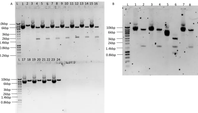
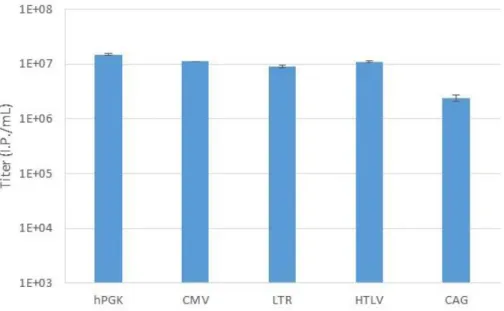
showing all necessary components for vector production. CMV is the cytomegalovirus promoter. Adapted from (Sakuma et al., 2012). ... 8 Figure 2.1 – Establishment of cell populations expressing Large T – S11 fusion protein. Infection with Large T – S11 refers to the infection with lentivirus carrying pRRL-SIN-LT-S11 and Infection with S10 refers to infection with lentivirus carrying pRRL-SIN-PGK-S10. ... 22 Figure 3.1 – Confirmation of the insertion of CMV, LTR, HTLV and CAG into pRRL-SIN. Four small scale plasmid extractions were made for each plasmid. (A) L lane is the ladder and lanes 1 through 8 correspond to pRRL-SIN-CMV ligated colonies, 9 through 16 to pRRL-SIN-LTR and 17 through 24 to pRRL-SIN-HTLV. Odd lanes present the non-restricted plasmids while even lanes present the restricted plasmids. Restrictions were performed with EcoRI and NdeI for pRRL-SIN-CMV, EcoRI and NheI for pRRL-SIN-LTR and EcoRI for pRRL-SIN-HTLV. Expected fragment sizes for pRRL-SIN-CMV were 1.7kbp and 5.7kbp, for pRRL-SIN-LTR were 1.9kbp and 5.5kbp and for pRRL-SIN-HTLV it was expected to be linear, with 7.4kbp. (B) Odd lanes correspond to non-digested pRRL-SIN-CAG and even lanes correspond to CAG digested with AgeI and NdeI. The expected fragments were 7.1kbp and 1.5kbp long. ... 24 Figure 3.2 – Titration of lentivírus produced in HEK293T. Titration of lentivirus carrying hPGK, CMV, LTR, HTLV and CAG promoters produced in HEK293T cell line. The presented titers were calculated from the average of the three best lentiviral vector productions with each promoter. Error bars correspond to the standard deviation (n=3). ... 25 Figure 3.3 - Transfection efficiency and lentiviral vector production of HEK293T,
HEK293, Vero, MDCK and Age1.CR cell lines. Transfection efficiency (A) and (B), and lentiviral vector production titer (C) of HEK293T, HEK293, Vero, MDCK and Age1.CR cell lines when transfected with pRRL-SIN-CMV (A) or pRRL-SIN-CMV, pMD2G, pMDLG/RRE
and pRSV-Rev (B). The error bars correspond to standard deviation (HEK293T n=6; other cell lines n=2). ... 26 Figure 3.4 – Infection efficiency of HEK293T, HEK293, Vero, MDCK and Age1.CR cell lines – flow cytometry analysis. Percentage and fluorescence intensity of GFP positive HEK293T, HEK293, Vero, MDCK and Age1.CR cell lines infected with lentiviral vectors carrying a GFP reporter gene driven by different promoters as indicated (hPGK, CMV, HTLV, LTR and CAG) at MOI 0.25 (A and B, respectively) and MOI 5 (C and D, respectively). RFU – Relative Fluorescence Units. ... 27
vii
Figure 3.5 – Infection efficiency of HEK293T, HEK293, Vero, MDCK and Age1.CR cell lines – fluorescence microscopy analysis. Representative images of fluorescence microscopy of 293T, HEK293, Vero, MDCK and Age1.CR cell lines infected with lentiviral vectors carrying a GFP reporter gene driven by different promoters as indicated (hPGK, CMV, HTLV, LTR and CAG). Infection was performed at MOI 5. The scale bar is 100µm long. ... 29 Figure 3.6 – Detection of Large T in Large T expressing populations – GFP
transcomplementation of LT-S11. Percentage of GFP transcomplemented cells of (A) HEK293.T, (B) Vero.T, (C) MDCK.T and (D) Age1.CR.T cells after infection with lentiviral vectors carrying pRRL-SIN-hPGK-S10 at MOI 50. S10 expressed after infection binds to the S11 part of the LT-S11 fusion protein, resulting in a functional GFP and yielding fluorescence. Values are also shown as normalized values () in relation to the average percentage obtained in a parallel infection with lentiviral vectors carrying pRRL-SIN-hPGK at MOI 50. The error bars represent the standard deviation (n=2). ... 30 Figure 3.7 – Detection of Large T in LT-S11 cell lines - Western Blotting analysis. Large T (94kDa) and Small T (21kDa) antigens from 293 S11 (A), Vero S11 (B), MDCK LT-S11 (C) and Age1.CR LT-LT-S11 (D) protein extracts. HEK293T, HEK293.T and Age1.CR.T extracts were made with 6.67x10³cells/µL of M-Per reagent and Vero.T and MDCK.T protein
extracts were made with 3.33x10³cells/µL of M-Per reagent. ... 31
Figure 3.8 – Transfection efficiency of HEK293, Vero, MDCK and Age1.CR derived cell lines. Percentage GFP positive () 293 LT-S11 (A), Vero LT-S11 (B), MDCK LT-S11 (C) and Age1.CR LT-S11 (D) cell lines transfected with pRRL-SIN-CMV. HEK293.T and Age1.CR.T were transfected at a concentration of 5 µg/106 cells and, due to being more difficult to
transfect, MDCK.T and Vero.T populations were transfected with the same plasmid at a concentration of 7.5 µg/106 cells. The error bars represent the standard deviation of 2 technical
replicates. The values are also shown as fold change in relation to MOI 0 (in the absence of LT-S11) cell lines (). ... 32 Figure 3.9 – Production of lentiviral vectors HEK293, Vero, MDCK and Age1.CR derived cell lines. Titration of lentivirus produced in HEK293.T (A), Vero.T (B), MDCK.T (C) and Age1.CR.T (D) and their respective production fold change (in red). Production of lentiviral vectors in HEK293T cells was used as control. The DNA mixes were prepared with pRSV-Rev, pMDLG/RRE, pMD2G and pRRL-SIN derived vectors at a ratio of 1:4:3.6:10 for each
concentration of DNA. The error bars correspond to the standard deviation (n=4). ... 33 Figure 3.10 – HEK293 () vs. HEK293T () Growth study - Growth curves, glucose consumption and lactate production. Growth curves (A), glucose consumption (B) and lactate
viii
production (C) curves. Specific growth, glucose consumption and lactate production rates were calculated form these curves. ... 35 Figure 6.1 – Representation of the plasmids constructed during the course of this work. A – pCI-neo-SV40LT; B – pRRL-SIN-CAG; C – pRRL-SIN-LTR; D – pRRL-SIN-HTLV; E – pRRL-SIN-CMV; F – pRRL-SIN-hPGK. ... 44
ix
A
BBREVIATION
L
IST
(+)RNA – Positive strand RNA; AAV – Adeno-associated virus; ADA – Adenosine Deaminase; AdV – Adeno virus; AU-rich – Adenylate and uridylate rich; bp – Base pairs; CA – Capsid proteins; CAG – Synthetic promoter composed by a modified chicken b-actin promoter connected to cytomegalovirus immediate-early promoter; CMV – Cytomegalovirus; cPPT – Central polypurine tract; cSIN – Conditional self-inactivating; CTE – Constitutive transport element; CTL – Cytotoxic T-cell; dsDNA - Double-stranded DNA; EF1 – Human elongation factor-1α; EIAV – Equine infectious anemia virus; EP – Eukaryotic promoter; GFP – Green Fluorescent Protein; HEK293 – Human Embryonic Kidney 293; HIV – Human immunodeficiency virus; HSV – Herpes simplex virus; HTLV - Human t-cell leukaemia virus; IN – Integrase; IP/mL – Infectious Particles per milliliter; Kbp – Kilobase pairs; LTR – Long terminal repeat; MA – Viral matrix; MDCK - Madin-Darby canine kidney; MMLV – Moloney murine leukemia virus; MOI – Multiplicity of infection; NC – Nucleocapsid; PCR – Polymerase chain reaction; PIC – Pre-integration complex; PR – Protease; PPT – Polypurine tract; RFU – Relative fluorescent units; RRE – Rev-responsive element; RT – Reverse transcriptase; SCID - Severe Combined Immunodeficiency Disorder; SIN – Self-inactivating; ssDNA – Single stranded DNA; SU – Surface proteins; SV40 – Simian vacuolating virus 40; T-Ag – Large T antigen; TAR – Transactivation-response element; TM – Transmembrane proteins; VSV-G – Vesicular stomatitis virus G protein; WPRE – Woodchuck Hepatitis Virus post-transcriptional regulatory element;
1
1 I
NTRODUCTION
1.1 A
BRIEF INTRODUCTION TO GENE THERAPYTreating a disease caused by defective genes has been a great challenge for modern medicine, to which an answer started to emerge in the beginning of the 1990’s: gene therapy. Many of these diseases significantly affect the bearer's quality of life and frequently lead to an early death. While the available means in classical medicine have shown to be unable to cure these conditions, treating and alleviating the symptoms has often remained as the only option.
The concept of gene therapy is based on the modification of an individual’s gene expression (through RNA interference, for instance) or the insertion of modified or corrected genes into the patient’s cells to treat a disease. The delivery of these genes, however, needs to be mediated by a vehicle, a vector. Among these vehicles those based on viruses have been found to be highly efficient, and consequently, the most frequently used for gene delivery. Although viruses are typically pathogenic agents, viral vectors take advantage of the specific abilities of viruses to deliver genes to target cells while removing their pathogenicity. Several characteristics specific to certain viruses can be used as advantages, such as being able to insert the viral genome into the host’s genome.
Although much progress has been made in gene therapy studies and various gene transfer systems have been developed to target genetic disorders, many problems still stand in the way, one of them being the lack of appropriate production methods. For some viral vectors such as the lentiviral based vectors, the main focus of this work, one of the problems is the production of high-titer clinical-grade preparations.
2
1.2 R
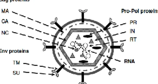
ETROVIRUS AND LENTIVIRUSRetrovirus are positive sense, single stranded RNA ((+)RNA) viruses with an icosahedral enveloped capsid and a genome of 7 to 13 kbp in length (9.3 kbp for the lentivirus genus, the focus of this work) (Blomberg et al., 2011). When outside the cell (the virion, represented in Figure 1.1), retroviruses carry two copies of the viral (+)RNA complexed with their nucleocapsid (NC) proteins. Along with the genome, they also carry several other viral proteins, such as the viral Protease (PR), the Integrase (IN) and a protein unique to this virus family and the Reverse
Transcriptase (RT), all of them inside a protein capsid (CA). Outside of the capsid there’s a layer of viral matrix proteins (MA) that interact with the host derived envelope containing viral envelope proteins (Env) which recognize the virus’ specific receptors in the host cell. Upon receptor mediated endocytosis, the viral (+)RNA is transcribed into dsDNA (into the provirus) by RT. Both RT and the provirus will then interact with IN, host proteins and proteins from the degraded viral core to form the pre-integration complex (PIC) nucleoprotein, which will later integrate the virus genome in the host genome (Coffin et al., 1997). After protein synthesis, the viral components assemble in lipid rafts, cholesterol and sphingolipid-rich parts of the membrane and the newly formed virion exits the cell by budding (Coffin et al., 1997).
Figure 1.1 – Retrovirus virion structure. MA – viral matrix; CA – capsid; NC – nucleocapsid; PR – protease; IN –
3
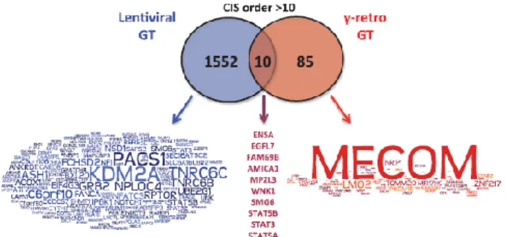
In simple retroviruses, such as gamma-retrovirus Moloney Murine Leukemia Virus (MMLV), the provirus cannot enter the nucleus through its pores. The dsDNA enters the nucleus only when the infected cell divides and the nuclear envelope disassembles, and will later be integrated into the host genome in certain patterns (depending on the virus). Integration patterns for lentivirus and gamma-retrovirus in gene therapy clinical trials are shown in Figure 1.2. Provirus integration can be potentially oncogenic and has shown to be one of the main limitations of vectors derived from gamma-retrovirus, establishing some negative landmarks in retroviral vector history (Raper, 2005). Despite this, retroviral vectors have many advantages. In addition to stably integrating the provirus into the host genome, retrovirus also present low immunogenicity, very low pre-existing immunity in the human body and gamma-retroviral and lentiviral vectors can carry up to 8 kbp and 9 kbp of heterologous gene content, respectively (Vannucci et al., 2013). Taking into account these obvious advantages, an effort to improve retroviral vector safety was employed.
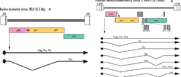
Gamma-retrovirus genome, represented in Figure 1.3A, is composed by 4 coding gene families flanked by 2 long terminal repeats (LTR): gag, pro, pol and env. Gag codes for capsid (CA), nucleocapsid (NC) and matrix (MA) proteins, pro codes for the viral Protease (PR), responsible for cleaving Gag-pol fusion protein and virion maturation, pol codes for Reverse Transcriptase (RT) and Integrase (IN), and finally, env codes for two envelope protein subunits, Transmembrane (TM) and Surface (SU), responsible for interaction with MA proteins and receptor binding, respectively (Coffin et al., 1997). Retroviral LTRs are composed of 3 regions: U3, containing viral promoters and enhancers, R, necessary for reverse transcription and replication, and U5, also important for reverse transcription, in particular its initiation. These
Figure 1.2 – Word cloud of common integration sites in gene therapy clinical trials with lentivirus (blue) and gamma-retrovirus (red). The size of the word represents the frequency of insertion sites in or close to each gene. In
4
regions are also necessary for stable integration of the provirus into the host’s genome (Coffin et al., 1997; Sakuma et al., 2012). Besides the LTRs, retroviral genome contains other non-coding,
cis-acting elements. These include a packaging signal (Ψ), responsible for packaging of unspliced
mRNAs (viral genomes) into the forming virion, the primer binding site (PBS), where the negative DNA strand primer will bind, and the polypurine tract (PPT) which primes the positive DNA strand synthesis (Coffin et al., 1997).
Belonging to the Retroviridae family (retroviruses) and to the Orthoretrovirinae sub-family (Blomberg et al., 2011), lentiviruses are closely related to gamma-retroviruses. Lentiviruses, however, are more complex retroviruses and their genome, represented by Figure 1.3B, codes for two regulatory proteins (Tat and Rev) and several additional proteins not required for viral replication (Nef, Vif, Vpr and Vpu)(Figure 1.3B and Figure 1.4). Lentiviruses also contain a second polypurine tract, the central polypurine tract (cPPT) which enhances transduction efficiency and provides a second DNA synthesis initiation site (Goff, 2007; Sakuma et al., 2012). Accessory proteins coded by nef, vif, vpr and vpu are mostly for host defense neutralization and so, are necessary for viral pathogenicity, but not for infectiousness or vector production (Sakuma et al., 2012). Tat and rev, code for the two additional regulatory proteins. Tat promotes the transcription of less spliced, longer transcripts (multiply spliced transcripts are necessary in the early infectious cycle to produce regulatory proteins such as Tat, Rev and Nef). This protein binds to transactivation-response element (TAR) in the viral mRNAs to promote and amplify structural protein transcription. Rev regulates splicing and nuclear exportation of singly spliced or unspliced viral transcripts by binding to a cis-acting element called Rev-responsive element (RRE) sequence present in the transcripts (Sakuma et al., 2012). These two proteins play
Figure 1.3 – Genome organization and transcripts of gamma-retrovirus and lentivirus. (A) represents MMLV genome
organization and transcripts and (B) represents HIV-1 genome organization and transcripts. In (Blomberg et al., 2011).
A
5
important roles in wild-type viruses: Tat greatly increases LTR activity (by more than two orders of magnitude) and Rev is necessary for the exportation of the viral genome to the cell’s membrane (unspliced mRNAs). The genomic RNAs assemble with the viral proteins and form an immature virion that will exit the cell through budding. Gag and Gag-pol will then form multimers that will activate the protease, mediating virion maturation after cellular release (Sakuma et al., 2012; Vannucci et al., 2013).
The main difference between lentivirus and gamma-retrovirus is the ability for lentivirus to infect non-dividing cells (Naldini et al., 1996). Furthermore, there is a great selection of lentiviruses that can be engineered into lentiviral vectors, both human viruses (Human immunodeficiency virus, HIV), which show an absence of pre-existing immunity, and non-human viruses (for instance, Equine infectious anemia virus, Feline Immunodeficiency Virus, etc.). The latter are apathogenic in humans, but, when modified, can infect human cells (Sauter & Gasmi, 2001; Stewart et al., 2009; Vannucci et al., 2013). Insertional mutagenesis is also reduced, since, unlike other retrovirus genera (gamma-retrovirus, Figure 1.2), insertion of lentiviral provirus occurs preferentially away from cellular promoters and oncogenes, avoiding the disruption of neighboring gene expression and LTR driven oncogene activation (Desfarges & Ciuffi, 2010; Vannucci et al., 2013). Also, provirus integration can happen in a wider number of genes, making it more arbitrary than other retrovirus, such as gamma-retrovirus. There is also the option of producing an integration defective vector by inactivating the integrase enzyme, which will infect the cell and transiently express the gene of interest (Vannucci et al., 2013). This approach is particularly useful when developing vaccines, since it allows expression of a gene for long enough to induce a cellular immune response against the required antigen.
6
1.3 F
ROM VIRUS TO VECTORViruses are natural gene delivery vehicles, probably the main reason for their high efficiency in gene therapy. However, the majority of wild type viruses are pathogenic and require to be engineered into harmless vectors, suitable for therapeutic applications.
The first approach taken to improve viruses as gene delivery agents was the progressive elimination of non-essential viral genes, making room for heterologous genes. Another reason for this approach was the elimination of the replication capacity, which could cause adverse effects, improving vector safety. The end result of this approach in adenoviral vectors, the most frequent viral vector system used in gene therapy clinical trials, was the creation of a gutless vector. These gutless vectors contain only the transgene and cis-acting elements necessary for replication and vector packaging, increasing their capacity up to 37 kbp (Danthinne & Imperiale, 2000; Schaffer et al., 2008). A similar approach was taken to develop Adeno-Associated Virus based vectors, substituting both viral genes, rep and cap, which encode for replication and structural proteins, with the transgene and an heterologous eukaryotic promoter (Vannucci et al., 2013).
The same rationale was applied to retroviruses and lentiviruses, eliminating as many non-essential genes as possible. Genes non-essential for vector packaging, called helper or packaging functions, are provided on separate plasmid constructs lacking the capacity to be packaged along with the transgene construct (Blomberg et al., 2011; Coroadinha, 2005).
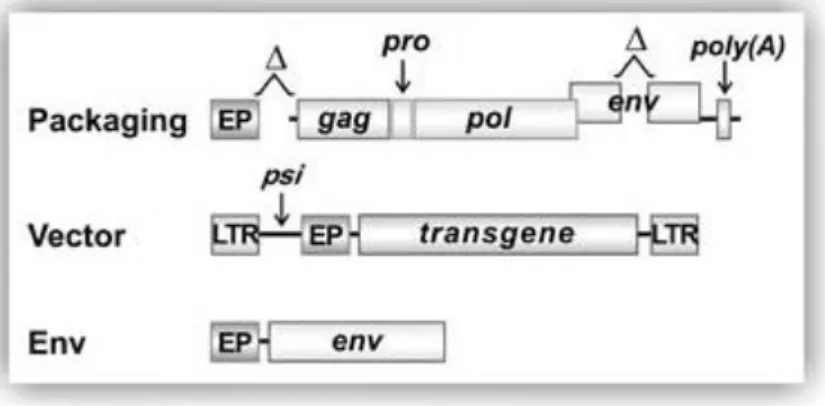
In gamma-retroviral vectors, the viral genome is separated into 3 plasmids reducing the probability of forming replication competent particles through homologous recombination and thus increasing the vector’s safety. This results in the constructs represented in Figure 1.5, where
Figure 1.5 – Schematic representation of the 3 necessary constructs for production of gamma-retroviral vectors. The
packaging construct contains all necessary components to form a functional capsid, env construct provides the envelope proteins, and the vector contains a packaging signal, the eukaryotic promoter (EP) and the transgene. The vector construct will then be packaged into a functional virion. In (Vannucci et al., 2013).
7
the packaging construct contains gag, pro and pol, necessary to produce a functional vector with all the necessary proteins and enzymes. The env construct contains the envelope proteins, TM and SU. The latter construct allowed for a great versatility of the resulting vectors, since it facilitated the exchange of the retroviral envelope proteins with envelopes from other viruses, changing the vector tropism, and allowing for its redirection to target cells (Sakuma et al., 2012). This process is called pseudotyping and is frequently used to retarget the vector to host cells other than the natural host and reduce the homology between the vector constructs and the wild type virus (Sakuma et al., 2012; Vannucci et al., 2013). One of the most frequently used envelopes is the G protein of the vesicular stomatitis virus (VSV-G). VSV-G binds to a common membrane phospholipid Low-Density Lipoprotein Receptor and its family of proteins granting the vector a much wider range of cells it can infect (Blomberg et al., 2011; Coroadinha, 2005; Finkelshtein et al., 2013; Sakuma et al., 2012). Tissue or cell specific transgene expression can also be mediated by using tissue or cell specific promoters. This allows the vector to transduce any susceptible cell, but the transgene will only be expressed in that specific tissue (Sakuma et al., 2012; Vannucci et al., 2013).
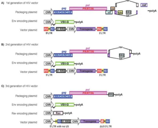
In the first generation of lentiviral vectors, the physical separation of the viral genome was accomplished into 3 transcriptional units Figure 1.6A. The vector plasmid contained the LTRs, the packaging signal (Ψ), RRE and the transgene with an eukaryotic promoter (EP). The envelope plasmid contained a receptor-binding protein and the packaging plasmid included all other viral proteins and RRE as well. The separation of env from the vector construct allows only one round of infection since the lack of a packaging signal avoids encapsidation of the env and packaging constructs into virions (Sakuma et al., 2012). Vectors produced with this configuration are replication-incompetent, unable to produce viral proteins in host cells and need two recombination events in the producer cell in order to create a replication competent virus. Yet the constructs still share many homologous sequences and coded unnecessary proteins (Sakuma et al., 2012; Vannucci et al., 2013). For the second generation of lentiviral vectors, these unnecessary protein coding sequences (vif, vpr, nef and vpu) were modified or deleted from the packaging vector (Figure 1.6B). However, this generation still had a problem with possible rescuing of the integrated vector into new virions by wild-type virus co-infection, spreading the transduction, and the activation of host genes by the LTR regulatory regions, such as enhancers (Sakuma et al., 2012). To avoid this, self-inactivating (SIN) viral vectors were developed by deleting the regulatory regions of the 3’ U3 region of the vector construct (Miyoshi et al., 1998; Zufferey et al., 1998). These regions are normally copied to the 5’ LTR when reverse transcription occurs such that the resulting provirus is transcriptionally inactivated and incapable of producing a full-length mRNA, avoiding LTR driven read-through (Yu et al., 1986). As well as being
self-8
inactivating, third generation vectors (Figure 1.6C) also brought with them tat independence through the use of a strong heterologous viral promoter instead of the 3’ U3 region. Also, Woodchuck Hepatitis Virus post-transcriptional regulatory element (WPRE) was included in the
vector plasmid, greatly improving transgene expression, and Rev was positioned in a fourth plasmid, enhancing biosafety by increasing to three the number of recombination events necessary to generate replication competent lentivirus (RCLs) (Sakuma et al., 2012).
For the latter generation of lentiviral vectors, further optimization can be carried out by codon optimization, since gag and pol are highly rich in adenylate and uridylate (AU-rich) destabilizing sequences, which strangely translates into an abnormal codon bias. This codon bias is quite different from highly expressed human genes codon usage (Kotsopoulou et al., 2000) leading to a reduced Gag-pol expression in the absence of Rev, which acts as a stabilizing agent. By performing codon optimization, Gag-pol showed increased expression. There is also a reduction in sequence homology with the native gag-pol sequence and the vector becomes independent given the reduction in destabilizing sequences (Kotsopoulou et al., 2000). Rev-independence allows for a substitution of Rev with another mRNA transport agent, such as constitutive transport element (CTE) from Mason–Pfizer monkey virus, that will allow to further
Figure 1.6 – Schematic representation of the three generations of lentiviral vectors, showing all necessary components for vector production. CMV is the cytomegalovirus promoter. Adapted from (Sakuma et al., 2012).
9
decrease homology with native lentivirus sequences (Sakuma et al., 2012). Although this process has its advantages, it has also proven to be challenging, often resulting in lower vector titers (Sakuma et al., 2012). Another way to augment vector safety is to add chromatin insulator sequences, which can protect neighboring cellular genes from transactivation by the vector’s promoters, and protect the vector’s promoters from cellular repression (Sakuma et al., 2012; Throm et al., 2009).
Several other systems have been developed to overcome most of the obstacles mentioned above, and to improve lentiviral vectors versatility. Among them is the development of non-integrating vectors in order to reduce the occurrence of insertional mutagenesis events. This was done by introducing mutations in the viral integrase gene. These vectors can be successfully used in transferring a transgene into a cell, but it is only expressed for long periods of time in non-dividing cells (Saenz et al., 2004; Sakuma et al., 2012). Another important vector modification is the development of inducible promoters such as a TetO-binding-sites containing promoter. Along with a chimeric transcription factor tTA transactivator, a fusion protein between a bacterial tetracycline repressor and the HSV activating domain, this system can induce the transgene expression (Tet-on) or silence the gene (Tet-off) (Xu et al., 2001). This approach has been used to create a stable cell line producing conditional SIN (cSIN) lentiviral vectors, in which instead of controlling transgene expression, the Tet-on/Tet-off system was used to control vector production (Hwang et al., 1997; Xu et al., 2001), partially overcoming the challenge of stable production, hampered by the cytotoxicity of some lentiviral proteins (Throm et al., 2009).
Gene therapy is the most demanding application of lentiviral vectors in terms of safety concerns and, as so, it is where this review focuses, but there are many other applications for this technology. These applications include: lentivirus based vaccines, cellular reprogramming (transforming a somatic cell into a multi or pluripotent cell) and monitoring of transfected cells (such as cancer cell metastasis)(Sakuma et al., 2012).
10
1.4 M
ETHODS AND MAIN CELL LINES USED FOR LENTIVIRAL VECTORPRODUCTION
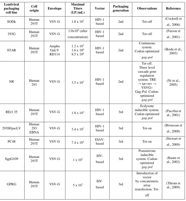
Ideally, a lentivector production system should consist on a stable producer cell line, both for the improved safety (due to the reproducibility of the system) and for the easy scalability to large-scale production (Schweizer & Merten, 2010). Current lentiviral vector production methods either involve transient expression of the vector which, although they have been used, present safety concerns (Schweizer & Merten, 2010), or, most commonly, conditional producer cell lines, summarized in Table 1.1. These current systems deliver insufficient titers (transient expression and conditional producer cell lines) for gene therapy or pose safety and batch-to-batch reproducibility issues (transient expression) in the production of lentiviral vectors (Throm et al., 2009).
Chemical methods such as cationic agents are commonly used to complex to negatively charged DNA to mediate cell entry for transient production of lentivirus. Among these, polyethylenimine is one of the most widely used, both for being relatively cheap and highly efficient. Cationic lipids, such as LipofectAMINE®, and the calcium phosphate method were also used in the past, but they are either expensive or difficult to scale up, although the calcium phosphate method is as efficient as polyethylenimine method (Rodrigues et al., 2011; Schweizer & Merten, 2010). The baculovirus system can also be used for transient lentivirus production, but it requires an extra effort to separate the baculovirus from the lentivirus preparation to achieve clinical-grade quality. Titers up to 1.4x10⁶ transducing units/milliliter (TU/mL)were obtained
before the necessary downstream processing (Lesch et al., 2008). This means that part of these transducing units are still going to be lost in the purification process, reducing the final titers. Although transient production produces higher titers (enough for Phase I clinical studies), this production is only temporary and lacks batch-to-batch reproducibility and, therefore, difficult to completely characterize, making this method highly undesirable for clinical use (Rodrigues et al., 2011; Segura et al., 2013). Stable producing cells are created by inserting, either by direct viral infection or by chemical means, the vector functions one by one into the cells. Each construct insertion is followed by a round of clonal selection of cells with high expression levels of each component. Typically the first construct to be inserted into the target cells is the packaging one, followed by the envelope construction and finishing with the stable integration of the transgene construct. This process, however is cumbersome and can take over a year to complete and characterize (Rodrigues et al., 2011). Also, in lentivirus, it has been hampered by the cytotoxicity of some of the viral components.
11
Many attempts at developing high titer lentiviral vector producer cell lines have been made with several human cells (Henrietta Lacks (HeLa), HT1080 and TE671, for instance). Monkey derived cells (CV-1, COS(CV-1 in origin carrying SV40)-1 and COS-7) were also tested in an attempt to avoid human cells, which carry an increased risk of retroviral activation, since 90% of non-coding mobile sequences in the human genome are endogenous retrovirus and there were concerns about possible human pathogen contamination (Pauwels et al., 2009; Zwolinska, 2006). Most of these, however, have proven to produce low titers, with the exception of COS-1 monkey kidney epithelial cells, which were capable of producing higher-quality vectors (higher infectious particles/total particles ratio) and the supernatant was almost free of cell debris (Schweizer & Merten, 2010; S. L. Smith & Shioda, 2009). These conclusions were taken by comparing vector production and quality with the most frequently used cell line, Human Embryonic Kidney 293T cells (HEK293T).
Most producer cell lines are based on variations of the adherently grown HEK293 cells, which are relatively easy to transfect (Segura et al., 2013). As mentioned before, the most frequent of these variations are HEK293T, which are HEK293 clones transformed with the oncogene Large T antigen from Simian vacuolating virus 40 (SV40 T-Ag). Stable expression of the Large T antigen allows for a faster growth, vector titers four times higher than their non-transformed counterparts under the same conditions and higher transfection efficiencies (Segura et al., 2013). Scaling-up production with HEK293T cells is also easier, since they can be grown in suspension in serum-free conditions more easily, reducing downstream processing costs (Ghani et al., 2007; Segura et al., 2013; H. S. Smith et al., 1971). Suspension culture also allows for a better control of the culture’s conditions, allowing for a more homogenous culture when a stirred tank reactor is used (Sadettin & Hu, 2006). However, these oncogene expressing cells still produce insufficient titers for clinical application, as 107 transfection units per milliliter (TU/mL) (shown in Table 1.1)
still demand about 10 to 100 L of cell culture volume to treat a single patient (Rodrigues, 2013 a)).
12
Table 1.1 – Examples of the developed stable packaging cell lines used for lentiviral production.
Although many of the problems associated with lentiviral vector production have been overcome and a stable producer cell line was established by Ikeda et al (2003), these cell lines still need the Simian Virus 40 Large T antigen (T-Ag) to achieve acceptable vector titers. The need for T-Ag is a major safety concern that needs to be avoided in order to produce safe, clinical-grade lentivector preparations.
Understanding the physiological changes induced by Large T antigen should offer a better view on potential cell engineering targets to improve vector titer and produce safer vector preparations, without the need of T-Ag.
Lentiviral packaging cell line Cell origin Envelope Maximal Titers (I.P./mL) Vector Packaging
generation Observations Reference
SODk Human 293T VSV-G 1.0 x 10 7 HIV-1 based 2nd Tet-off (Cockrell et al., 2006) 293G Human 293T VSV-G 1.0x10⁹ (after concentration) HIV-1 based 2nd Tet-off (Farson et al., 2001) STAR Human 293T Ampho GaLV RD114 1.2 x 10⁷ 1.6 x 10⁶ 8.5 x 10⁶ HIV-1 based 2nd Continuous system. Codon-optimized gag-pol (Ikeda et al., 2003) NR Human 293 VSV-G 3.5 x 10 7 HIV-1 based 2nd Tet-off. Three level cascade gene regulation system: TRE → tat+rev → VSVG+ Gag-Pol. Codon-optimized gag-pol (Ni et al., 2005) REr1.35 Human 293T VSV-G 1.8 x 10⁵ HIV-1 based 3rd Ecdysone inducible system. Codon-optimized gag-pol (Pacchia et al., 2001) 293SFpacLV Human 293 EBNA VSV-G 3.4 x 10⁷ HIV-1 based 3rd Tet-on (Broussau et al., 2008) PC48 Human 293T VSV-G 7.4 x 10⁵ EIAV- based 3rd Tet-on (Stewart et al., 2009) SgpG109 Human 293T VSV-G 1 x 10⁵ SIV-based 3rd Ponasterone inducible system. Codon-optimized gag-pol (Kuate et al., 2002) GPRG Human 293T VSV-G 5 x 10⁷ SIV-based 3rd Introduction of vector by concatemeric array transfection. Tet-off (Throm et al., 2009)
293EBNA – HEK 293 cells transformed with the Nuclear Antigen of Epstein Bar virus. NR – not reported. Adapted from (Rodrigues et al., 2011).
13
1.5 C
ELLULAR CHANGES INDUCED BYL
ARGET
ANTIGENSV40 is a virus from the poliomavirus family endogenous in Rhesus monkeys (Zheng et al., 2009). Although infection of its natural host is asymptomatic, in human and mouse cells it induces neoplastic transformation and, consequently, tumor formation. The major player in cellular transformation by SV40 is the Large T antigen. The Large T antigen contains an helicase domain that overlaps a p53 binding domain and a Rb(retinoblastoma protein)-binding domain. Rb is a protein involved in cell proliferation control (it has several functions in the cell cycle), genome maintenance and in apoptotic cell death (Dick & Rubin, 2013). P53 is a tumor suppressor protein with functions in cell cycle arrest, DNA damage response and apoptosis (Ozaki & Nakagawara, 2011). Also, two regions in Large T, one in the amino terminus end and the other in the carboxyl terminus end, interact with cellular transcriptional coactivators p300 and CBP (CREB binding protein), increasing their mRNA loading into polysomes (Ali & DeCaprio, 2001; Saenz Robles et al., 2013). Increased levels of these proteins are linked to increased histone acetyltransferase activity, which will alter histone acetylation patterns (Saenz Robles et al., 2013). Acetylation of histones removes their positive charge, decreasing their affinity for DNA, consequently increasing the accessibility of transcriptional and regulatory proteins to chromatin (Struhl, 1998). Taking these binding abilities into account, the Large T antigen promotes cellular proliferation by avoiding cell cycle arrest. Additionally, T-Ag provides anchorage-independent growth and allows for transformed cells to grow in low serum conditions (or no serum at all) (Ahuja et al., 2005). These changes appear to be induced by the N-terminal of this protein, and are complemented by Small T antigen, a protein resulting from an alternative splicing of the T-Ag sequence (Tevethia et al., 1997). Small T may activate integrin signaling pathways, leading to anchorage independent growth in aggregates (Moreno et al., 2004).
Even though it is known that T-Ag induces increased lentiviral vector productivity, it can only do so when the packaging cell stably expresses it. This was shown in a study carried out by Gamma-Norton et al. (2011) where HEK293 derived cell lines were used to produce lentiviral vectors containing a Green Fluorescent Protein (GFP) gene. To compare vector production titers, three HEK293 derived cell lines were used: HEK293, 293T and several TAR293LV clones. The latter were previously shown to produce high titers of retroviral vectors, while stably expressing T-Ag. These were transduced to express Large T antigen either transiently or stably and lentiviral vector production was characterized both by Real Time Polymerase Chain Reaction (qPCR) amplification and quantification of the packaging signal and by quantification of viral titers. Their results demonstrate that short-term expression of T-Ag does not alter lentivector production while showing a slight increase in lentivector production when it is integrated into the host’s genome.
14
Although TAR293LV vector production was increased upon long-term expression of Large T antigen, this increase was not related to increased viral mRNA levels, suggesting that additional mechanisms may be involved in vector titer increase (Gama-Norton et al., 2011).
A study to clarify the oncogenic role of T-Ag in eye lens carcinogenesis in mice was performed by Zheng et al. Mice expressing Large T antigen gene driven by α A-crystalin promoter, a major soluble protein of the lens which is specific for the lens cells, were used. Briefly, histopathological observation, immunohistochemical labeling of several proteins involved in cell cycle, proliferation, apoptosis, signal transduction, transcriptional activation, protein folding and cell mobility and adhesion, and a commercial gene chip representing over 22,600 mouse transcripts were used. Histopathological observation revealed an aggressive tumor progression. The gene-chip showed significant alterations in 404 proliferation related genes and 628 cell death related genes after the carcinoma began to invade. 2158 cell growth related genes, 1730 cell-to-cell signaling related genes, 889 cell cycle and 9010 metabolism related genes were altered after the tumor started to invade the outside of the eyeball. These results were backed up by the immunohistochemical labeling where some of these genes’ products were monitored. For instance, the gene chip indicated remarkable alterations in calcium signaling and glucose metabolism, and monitoring of glucose-related protein-78, a stress-responsive gene product, showed increased expression, indicating that its up-regulation is important for carcinoma progression (Zheng et al., 2009). These results have shown that Large T antigen can play an important role in lens carcinogenesis and subsequent development of the cancer, making this a safety issue for the use of viral vectors produced by T-Ag expressing cells.
The reasons behind the higher viral titers by Large T antigen expressing cells are poorly understood, but, recently, a metabolic analysis comparing two retroviral vector producer cell lines and their parental lines was performed by A. F. Rodrigues et al. This study brought new light to the metabolisms involved in retroviral vector production and these may be linked to the cellular changes caused by Large T antigen. It was shown that stable retroviral vector producer cell lines suffer a down-regulation of lipid synthesis pathways, balanced by the up-regulation of lipid uptake mechanisms (Rodrigues et al., 2013 b)). Therefore, changes at serum dependence by Large T antigen suggest that a lipid metabolism pathway may be altered. Up-regulation of these pathways could explain why the cell no longer needs to uptake serum (a lipid source) from the medium. Clarification of these changes is needed in order to enhance lentiviral vector production. The same study also showed that HEK293 derived low producer cell line 293 FLEX, when compared to a high producer cell line, had lower glycolytic fluxes, down-regulated polyamine (a substrate for retroviral vector production) and glutathione (an antioxidant) metabolism. The low
15
producer cell line also relied on nucleoside uptake to produce nucleotides (by the salvaging pathway) and the high producer cell line had a higher amino acid uptake, both generating more energy from them and increasing protein synthesis by up-regulation of elongation and initiation factors. Supplementation of each of these substances in the growth medium showed increased vector titers, with higher titer increases when nucleosides were added to the medium and lower increases when amino acid supplements were added (Rodrigues et al., 2013 b)). With this study in mind, increases of vector production by Large T expression may be linked to some of these metabolic targets. Further investigation should be performed in order to find cell and metabolic engineering targets that may render Large T antigen unnecessary, enabling the production of safer vector preparations.
1.6 A
IM OF THIS THESISLentiviral vectors are an important tool for therapeutics, particularly in treating monogenic diseases, but their production still has some obstacles to overcome. Currently, the most prominent ones are the sub-optimal titers obtained and the need to use of SV40 Large T antigen to support high titer productions. In an attempt to surpass these problems, a human (HEK293) and three non-human cell lines (Vero, MDCK and Age1.CR) were transformed with Large T antigen. These were then transfected with a GFP expressing lentiviral construct and their transfection efficiency and vector production was characterized and compared to their parental cell lines. HEK293 and HEK293T cells were also compared in terms of cell growth and glycolysis. These approaches gave us insight as to whether non-human alternative substrates are suitable for high titer vector production and a deeper understanding of Large T antigen induced cellular alterations.
Knowledge derived from this study has the potential to allow the establishment and engineering of new cell substrates for the production of high titers of safer clinical-grade lentiviral vectors.
16
2 M
ATERIALS AND METHODS
2.1 P
LASMIDSFor all the vectors constructed in this work the cloning sites, primers and templates are listed in Table 6.1. (Underlined plasmids). A schematic representation can be found in Figure 6.1.
pSelect-Blasti-mcs (Invivogen, San Diego, California, USA) is a plasmid containing a multiple cloning site (MCS) downstream of a composite EF1/HTLV promoter, and a blasticidine resistance marker, driven by the CMV (cytomegalovirus) promoter. EF1/HTLV is composed of the human elongation factor-1α (EF1) promoter coupled with the R segment and part of the U5 region of human t-cell leukaemia virus (HTLV) type 1’s Long Terminal Repeat (LTR). This plasmid was used for the amplification of CMV and hEF1/HTLV promoters, to replace the hPGK promoter in pRRL-SIN-PGK.
pEM-MFG is a recombinase mediated cassette exchange plasmid with an wild type FTR site and a mutated F5 FTR site next to an ATG which follows an encephalomyocarditis virus (ECMV) internal ribosome entry site (IRES). This ECMV-IRES follows an Murine Leukemia Virus (MLV) based retroviral vector MFG-LTR (Coroadinha et al., 2006). This plasmid was used for the amplification of the 5’LTR promoter, in order to replace the hPGK promoter in pRRL-SIN-PGK.
pCAG-DsRed, kindly provided by Dr. Gonçalo Real (Animal Cell Tecnology Unit, iBET, Portugal) comprises the DsRed fluorescent protein from Discosoma sp, driven by the CAG promoter. The CAG synthetic promoter was constructed by modifying the chicken b-actin promoter with a 3’ part of the second intron and a 5’ part of the third exon of the rabbit b-globin gene connected to the CMV immediate-early enhancer sequence as described in (Miyazaki et al., 1989) and (Hitoshi et al., 1991). This plasmid was used to isolate the CAG promoter in order to replace hPGK in pRRL-SIN-PGK to construct pRRL-SIN-CAG.
pRRL-SIN-PGK is a 3rd generation self-inactivating (SIN) lentiviral backbone with an
eGFP transgene driven by a human phosphoglycerate kinase (hPGK) promoter, described in Dull et al (1998) and provided by Didier Trono through the Addgene plasmid repository (Cambridge, MA, USA). pRRL-SIN-CMV, pRRL-SIN-HTLV, pRRL-SIN-LTR and pRRL-SIN-CAG are four plasmids derived from pRRL-SIN-PGK lentivirus backbone.
pMD2G is an envelope plasmid, provided by Didier Trono through the Addgene plasmid
17
CMV promoter. This plasmid was used to express the envelope proteins necessary for lentivirus production.
pMDLG/pRRE is a 3rd generation packaging plasmid containing gag (responsible for the
virion’s main structural proteins) and pol (coding for retrovirus specific enzymes) genes and rev-responsive element (a binding site for Rev protein, improving the exportation of RNA from the nucleus). This plasmid was used for the expression of the structural proteins and essential enzymes for the production of lentivirus.
pRSV-REV is a 3rd generation packaging plasmid, containing the second and third exons
of HIV-1’s Rev under transcriptional control of Rous Sarcoma Virus (RSV) U3 promoter. pJSATIR is a plasmid composed by the Large T antigen coding sequence driven by a tet-on promoter and a neomycin resistance gene fused with an eGFP. This plasmid was used to amplify and isolate Large T for subsequent cloning in pCI-neo and pRRL-SIN-LacZ-S11 and was kindly provided by Dr. Dagmar Wirth (HZI, Germany).
pCI-neo (Promega, Madison, WI, USA) is a mammalian expression vector with a neomycin resistance gene and multiple cloning site (MCS) downstream of a CMV promoter, driving the expression of a gene of interest. This plasmid was used to construct pCI-neo-SV40LT by cloning the previously amplified Large T coding gene into the MCS.
In order to quantify the amount of Large T, a split-GFP® SandiaBiotech (Albuquerque, NM, U.S.A) system was employed. This system is based on splitting GFP into two fragments: S10, which codes for the first 214 amino acids of GFP and S11, which codes for the last 15. These fragments yield no fluorescence unless they are both present in the same cell and so, fusing Large T with the S11 fragment allows for relative quantification of Large T-S11 fusion protein by transcomplementation. The resulting fluorescence is proportional to the amount of Large T in a given sample. To employ this system, pRRL-SIN-PGK-S10 and pRRL-SIN-LacZ-S11 were kindly provided by Ana Oliveira (a co-worker). pRRL-SIN-PGK-S10 was constructed from PGK by replacing GFP from the lentiviral backbone with GFP S10, while pRRL-SIN-LacZ-S11 was constructed by replacing GFP with pRRL-SIN-LacZ-S11 fusion protein (Oliveira, 2012). pRRL-SIN-PGK-S10 was used to quantify Large T antigen along with pRRL-SIN-CMV-LTS11, explained below. pRRL-SIN-LacZ-S11 was used to fuse the Large T coding sequence with the S11 fragment.
pRRL-SIN-CMV-LTS11, was constructed from pRRL-SIN-CMV and pRRL-SIN-Lacz-S11, first by exchanging LacZ with Large T antigen coding sequence in pRRL-SIN-LacZ-S11 and then by replacing the GFP gene in pRRL-SIN-CMV with the resulting LargeT-S11 fusion protein.
18
2.2 C
ELL LINES AND CULTURE CONDITIONSHEK293 is a human cell line (ATCC® CRL-1573™) used as a negative control for Large T transformation assays for the other cell lines, to compare promoter strength driving the transgene between cell lines, for the establishment of 293.T and 293 LT-S11 populations and for growth studies.
HEK293T cell line (ATCC® CRL-3216™), is a HEK293 derived cell line, expressing SV40 Large T antigen. It was used to produce and titrate lentivirus carrying the different promoters, to assess the promoters strength driving the transgene and for growth studies.
MDCK (Madin-Darby canine kidney) cell line (ATCC® CCL-34™) is and animal cell line derived from Canis familiaris (dog) kidney epithelial cells. It was used to assess its potential as a substrate for lentiviral vector production.
Vero cell line (ATCC® CCL-81™) is derived from African Green Monkey (Cercopithecus aethiops) kidney tissue. It was used to assess its potential as a substrate for lentiviral vector production.
Age1.CR cell line (Probiogen, Berlin, Germany) is derived from Muscovy duck (Cairina
moschata) retina (Jordan et al., 2009). It was used to assess its potential as a substrate for lentiviral
vector production.
All cell lines were maintained in Dulbecco’s Modified Eagle Medium (DMEM) (Gibco, Paisley, UK) with 25mM of glucose, 4mM of glutamine and supplemented with 10% (v/v) Foetal Bovine Serum (FBS) (Gibco, Paisley, UK). All cell lines were cultured under adherent conditions in a humidified incubator at 37ºC and 7% CO2. Cell dissociation from adherent conditions (for
passaging and seeding) was performed with 0.05% (w/v) Trypsin solution (Gibco).
2.3 B
ACTERIAL STRAINSEscherichia coli (E. coli) Stellar™ (Clontech, California, USA) competent bacteria were used for the production of the constructed lentiviral backbone plasmids and Library Efficiency® DH5α™ (Invitrogen, Carlsbad, CA, U.S.A) competent bacteria were used for the production of all other plasmids. Transformation procedures were performed according to the manufacturer’s instructions.
The liquid bacterial cultures were performed with Terrific Broth media (TB) (Fast-Media® TB from Invivogen) supplemented with the appropriate antibiotic (Ampicillin or Blasticidin). The media were prepared using ultrapure water (Millipore, Billerica, MA, U.S.A.), according to the manufacturer's instructions.
19
2.4 C
LONING PROCEDURESPolymerase chain reactions (PCRs) were performed using custom made primers (Sigma-Aldricht, St.Louis, MO, U.S.A) and Phusion High Fidelity DNA polymerase (Finnzymes OY, Espoo, Finland) (Table 6.1). The reaction conditions were as follows: 30 seconds at 98ºC for the initial denaturation step followed by 30 cycles of denaturation at 98ºC for 10 seconds, 30 seconds of annealing at the appropriate melting temperature and elongation at 72ºC with time depending on the fragment length (30 seconds per kbp), and a final elongation step at 72ºC for 10 minutes. Fragments generated either by PCR or enzyme restriction were separated with 0.7% (w/v) agarose gels and then purified using Ilustra™ GFX™ PCR DNA and Gel Band Purification Kit (GE Healthcare Life Sciences, Little Chalfont, UK).
pRRL-SIN-CMV, pRRL-SIN-LTR, pRRL-SIN-HTLV and pRRL-SIN-CAG, were established by removing hPGK from pRRL-SIN-PGK by enzyme restriction (AgeI and XhoI), flanking the promoter, and it was exchanged by: CMV promoter, hEF1/HTLV fusion promoter, Moloney murine leukaemia virus’ 5’LTR and CAG promoter, respectively.
CMV and EF1/HTLV promoters were amplified by PCR from pSELECT-blasti-mcs plasmid, 5’LTR was isolated from pEM-MFG plasmid and Large T antigen coding sequence was amplified from pJSATIR, using primers designed for the In-Fusion® HD cloning kit (Clontech, California, USA), (Table 6.1). The amplified fragments were then ligated into the linearized pRRL-SIN following the In-Fusion cloning procedure. CAG promoter was isolated from pCAG-dsRed plasmid using SalI and AgeI restriction enzymes, the resulting fragment was purified, phosphorylated with Antarctic Phosphatase (New England Biolabs, Ipswich, MA, USA) and was ligated to pRRL-SIN backbone with T4 DNA ligase following the manufacturer’s instructions. The resulting plasmids were then cloned into Stellar™ competent cells following a heatshock transformation protocol, according to the manufacturer’s instructions.
2.5 P
LASMID PURIFICATION AND QUANTIFICATIONPlasmid extraction was performed on two different scales, a small scale production using QIAprep® miniprep kit (QIAgen, Hilden, Germany) and a large scale production using Genopure Plasmid Maxi Kit (Roche, Mannheim, Germany) following the manufacturer’s instructions. DNA working banks were generated from the large scale production and stored at -20ºC.
Plasmid DNA concentration and purity was determined using a spectrophotometer (Nanodrop2000C spectrophotometer, Thermo Scientific, USA) and plasmid integrity was verified
20
with and without enzymatic restriction in 0.7% (w/v) agarose gels. All plasmids were sequenced by Macrogen Europe (Amsterdam, The Netherlands).
2.6 D
ETERMINATION OF CELL CONCENTRATION AND VIABILITYCell concentration and viability was determined by trypan blue exclusion method using a 0.1% (v/v) trypan blue solution prepared in phosphate buffer saline (PBS). Cell counting was performed with a Fuchs-Rosenthal hemacytometer (Brand, Wertheim, Germany) using an inverted microscope.
2.7 C
ELL TRANSFECTIONFor transfection procedures cells were seeded in 6-well plates (Nunc, Rocherster, NY, U.S.A) at 5x10⁴ cell/cm2. 24 hours later transfection was performed using polyethylenimine (PEI,
Linear 25 kDa from Polysciences, Eppelheim, Germany) at 1:3 ratio of DNA:PEI). 5 μg of DNA per 1x10⁶ cell were used. PEI transfection solution was prepared in fresh serum-free-media. All
DNA solutions were filtered through a 0.22 µm filter.
2.8
LENTIVIRUS PRODUCTION AND TITRATION 2.8.1 ProductionFor lentiviral transient production a third generation of lentiviral system was used (Dull et al., 1998). HEK293T cells were seeded at 8x10⁴ cells/cm2. PEI transfection was performed 24
hours later as described in section 2.7 with a mixture of: pREV and pMDLG RRE (providing the packaging functions), pMD2G (for the envelope) and pRRL-SIN derived vectors (section 2.1) providing the transfer vector (transgene). The DNA ratio used was 1:4:3.6:10 (Dull et al., 1998). Except for transfer vectors, all plasmids were kindly provided by D. Trono through Addgene (Cambridge, MA, U.S.A). After 24 hours, the medium was replaced with 2/3 of the original volume to concentrate lentiviral particles stock. The medium containing the viral vectors was harvested after an additional 24 hours production period, filtered through 0.45 μm cellulose acetate filter for clarification, aliquoted and stored at -80ºC. Transfection efficiency of producer cells was assessed by flow cytometry (CyFLow-space, Partec, Münster, Germany).
2.8.2 Titration
To titrate the lentiviral vectors produced, HEK293T cells were seeded at a 5x10⁴ cell/cm2
concentration in 24-well plates 24 hours before infection. At the time of infection, cell concentration was determined. Infection was performed in duplicates by removing the cell
21
supernatant and infecting with 200 µL of viral suspension using several dilutions in fresh DMEM with 10% (v/v) FBS and 8 µg/mL of polybrene. Cells were incubated at 37ºC for 4 hours and then the supernatant was exchanged with 500 µL of DMEM with 10% (v/v) FBS. Two days post-infection the cells were harvested and analysed by flow cytometry (CyFLow-space, Partec, Münster, Germany). The resulting titers were then calculated using the following equation:
𝐼. 𝑃. 𝑚𝐿 =
% 𝑜𝑓 𝐺𝐹𝑃 𝑝𝑜𝑠𝑖𝑡𝑖𝑣𝑒 𝑐𝑒𝑙𝑙𝑠
𝑖𝑛𝑓𝑒𝑐𝑡𝑖𝑜𝑛 𝑣𝑜𝑙𝑢𝑚𝑒 × 𝑑𝑖𝑙𝑢𝑡𝑖𝑜𝑛 𝑓𝑎𝑐𝑡𝑜𝑟 × 𝑐𝑒𝑙𝑙 𝑛𝑢𝑚𝑏𝑒𝑟
2.9 P
ROMOTER STRENGTH ASSESSMENTMDCK cells were seeded at 1.5x10⁴ cells/cm2, 293T and Vero cells were seeded at 3x10⁴
cells/cm2, Age1.CR cells were seeded at 4x10⁴ cells/cm2 and HEK293 cells were seeded at 6x10⁴
cells/cm2 in 6-well plates. Cells were infected with lentivirus carrying pRRL-SIN derived vectors
24 hours after seeding, using two different Multiplicities of infection (MOIs, given by the number of infectious particles per cell): 0.25 and 5. All viral dilutions were made in DMEM with 10% (v/v) FBS and 8 µg/mL of polybrene. Cells were analysed by flow cytometry (CyFLow-space, Partec, Münster, Germany) 48 hours post-infection.
2.10 E
STABLISHMENT OF CELL LINES EXPRESSINGL
ARGET
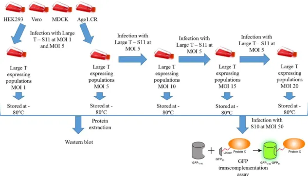
ANTIGENCell populations of HEK293, Age1.CR, Vero and MDCK cells expressing Large T – S11 were established by lentiviral vector infection (Figure 2.1). Briefly, HEK293 and Age1.CR cells were seeded in 6-well plates and MDCK and Vero in T25 flasks at a concentration of 1x10⁵
cells/well and 1x10⁵ cells/T25, respectively. After 24 hours cells were infected with lentivirus
carrying pRRL-SIN-CMV-LTS11 at MOIs 1 and 5. After 48 to 72h, the infected cells were amplified (from 6-well plates to T75 flasks and from T25 to T150 flasks) for cell banking and
re-22
seeding. Cells infected at MOI 5 (Large T expressing MOI 5 populations) were re-seeded at the same cell concentrations and infected with MOI 5 again, resulting in Large T expressing MOI 10 populations. This process was repeated twice, resulting in HEK293.T, Age1.CR.T, Vero.T and MDCK.T MOI 15 and LT-S11 MOI 20 populations. In order to evaluate expression of Large T antigen in LT-S11 cell populations, HEK293 and Age1.CR derived cells were seeded at a concentration of 2.5x10⁴ cells/cm2 in 24-well plates and MDCK and Vero derived cells were
seeded at a concentration of 1.3x5x104 cells/cm2 in 6-well plates. The next day, cells were infected
with lentivirus carrying pRRL-SIN-hPGK-S10 (and pRRL-SIN-hPGK, as a control) at MOI 50. GFP transcomplementation was analysed 48 hours post-infection by flow cytometry (CyFLow-space, Partec, Münster, Germany).
2.11 D
ETECTION OFL
ARGET
ANTIGEN BYW
ESTERNB
LOTTINGCell extracts were prepared in m-PER Mammalian Protein Extraction Reagent (Thermo Scientific, Waltham, MA, USA) at a concentration of 6.67x10³ cells/µL for Age1.CR and
HEK293 derived cell lines and 3.33x10³ cells/µL for Vero and MDCK derived cell lines. Large
T antigen expression was detected by Western blotting, after separation in a 4-12% (w/v) acrylamide NuPAGE gradient pre-cast gel (Invitrogen, Paisley, UK) on reduced and denatured samples using MES running buffer (Invitrogen). Primary antibody was a mouse monoclonal
Figure 2.1 – Establishment of cell populations expressing Large T – S11 fusion protein. Infection with Large T –
S11 refers to the infection with lentivirus carrying pRRL-SIN-LT-S11 and Infection with S10 refers to infection with lentivirus carrying pRRL-SIN-PGK-S10.
23
antibody against Large T antigen C-terminus from SV40 (Santa Cruz Biotechnology, CA, USA). Detection was performed with the corresponding secondary antibodies (GE Healthcare Life Sciences, Little Chalfont, UK), conjugated with horseradish peroxidase and detected by Amersham™ ECL Select™ (GE Healthcare Life Sciences).
2.12 C
ELL GROWTH STUDIES AND METABOLITE ANALYSISFor growth studies and subsequent metabolite consumption and production analysis, HEK 293 and HEK293T cell lines were seeded at 2x10⁴ cells/cm2 in T25 flasks. These were cultured under
standard conditions (7% CO2, 37ºC) for up to 9 days. Two samples were collected per day: culture
supernatant was harvested, filtered through 0.45μm for clarification, aliquoted and stored at -85ºC until analysis. Culture supernatants were analysed using automated enzymatic assays to determine glucose and lactate concentrations (YSI 7100 Multiparameter Bioanalytical System, USA) and Cell concentration and viability was determined by the trypan blue exclusion method.
To determine specific rates of cell growth and metabolite production/consumption, the Boltzmann equation was considered:
𝑑𝑃
𝑑𝑡 = µ𝑝𝐶
where P represents the different parameters, either viable cell concentration or metabolite concentration, C is the viable cell concentration, t is the culture time and μP is the cell specific