Universidade de Lisboa
Faculdade de Medicina
The role of regulatory T cells in the control
of B cell mediated immune responses
Ivonne Wollenberg
Ph.D. thesis in Biomedical Science
(Speciality Immunology)
2011
Universidade de Lisboa
Faculdade de Medicina
The role of regulatory T cells in the control
of B cell mediated immune responses
Ivonne Wollenberg
Thesis Supervisor: Luis Graça
Thesis Co‐Supervisor: Jose Faro
Ph.D. thesis in Biomedical Science
(Speciality Immunology)
2011
Todas as afirmações efectuadas no presente documento são da exclusiva responsabilidade do seu autor, não cabendo qualquer responsabilidade à Faculdade de Medicina de Lisboa pelos conteúdos nele apresentados.
A impressão desta dissertação foi aprovada pelo Conselho Cientifico da Faculdade de Medicina de Lisboa em reunião de 21 de Junho de 2011. The print of this thesis was approved by the Scientific Committee of the Faculty of Medicine Lisbon in June 21st 2011 meeting.
This work was supported by the fellowship SFRH/BD/29638/2006 from the Fundação para Ciênca e Tecnologia.
Table of Contents
ACKNOWLEDGEMENTS III SUMÁRIO VIII ABSTRACT X 1. GENERAL INTRODUCTION 1 1.1 The immune system 1 1.1.1 The innate Immune Response 1 1.1.2 The adaptive Immune Response 2 1.2 B cells 3 1.2.1 B cell subpopulations 4 1.2.2 Isotype switching and affinity maturation 6 1.2.3 Structure and diversity of antibodies 7 1.3 T cells 9 1.3.1 CD4+ T cells 10 1.4 CD4+ T cell activation and costimulation 13 1.4.1 OX40 14 1.4.2 OX40L 15 1.5 T cell independent immune responses 16 1.6 T cell dependent immune responses 16 1.6.1 Germinal center reaction 17 1.6.2 Affinity maturation 19 1.6.3 Follicular helper T cells 20 1.6.4 OX40L signaling and follicular helper T cells 22 1.6.5 Other Cells involved in the germinal center reaction 23 1.6.6 Germinal center regulation 24 1.7 Immune Tolerance 26 1.7.1 Ignorance mechanisms to maintain peripheral tolerance 26 1.7.2 Role of DCs in peripheral tolerance 27 1.7.3 Coinhibitory signals control peripheral tolerance 28 1.7.4 Blockade of costimulatory molecules as a therapeutic tool 29 1.7.5 Blockade of OX40‐OX40L signalling 31 1.8 Regulatory cells 32 1.8.1 CD4+Foxp3+ regulatory T cells 32 1.8.2 Mechanisms of Foxp3+CD4+ Treg cell function 33
II 1.9 Aims of this thesis 37 2. THE INFLUENCE OF OX40L BLOCKADE IN A MODEL OF ALLERGIC AIRWAY DISEASE 39 2.1 Background 39 2.2 Materials and Methods 41 2.3 Results 43 2.3.1 Anti‐OX40L treatment prevents allergic AHR 43 2.3.2 Anti‐OX40L treatment reduces allergic airway inflammation in pre‐sensitized mice 45 2.3.3 Anti‐OX40L treatment does not lead to long term tolerance 45 2.4 Discussion 47 3. IDENTIFICATION OF FOXP3+ FOLLICULAR T CELLS 49 3.1 Background 49 3.2 Materials and Methods 51 3.3 Results 53
3.3.1 Follicular CD4+ T cells contain a Foxp3+ subset 53
3.3.2 Follicular Foxp3+ T cells share properties of Foxp3+ Treg cells and TFH cells 54
3.3.3 Specificity of follicular Foxp3+ T cells 55 3.3.4 Origin of follicular Foxp3+ T cells 56 3.4 Discussion 59 4. REGULATION OF GCR BY FOXP3+ FOLLICULAR T CELLS 61 4.1 Background 61 4.2 Materials and Methods 63 4.3 Results 65 4.3.1 Co‐development of the GCR and GC T cells 65 4.3.2 Foxp3+ GC T cell concentration increases during GCR 66 4.3.3 GC Foxp3+ T cells are highly proliferative 66 4.3.4 Absence of Foxp3+ T cells enhances the magnitude of the GCR 67 4.3.5 Foxp3+GC T cells regulate the magnitude of GCR 69 4.4 Discussion 71 5. GENERAL DISCUSSION 73 6. REFERENCES 79 7. APPENDIX 101
Acknowledgements
I would like to thank: My two supervisors Luis Graça and Jose Faro who supported the development of this thesis with their combined effort. My thesis committee Henrique Veiga‐Fernandes, Ana Espada‐Sousa and Bruno Silva‐Santos for all the wise suggestions and helpful discussions. Ana for not having enough time for the GC project, which gave me the opportunity to start it. ;‐) Joana for being such a nice college, always there to lift up the spirit with a silly joke. Catarina for being so passionate about my project, you were a big help to me. Vanessa for all the fruitful discussions and the support. Thanks, for challenging me and not believing in TFH cells ;‐). Marta Monteiro for challenging me with a fascinating side project. Patricia for helping me to stay calm and keep on writing, instead of throwing my computer out of the window. Alex for making the effort to learn from me, to support this project, when I am gone. Marta Caridade, my neighbor, who was at the end always there for me when I needed her. The LSM710, dear friend we spend so many nights together. I will truly miss you. The IMM community, it was a pleasure working in this institute. I never before saw a science community being so open, helpful to each other and humble. Keep that spirit; unfortunately it is a rare thing in the science world. Jose Rino for teaching me everything about confocal microscopes. My Portuguese family for all their support, especially for the soup at midnight, when coming from the confocal.Á minha família portuguesa pelo apoio, especialmente pela sopa da meia‐noite depois do confocal.
My German family for sending all the care packages with tones of chocolates.
Meiner deutschen Familie; Danke Mama und Oma fuer all die Schokolade.
David for going with me through all the ups and downs of the last 4 years and being patient enough to teach me how to handle photoshop.
IV Thank you all so much!
Abbreviation List
Ab – antibody AHR ‐ airway hyperreactivity AID ‐ activation‐induced cytodine deaminase APC – antigen presenting cell BCR – B cell receptor C – constant (region) CCR7 – C chemokine receptor 7 D – diversity (gene segment) CD – cluster of differentiation CDR – complementary determining regions CSR – class switch recombination CTLA‐4 ‐ cytotoxic T cell associated antigen‐4 CXCR5 – CX chemokine receptor 5 DC – dendritic cell FDC – follicular dendritic cell FR – framework regions FRC – fibroblastic reticular cells GC – germinal center GCR – germinal center reaction H – heavy (chain) ICOS – inducible T cell costimulator iTreg – induced regulatory T (cell) Ig – immunglobulin IFN – interferon IL – interleukin i.p. – intraperitoneal i.v. – intravenous J – joining (gene segment) L – light (chain) Lck – lymphocyte‐specific protein tyrosin kinase LPS ‐ lipopolysaccharide ko –knockout LN – lymph node mAb – monoclonal antibody mLN – mesenteric lymph node MHC – major histocompatibility complex mTEC – medullary thymic epithelial cell NK – natural killer (cell) NKT – natural killer T (cell) nTreg – natural regulatory T (cell) OVA – ovalbumin PAMPs – pathogen associated molecular patterns PD‐1 – programmed death‐1 PRR – pattern recognition receptor SHM – somatic hypermutation SLO – secondary lymphoid organs SPF – specific pathogen free Src ‐ sarcoma TCR – T cell receptor TD – T cell dependent TdT – terminal desoxyribonucloetidyl transferase TFH – follicular helper T (cell) TFreg – follicular regulatory T (cell) Th – T helper (cell) TI – T cell independent TLR – Toll like receptor TNFRSF – TNF receptor superfamily Treg – regulatory T (cell)VI V‐ variable (region) VH – variable heavy (chain) VL – variable light (chain)
VIII
Sumário
Esta tese descreve o estudo da regulação de respostas imunitárias que conduzem à produção de anticorpos. Este tipo de respostas imunitárias depende de interações T‐B. A primeira parte da tese descreve o papel do bloqueio do ligando do OX40 (OX40L) na prevenção do desenvolvimento da asma alérgica num modelo animal. A asma alérgica é uma patologia dependente de células Th2 associada à produção de IgE e IgG1. A segunda parte desta tese descreve a regulação da reacção dos centros germinativos, um evento chave na produção de anticorpos e células B de memória. Este estudo levou à identificação de uma população funcionalmente relevante de células T foliculares com fénotipo regulador, isto é, células que expressam o factor de transcrição Foxp3 para além dos marcadores característicos de células T foliculares (PD‐1, CXCR5 e Bcl‐6).
A produção de anticorpos nos tecidos linfóides secundários, tal como os gânglios linfáticos e baço, em resposta a antigénios requer a rápida expansão de células CD4 específicas para esse antigénio e o seu recrutamento para os locais onde vão colaborar com as células B. Na zona T, as células T providenciam ajuda às células B, permitindo a rápida formação de plasmócitos em locais extrafoliculares. Nos folículos linfóides, as células T CD4 são necessárias para o desenvolvimento dos centros germinativos, importantes para a formação dos linfócitos B de memória e dos precursores de plasmócitos. Além disso, após re‐exposição ao mesmo antigénio, as células T de memória fornecem ajuda às células B, tanto de memória como naïve, de modo a obter uma resposta secundária mais rápida. As interacções celulares e moleculares que direccionam as células T para auxiliarem as células B durante uma resposta humoral, bem como a sua regulação, ainda não são bem compreendidas. O sinal co‐estimulatório fornecido pelas células dendríticas através de CD28 às células T é essencial ao desenvolvimento dos centros germinativos. As células CD4 activadas com CD28 passam a expressar OX40, uma molécula que não é expressa nas células T naïve, o que permite a obtenção de sinais secundários através de OX40L. Esta molécula é expressa nas células dendríticas activadas por CD40. Foi descrito que OX40 promove o desenvolvimento de células Th2 e a expressão do receptor de quimiocina CXCR5, que direcciona a migração de células CD4 para os folículos linfóides onde o seu ligando é expresso.
Neste trabalho examinámos o papel do OX40L no desenvolvimento da inflamação alérgica das vias aéreas, mediada por células Th2, usando um anticorpo monoclonal que bloqueia OX40L. A sensibilização e re‐exposição intra‐nasal com ovalbumina em ratinhos BALB/c induz características típicas da asma alérgica, nomeadamente a hiperreactividade das vias aéreas, infiltrados eosinofílicos, hiperplasia das células caliciformes e produção de citocinas Th2 nos pulmões. Observámos que a administração do anticorpo monoclonal bloqueante anti‐OX40L preveniu a indução da inflamação das vias aéreas. No entanto, este tratamento não levou à indução de tolerância específica para o antigénio administrado. Estes resultados mostram que OX40L tem um papel importante na fase de indução da doença.
Recentemente, a expressão de CXCR5, PD‐1 e do factor de transcrição Bcl‐6 permitiu a identificação de uma subpopulação de células T especializadas em providenciar ajuda às células B nos folículos linfóides. Estas células foram denominadas de células T auxiliares do folículo (TFH). As células TFH participam na resposta humoral providenciando sinais
importantes para a ocorrência de hipermutação somática e maturação da afinidade das células B dos centros germinativos.
Encontrámos uma sub‐população de células T foliculares, com características fenótipicas de células TFH que co‐expressam Foxp3 e que são recrutadas durante a reacção do centro
germinativo. Mostrámos que estas células T foliculares Foxp3+ derivam da população de células T reguladoras naturais. Com o propósito de estabelecer a importância fisiológica das células T foliculares Foxp3+ in vivo, usámos células Foxp3+ deficientes em CXCR5, que deste modo não conseguem aceder à região folicular. A transferência destas células Foxp3+ deficientes em CXCR5 mostrou que as células T foliculares Foxp3+ são importantes na regulação da reacção do centro germinativo depois da imunização com um antigénio T‐ dependente. Os nossos resultados in vivos mostraram que as células T foliculares Foxp3+ podem limitar a magnitude da reacção do centro germinativo, bem como a quantidade secretada de IgM, IgG1, IgG2b e IgA específicas para o antigénio. Como tal, as células T foliculares Foxp3+ parecem combinar características das células T auxiliares do folículo, com as características das células T reguladoras para controlar as respostas imunes humorais. No seu conjunto, os dados desta tese descrevem a identificação de mecanismos chave na regulação da reacção do centro germinativo que, em última análise, previnem as patologias mediadas por anticorpos.
X
Abstract
This thesis reports research on the regulation of immune responses leading to a humoral immune reaction. This type of immune phenomena is based on B‐T cell interactions. The first part of the thesis is devoted to study the effect of OX40‐ligand blockade in preventing allergic airways disease in mice. Allergic airways disease is a Th2‐dependent pathology associated with production of IgE and IgG1 specific to the allergen. In the second part of the thesis the regulation of germinal centre reaction, a key event for the production of antibodies and B cell memory, is investigated leading to the identification of a follicular population of Foxp3+ regulatory T cells.
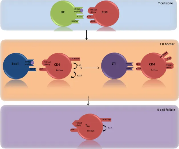
To mount a successful antibody response to antigens, rare antigen‐specific CD4 T cells have to be recruited to the secondary lymphoid tissues, enabling cognate B cell/T cell interactions. In the T cell zone, T cells provide help for B cells, allowing first a fast antibody response by the generation of plasma cells in extrafollicular foci. In the B cell zone, CD4 T cells are required for the development of germinal centers, which subsequently give rise to memory B cells and the precursors of long‐lived plasma cells. Moreover, after re‐exposure to antigen, memory CD4 T cells provide help to both memory and naive B cells for more efficient secondary immune responses. The cellular and molecular interactions that direct T cells to encounter with B cells for cognate interaction to provide survival and differentiation signals in secondary immune responses, are still incompletely understood. The CD28 costimulatory signal that T cells receive from dendritic cells, during priming in the T cell zone, are essential for GC development. Activation of CD4 T cells through CD28 upregulates OX40, which is not expressed on naive T cells, allowing CD40‐activated dendritic cells to provide secondary signals through OX40 ligand. It has been reported that OX40 signals can promote Th2 development and induce expression of the chemokine receptor CXCR5 by CD4 T cells, which directs their migration to B cell follicles following a CXCL12 gradient, which is the ligand of CXCR5.
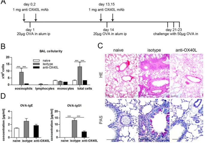
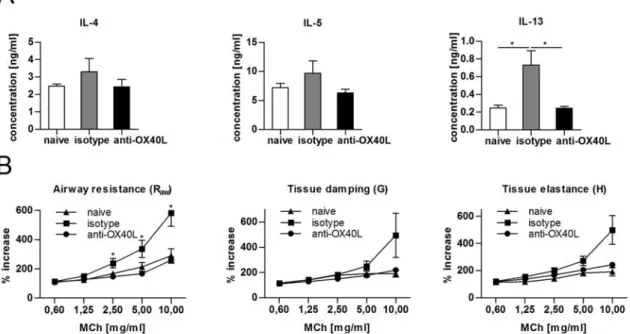
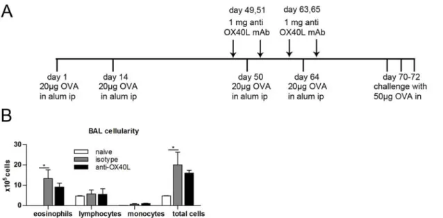
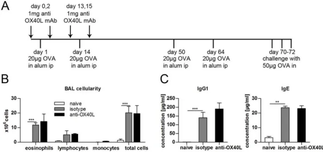
We examined the role of OX40L in the development of Th2‐mediated airway inflammation by utilizing a blocking anti‐OX40L monoclonal antibody (mAb). Sensitization and airway challenge with ovalbumin in BALB/c mice induced typical features of allergic asthma, namely airway hyperreactivity, eosinophilic infiltrates in the airways, hyperplasia of goblet cells with increased mucus production, and high levels of Th2 cytokines in the lung. Administration of blocking anti‐OX40L mAb at the time of sensitization prevented the induction of airways inflammation. However, treatment with anti‐OX40L mAb did not lead to long‐term tolerance against the administered allergen. These results indicate a critical role for OX40L in the induction phase, which leads to the development of pathogenic Th2 cells, but not in the induction of tolerance.
Recently, expression of CXCR5, PD‐1, and the transcription factor Bcl‐6 has allowed the identification of a defined T cell subpopulation specialized in providing B cell help in lymphoid follicles. These cells have been named follicular helper T cells (TFH). TFH cells
participate in humoral responses providing signals required for somatic hypermutation and affinity maturation of germinal center B cells. We found that a proportion of follicular T cells, with phenotypic characteristics of TFH cells but expressing Foxp3 are recruited into the germinal center during the course of a germinal centre reaction. We found that, these follicular Foxp3+ T cells derive from natural regulatory T cells. In order to establish the in vivo physiological importance of Foxp3+ follicular T cells we used CXCR5‐deficient Foxp3+ cells, which do not have access to the follicular region. Adoptive cell transfers of CXCR5‐deficient Foxp3+ cells showed that Foxp3+ follicular T cells are important regulators of the germinal center reaction following immunization with a thymus‐dependent antigen. Our in vivo data show that Foxp3+ follicular T cells can limit the magnitude of the germinal center reaction and also the amount of secreted antigen‐specific IgM, IgG1, IgG2b and IgA. Therefore, Foxp3+ follicular T cells appear to combine characteristics of follicular helper T cells and regulatory T cells for the control of humoral immune responses. Taken together, the data in this thesis report the identification of key mechanisms regulating the germinal center reaction, and ultimately preventing antibody‐mediated pathology.
1
1. General Introduction
1.1 The immune system
Early vertebrates evolved throughout millennia a complex immune system that protects individuals from a broad range of dangerous pathogens. It can be traditionally divided into innate and adaptive immune system, each with different function and role.
The innate immune response involves several different cellular players, as well as specific molecules, allowing a fast, but unspecific response, being the first line of defense against foreign pathogens. The adaptive immune response takes around 2‐3 days to start, but has the advantage, that the response is specific for each pathogen and by that very effective. As a consequence, the adaptive immune response represents the second line of defense during an infection. Another main feature of the adaptive immune response is to generate memory cells after the encounter with a pathogen. These memory cells persist in the host under steady state conditions for months to years and allow the body to mount a fast specific response (secondary immune response) when exposed a second time to the same pathogen. This secondary immune response represents a powerful tool in the host defense against pathogens and is the basis for prophylactic vaccination.
1.1.1
The innate Immune Response
The effector mechanisms of innate immunity, which include antimicrobial peptides, phagocytes, and the alternative complement pathway, are activated immediately after infection to fast and efficiently control the infecting pathogen.
During evolution, the innate immune system appeared before the adaptive immune system, and some form of innate immunity probably exists in all multicellular organisms. This system consists of a humoral and a cellular part. The cellular part of the innate immune system is performed by cells of hematopoetic and nonhematopoetic origin. Hematopoetic cells involved in innate immunity include macrophages, dendritic cells (DCs), mast cells, neutrophils, eosinophils, natural killer (NK) cells and natural killer T (NKT) cells. Albeit DCs and NK T cells are classically seen as part of the innate immune system, they also show features of the adaptive immune system, hence they represent a bridge between innate and adaptive immune response. In addition to hematopoetic cells, innate immune response also includes nonhematopoetic cells like for example the epithelial cells of the skin. In contrast to the adaptive immunity, innate immune recognition is mediated by receptors with a genetically predetermined specificity (expressed mainly by macrophages and DCs). The advantage of these germ‐line‐encoded receptors is that they evolved to have defined specificities for common molecular structures represented by infectious microorganisms. The disadvantage is, that microorganisms can mutate at much higher rates than any of their hosts. Therefore the strategy of the innate immune response is to focus on a few, highly conserved structures present in large groups of microorganisms, rather than recognizing
every possible antigen (Janeway, 1989). These structures are referred to as pathogen‐ associated molecular patterns (PAMPs), and the receptors of the innate immune system that evolved to recognize them are called pattern‐recognition receptors (PRRs). Examples of PAMPs are bacterial lipopolysaccharide (LPS), peptidoglycan, lipoteichoic acids, mannans, bacterial DNA, double‐stranded RNA, and glucans. Eventhough these molecules are chemically quite distinct, all PAMPs share certain features (Janeway, 1992; Medzhitov and Janeway, 1997). PAMPs are produced only by microbial pathogens, and not by their hosts. For example, LPS is synthesized only by gram‐negative bacteria. Furthermore, the molecular structures recognized by the innate immune system are usually essential for the survival or pathogenicity of the microorganism, as a consequence those structures are more conserved between different species and cannot be easily altered to escape the immune defense. For example, all gram‐positive bacteria have lipoteichoic acids, and therefore, the lipoteichoic acid PRR of the host can detect the presence of virtually any gram‐positive bacterial infection. Signaling receptors recognize PAMPs and activate signal‐transduction pathways that induce the expression of a variety of immune‐response genes, including inflammatory cytokines. The receptors of the toll‐like family (TLR) have a major role in this induction. To support and increase these cellular defenses, innate immunity also has a humoral component. Those proteins, such as complement proteins, LPS binding protein, C‐reactive protein and others circulate through the body and are involved in both sensing microbial components and effector mechanisms to facilitate clearance of infection.
1.1.2
The adaptive Immune Response
The adaptive component of the immune system is organized around two classes of specialized cells, T cells and B cells. The T‐cell receptor (TCR) and the B‐cell receptor (BCR) are, not like in the innate immune system germline‐encoded, but rather generated somatically. Given the fact that these receptors are not genetically encoded, they are not predestined to recognize any particular antigen and therefore an extremely diverse repertoire of receptors has to be generated. Given that each lymphocyte displays a single kind of structurally unique receptor, that the number of lymphocytes is very large (>108 in mice) and that the average lymphocyte clonal size is small (<100 cells), it follows that the repertoire of antigen receptors in the entire population of lymphocytes is very large and extremely diverse. The dimension and diversity of this repertoire increases the possibility that an individual lymphocyte will encounter an antigen that binds to its receptor, leading to activation and proliferation of this cell.
As each T and B cell owns a unique kind of receptor, clonal expansion of lymphocytes in response to infection is absolutely mandatory for the generation of an efficient immune response. After having contact to a foreign pathogen the lymphocyte gets activated and goes through multiple rounds of proliferation building a clonal army against this specific pathogen. However, since the binding sites of the receptor arise from a random process, these binding sites also possess the risk of generating receptors that recognize self antigens.
3
1.2 B cells
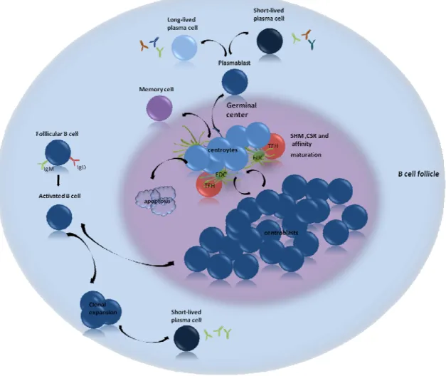
In mammals, B cells arise from hematopoetic cells in the bone marrow or fetal liver and achieve maturity in peripheral lymphoid organs. In the bone marrow of adults B cells pass through several distinct developmental stages, during which they acquire their antigen‐ specificity. At an immature stage, B cells exit the bone marrow and complete their development to the mature or naive stage in the periphery. Those naive B cells express IgM together with IgD on their surface. The development in the bone marrow occurs in the absence of any exogenous antigen. Thus this stage is called antigen‐independent B‐cell development (Fig. 1).
After maturing in the secondary lymphoid organs (SLOs) naive, mature follicular B cells recirculate through the body and migrate repeatedly through the B cell area of the spleen and the lymph nodes, waiting to get activated as a consequence of antigen recognition. In the lymph nodes B cells are concentrated in the cortex in primary follicles, in contact with follicular dendritic cells (FDC). B cells, unlike T cells, recognize antigens in their native form. Low‐molecular‐weight antigens might diffuse directly into B‐cell areas in secondary lymphoid tissues. Larger molecules require active cellular mechanisms that are still being examined (Batista and Harwood, 2009; Cyster, 2010). Antigens complexed with IgM, IgG, and complement might be carried on the surfaces of specialized macrophages, FDCs and even B cells themselves, collectively expressing receptors for IgG Fc and complement fragments on their surface (Elgueta et al., 2010). Antigen presented on the surface of these cells can stimulate B cells through B cell receptor (BCR) crosslinking, expression of other interacting surface molecules, and secreted cytokines.
The surface immunglobulin that serves as the BCR has two functions in B‐cell activation. First, like the antigen receptor on T cells, it transmits signals directly to the cell's interior when it binds antigen. Second, the BCR delivers the antigen to intracellular sites where it is processed and subsequently returned to the B‐cell surface as peptide bound to major histocompatibility complex (MHC) class II molecules. B cells require two principal types of signals to become activated. Signal 1 is delivered by cross‐linking of the immunoglobulin receptor. This cross‐linking leads to activation of intracellular signaling pathways allowing the cell to interact with T cells that recognize the same antigen and thus deliver the signal 2 through CD40. The cognate interaction between T cells and B cells is analogous to the interaction between T cells and DCs, which are usually referred to as professional antigen presenting cells (APCs). B cells express many of the same costimulatory molecules found on DCs, such as CD40, B7‐1 (CD80), and B7‐2 (CD86). T cells and B cells form an analogous immunologic synapse, which is followed by a signaling pathway. This initial interaction takes place at the boundary between primary follicles and T‐cell areas in SLOs. The activated B cells enter one of two pathways: either they immediately start secreting low‐affinity antibodies as short lived plasma cells, or they enter a follicle to establish a germinal center (GC) (Batista and Harwood, 2009). Some microbial antigens can activate B cells directly in the absence of T cell
help. The ability of B cells to respond directly to these antigens provides a rapid response to many important bacterial pathogens. However, somatic hypermutation and switching to certain immunoglobulin isotypes depends on the interaction of antigen‐activated B cells with helper T cells.
B cells are active as APCs and express peptides along with MHC class II on their surface. These peptides, as already mentioned above, originate from processed antigen internalized after binding to the BCR (Huston, 1997). When the B cell contacts a CD4+ T cell specific for such a MHC class II/peptide complex and having been previously activated by a professional APC, the T cell is able to provide cognate help and activate the B cell for further differentiation into memory or plasma cell.
1.2.1
B cell subpopulations
Newly formed immature B cells are defined by their short half‐lives and their tendency to undergo apoptosis rather than proliferating following BCR engagement and are usually classified as transitional (T)1 and T2 B cells (LeBien and Tedder, 2008) (see Fig. 1). Naive mature B cells can be commonly divided into three subsets, B‐1 B cells (that are typically subdivided into B‐1a and B‐1b), follicular B cells and marginal zone B cells. The cells of the different subsets vary in terms not only of their location, but also of their ability to migrate and likelihood to be activated in a T‐dependent (TD) or T‐independent (TI) fashion.
Marginal zone B cells are positioned at the marginal zone by the activity of S1P1 and S1P3, receptors for sphingosine1‐phosphate (Cinamon et al., 2004; Cinamon et al., 2008; Vora et al., 2005) and have a particular role in responding to TI antigens type 2 (Steiniger et al., 2006). They are involved in TD B cell responses, can mediate the transport of antigen in form of immune complexes into splenic follicles, but they may also participate in immune responses to lipid antigens. Marginal zone B cells can be induced to differentiate into short‐ lived plasma cells in the absence of ligation through their BCR, hence they are considered to be innate‐like B cells. It is to date not fully understood whether a distinct population of marginal‐zone B cells also exists in humans, as the histological structure of the spleen is distinct from the murine one.
5
Figure1. B cell development and B cell subpopulations. B cell development starts in the fetal liver or bone
marrow. The terminal differentiation of B cells occurs in the B cell follicle. B‐1a, B‐1b and B10 populations are still very poorly characterized.
In mice the presence of the surface marker CD5 distinguishes a B cell population with distinct characteristics, called B‐1 cells. They develop early in ontogeny, they tend not to undergo somatic hypermutation (SHM), and they secrete IgM antibody with polyspecificity, including binding to self‐antigens (Dorshkind and Montecino‐Rodriguez, 2007). B‐1 cells are so far the best characterized in mice. B cells expressing CD5 were also found in humans , and at least a subset of these cells might have similarities to those of murine B1 cells. Still, phenotypically and functionally distinct sublineages are not well described. Murine B‐1a and B‐1b cells seed the peritoneal and pleural cavities, however their origin and their precursors have been controversial. The pleuroperitoneal milieu appears to influence the functional characteristics of both B‐1a and B‐1b B cells as well as of the relatively small proportion of B‐ 2 cells that reside in these sites (Berberich et al., 2007; Hastings et al., 2006). LPS from commensal bacteria presented by DCs can induce both the proliferation of B‐1 B cells as well as their differentiation into IgM‐secreting short‐lived plasma cells. Antigen‐specific B‐1 B cells can be induced to switch in a T‐cell‐independent manner into IgA‐secreting cells (Fagarasan and Honjo, 2003).
The CD5‐ B cell population is called B2 or conventional B cells, which include a number of subpopulations that represent different maturation stages. Depending on surface markers and the immunoglobulin isotype expressed conventional mature B cells can be distinguished as naive B cells (IgD+IgM+CD27+), GC B cells (Fas+GL7+), switched memory B cells (IgM‐IgD‐ CD27+), and plasmablast (CD38high, IgM‐) (Allman and Pillai, 2008; Chung et al., 2003).
Additionally, B cells with regulatory functions have been described (Mizoguchi and Bhan, 2006) and one phenotypically distinct subset, called B10 B cells, has been shown to regulate T cell‐mediated inflammatory responses by means of IL‐10 production (Yanaba et al., 2008).
1.2.2
Isotype switching and affinity maturation
The genes encoding immunoglobulins are assembled from 4 heavy chain segments (VH, D, JH and CH) and 3 light chain segments (VL, JL, and CL). There are 9 different heavy chain types (, , 1‐4, 1, 2 and ), determining the isotype of the immunoglobulin and 2 light chain types ( and ). The gene encoding the constant region lies closest to the JH gene segment and therefore closest to the assembled V‐region exon after DNA rearrangement which makes IgM the first immunoglobulin isotype to be expressed during B‐cell development. The gene encoding is followed by the gene encoding the constant region, consequently the next immunoglobulin to be expressed in B cell development is IgD.
IgM and IgD are expressed by alternative splicing of the same VHDHJH exon to the and heavy chain exons (Fig. 2 upper panel). Th cells are involved in the maturation process of B cells outside the bone marrow. T cell‐derived cytokines induce isotype switching, which is a process of DNA rearrangement. Switching moves the rearranged VHDHJH exon into a position upstream, bringing alternative exons of the C region in close proximity of the V region (Fig. 2, lower panel). This permits a functionally rearranged VHDHJH exon to be used to produce antibodies of different isotypes but the same antigenic specificity (Malisan et al., 1996). T cell–derived IL‐4 causes switching to IgG1. IL‐5 and TGF‐β cause switching to IgA. IFN‐γ appears to induce switching to IgG2 (Chaudhuri and Alt, 2004). Simultaneously, as B cells undergo isotype switching, an active process produces mutations, apparently randomly, in the antigen‐binding portions of the heavy and light chains. If these mutations result in loss of affinity for the antigen, the cell loses access to important growth signals and dies. However, if the mutations result in increased affinity for the antigen the cell producing that antibody will have a proliferative advantage in response to antigen and grows to dominate the pool of responding cells. Somatic mutation and clonal expansion of mutated cells occurs in the GCs of SLOs (Schmidlin et al., 2009).
7
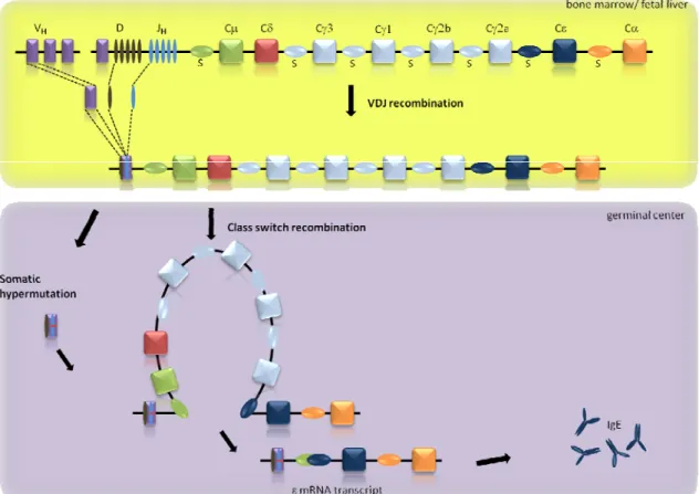
Figure 2. Rearrangement of the immunoglobulin heavy chain.. V(D)J recombination takes place in the bone
marrow, while somatic hypermutation and CSR occur in the peripheral lymphoid tissues. V(D)J recombination selects one segment for each of the V, D and J segments from a pool of gene fragments and combines them into a variable (V)‐region exon. Somatic hypermutation introduces mutations in the rearranged V exon., CSR brings the downstream constant (C) region exon in the proximity of the V exonen abling the production of antibodies with different isotypes.
1.2.3
Structure and diversity of antibodies
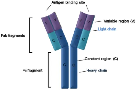
All antibodies share the same basic structure but exhibit high variability regarding the antigen binding region. The heterodimeric structure of an antibody is composed of two heavy (H) and two light (L) chains that are covalently connected via disulfide bonds. Each heavy and light chain consists of an amino terminal variable (V) and a carboxy terminal constant region (C). As mentioned above, the constant region of an antibodies H chain assigns the molecule its Ig class (IgM, IgD, IgA, IgG and IgE) conferring different effector functions, such as complement activation and mediation of cell cytotoxicity, depending on the isotype. On the other hand, the variable region of both, heavy and light chain, defines the antigen‐specificity of the antibody and thereby accounts for the recognition of antigens. In any given antibody the two heavy and light chains are always identical, resulting in an antibody molecule with two identical binding sites and by that the ability to bind simultaneously two identical structures (Fig. 3). Proteolytic enzymes have been used to dissect the structure and function of the antibody molecule. Digestion with the protease
papain cleaves antibodies into three fragments. Two of them are identical and are called Fab fragments (fragment antigen binding). The Fab fragments correspond to the two identical arms of the antibody molecule, consisting of the light chain and the variable region together with one domain of the constant region from the heavy chain (Fig. 3). The third fragment is called Fc fragment and contains the two other domains from the constant region. As already mentioned, the Fc fragment is the part of the antibody able to interact with other molecules and cell receptors and therefore responsible of antibody effector functions.
The variable regions of H and L chain consist of three highly divergent segments called hypervariable regions that are flanked by conserved framework regions (FR). In three‐ dimensional space, the three hypervariable segments of the (VH) chain and the three hypervariable segments of the (VL) chain are brought together to form the antigen binding surface. The antigen binding surface is complementary to the three‐dimensional structure of bound antigen, therefore the hypervariable segments are also referred to as complementarity‐determining regions (CDR) (Janeway and Travers, 2001).
Figure 3. Structure of an IgG molecule. The protease papain cleaves immunoglobulins into three segments:
Two Fab fragments which consist of the light chains (light blue) and a part of the heavy chain (dark blue) and the Fc fragment which consists of a part of the heavy chains. The Fab fragments contain the antigen binding site, the Fc fragment the effector region. The enormous diversity of the antibody repertoire is generated by somatic recombination ‐ also referred to as VDJ recombination ‐ of Ig genes during the development of B lymphocytes in the bone marrow (Figure 2 upper panel). Three separate loci encode the two Ig light chains and the Ig heavy chain. Each light chain locus is composed of three different clusters of gene segments, referred to as variable (V), constant (C) and joining (J) gene segments. The IgH locus bears an additional cluster of diversity (D) gene segment situated between V and J clusters. The genes within a cluster are each separated from another by regions of non‐ coding DNA that vary in length. The somatic recombination of gene segments within each Ig
9
locus is a requisite step for the production of a functional antibody molecule and follows a precise order. The first recombination, occurring in the IgH locus results in joining of one of the D to one of the J gene segments. Thereafter, one of the V gene segments is joined to the DJ complex. Due to the lack of D gene segments within the light chain loci, somatic recombination directly joins one of the V to one of the J gene segments. The somatic recombination of gene segments within each locus occurs randomly. Therefore, the diversity that can be generated at each locus depends on the number of genes within its clusters. The diversity of antibodies is further enhanced by the so‐called junctional diversity that is due to "non‐precise" joining of gene segments. During somatic recombination, nucleases may remove nucleotides of the recombining gene segments. In addition, the enzyme terminal deoxyribonucleotidyl transferase (TdT) mediates the random addition of up to 20 non‐ germline encoded nucleotides at the junctions (Krangel, 2003). Taken together, theoretically the potential murine antibody repertoire comprises 1012‐ 1014 different specificities. Because formation of the B lymphocyte repertoire in the bone marrow is antigen independent, it is also referred to as pre‐immune repertoire.
1.3 T cells
T cells express an antigen‐receptor, referred to as TCR that is related to immunoglobulins but strongly differs structurally from them. As all haematopoietic cells, T cells originate in the bone marrow, but develop in the thymus. In the thymus they recombine the TCR segments, of which there are four: α,β,γ and δ. Recombination begins at the γ, δ and β loci, and if expression of the γδTCR is successful, commitment to the γδT‐cell lineage results (Lauritsen et al., 2006). γδ T cells leave the thymus to populate the lymphoid tissue and epithelia. Alternatively, successful β loci recombination results in βTCR expression, which pairs with the surrogate α receptor (pre‐Tα) and forms the pre‐TCR. It follows the recombination of the α‐loci which generates, if successful, the αβTCR. Those thymocytes express CD4 and CD8 co‐ receptor molecules and undergo clonal selection by binding to peptide‐loaded MHC molecules (peptide‐MHC) expressed on thymic cortical epithelia. The interaction of TCR complex with peptide‐MHC complexes is restricted by the specificity of the TCR and T‐cell coreceptor. CD4 restricts interaction to class II MHC and CD8 to class I MHC molecule. T cell clones that bind with sufficient affinity receive survival signals and get positively selected. Surviving cells then lose the CD4 or CD8 coreceptor not involved in MHC recognition. These single‐positive cells migrate to the thymic medulla, and those that react too strong with self‐ antigens presented by medullary thymic epithelial cells (mTEC) and APCs are deleted by apoptosis mechanisms (negative selection) (Carpenter and Bosselut, 2010). The CD4‐ expressing T cells have been classically designated as the helper lineage of the T cells and the CD8‐expressing T cells are the cytotoxic lineage. Both represent the two major lineages among mature T cells.
Mature T cells are activated on interaction of their TCRs with peptide‐MHC complexes. CD8 T cells can interact with peptides (9‐11 amino acids in length) on almost any cell expressing
MHC class I. These MHC class I ‐ restricted peptides are usually produced from proteins translated within the cell encoded either in the host genome or by infecting viruses or other intracellular pathogens. In contrast, the TCRs of CD4 T cells engage peptides complexed to MHC class II. In contrast to MHC class I, which is constitutively expressed in all nucleated cells, MHC class II molecules are only present on the surface of APCs and their membrane levels are increased by innate immune stimuli, including ligands for TLRs. As a consequence of their activation, after antigen encounter, APCs start migrating from the skin and mucosal sites to nearby lymph nodes, where interaction with T cells will initiate a immune response. T‐cell activation is initiated when the TCR and associated proteins recognize a peptide‐MHC complex on an APC, leading to a rapid clustering of TCR‐associated molecules at the interface between T cells and APCs and the formation of a immunological synapse (Dustin, 2009). At the T‐cell side the synapse is formed around a central cluster of CD3 and TCR, which bind specifically to the peptide‐MHC complex, as well as CD4/CD8 molecules, which stabilize this interaction. Binding to MHC/peptide on the APCs by TCRs and at the same time CD4/CD8 in the synapse brings the cytosolic domains of these molecules into proximity. As a result, the CD4‐ and CD8‐associated Src family protein tyrosine kinase Lck is able to phosphorylate tyrosine residues contained in cytoplasmic immunoreceptor tyrosine‐based activation motifs of the TCR‐associated CD3 chains. This results in the start of a signaling cascade leading to the activation of the T cell (Dustin, 2009).
Elimination of intracellular pathogens and tumors relies on cell‐mediated immune response. CD8 effector T cells are the cells pivotal in this response and their function is distinguished by antigen‐specific cytotoxicity restricted by MHC class I. After activation, CD8 T cells produce cytotoxic proteins including perforin and granzymes and secrete them at the point of contact with the target cell, the immunological synapse, therefore resulting in specific killing of the target cell without bystander cell damage. Perforin is a membrane‐disrupting protein that allows granzymes to enter the cell and induce apoptosis. In addition to cytolysis, CD8 effectors produce mainly IFN‐γ and TNF, which are pro‐inflammatory cytokines.
1.3.1
CD4
+T cells
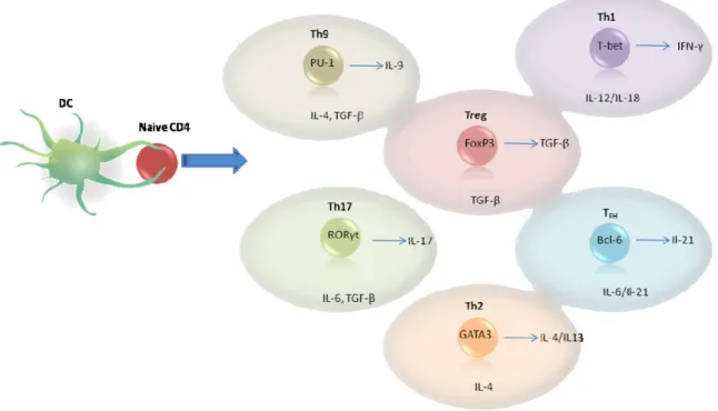
Many aspects of the adaptive immune response start with the recognition of foreign MHC class II complexes on the surface of APCs by CD4 T cells (Banchereau and Steinman, 1998). Naive CD4+ T cells can differentiate into at least five main functional subsets: T helper‐1 (Th1), Th2, Th17, regulatory T cells (Treg) and the B follicle‐residing follicular helper T cells (TFH) (Fig.4). Moreover recent studies showed an IL‐9 producing subset called Th9 cells(Veldhoen et al., 2008) and a IL‐22 producing subset called Th22 was found in human skin samples (Duhen et al., 2009). In addition, other CD4+ T cells may also contribute to the adaptive immune response namely, Tr1 and NKT cells.
The differentiation of naive CD4+ T cells into effector cell lineages underlies successful adaptive immune responses aimed at distinct categories of pathogens. Their functional specialization is coordinated by genetic programs that use different transcription factors to
11
direct expression of distinct soluble mediators and surface molecules that support interactions with other immune cells. The first paradigm for this functional diversification was the description of Th1 and Th2 CD4+ effector subsets by Mosmann and Coffman in 1986 (Mosmann et al., 1986). Th1 cells were thought to be responsible for delayed‐type hypersensitivity, activating macrophages through release of interferon (IFN)‐γ and enabling them to kill intracellular pathogens. Th2 cells were considered the classical helper T cells providing help to B cells to generate class‐switched antibodies. GATA‐3 was identified as the transcription factor for the Th2 lineage, while for the Th1 lineage Tbet was described as the key transcription factor (Murphy et al., 2000). In Th2 cells, the transcriptional activation of GATA‐3 provides a self‐reinforcing feedback circuit (Ouyang et al., 2000). Likewise, T‐bet induces its own expression, either directly (Mullen et al., 2001) or indirectly (Afkarian et al., 2002). Furthermore the most characteristic cytokines those subsets produce, further the transcription of those factors, while inhibiting the expression of other transcription factors.
Figure 4. CD4 T cell lineages. Classical view of lineages and master regulators. These subsets express lineage
defined transcription factors and produce selective cytokines. Environmental factors will support the development into the different lineages.
In 2003 the requirement for IL‐23 in IL‐17‐producing CD4+ T cells was recognized and with that, the classical Th1/Th2 model had to be revised. IL‐17‐producing cells, rather than Th1 cells, were established to play an important role in the animal model of multiple sclerosis (MS) and experimental autoimmune encephalomyelitis (EAE) (Murphy et al., 2003). Initially presumed to diverge from a common Th1 precursor (Bettelli and Kuchroo, 2005), the IL‐17‐ producing cells, named Th17, were classified as a new subset on the basis of being
independent of the transcription factors GATA‐3 and T‐bet (Harrington et al., 2005; Park et al., 2005). The robust inducing conditions of IL‐6 and TGF‐β (Veldhoen et al., 2006) and the identification of RORγt and RORα as lineage‐defining transcription factors (Ivanov et al., 2006; Yang et al., 2008) provided definitive support of Th17 as a separate subset.
Another main subset of CD4+ T cells are Treg cells (Josefowicz and Rudensky, 2009), characterized by expression of the transcription factor forkhead box protein 3 (Foxp3). Treg cells derived from the thymus are thought to be a stable subset. However, Treg cells can be induced in the periphery from naive CD4+ T cells by activation in the presence of TGF‐β. Like the natural Treg cells (nTreg), induced Treg (iTreg) cells express Foxp3, but may be less stable and share circuitry with Th17 cells, which also require TGF‐β for their differentiation (Curotto de Lafaille and Lafaille, 2009; Komatsu et al., 2009).
TFH cells are localized in B cell follicles and essential for the generation of high‐affinity and
isotype switched antibodies and B cell memory (Breitfeld et al., 2000; Kim et al., 2001; Schaerli et al., 2000; Vinuesa et al., 2005). Although all activated CD4+ T cells can migrate to follicular regions, TFH cells preferentially reside there by virtue of their continuous expression
of the chemokine receptor CXCR5. TFH cells have the potential to secrete Th1, Th2 or Th17
cytokines (Crotty, 2011) and produce large amounts of IL‐21, which acts in an autocrine manner together with IL‐6 on their differentiation and expansion. The differentiation of TFH
cells depends also largely on the transcription factor Bcl‐6, which is the key regulator of the TFH lineage (Vogelzang et al., 2008; Yu et al., 2009b).
The CD4+ T cell subpopulation are defined according to the lineage‐indicating cytokine profile, their function and associated key transcription factors. Besides the aforementioned established CD4+ T cell lineages there are various recent reports about additional new subsets.
A population of IL‐9‐producing cells, derived from Th2 cells by treatment with TGF‐β, has been described (Veldhoen et al., 2008). IL‐9 was once considered a Th2 cytokine but is now recognized as not being expressed together with IL‐4, IL‐5 or IL‐13. Although suggested to be produced by Th17 or iTreg cells (Elyaman et al., 2009; Lu et al., 2006; Nowak et al., 2009), IL‐ 9 is not expressed together with IL‐17 or IL‐22 and is not expressed by nTreg cells (Veldhoen et al., 2008). As this population has only been examined in vitro, it is unclear whether IL‐9 producers should be considered a new subset, to be called Th9, or whether expression of this cytokine reflects adaptation of Th2 cells to a change in the microenvironment in the course of a response triggered by a pathogen or allergen. Recently the transcription factor PU‐1 was identified as to be important for Th9 differentiation (Chang et al., 2010).
In another recent work, human, but not mouse, Th22 T cells (expressing IL‐22 but not IL‐17 or RORγt) were described (Duhen et al., 2009; Trifari et al., 2009) and may represent a skin‐ homing subset responsible for skin inflammation such as psoriasis. These cells preferentially develop when cultured with plasmacytoid DCs, which infiltrate psoriatic skin, but are independent of (and even inhibited by) IFN‐α (Duhen et al., 2009), making their link to skin inflammation still uncertain.
13
1.4 CD4
+T cell activation and costimulation
T cell activation begins with the binding of antigenic peptides to MHC molecule on the surface of an APC which is then, in its processed form, recognized by the TCR. The TCR has no intracellular signaling domain, but its membrane expression requires being physically associated to accessory molecules known as the CD3 complex (Salmond et al., 2009). The CD3 complex consists of three types of transmembrane molecules named CD3γ, CD3δ and CD3ε plus a mainly intracytoplasmic homodimer of 2 CD3ζ chains. TCR and CD3 complex together are termed the TCR complex.
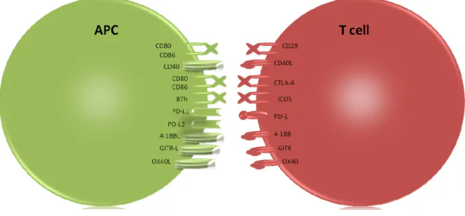
The activation signal delivered by a ligand bound to the TCR complex, known as signal 1, is not sufficient by itself to completely activate a T cell. The TCR‐peptide‐MHC interaction is stabilized by the CD4 co‐receptor. Besides the co‐receptor a number of other costimulatory molecules are additionally required to enhance signal 1 and are referred to as signal 2. Costimulatory molecules on T cells (Fig. 5) can be divided, on the basis of their structural relationships, into three groups: immunoglobulin (Ig)‐like receptors, integrins, and tumor necrosis factor receptor superfamily (TNFRSF) members. The majority of these molecules is expressed following TCR engagement and provides positive signals to the T cell encouraging maturation, proliferation, survival, and cytokine production.
Ig‐like receptors, including the inducible costimulator (ICOS) as well as the constitutively expressed CD28, interact with ligands expressed on the surface of APCs. CD28 provides strong costimulation and, in almost all cases, is necessary for full T cell activation. Signaling through CD28 enhances protein tyrosine phosphorylation (Vandenberghe et al., 1992) and promotes cytoskeletal reorganization (Sedwick et al., 1999) and TCR association with lipid rafts (Viola et al., 1999). Furthermore CD28 signaling augments gene expression and stabilizes mRNA molecules from pro‐inflammatory and survival genes (Cerdan et al., 1992; Wu et al., 2005). These signals continue until CD28 is displaced by cytotoxic T lymphocyte antigen‐4 (CTLA‐4), which arrests signaling and ends the activation process. Without the CD28 signal, T cells become anergic, which means they lose the ability to respond to antigen and acquire a passive, non‐proliferative phenotype (Gimmi et al., 1993). This state of anergy has been claimed to be important to the development of peripheral tolerance ensuring that harmless antigens, encountered in the absence of other “danger” signals, do not trigger a potentially damaging autoimmune or allergic response (Gallucci and Matzinger, 2001). In contrast, T cells that receive full costimulation start proliferating, along with the production of cytokines, and ultimately survive and progress into the memory pool. The CD28 signal is so vital to full activation of naive T cells that it is known as signal 2 (Herrick and Bottomly, 2003).
Figure 5. Costimulatory molecules. Costimulatory molecules can have either positive or negative stimulatory
function. Depicted are some of the costimulatory receptors and their ligands expressed by APCs and T cells.
Integrins, such as leukocyte function‐associated antigen‐1 (LFA‐1), also provide T cells with activation signals following interaction with their ligands on APCs, but are reliant on the presence of other costimulators, in particular CD28, to exert this effect (Dubey et al., 1995). They also play an important part in T cell migration, interacting with adhesion molecules on endothelial cells to allow T cell extravasation to the site of inflammation (Mentzer et al., 1986).
The TNFRSF includes a number of late costimulatory molecules, like OX40 and 4‐1BB . Characteristically, these molecules are not constitutive expressed and provide positive signals to T cells inducing their survival and proliferation. In contrast to constitutive molecules, such as CD28, and early costimulators, such as ICOS, the TNFRSF members are mostly expressed on fully activated inflammatory T cells (Croft, 2003).
1.4.1
OX40
The OX40 molecule (also known as CD134) has a molecular weight of 50 kDa and is a glycosylated protein with three extracellular cysteine rich domains that act with OX40L on APCs. OX40 is a member of the TNFR superfamily and is expressed by T cells after activation (Takasawa et al., 2001). Both molecules, OX40 and its ligand, are membrane bound and trimerization, a common feature of TNF/TNFR superfamily members, is required for signaling (Compaan and Hymowitz, 2006). After ligation with OX40L, OX40 transmit signals to the cytoplasm through TNFR‐associated factor (TRAF) molecules 2 and 5, which will activate NFκB‐inducing kinase (NIK) and IκB kinases α and β which, in turn, phosphorylate and
15
degrade IκBα allowing its dissociation from NFκB. This results in the activation and translocation of transcripton factors like NFκB to the nucleus. Following this signaling cascade is the activation of the transcription of anti‐apoptotic factors, such as Bcl‐2 and BcL‐ XL (Rogers et al., 2001), and the increase in expression of cytokine receptors such as the IL‐12
receptor (Ruby et al., 2008).
Furthermore, up‐regulation of CD25, the IL‐2 receptor’s α chain (Redmond et al., 2007), increases IL‐2 signaling. OX40 signals in this fashion augmenting T cell survival and may also down‐regulate the expression of pro‐apoptotic molecules, like Bad and Bim, via phosphatidylinositol‐3 kinase, further ensuring T cell survival and progression into the memory pool (del Peso et al., 1997; Rogers et al., 2001). The competitive recruitment of TRAF3 to the intracellular domain of OX40, arrests these signals by displacement of TRAF2 and prevents signaling to NIK (Takaori‐Kondo et al., 2000). As well as its role in the activation and subsequent survival of naive T cells, OX40 provides several other signals that contribute to the inflammatory environment. Treg cells may express OX40 to a high level (Takeda et al., 2004), however, signaling through OX40 has been claimed to reduce the regulatory activity of Treg cells, abolishing their production of IL‐10 (Ito et al., 2006) and reducing levels of the transcription factor Foxp3 (Vu et al., 2007). In addition, OX40 signaling breaks T cell anergy (Lathrop et al., 2004) and leads to activation of auto‐reactive T cells that can then cause extensive tissue damage and potentially initiate autoimmunity.
In addition, it became clear that on OX40‐OX40L interaction, signals are also transmitted to APCs via OX40L (Matsumura et al., 1999).
1.4.2
OX40L
OX40L (also called CD252) is part of the TNF superfamily and was first identified as glycoprotein 34 on human T‐lymphotropic virus‐I (HTLV‐I)‐ transformed cells. It is a type II glycoprotein with a 23 amino acid cytoplasmatic tail and a 133 amino acid extracellular domain (Miura et al., 1991; Tanaka et al., 1985). Later it was found to be the ligand for OX40 (Baum et al., 1994; Godfrey et al., 1994). It is expressed as a trimer and has a TNF homology domain; thus, it is structurally similar to other molecules of the TNF superfamily. Although a number of the TNF family members can bind to several partners, until now there are no indications that OX40L can complex with anything other than OX40 (Croft, 2010).
OX40L which is expressed by various cell types, like airway smooth muscle cells, DCs and LTi, can be constitutively expressed or induced via TLR, as well as cytokine signaling (Burgess et al., 2004; Calderhead et al., 1993; Imura et al., 1996; Ohshima et al., 1997). The signaling pathways of OX40L are less well studied then those of OX40, but it is known, that c‐fos and c‐jun mRNA levels increase in endothelial cells following OX40L ligation (Matsumura et al., 1999). Additionally there are a number of functional outcomes of binding depending on the type of cell investigated. OX40L ligation on DCs leads to increased production of pro‐ inflammatory cytokines (Ohshima et al., 1997), B cells receive maturation signals for differentiation into plasma cells and increase Ig production (Stuber and Strober, 1996), and