DNA-DEPLETED HUMAN HUNTINGTON’S DISEASE AND
CONTROL DERIVED LYMPHOBLASTS
Dissertação de Mestrado
Biotecnologia para as Ciência da SaúdeMARIA JOÃO RODRIGUES FERREIRA RIBEIRO
Universidade de Trás-os-Montes e Alto Douro Vila Real, 2010
i
A
A
GGRRAADDEECCIIMMEENNTTOOSSFinalizada uma etapa particularmente importante na minha vida, não poderia deixar de expressar o mais profundo agradecimento a todos aqueles que me apoiaram, tornando possível a realização desta tese. Assim queria agradecer:
Ao Centro de Neurociências e Biologia Celular na pessoa da Professora Doutora
Catarina Resende de Oliveira, por me ter acolhido proporcionando-me as condições
necessárias para a execução deste trabalho.
À Professora Doutora Ana Cristina Rego, pela orientação deste trabalho, pelas palavras sempre tão sábias. O seu apoio, incentivo, rigor e paciência que sempre me fizeram acreditar que sim é possível. Agradeço também, pela forma de transmitir, estimular o meu gosto pela ciência e contribuir tão activamente para a minha formação profissional.
À Doutora Ildete Luísa Ferreira por me ter acompanhado ao longo deste ano, pelo tempo que me dedicou, pela paciência, confiança, boa disposição, assim como ajuda fundamental no progresso do meu trabalho.
À Professora Doutora Amélia Sílvia, minha co-orientadora de estágio, pela disponibilidade, apoio e motivação.
À Ana Silva um agradecimento muito especial, pela ajuda, carinho, preocupação, apoio incondicional, por estar sempre presente e sobretudo pela amizade que sempre me demonstrou.
Aos restantes elementos do grupo Mitochondrial Dysfunction and Signaling in
Neurodegeneration, especialmente à Rita Perfeito, Sandra Mota, Tatiana Rossentock, Márcio Ribeiro e Mário Laço, por me apoiarem incondicionalmente,
pela partilha de conhecimentos, disponibilidade demonstrada, boa disposição, espírito de equipa e principalmente pela amizade. É um privilégio pertencer a este grupo e conviver com investigadores assim.
ii À Luana Naia e Carla Lopes, minhas colegas, pela força, alegria e espírito de entreajuda. Pelas horas de diversão que sempre partilhamos, assim como pelos maus momentos, onde estiveram sempre presentes com uma palavra ou gesto, prontas para me ajudar.
À Ana Plácido, Sílvia Gomes, Diana Silva e Raquel Esteves pela força, disponibilidade, companheirismo e pela boa disposição.
Aos meus amigos, Filipe Pinto, Filipa Guedes e Diana Sousa pelo carinho e amizade com que sempre me acolheram, apesar de longe estiveram sempre perto.
À minha família, em especial aos meus pais e irmão “fonte da minha existência” por todo amor, compressão, paciência, animo e apoio incondicional, que tornou possível este percurso. O melhor de mim, devo a vocês.
Um agradecimento final, ao meu querido sobrinho Afonso, o miúdo da minha vida, por me fazer sorrir e simplesmente por existir.
A todos os que directa ou indirectamente contribuíram para a realização deste trabalho, o meu
muito obrigado
.iii
A
A
o
o
m
m
e
e
u
u
a
a
v
v
ô
ô
A
iv ABBREVIATIONS ... V RESUMO ... VII ABSTRACT ... IX --CCHHAAPPTTEERR 11.. IINNTTRRODODUUCCTTIIOONN-- ... 1 1.1. HUNTINGTON´S DISEASE ... 2 1.2. HUNTINGTIN ... 5 1.2.1. Function ... 6
1.3. MECHANISMS OF HD PATHOLOGY: GAIN OR LOSS FUNCTION? ... 7
1.3.1. When Htt becomes toxic ... 8
1.4. MITOCHONDRIAL DYSFUNCTION IN HD ... 10
1.4.1. Mitochondria ... 10
1.4.2. Possible mechanisms of Mitochondrial dysfunction ... 12
1.5. RHO ZERO AS A MODEL FOR STUDYING MITOCHONDRIAL DYSFUNCTION ... 20
1.5.1. Cybrids... 21
1.6. OBJECTIVE ... 23
--CCHHAAPPTTEERR 22.. MMAATTEERRIIAALL && MMEETTHHOODDSS-- ... 24
2.1. MATERIAL ... 25
2.2. LYMPHOBLASTOID CELL LINES ... 25
2.3. CELL CULTURE ... 26
2.4. PROLIFERATION CURVES ... 27
2.5. CREATION OF Ρ0 CELL LINES ... 29
2.6. SUBCELLULAR FRACTIONATION ... 29
2.6.1. Total extracts ... 29
2.6.2. Mitochondrial extracts ... 30
2.7. MITOCHONDRIAL RESPIRATORY CHAIN COMPLEXES ACTIVITIES ... 30
2.7.1. NADH-ubiquinone oxidoreductase assay ... 30
2.7.2. Cytocrome c oxidase assay ... 30
2.8. WESTERN BLOTTING ANALYSIS ... 31
2.9. DATA ANALYSIS AND STATISTICS ... 31
--CCHHAAPPTTEERR 33.. RREESSUULLTTSS-- ... 32
3.1. ELECTRON RESPIRATORY CHAIN FUNCTION IN CTR AND HD LYMPHOBLASTS ... 33
3.1.1. Effect of ethidium bromide in the expression of mitochondrial chain subunits encoded by mtDNA ... 35
3.1.2. Ethidium bromide treatment does not affect expression of mitochondrial respiratory chain subunits encoded by nDNA ... 38
3.2. ETHIDIUM BROMIDE TREATMENT DOES NOT INFLUENCE THE LEVELS OF NDNA-ENCODED MITOCHONDRIAL PROTEIN HSP60 ... 41
3.3. CELL REPOPULATION – RECOVERED EXPRESSION OF MITOCHONDRIAL ENCODED SUBUNITS OF COMPLEX I AND IV AFTER ETHIDIUM BROMIDE WITHDRAWAL ... 45
--CCHHAAPPTTEERR 44.. DDIISSCCUUSSSSIIOONN-- ... 47
v
A
A
BBBBRREEVVIIAATTIIOONNSSANOVA, One-way analysis of variance ATP, adenosine triphosphate
bp, base pair
BDNF, brain derived neurotrophic factor CAG, cytosine-adenine-guanine
CIs, cytoplasmic inclusions CNS, central nervous system
CREB, cyclic-adenosine monophosphate response element binding protein CoQ, coenzyme Q
Cx, complex
ddC, 2’,3’dideoxycytidine DTT, DL-dithiothreitol ER, endoplasmatic reticulum EtBr, ethidium bromide ETC, electron transport chain FBS, fetal bovine serum GABA, γ–aminobutyric acid GOF, Gain of function
HAP1, Htt-associated protein 1 HD, Huntington Disease
HEAT, Htt, elongation factor 3, the regulatory A subunit HIP1, Htt-interacting protein 1
Htt, huntingtin
Hsp60, heat shock protein 60
IP3Rs, type 1 inositol (1, 4, 5)-trisphosphate INIs, intracellular inclusions
IT15, Interesting Transcript 15 KI, knock-in
KO, knockout
LOF, Loss of function mHtt, mutant huntingtin
vi
MRS, magnetic resonance spectroscopy mtDNA, mitochondrial DNA
NADH, nicotinamide-adenine dinucleotide nDNA, nuclear DNA
NMDAR, N-methyl-D-aspartate receptor NR2, NMDA receptor subunit
NES, nuclear export signal NLS, nuclear localization signal
NRSFs: neuron restrictive silence factor OXPHOS, oxidative phosphorylation PBS, phosphate buffered saline
PGC-1α, peroxisome proliferator-activated receptor γ coactivator 1-alpha PMSF, phenylmethanesulfonyl fluoride
PET, positron emission tomography POLG, polymerase gamma
PolyQ, polyglutamine (s)
PTP, permeability transition pore PVDF, polyvinylidene difuoride
REST, repressor element 1 transcription ROS, reactive oxygen species
rRNA, ribosomal RNA tRNA, transfer RNA SP1, specificity protein 1 SOD1, superoxide dismutase 1 TAFII, TATA-associated factors TBS, Tris-buffered saline
TBP, TATA binding proteins tRNA, transfer RNA
UPS, ubiquitin-proteasome system YAC, yeast artificial chromosome ΔΨm, membrane potential
ρ0
vii
R
R
EESSUUMMOOA doença de Huntington (HD) é uma doença neurodegenerativa autossómica dominante, caracterizada por perda neuronal selectiva dos neurónios do estriado e córtex. Os seus sintomas incluem movimentos corporais involuntários (nomeadamente coreia e distonia), alterações de personalidade, perda da habilidade cognitiva e demência. A sua causa deve-se à presença de expansões trinucleótidicas CAG no gene
IT15 ou HD que, neste caso, codifica uma proteína com um número de poliglutaminas,
aumentado, a huntingtina mutante (mHtt). A mHtt interfere directa ou indirectamente em vários mecanismos inerentes à doença, incluindo a disfunção mitocondrial. Sabe-se que a presença de mHtt provoca um decréscimo na capacidade de tamponização do Ca2+, no potencial de membrana (ΔΨm), na actividade do complexo II, aumento de espécies reactivas de oxigénio (ROS), tráfego anormal de vesículas e alteração da dinâmica mitocondrial, o que culmina em morte neuronal. No entanto, desconhece-se a causa específica e a natureza da disfunção mitocondrial, associada à degeneração selectiva, presente na doença de Huntington. Nós últimos anos vários modelos, têm vindo a ser desenvolvidos, nomeadamente a criação de células rho zero (ρo
), com intuito de avaliar a disfunção mitocondrial, assim como defeitos bioenergéticos característicos da doença de Huntington. O brometo etídeo (EtBr), é um agente que se intercala no DNA, que quando presente em concentrações baixas, inibe a replicação do mtDNA, sem afectar o nDNA, sendo, portanto, amplamente utilizado na obtenção de células ρo. Este trabalho teve como principal objectivo, criar e caracterizar células ρo
a partir de linfoblastos controlo (CTR) ou doentes (HD). Neste trabalho as linhas de linfoblastos CTR ou HD foram incubadas com concentrações definidas de EtBr, 25 e 50 ng/ml durante 15 ou 30 dias, sendo posteriormente analisada a actividade dos complexos da cadeia respiratória mitocondrial, assim como a expressão de subunidades dos complexos I, II e IV. Os nossos resultados demonstram que a exposição a 25 e/ou 50 ng/ml de EtBr promove uma diminuição significativa na expressão da subunidade 20 kDa do Cx I e uma redução moderada na expressão da subunidade 57 kDa do Cx IV, ambas codificadas pelo mtDNA. Estas alterações são acompanhadas, pelo decréscimo da actividade do Cx I e do Cx IV. Em contraste, a expressão da subunidade 30 kDa do Cx I e da subunidade 70 kDa do Cx II ambas codificadas pelo nDNA, não são afectadas. No entanto, 60 dias após a remoção de EtBr, ocorre uma recuperação da
viii expressão das subunidades 20 kDa do Cx I e 57 kDa do Cx IV, ambas codificas pelo mtDNA, aumentam. Verificamos também que a expressão da Hsp60, uma proteína mitocondrial codificada pelo nDNA, permanece inalterada, após tratamento com EtBr, tanto em linfoblastos CTR como HD. Os resultados obtidos com este trabalho indicam que a exposição a concentrações definidas de EtBr inibe selectivamente a replicação do mtDNA, sem afectar o nDNA, fornecendo evidências para a possibilidade de criação de células ρo CTR e HD a partir de linfoblastos humanos.
Palavras Chave: Doença de Huntington; huntingtina mutante; disfunção mitocondrial;
ix
A
A
BBSSTTRRAACCTTHuntington disease (HD) is an autosomal dominant neurodegenerative disease characterized essentially by selective neuronal loss of neurons in the striatum. Symptoms include involuntary body movements (e.g chorea and dystonia), personality changes, loss of cognitive ability, leading to dementia. HD is caused by the presence of trinucleotide CAG expansion in IT15 gene or HD gene, which encodes a protein with an increased number of polyglutamines, namely mutant huntingtin (mHtt). mHtt interferes directly or indirectly in various mechanisms of disease, including mitochondrial dysfunction. It is know that presence of mHtt cause a decrease in Ca2+ buffering capacity, decrease in mitochondrial membrane potential (ΔΨm), defective bioenergetics, decrease of complex II activity, increase generation of reactive oxygen species (ROS), induce abnormal traffic of vesicles and impairment in mitochondrial dynamics, leading to neuronal death. However, the precise nature and cause underling mitochondrial dysfunction selective degeneration in HD is still unknown. In the last years, several models have been developed, including rho zero cells (ρo), to evaluation of mitochondrial dysfunction and bioenergetics defects that play an important role in HD. Ethidim bromide (EtBr) is the most frequent DNA intercalating agent used, which in low concentrations, inhibits mtDNA without affecting nDNA. In the present work CTR or HD lymphoblast cell lines were cultured in the presence of 25 and 50 ng/ml EtBr for 15 or 30 days, and further assayed for mitochondrial respiratory chain (MCR) complexes activities and associated subunits expression of complexes I, II and IV by western blotting analysis. However, 60 days after EtBr withdrawal, recovery in mtDNA-encoded Cx I 20 kDa and Cx IV 57 kDa subunits was observed. In addition we observed the expression of Hsp60. A mitochondrial protein encoded by nDNA, remains unchanged, that after treatment with EtBr. These data indicate that exposure to defined concentrations of EtBr, selectively inhibits the replication of mtDNA without affecting nDNA, providing evidence for the possible creation of ρo cells from CTR or HD human lymphoblasts.
Keywords: Huntington Disease; mutant huntingtin; mitochondrial dysfunction
mitochondrial; ρ0 cells; ethidium bromide; lymphoblasts; mitochondrial respiratory chain.
2
“I have drawn your attention to this form of chorea, gentleman, not that I considered it of any great practical importance to you, but merely as a medical curiosity, and as such it may have some interest”. George Huntington, 1872”
1
1
.
.
1
1
.
.
H
H
UUNNTTIINNGGTTOONN´
´
SSD
D
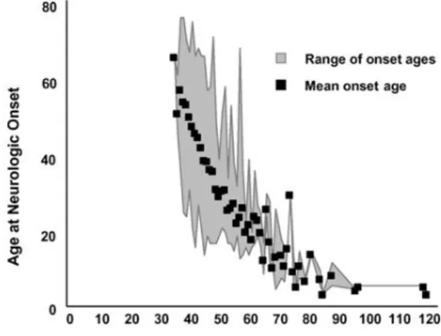
IISSEEAASSEEHuntington´s disease (HD) was first described in 1872 by George Huntington, who identified both clinical features and pattern of familial transmission. HD is an autosomal, dominantly inherited neurodegenerative disease with a prevalence of 5-10 per 100,000 individuals in Europe and North of America (Gusella & Macdonald, 2006). The HD gene initially labeled IT15 (Interesting Transcript 15) was identified for the first time in 1993 by a multicenter consortium, organized by the Hereditary Disease Foundation and a tremendous progress has been made since this discovery (Huntington´s Disease Collaborative Research Group, 1993). The molecular basis of the disease involves the expansion of the trinucleotide CAG (cytosine-adenine-guanine) in the first exon of the HD gene, located in chromosome 4 (4p16.3). The HD gene encodes for a widely expressed protein, huntingtin (Htt) with a molecular weight ~350 kDa, which in the mutant form (mutant huntingtin, mHtt) contains an elongated polyglutamine (polyQ) stretch in its N-terminal (The Huntington´s Disease Collaborative Research Group, 1993;(Gil & Rego, 2008). In the unaffected population, the number of CAG varies from 6-35 CAG units, while affected population have 36 or more CAG repeats (Gusella & Macdonald, 2007, Reddy et al., 1999) (Fig. 1.1). The presence of 60 or more CAG repeats causes the juvenile-onset disease (Nance & Myers, 2001). In general, polyQ repeats are highly polymorphic and their length increase in every generation when expanded polyQ repeats are inherited through males, a phenomenon referred as genetic anticipation (Reddy et al., 1999). There is an inverse correlation between the age of onset and the length of polyQ tract, whereby more CAG repeats are associated with an earlier disease onset (Fig. 1.1). However, the relationship between CAG repeat length and the age of onset differs when considering HD patients with juvenile onset or adult onset. The influence of each CAG appears to be stronger in the adult-onset range of CAG repeats than in juvenile-onset range (Andresen et al., 2007). There are two chromosomal loci - one at 6q23–24 and the other at 18q22—that are capable of modifying the age of onset of HD. Interesting candidate
3 genes in these loci are serum and glucocorticoid regulated kinase gene (SGK) and metabotropic glutamate receptor gene (GRM1), for 6q23–24 and gene which encodes developmentally down-regulated 4-like gene (NEDD4L) for 18q(Li et al., 2006)
Figure 1.1: The HD CAG trinucleotide repeat mutation and its relationship with age at neurological onset. The mean age at neurological onset and the range of ages at onset associated with different HD
expanded CAG repeats. Adapted from (Gusella & Macdonald, 2007).
HD typically manifests in mid-life, and terminates in death 10–20 years after the initial symptoms. Symptoms develop gradually over the disease progression and are categorized into 6 onset periods (Kirkwood et al., 2001). Early symptoms vary from person to person but disease onset is generally marked by involuntary movements of the face, fingers, feet or thorax. Psychiatric symptoms are more heterogeneous but can occur before onset; they include depression, anxiety, apathy, and irritability (Duff et al., 2007). The late stages are characterized by a variety of motor, emotional/behavioral, and cognitive symptoms, such as unsteadiness, trouble holding onto things, trouble walking, changes in sleeping patterns, hallucinations, intellectual decline, memory loss, difficulty in speech and weight loss. In the late stage patients lose bowel and bladder control (Kirkwood et al., 2001).
The neuropathology markers involves the selective dysfunction and death of specific neuronal subpopulations within the central nervous system (CNS), namely GABAergic (γ–aminobutyric acid) projection medium/spiny neurons of the striatum (caudate and putamen), neurons in the cerebral cortex and, to lesser extent, in hippocampus (Spargo et al., 1993, Vonsattel & Difiglia, 1998). However, with the disease progression, there is a general neuronal loss in several brain regions, such as globus pallidus, subthalamic nuclei, substantia nigra, cerebellum and the thalamus (Gil
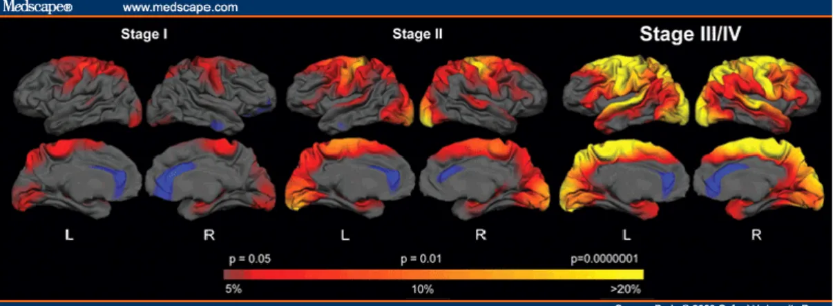
4 & Rego, 2008, Spargo et al., 1993). The extent of neuropathology and clinical symptoms was used to distinguish between 5 grades (0-4) of disease progression (Vonsattel et al., 1985) (Fig. 1.2).
Figure 1.2. Model of disease progression. Selective dysfunction and death of striatal neurons and to
lesser extent neurons within cerebral cortex. HD subjects were grouped according to stage. The colour scale at the bottom represent the thickness difference, with red to yellow indicating regions of more significant thinning in HD, compared to age matched controls. The magnitude of the brain thickness change is displayed as well, transitioning from red (5% loss) to yellow (>20% loss). Adapted from http://www.medscape.com/viewarticle/573134_3.
A characteristic feature of HD disease is the formation of intracellular aggregates, forming neuronal intranuclear inclusions (INIs) or cytoplasmic inclusions (CIs) in the affected brains (Ross & Poirier, 2005) (Fig. 1.3). The role of these protein aggregates remains controversial, since their formation is correlated with disease progression, but not associated with neuronal degeneration (Kuemmerle et al., 1999). Thus, both protective (Arrasate et al., 2004, Kuemmerle et al., 1999) as well as toxic functions (Bates, 2003) have been described in the last years, for this aggregates.
In HD patients neurological symptoms predominate but, they are not the sole manifestations of the disease. Early reports described pathological phenotypes in peripheral tissues of HD patients, including weight loss, muscle wasting and altered glucose homeostasis (Sassone et al., 2009). This suggests that cells from peripheral tissues of HD patients bear abnormalities related to expression of mHtt. Other reported changes included sub-cellular abnormalities in both fibroblasts and erythrocytes from HD patients (Sassone et al., 2009) .
5
Figure 1.3 Intracellular aggregates in HD. A) Intranuclear inclusion and cytoplasmic inclusions B)
Intranuclear inclusion. Visualized by light microscopy in the motor cortex of HD brain. Adapted from (Ross & Poirier, 2005).
1
1
.
.
2
2
.
.
H
H
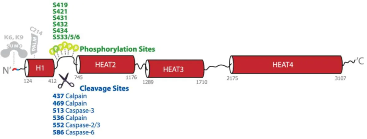
UUNNTTIINNGGTTIINNHuman Htt is a soluble large protein consisting of 3144 amino acids that has no similar sequence with other proteins. It has many potential domains whose boundaries and activities are not fully understood (Cattaneo et al., 2001). The sequence is phylogenetically highly conserved, except the polymorphic proline-rich region adjacent to the polyglutamine tract (Faber et al., 1998). Sequence analysis revealed that Htt contains multiple HEAT (Htt, elongation factor 3, the regulatory A subunit (PR65/A) of protein phosphatase 2A, and the lipid kinase TOR1) repeat sequences (Andrade & Bork, 1995), which are clustered into 4 major HEAT domains. Many phosphorylation and caspase-cleavable sites are located between the first two HEAT domains and present multiple targets for modulation and regulation of some events in HD pathogenesis (Warby et al., 2009, Wellington et al., 1998) (Fig. 1.4).
A functionally active C-terminal nuclear export signal (NES), sequence and nuclear localization signal (NLS) are present too. The NES defines a potential role of Htt as a member of nucleocytoplasmic dynamic protein complex, which may be important in HD because this fragment of protein is proteolytically cleaved in the disease (Xia et al., 2003).
Various types of post-translational modifications may occur in Htt, including phosphorylation, ubiquitylation, SUMOylation, palmitoylation, transglutamination and proteolytic cleavage. The protein context and post-translational modifications influence Htt neurotoxicity (Pennuto et al., 2009).
6
Figure 1.4. Schematic diagram of Htt. Htt is predominantly composed of HEAT repeats that comprise
four major HEAT domains (red barrels). Several phosphorylation sites (green) are clustered in the intervening sequence between HEAT 1 and 2. Numerous cleavage sites (blue) are also found in this region. The location of characterized Htt SUMO and palmitoylation sites are also indicated. Adapted from (Warby et al., 2009).
1.2.1.FUNCTION
The ubiquitous expression of Htt throughout the body, in neuronal and non- neuronal tissues, and its widespread localization at the subcellular level makes difficult to determine its function. It is not surprising that this protein is considered a scaffolding protein mediating protein–protein interactions playing a role in many cellular pathways (Macdonald, 2003). Several roles are assigned, depending on its subcellular localization and interaction with others proteins (Borrell-Pages et al., 2006, Cattaneo et al., 2005, Orr & Zoghbi, 2007). Consistent with this, it is known that Htt interact with a variety of proteins that can be grouped according to whether they are involved in gene transcription, intracellular signaling trafficking, endocytosis or metabolism (Harjes & Wanker, 2003, Li & Li, 2005).
Htt interacts directly with transcription factors, and might therefore act in the CNS as a general facilitator of neuronal transcription. Htt binds to the transcriptional repressor element 1 transcription/neuron restrictive silence factor (REST/NRSFs) in the cytoplasm, thereby preventing it from forming the nuclear co-repressor complex at RE1/NRSE nuclear site and allowing gene transcription (Zuccato et al., 2003). Furthermore, in vivo data show that Htt stimulates cortical production of brain derived neurotrophic factor (BDNF), an important neurotrophin produced by projecting cortical neurons projecting to the striatum, necessary for the survival of striatal neurons (Zuccato et al., 2001).
7 Another function that has been described for Htt is its involvement in intracellular trafficking. Htt was shown to be associated with proteins present in vesicle membranes (Difiglia et al., 1995) and microtubules (Gauthier et al., 2004), such as Htt-associated protein 1 (HAP1) and Htt-interacting protein 1 (HIP1) (Li & Li, 2005). Although both are associated with the trafficking, the HAP1 is associated with molecular motors dynein/dynactin (subunit p150Glued), which is involved in microtubule dependent retrograde transport (Engelender et al., 1997) and kinesin light chain, which is involved in anterograde transport 2 (Mcguire et al., 2006). HIP1 is also important for assembly and function of the cytoskeleton, for endocytosis and binding of clathrin (a protein involved in the formation of coated vesicles) and alpha-adaptin subunit AP-2 (Waelter et al., 2001). In addition, Htt directly promotes the microtubule-based transport of BDNF in neurons through this interaction (Gauthier et al., 2004). Recently, studies in vitro and in vivo suggest that Htt may play a role in post-transcripcional transport/targeting of mRNA through association with neuronal RNA granules. These findings implicate a role of Htt in maintaining neurotrophic support and neuronal survival via delivery and processing of BDNF mRNA (Savas et al., 2010).
Finally, Htt is an indispensable protein having anti-apoptotic properties, protecting neurons against apoptotic stimuli like serum deprivation, mitochondrial toxins or transfection of death genes (Cattaneo et al., 2005, Rigamonti et al., 2000). Htt acts downstream of mitochondrial cytochrome c release, preventing the formation of a functional apoptosome complex and the consequent activation of caspase-9 (Rigamonti
et al., 2001) and caspase 3 (Rigamonti et al., 2000). In addition, Htt is essential for
embryonic development and neurogenesis, as defined in different Htt knockout (KO)-mice, since complete inactivation of Htt in KO mice (Hdh -/-) causes embryonic death before day 8.5 (Nasir et al., 1995, Zeitlin et al., 1995). However, heterozygous KO mice appear either phenotypically normal (Duyao et al., 1995) or display increased motor activity and cognitive deficits (Nasir et al., 1995).
1
1
.
.
3
3
.
.
M
M
EECCHHAANNIISSMMSSOOFFH
H
D
D
PPAATTHHOOLLOOGGYY:
:
G
G
AAIINNOORRLLOOSSSSFFUUNNCCTTIIOONN?
?
Since the discovery of the HD gene in 1993, a large number of studies on post
mortem HD brain, cellular cultures and animal models revealed that a great number of
cellular and, in particular, neuronal pathways and functions are abnormal in HD. However, the mechanism(s) responsible for triggering HD pathogenesis still remains
8 unknown. There are two hypotheses that explain the mechanism behind neuronal degeneration in HD: i) the gain of function (GOF) of mHtt and, ii) the loss of function (LOF) of normal Htt (Borrell-Pages et al., 2006, Cattaneo et al., 2001, Cattaneo et al., 2005). According to the GOF hypothesis, the expanded polyQ causes a conformational change and confers a new function to Htt that is toxic to the cell (Fig. 1.5). This hypothesis is supported by the fact that patients with Wolf-Hirshhorn syndrome, a rare condition in which a deletion on chromosome 4 that comprises the CAG triplet repeats region (HD gene), occur do not develop HD (Gottfried et al., 1981). This suggested that the presence of one fully functional allele is compatible with life in humans and that HD is not caused by a simple loss of function of HD gene. In contrast, in LOF hypothesis the decrease in expression of Htt due to interaction with mutant protein is thought to contribute to the disruption of intracellular homeostasis, culminating in neuronal dysfunction and death (Gil & Rego, 2008). As mentioned previously, Htt is an anti-apoptotic protein and promotes the transcription (Zuccato et al., 2001) and microtubule-dependent transport (Gauthier et al., 2004) of BNDF. Actually it is believed that HD pathology result of combined effect of GOF and LOF of normal Htt, leading to deregulation of relevant intracellular pathways that culminate in neurodegeneration and cell death.
1.3.1.WHEN HTT BECOMES TOXIC
In the last years there have been many attempts to determine the mechanisms by which the polyQ tract causes neurodegeneration in HD. It is known the pathogenic process begins with the synthesis of Htt with an expanded polyQ with a tract, altered native conformation that interferes with several pathways (Fig. 1.5). A fraction of abnormally folded protein is degradated by the proteasome, however expanded proteins are prone to misfolding and resistant to proteolysis by the ubiquitin-proteasome system (UPS), leading their accumulation within cell (Cummings & Zoghbi, 2000) and interference with proteosome function, leading to inhibition of proteosome activity (Venkatraman et al., 2004). On the other hand, as described before, mHtt might undergo proteolytic cleavage by caspases, such as caspase 3, and also calpains, producing a toxic short N-terminal fragments that favours the aggregation process (Schilling et al., 2006, Wellington et al., 2000) (Fig. 1.4). This is supported by the fact that prevention of proteolysis by inhibiting caspases or calpain activation or by modifying the consensus cleavage sites in Htt reduces mHtt toxicity in in vitro and in vivo models (Gafni &
9 CAG CAG n CAG CAG CAG n CAG
Ellerby, 2002, Wellington et al., 2000). In addition, once cleaved, Htt fragments can translocate into the nucleus, where they have a greater affinity to bind to other proteins, such as nuclear proteins and transcription factors, forming aggregates, which affect transcriptional activity and may cause several deleterious events (Riley & Orr, 2006). Many proteins have been reported to interact with mHtt, such as TATA binding proteins (TBP), CREB binding proteins (CBP) (Schaffar et al., 2004), specificity protein 1 (SP1) (Li et al., 2002) and components of the basal machinery, such as essential subunits of RNAII complex, TBP, TFIIF, TAFII130 (Zhai et al., 2005). For example one study demonstrated that the occupancy of RE-1/NRS loci by REST/NRSF is higher in HD, leading to decreased transcription of BDNF in HD cells, mice and humans post mortem samples, while inhibition of REST/NRSF binding restored BDNF levels (Zuccato & Cattaneo, 2007).
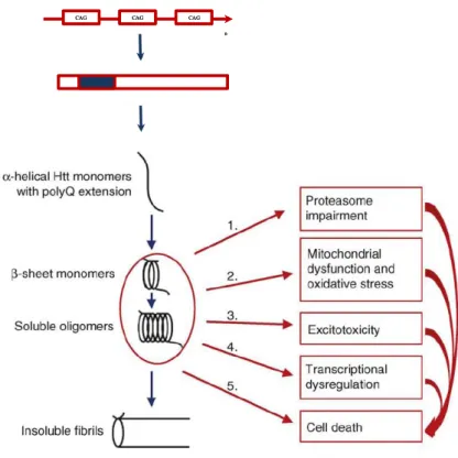
Figure 1.5. Model of pathogenic mechanisms in HD. Huntingtin proteins with >35 glutamine repeats
fold into β-sheet structures. This might facilitates intermolecular cross-links by transglutaminases, leading to the accumulation of aggregates of misfolded Htt in the cytoplasm. Aggregates are toxic by a variety of mechanisms: 1. mHtt cannot be cleaved by the proteosome, leading to the accumulation of misfolded proteins 2. mHtt can interact with mitochondria causing dysfunction complexes of mitochondrial electron transport chain and decreased Ca2+ buffering (section 1.4.2.2), 3. Evidence from animal models and patients supports a role for excessive glutamatergic input (excitotoxicity) in HD pathogenesis but the molecular mechanism is not completely clear, 4. mHtt can be translocated into the nucleus and disrupt transcription, 5. mHtt can directly initiate pro-apoptotic signaling with activation of caspases and release of cytochrome c by mitochondria and cell death. However, all of these mechanisms may culminate in cell death. Adapted from (Fecke et al., 2009).
10 As mentioned earlier, the presence of expanded protein in the cytoplasm can also interfere with microtubules BDNF. Indeed mHtt binds with high affinity to HAP1 and the p150 (Glued), causing an impaired association between motor proteins and microtubules leading to a reduced transport of BDNF vesicles along microtubules and loss of neurotrophic support (Gauthier et al., 2004). In addition, mHtt (monomers and oligomers) can interact directly with mitochondria leading to caspases activation and mitochondrial dysfunction (Panov et al., 2002).
Currently, there are two mechanisms that attempt to explain the formation of aggregates of mHtt: the polar zipper model (Perutz et al., 1994) and the transglutamine model (Kahlem et al., 1998). Polar zipper model refers the capacity that polyQ chains could theoretically form polar zippers, due to hydrogen bonds (Perutz et al., 1994). In transglutamine model, transglutaminases to catalyze aggregation of Htt protein, especially in the expanded form (Kahlem et al., 1998).
1
1
.
.
4
4
.
.
M
M
IITTOOCCHHOONNDDRRIIAALLDDYYSSFFUUNNCCTTIIOONNIINNH
H
D
D
1.4.1.MITOCHONDRIA
Mitochondria are highly dynamic organelles involved in multiple cellular processes of being ATP (adenosine triphosphate) production by oxidative phosphorylation (OXPHOS) the most prominent one (Schatz, 1995). However, mitochondria are also central to intracellular Ca2+ homeostasis (Celsi et al., 2009), Krebs cycle and oxidation of fatty acids (Van Der Giezen & Tovar, 2005), generation of reactive oxygen species (ROS) (Benard et al., 2007, Droge, 2002) and apoptotic pathways, involving proteins of Bcl-2 family of proteins and the release of cytochrome c and other pro-apoptotic factors (Spierings et al., 2005).
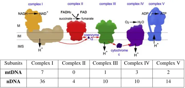
The human mitochondrial DNA (mtDNA) genome is small 16,569 base pair (bp) in a long circular chromosome composed of double-stranded DNA, and 37 genes which encode for RNA components of the mitochondrial translation apparatus, 22 transfer RNA (tRNAs) genes and 12S and 16S ribosomal RNA (rRNA) genes, as well as 13 polypeptide-encoding genes (mRNAs). All 13 polypeptides are essential components of four of the five complexes that form the mitochondria OXPHOS complex (Cx) (Attardi & Schatz, 1988). Seven polypeptides, ND1, ND2, ND3, ND4, ND4L, ND5, ND6, are subunits of Cx I (NADH-dehydrogenase-ubiquinone reductase); cytochrome b is part of Cx III (ubiquinol-cytochrome c reductase); COI, COII and COIII are catalytic subunits
11 of Cx IV (cytochrome c oxidase), and ATPase 6 and 8 are subunits of Cx V. All the subunits of Cx II (succinate dehydrogenase-ubiquinone reductase) are encoded by nuclear DNA (nDNA) (Fig. 1.6) (Zeviani & Antozzi, 1997). Therefore, each Cx of the mitochondrial respiratory chain (MRC) (except Cx II) contains subunits encoded by nuclear genes, which are assembled together with the mtDNA-encoded subunits into the respective haloenzymes, located in the inner mitochondrial membrane (Scarpulla, 1997). Human mtDNA lacks the protection by histones, DNA binding proteins, and is replicated without efficient proofreading and a DNA repair system. Moreover, mtDNA is highly exposed to ROS that are continually generated by the MRC and to other free radicals. The random hit of the naked mtDNA by ROS or free radicals is likely to cause oxidative damage or mutations (Wang et al., 2003). Accumulation of mutations and oxidative damage to mtDNA result in MRC dysfunction, leading to increased production of ROS in mitochondria and induction of further mtDNA mutations. mtDNA damage due to oxidative stress has been observed in cortex of pos tmortem HD brains (Polidori et al., 1999), and in animal models such as transgenic mice R6/2 (Acevedo-Torres et al., 2009).
12
Subunits Complex I Complex II Complex III Complex IV Complex V
mtDNA 7 0 1 3 2
nDNA 36 4 10 10 14
Figure 1.6. Energy production through the coupling of MRC with OXPHOS in the mitochondria.
The reducing equivalents in NADH or FADH2 enter the electron transport chain thought Cx I and Cx II,
respectively. During the transfer of electrons from NADH to coenzyme Q (CoQ), from CoQ to Cx III, and then from cytochrome c to Cx IV, protons are translocated from the matrix to the intermembrane space. A proton gradient is thus established across the mitochondrial membranes, which is the driving force for ATP synthesis catalyzed by membrane-located ATP synthase (Cx V). The subunits of complexes are encoded by two genetic system: the nDNA and mtDNA. Adapted from (Dudkina et al., 2010).
1.4.2.POSSIBLE MECHANISMS OF MITOCHONDRIAL DYSFUNCTION
Mitochondria are organelles of great importance in the cell, being mitochondrial dysfunction a hallmark of many common neurodegenerative diseases (Beal, 2005, Knott
et al., 2008). Evidences of mitochondrial dysfunction associated to the pathogenesis of
HD have been accumulated over the last 30 years (Section 1.4.2.1.). Many HD models have been generated using mitochondrial toxins (Brouillet et al., 2005), which stemmed from post mortem brain data (Gu et al., 1996).
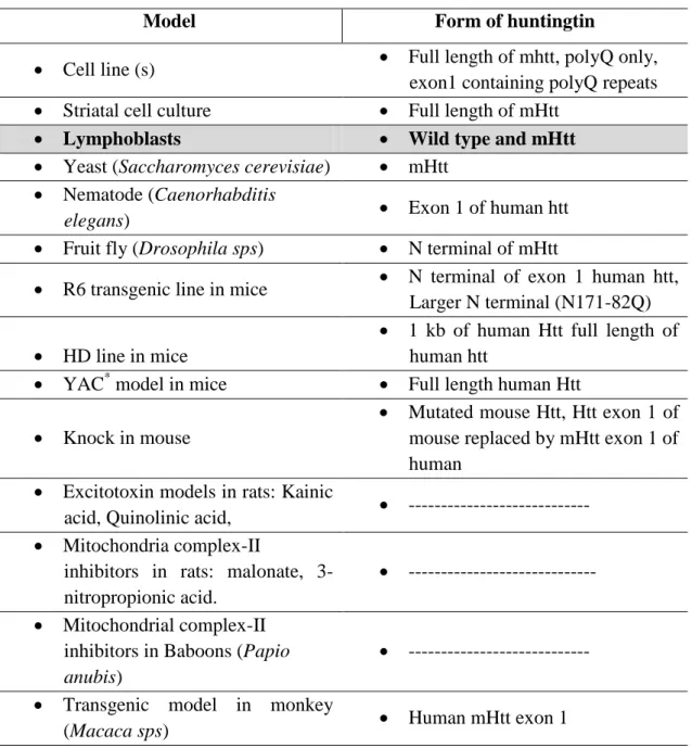
Several different models are available for HD research as represented in Table I. Thus, it is unquestionable the involvement of mitochondrial dysfunction in the process of disease, but its precise nature and cause remain uncertain (Browne & Beal, 2004, Oliveira, 2010).
It is known that mHtt interacts directly or indirectly with mitochondria, interfering with mitochondrial function, including a reduction of Ca2+ buffering capacity, loss of mitochondrial membrane potential (ΔΨm), impairment in MRC complexes, increased generation of ROS, abnormal vesicle trafficking and impairment
13 in mitochondrial dynamics, leading to neuronal death. The role of mitochondrial dysfunction in HD has been recently reviewed (Bossy-Wetzel et al., 2008, Browne, 2008, Pandey et al., 2010, Reddy et al., 2009).
Table I. Different models used in the study of mitochondrial dysfunction in HD. Model Form of huntingtin
Cell line (s) Full length of mhtt, polyQ only,
exon1 containing polyQ repeats
Striatal cell culture Full length of mHtt
Lymphoblasts Wild type and mHtt
Yeast (Saccharomyces cerevisiae) mHtt
Nematode (Caenorhabditis
elegans) Exon 1 of human htt
Fruit fly (Drosophila sps) N terminal of mHtt
R6 transgenic line in mice N terminal of exon 1 human htt, Larger N terminal (N171-82Q)
HD line in mice
1 kb of human Htt full length of human htt
YAC* model in mice Full length human Htt
Knock in mouse
Mutated mouse Htt, Htt exon 1 of mouse replaced by mHtt exon 1 of human
Excitotoxin models in rats: Kainic
acid, Quinolinic acid, ---
Mitochondria complex-II
inhibitors in rats: malonate, 3-nitropropionic acid.
---
Mitochondrial complex-II inhibitors in Baboons (Papio
anubis)
---
Transgenic model in monkey
(Macaca sps) Human mHtt exon 1
Different models and correspondent form of mhtt. *Yeast artificial chromosome
14
1.4.2.1. Respiratory chain impairment
Since early biochemical studies, defects in enzymes of oxidative metabolism have been observed in HD, and thus respiratory chain impairment was proposed as a primary triggering event in HD pathogenesis. Despite controversial results obtained in different models, it is nowadays believed that mitochondrial impairment is a secondary event in the pathogenesis of HD (Oliveira, 2010).
Energetic impairment in HD patients has been observed by a variety of methods, such as, Positron Emission Tomography (PET) that showed a significant reduction in glucose uptake in cortex and striatum of HD patients (Gil & Rego, 2008). Through the use of Magnetic Resonance Spectroscopy (MRS), it was found that levels of lactate are diminished in cortex of symptomatic patients and in striatum of presymptomatic HD patients (Jenkins et al., 1998). Impairment in MRC enzyme activities has been demonstrated such as, activity of succinate dehydrogenase, Cx II, cytochrome oxidase, Cx IV (Brennan et al., 1985), pyruvate dehydrogenase (Butterworth et al., 1985) and aconitase (Sorolla et al., 2008, Tabrizi et al., 1999) in striatum of HD patients.
Post mortem samples of patients with striatum atrophy revealed reduced activity
in complexes II/III and a mild reduction in Cx IV (Browne, 2008, Gu et al., 1996). Accordingly, expression of two subunits of Cx II (Ip and Fp) are preferentially decreased in the striatum of HD patients compared with controls (CTR) subjects and these alteration affect the dehydrogenase activity of the Cx (Benchoua et al., 2006). Still, in these studies, the activity of Cx I was not altered. Nevertheless, deficits in Cx I have been described in different peripheral tissues from HD patients, such as platelets (Parker et al., 1990) and muscle (Arenas et al., 1998). However, several different studies could not reproduce such deficit in post mortem HD brain or in HD patient’s platelets (Gu et al., 1996, Powers et al., 2007), muscles (Turner et al., 2007) and lymphoblasts (Sawa et al., 1999). In genetic HD models, particularly mice expressing full-length mHtt, no significant alterations were found in measurements of MRC Cx I-IV in striatum and cerebral cortex (Browne & Beal, 2004, Guidetti et al., 2001). It should be noted that in digitonin-permeabilized striatal cell lines that exhibited a significant decrease in MRC rates, no impairment in individual respiratory complexes was detected (Milakovic & Johnson, 2005). The same authors observed that differences in MRC rates disappeared when using isolated mitochondria from the same cell lines
15 (Milakovic et al., 2006), suggesting that detection of mitochondrial deficits requires a preserved cellular context.
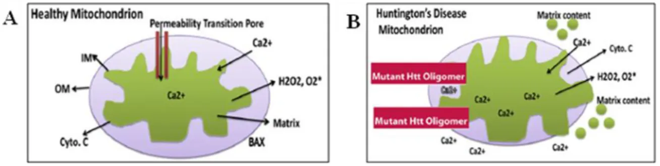
1.4.2.2. Mitochondrial Ca2+ buffering capacity in HD
Mitochondria are involved in the maintenance of Ca2+ homeostasis mainly because of their capacity to buffer cytosolic Ca2+ (Budd & Nicholls, 1996). Several lines of evidences suggested that abnormal Ca2+ uptake occurs in HD neurons (Reddy et
al., 2009) (Fig. 1.7). It is believed that mHtt interacts directly with mitochondrial
membranes causing deleterious effects. A decreased mitochondrial Ca2+ loading capacity and lower ΔΨmwere observed in lymphoblasts from HD patients (Panov et al., 2002). This vulnerability was proportional to mHtt levels (Panov et al., 2002).Similar defects were observed in brain mitochondria from transgenic yeast artificial chromosome (YAC) mice expressing full-length mHtt (Panov et al., 2002). Subsequent studies, using knock-in (KI) HD mice (50Q, 92Q, 111Q and 150 Q) revealed either no differences or decreased susceptibility to Ca2+ loads, expressed as increased Ca2+ buffering capacity (Bezprozvanny & Hayden, 2004). The same was observed in brain mitochondria isolated from diverse HD mice such as R6/2, revealing either no differences or decreased susceptibility to Ca2+ loads, expressed as increased Ca2+ buffering capacity (Brustovetsky et al., 2005, Oliveira et al., 2007). However, more recently brain mitochondria from HD rats exhibited diminished ΔΨm stability in response to Ca2+, lower capacities and rate of Ca2+ accumulation when compared to CTR rats (Gellerich et al., 2008). One explanation for these results is that the buffering capacity may change between different models and isolated mitochondria may be influence methodological approaches (Oliveira et al., 2007).
Figure 1.7.Mitochondrial Ca2+ buffering capacity. A) In healthy mitochondria, the inner mitochondrial
membrane provides a highly efficient barrier to ionic flow and protects mitochondria from toxic insults B) Excess mitochondrial Ca2+ load may open the mitochondrial permeability transition pore in mitochondria from HD patients, leading to cytochrome c release. Adapted from (Reddy et al., 2009).
16 Therefore these studies suggest that ΔΨm and mitochondrial Ca2+ regulation are directly impaired by mHtt and that increased ROS generation may be driving these alterations (Panov et al., 2002, Puranam et al., 2006).
Cytosolic Ca2+ can also be mobilized by the endoplasmic reticulum (ER), which is another major store for intracellular Ca2+. mHtt forms a ternary complex with Huntingtin associated protein 1 (HAP-1A) and type 1 inositol 1,4,5-trisphosphate (IP3Rs). In this complex, mHtt facilitates the release of Ca2+ from the ER and renders
neurons more sensitive to Ca2+ mediated cellular dysfunction (Bezprozvanny & Hayden, 2004). Additionally, mHtt enhances Ca2+ entry through the N-methyl-D-aspartate (NMDA) receptors, leading to Ca2+.desregulation and consequent activation of caspases and calpain, leading to cell death (Bezprozvanny & Hayden, 2004). Indeed, several approaches have shown that mHtt can directly modify NMDA-receptor function through its interaction with PSD-95 (Song et al., 2003, Sun et al., 2001). In particular, mHtt increases the sensibility of neurons to excitotoxicity associated to the stimulation of NMDA receptors contain the NR2B subunits (Zeron et al., 2002).
According to Choo et al. (2004), mHtt induces mitochondrial permeability transition pore (PTP) (Fig. 1.7) in isolated mouse liver mitochondria. Moreover from studies in mouse striatal neurons, where authors observed the opening of PTP in permeabilized polyQ expressing cells (Lim et al., 2008). Therefore, the PTP appears to be a final commitment step in a number of cellular stress conditions, with Ca2+ acting as a potent sensitizing factor (Lim et al., 2008).
1.4.2.3 Mitochondrial trafficking deficits and dynamics
Mitochondrial trafficking deficits are a recent proposed mechanism for mitochondrial dysfunction in HD. The first study was conducted by Trushina and co-workers (2004) who analysed the impact of mHtt on mitochondrial transport. Accordingly, mHtt impair movement indirectly by the sequestration of machinery components and Htt, which is essential for axonal transport (Trushina et al., 2004) or by physical blockage of axonal transport (Chang et al., 2006).
Accordingly, Chang et al. (2006) reported that mHtt aggregates act as physical roadblocks for mitochondrial transport in cortical neurons; consequently, in the narrow neuronal projections these aggregates prevent passage of mitochondria and fragmented mitochondria accumulate around mHtt. They proposed that this impairment in mitochondrial movement was an early pathogenic event, occurring before mitochondrial
17 and cellular dysfunction in cortical neurons (Chang et al., 2006). In addition, it was also reported that mHtt induces changes in mitochondrial morphology from elongated to a round phenotype (Chang et al., 2006). More recently, another study demonstrated that mHtt associates with microtubule based transport proteins decreasing mitochondrial transport in striatal neurons (Orr et al., 2008). This mechanism may be behind the vulnerability of striatal neurons, in HD disease.
Therefore, mHtt change the mitochondrial trafficking by several ways; in other, mHtt aggregates may block mitochondrial movement or/and mHtt may heavily interact with trafficking proteins, which may block/derail mitochondrial movement in the axon (Reddy et al., 2009). In addition, a large number of defective mitochondria accumulate due to excessive mitochondrial fragmentation in HD neurons; mHtt may create an imbalance between mitochondrial fission and fusion, leading to decrease in overall mitochondria dynamics in neurons. All these events may be responsible for low ATP production, mitochondrial dysfunction, and damaged medium spiny neurons in HD (Reddy et al., 2009).
1.4.2.4 Transcriptional dysregulation in HD
As previously described, mHtt induces transcriptional deregulation via interference with transcriptional factors occupation of genes promotes, and even direct DNA binding (Benn et al., 2008, Zhai et al., 2005). Because p53 regulates many apoptotic mitochondrial (Bax and Puma) and oxidative stress responsible genes (Vogelstein et al., 2000), the strong interaction between mHtt and p53 accumulation in the nucleus and thus induction of p53-dependent transcription (Bae et al., 2005) (Fig. 1.7). However, in intracellular polyQ aggregates p53 soluble levels are decrease in HD. (Suhr et al., 2001).
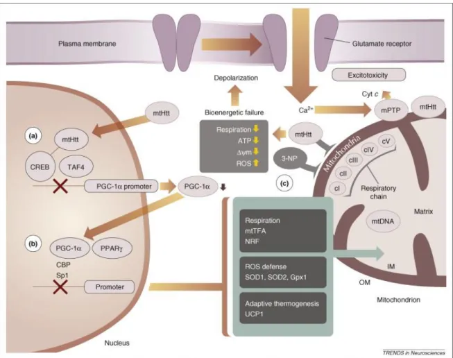
Another very interesting mechanism that has recently been proposed is the peroxisome proliferator-actived receptor γ coactivador-1α (PGC-1α) pathway, an important orcheastrator of mitochondrial function via integration of signals that regulate mitochondrial respiration, oxidative stress defense and adaptive thermogenesis (Cui et
al., 2006, Puigserver & Spiegelman, 2003, St-Pierre et al., 2006). PGC-1α promoter has
been shown to associate with mHtt, and interfere with CREB/TAF4 dependent transcriptional pathway, which is important for PGC-1α expression (Cui et al., 2006). mHhtt induces PGC-1α transcriptional repression, which is associated with mitochondrial dysfunction and neurodegeneration. A very recent study reports that
18 PGC-1α levels are reduced in muscles of HD patients and in transgenic HD mice NLS N171-82Q (containing 82 polyQ repeats), as compared to wild type littermates, confirming the active involvement of this transcriptional co-activador in HD pathology (Chaturvedi et al., 2009). It was found that PGC-1α decreased in HD post mortem brains, in cell lines expressing mHtt, and in HD mouse models, suggesting that mHtt promotes the increased production of ROS due to an increase in PGC-1α. Related with this increase in PGC-1α is an increase in scavenging enzymes such as superoxide dismutase (SOD)1 (Cu/Zn-SOD) or 2 (Mn-SOD), catalase and glutathione peroxidase (Arany et al., 2008, Cui et al., 2006, St-Pierre et al., 2006, Weydt et al., 2006). Although the findings of transcriptional dysregulation by mHtt are of high relevance, they can not entirely explain all the mitochondrial defects observed in HD. Therefore, more studies will be necessary to better understand this and other mechanisms.
19
Figure 1.8. mHtt and mitochondrial dysfunction in HD. Representative direct and indirect
mechanisms involving mHtt and mitochondria. A) mHtt blocks the PGC-1α promoter via inhibition of the CREB transcriptional activator, resulting in decreased PGC-1α expression. B) Lowered PGC-1α decrease PPARγ-mediated expression of nuclear-encoded mitochondrial proteins that are necessary for respiration and oxidative-damage defense. C) Direct interaction with mitochondria blocks the respiratory complex II, similarly to 3-nitropropionic acid (3-NP). Respiratory chain, in turn, leads to decreased energy production and decreased ΔΨm in addition to increased ROS generation. The bioenergetic declines caused by transcriptional deregulation and direct effects on mitochondria can cause increased vulnerability to excitotoxic stimuli and amplification of the mitochondria damage Ca2+-mediated PTP opening. Adapted from (Bossy-Wetzel et al., 2008).
20
1
1
.
.
5
5
.
.
R
R
HHOO ZZEERROO AASS AA MMOODDEELL FFOORR SSTTUUDDYYIINNGG MMIITTOOCCHHOONNDDRRIIAALL DDYYSSFFUUNNCCTTIIOONN
Several mtDNA depleted mammalian cells lines have been generated to investigate the role of mitochondria in aging and age related disorders , such as aging and age-related, namely Alzheimer's and Parkinson's disease (Chomyn et al., 1994, Marusich et al., 1997, Miller et al., 1996). The development of cells depleted of mtDNA has provided a suitable model to study some of the molecular mechanisms governing mitochondrial defects. Such cells, defined as rho-zero (ρ0) have been produced by long-term culture with compounds such as ethidium bromide (EtBr) a cationic and lipophilic agent that damage and inhibits mtDNA replication and transcription when present under lower concentration (King & Attardi, 1989). Additionally, other methods were tested to generated ρ0
cells, such as dideoxynucleoside analogs (i.e 2’,3’dideoxycytidine (ddC)) (Nelson et al., 1997), a antiviral nucleoside analog that inhibits mtDNA replication (Martin et al., 1994), exposure to rhodamine 6-G, a lipophilic dye which degrades mammalian mitochondria (Trounce & Wallace, 1996) and more recently based on an enzymatic approach (Kukat
et al., 2008). This method destroy endogenous mtDNA in vivo was also based on a
restriction endonucleases that was target to the matrix of mitochondrial thereby cleaving the genome and allowing endogenous enzymes to fully disintegrate the DNA molecules (Kukat et al., 2008). EtBr inhibits mtDNA polymerase gamma (POLG) more strongly than DNA polymerase alpha and beta, thus inhibiting the replication and transcription of mtDNA without substantially affecting nDNA (Qian & Van Houten, 2010).
Rho-zero cells are devoided of mtDNA and electron transport chain (ETC) activity and are dependent on uridine and pyruvate, for growth because of the absence of a functional respiratory chain (Desjardins et al., 1985, King & Attardi, 1989) i. e, these cells present inability to synthesize a particular compound (in this case uridine and pyruvate) required for its growth (auxotrophic). The growth medium needs supplementation with nutrients to sustain viability. This is achieved by adding pyruvate (to regenerate NAD+ following its conversion to NADH (nicotinamide adenine dinucleotide) in glycolysis and thus anaerobic ATP generation) and uridine (to facilitate pyrimidine synthesis, which becomes ineffective under conditions of ETC failure), in order to prevent energy demand of cell is satisfied (King & Attardi, 1989, Miller et al.,
21 1996, Swerdlow et al., 1997, Swerdlow et al., 1999). After EtBr treatment, these authors observed a reduction in mtDNA amount. However, if the DNA-intercalating agent is removed before complete depletion of mtDNA, cells repopulate with residual genomes in a period that will depend on the size of the mtDNA (Moraes et al., 1999). Cells with large deletions, but not with pathogenic point mutations, repopulate organelles faster than wild-type genomes in the same cell, particularly during relaxed copy number control (Diaz et al., 2002)
1.5.1.CYBRIDS
One of the applications for the creation of ρ0 cells is the ability to create cytoplasmic hybrids (cybrids), first described in mammals by King and Attardi, in 1989 as a new model of disease for mitocondrial researches. In general, the cybrids are created when cytoplasmic contents of two different cells are processed within a single plasma membrane.This presents distinct applications in the study of mitochondrial function, including the study of mutations in mtDNA, assessing the integrity of the transferred mtDNA or compatibility biogenomic (Khan et al., 2007, Swerdlow, 2007) and more recently as a platform for the development of new therapies (Trimmer & Bennett, 2009).
Despite the various approaches to create cybrid lines, the most commonly used technique involves the transfer of mitochondria from non-nucleated cells (usually platelets) to ρ0 cells, resulting in cybrids containing nDNA from ρ0 cells and mtDNA from patient’s or donor’s platelets (Chomyn et al., 1994) (Fig. 1.9). After fusion, host cells repopulated with platelet-derived mitochondria undergo metabolic selection to eliminate cells with incomplete repopulation (Swerdlow et al., 1997).
Presently this is a technique widely used in the study of many neurodegenerative diseases, such as Parkinson´s disease (Esteves et al., 2008, Trimmer & Bennett, 2009), Alzheimer's disease (Cardoso et al., 2004, Swerdlow et al., 1997) and Huntington’s disease (Ferreira et al., 2010, Swerdlow et al., 1999).
22
Figure 1.9. Cybrids technique. ρ0 cell produced by long term culture with compounds that damage mtDNA. Transfer of mitochondria from platelets to ρ0 cells, resulting in hybrids cells (cybrids) containing nDNA from ρ0 cells and mtDNA from donor platelets.
23
1
1
.
.
6
6
.
.
O
O
BBJJEECCTTIIVVEEDespite major research efforts on HD, the underlying mechanisms leading to selective degeneration of striatal neurons in HD are still largely unknown and no therapy is currently available for this fatal disease. Several mechanisms of mHtt toxicity have been proposed, which partially fit with clinical data gathered from HD patients as well as from molecular, cellular and animal experiments. Currently, it is not known if one of these different mechanisms previously described triggers the other or if these different mechanisms, involved in many pathways, could participate synergistically in the pathology. It is well accepted that mHtt is widely expressed not only in the brain but also in peripheral tissues, suggesting that an adverse effect of mHtt is not limited to neurons (Rosenstock et al., 2010, Sassone et al., 2009). Several reports described alteration in peripheral tissues of HD patients, including platelets (Parker et al., 1990), lymphocytes (Almeida et al., 2008, Sawa et al., 1999) and muscles (Arenas et al., 1998, Turner et al., 2007).
Human HD lymphoblasts have been used in many studies as a cellular model of HD in mitochondrial dysfunction (Sassone et al., 2009). Many alterations present in HD neurons are present in lymphoblasts from HD patients, namely decreased ΔΨm (Panov
et al., 2002), impaired Ca2+ buffering (Panov et al., 2002), mitochondrial morphological alterations (Squitieri et al., 2006) and the presence of genetic instability (Cannella et al., 2009, Squitieri et al., 2006).
To gain further insight into the pathology, the main aim of this thesis was to create and characterize a new human cell model of mitochondrial dysfunction in HD, namely HD versus control (CTR) ρ0 lymphoblast cells. For this purpose, different lymphoblast cell lines were cultured in the presence of 25 and 50 ng/ml and EtBr for 15 or 30 days. This model was further characterized by analyzing several proteins encoded by mtDNA and nDNA, namely subunits of complexes I, II and IV, and the activity of MRC complexes I and IV.
This model will allow a better understanding of the role of mitochondria in HD, as well as the effects of mitochondrial dysfunction in this devastating neurodegenerative disease.
25
2
2
.
.
1
1
.
.
M
M
AATTEERRIIAALLLymphoblastic cell lines were obtained from NIGMS Human Genetic Cell Repository (CORIELL Institute for Medical Research, New Jersey, and USA). RPMI-1640 medium, phenylmethanesulfonyl fluoride (PMSF), DL-Dithiothreitol (DTT), protease inhibitor cocktail, FBS, EtBr, uridine and pyruvate were obtained from Sigma Chemical Co, St Louis, MO, USA. Antibodies against-Cx I 20 kDa subunit and Cx IV 57 subunit were obtained from Invitrogen (Carlsbad, USA). Cx I 30 kDa and Cx II 70 kDa subunit were obtained from Molecular Probes, Leiden, Netherlands and antibody against heat shock protein (Hsp60) was obtained from Chemicon, Hampshire, UK. ChemiFluorescence reagent ECF and anti-mouse secondary antibody were obtained from GE Healthcare (Little Chalfort, UK). All other reagents were of analytical grade.
2
2
.
.
2
2
.
.
L
L
YYMMPPHHOOBBLLAASSTTOOIIDDCCEELLLLLLIINNEESSLymphoblast cell lines were produced by CORIELL Institute. Accordingly to data sheet, lymphocytes were obtained from peripheral blood of HD patients or control (CTR) subjects, cultured by using phytohemagluttinin as a mitogen and then infected by Epstein-Barr virus in order to obtain lymphoblastoid cell lines. In this study, lymphoblast cell lines were obtained from HD affected patients containing heterozygous expansion mutation (n=4, three males (43/15, 45/15, 42/18) and one female (47/18) (Table II), or from unaffected aged matched voluntary subjects (control siblings) (n=2, one male and one female), used in this work as CTR lymphoblasts (Table III). Human peripheral blood was obtained after informed consent.
26
Table II. Demographic and genetic characteristics of HD lymphoblast cell lines.
Cell line Gender Age at onset
Age at Sampling
Expanded
CAG Race
#4798 Male 42 yrs 47 yrs 43/15 Caucasian
#5610 Female 40 yrs 52 yrs 47/18 Caucasian
#5622 Male 38 yrs 41 yrs 45/15 Caucasian
#5678 Male 48 yrs 58 yrs 42/18 Caucasian
Catalog number of caucasian HD lymphoblasts and detailed information about gender, age of disease onset, age of sampling and respective number of CAG repeats.
Table III. Demographic characteristics of the CTR lymphoblast cell lines.
Cell line Gender Age at onset
Age at Sampling
Expanded
CAG Race
# 4800 Female --- 45 yrs --- Caucasian
#4808 Male --- 42 yrs --- Caucasian
Catalog number of caucasian CTR lymphoblasts and detailed information about gender and age of sampling.
2
2
.
.
3
3
.
.
C
C
EELLLLC
C
UULLTTUURREELymphoblast cells were shipped in T25 tissue culture flasks that have been filled to capacity with carbon dioxide-equilibrated medium to provide sufficient nutrients for extended transport times. Upon receipt, flasks containing lymphoblasts were incubated unopened overnight at 37°C in upright position, with vented or loose caps. Lymphoblast cultures were counted in the next day and split if sufficient growth has occurred. Alternatively, the volume of the culture medium was decreased to yield a cell density of 200,000 - 500,000 viable cells/ml. Lymphoblasts were then cultured in RPMI 1640 medium supplemented with 15% of non-inactivated FBS plus 2 mM glutamine and 50 µg/ml streptomycin plus 100 IU/ml penicillin in T25 or T75 flasks, in upright position, by using an incubator chamber containing 5% CO2, 95% air, 100% humidity at 37ºC. In these conditions, lymphoblastoid cell lines grew in suspension with cells clumped in loose aggregates (Fig. 2.1 A and B). When desired, these aggregates were dissociated by gently agitating the culture or by gentle trituration with a pipette. In three to four days, the culture was either re-fed with fresh medium or split again taking into account
27 how fast the particular line grows, or the desired number of cells needed for the experiments.
A
B
Figure 2.1. Morphological features of lymphoblast cell lines. Representative images of aggregates of
CTR (A) or HD (B) lymphoblast cell lines in culture, visualized by PALM MicroBeam inverted microscope.
2
2
.
.
4
4
.
.
P
P
RROOLLIIFFEERRAATTIIOONNCCUURRVVEESSTrypan blue test is commonly used to evaluate cell viability in a cell suspension It is based on the principle that live cells having intact plasma membranes exclude the dye, whereas dead cells do not. In order to study the rate of division of the lymphoblast cell lines, cells were seeded at a density of0.2x106 cell/ml in 48-well plates and cultured for 5 days. Every other day, an aliquot of cell suspension was two times diluted in 0.1 % trypan blue and counted by using a hemocytometer under inverted light microscopy (Fig. 2.2 A-E). The plateau level for most cultures was reached at about 1 x 10⁶ viable cells/ml three to five days after sub-culturing. The pH of cultures was shown to be quite acidic, appearing distinctly yellow at this point since phenol red was used in the culture medium as pH indicator. Cultures left in the plateau phase exhibited a decrease in viability accompanied by a lengthening of the doubling time. All our experiments were performed in cultures presenting viability over 95%.
28
Figure 2.2. Proliferation curves of CTR or HD lymphoblast cell lines. CTR (A and C) or HD
lymphoblasts (B, D and E) were seeded at a density of 0.2x10⁶ cells/ml and cultured for 5 days in the incubator chamber. Every day the cells were evaluated by trypan blue assay and counted by using an hemocytometer. Results are presented as mean ± SEM of 3-4 independent experiments performed in duplicates.