A new look into the archaeological materials from the Museum of Évora, Portugal – the case study of the Zambujeiro Dolmen
Ana Manhita1, Cristina Barrocas Dias1,2, Sérgio M artins1
,
Joana Costa1, Dora Teixeira1 ,2, Mafalda Costa1, Massimo Beltrame1, José M irão
1,3, Jorge O liveira4, Leonor Rocha4Abstract
1Laborat ório HERCULES, Universidade de Évora, Largo Marquês de M arialva 8, 7000-809 Évora, Portugal 2Escola de Ciências e Tecnologia, Departamento de Quí mica, Universidade de Évora, Rua Romão Ramalho 59,
7000-671 Évora, Portugal
3Escola de Ciências e Tecnologia, Departamento de Geologia, Universidade de Évora, Rua Romão Ramalho 59,
7000-671 Évora, Portugal
4Escola de Ciências Sociais, Departamento de História, CHAIA, Universidade de Évora, Largo Marquês de Mari alva
8, 7000-809 Évora, Portugal
This paper presents the results of a multidisciplinary and multi-analytical study of the amber beads, red pigments, lithic arrowheads and selected ceramics from the Museum of Évora’s collection of the Zambujeiro Dolmen.
Amber beads were studied by Attenuated T otal Reflectance Fourier Transformed Infrared Spectroscopy (AT R-FT IR) and Pyrolysis coupled to Gas Chromatography and M ass Spectrometry (Py-GC /MS) to confirm their chemical nature and provenance. T he red pigments, frequently found in funerary Neolithic context of the Iberian Peninsula, were studied with micro-Raman, and Scanning Electron Microscopy coupled to Energy Dispersive X-Ray Spectroscopy (SEM-EDS) to identify their chemical nature and provenance. The lithic arrowheads were analysed by portable X-Ray Fluorescence (p-XRF), micro X-Ray Diffraction (XRD), SEM -EDS, and Laser Ablation Inductively Coupled Plasma Mass Spectrometry (LA-ICP-MS). The ceramic materials were studied to infer provenance and production technology by p-XRF, XRD and SEM-EDS; ceramic contents were evaluated by GC/MS.
The studies have shown that while some materials travel hundreds or thousands of kilometres to arrive to the Zambujeiro Dolmen, local materials were also used in the items selected by the communities to honour their deceased.
1. Introduction
Figure 1.
Zambujeiro Dolmen (Figure 1), located near Évora (30º32´15´´N; 8º0´47´´W), is the biggest chambered megalithic monument of Portugal, and one of the largest in the Iberian Peninsula. T his megalithic monument was built by the end of the 4thmillennium BC/transition to the 3r dmillennium, but was kept in use until the Bronze age. A second ritual deposit, at the entrance of the monument, was dated to the late Chalcolitic (2480-2290 BC). Zambujeiro Dolmen was identified and excavated between 1964 and 1968 and is classified as a National M onument since 1974. During the 1980s, due to the monument degradation, some conservation works and archaeological research were carried out [1]. The Zambujeiro Dolmen still preserves the grave (burial chamber and hall), much of the tomb hill, and on its periphery two large stelae-menhirs can be found. Collective inhu mation was practiced in Zambujeiro, accompanied by the deposit of articles of great quality, indicating that they were most likely a group of distinguished individuals. Several archaeological campaigns unearthed an exceptional rich array of assets of different typologies (lithic, engraved stone plaques, ceramic, jewelry, etc.). Despite the lack of important information that should have been collected during the initial excavations in this archaeologic site, the architectural monumentality of Zambujeiro Dolmen and the extent and variety of its spoil are sufficient reasons to try new types of studies that can bring more information about the life of people who were buried in this location.
1.1. Ceramics
The study of ceramics in archaeology is mainly concerned with the vessels’ provenance, manufacture and usage. The recent development of very sensitive and non-destructive analytical techniques can provide information at the microscopic level that can elucidate and complement the archaeological inventory of the spoil. T hrough the use of different physical and chemical techniques, it is possible to identify the mineralogical and chemical composition of the ceramics, and thus obtain information on its provenance (eg., to distinguish the local ceramic from imported) and the production technology used (eg., identify kiln temperature) [2-4].
The precise identification of the mineralogical composition of the ceramic requires X-Ray Diffraction (XRD) and thermoanalytical experiments [5]. The chemical composition can be obtained by Scanning Electron Microscopy coupled with Energy Dispersion X-ray Spectrometry (SEM-EDS), X-ray fluorescence (XRF) and Inductively Coupled Plasma Mass Spectroscopy (IC P-MS).
Scanning Electron M icroscopy coupled with Energy Dispersion X-ray Spectrometry (SEM -EDS) analysis allows further insight on the paste texture and the textural interrelationships of the mineral phases present that are too small to observe by optical microscopy. T he EDS spectra can provide the elemental analysis of the paste and temper components complementary with in-situ X-Ray Fluorescence Spectroscopy (XRF). Although the major element composition stays constant during the firing, the possibility of chemical element inward or outward diffusion during the firing or the buried, inducing also mineralogical and textural transformations, must be considered. T he analysis of the inner part of ceramics by XRF can provide information about this aspect.
With XRD it is possible to identify the phase (i.e. mineralogical) composition of the ceramic in what is concerned to the paste and the temper. The particularity of XRD is that it can distinguish the minerals within the clay group [6]. Also, since it is known that the original clay components alter its original structure when subjected to certain intensity and heating rate [7,8], it is possible to infer the approximate temperature ranges [9].
The preservation of organic molecules inside the porous structure of the ceramic, which can serve as biomarkers, allows the identification of certain food products or others that can provide information on the type of the ceramics use.
The study of vessel usage using the recovered organic compounds from the porous ceramic is based on the archaeological biomarker concept which relies upon matching the chemical structures of the organic materials and their distribution on the archaeological samples, the ‘chemical fingerprinting’, with the presence of chemicals in organisms known to have been exploited in the past. T he archaeological biomarker concept can be applied to any class of biomolecules, such as ancient DNA, proteins, carbohydrates, and fats,
even if, in most cases, these are only present in a degraded form [10]. The application of the archaeological biomarker concept requires that the total extract recovered from the ceramic is separated into individual compounds, and that these compounds are identified. T his requires that the analytical techniques employed are able to provide molecular-level resolution, achievable by the combination of chromatographic techniques (gas or liquid chromatography, GC or LC) with mass spectrometry detection (GC/MS or LC/M S).
Amber is a fossilized tree resin, very much appreciated by its colour and natural beauty. The process of fossilization of plant resins, mainly from conifers and some angiosperms, occurring along millennia, involves the loss of volatile components and polymerization of the diterpenoid (C20) and triterpenoid (C30) constituents of the resin, ultimately forming masses of amber.
Artefacts made with this soft vegetal resin are documented since Neolithic times, although they are quite rare in Portuguese Prehistoric contexts: usually, amber artefacts are considered as prestige indicators, being connected with long-distance trade and related with the emergence of social complexity.
One of the most known prehistoric sources of amber is located in the Baltic Sea ( ): beads made with raw material from this region have been identified in some Late Bronze Age sites from Central Portugal. Nevertheless, sources of geological amber are also documented in other regions, namely in Romania ( ), in Sicily ( ), as well as in the Iberian Peninsula (i.e. along the Cantabrian coast and in the Barcelona, Guadalajara and Cadiz regions).
The determination of the geological origin of the ambers has attracted much attention from researchers since the 19th century. Fourier transformed infrared spectroscopy, together with pyrolysis coupled to gas chromatography and mass spectrometry, has been widely used in the analysis of these resins.
The presence of red pigments occurs frequently in pre-historic sites, but these are usually identified as ochre (iron oxide), based solely on their morphology and macroscopic characteristics.
The presence of cinnabar associated with funerary contexts in the Iberian Peninsula is known since the end of last century, due to the contribution of the exact sciences and the use of analytical instrumentation, such as micro-Raman and SEM -EDS. In fact, the use of cinnabar (HgS) during the Neolithic and C halcolithic periodsis already registered in several monuments in Spain, because of the new analytical methodologies applied to the objects [11-16].
1.2. Amber beads
succinite
rumanite simetite
Within the study of the archaeological materials from the Museum of Évora, it was found that there were large amounts of red pigments and some lithic and ceramic materials with red colour, which is why it was decided to chemically analyse these objects.
The lithic assemblage of a burial site is usually dominated by bifacial tools [17]. Bifacial tools or bifaces are tools in which both faces show evidence of flake removal. T hese tools are made out of knappable stones: brittle and homogenous stones that do not have fissures, inclusions or any other properties that can cause them to crack in an unpredictable matter. Obsidians and other siliceous stones, such as cherts, are usually used to produce bifaces due to both their fracture mechanical properties and their relatively widespread availability [18].
Arrowheads constitute the final stage of the bifacial reduction sequence. Bifacial reduction is a gradual and continuous process, and the biface can be used as a functional tool throughout the various stages [17,18].
Amber beads spectra were obtained using a Bruker ALP HA FTIR spectrometer equipped with a diamond crystal ATR module. All spectra were acquired between 4000 and 500 cm-1, with 128 accumulations and a spectral resolution of 4 cm-1.
Spectra of red pigments were recorded with a Horiba XploRA ONE micro-Raman spectrometer, using a diode laser of 532 nm at 0,1% intensity. T he laser was focused on the samples using an objective lens of 50x and a grating of 1200 lines/mm. Spectra were acquired between 100 and 1000 cm-1.
For red pigments, EDS analyses were performed with a scanning electron microscope HITACHI 3700N coupled with an energy-dispersive X-ray spectrometer Bruker Xflash 5010. T he analyses were done at 20 1.4. Lithic arrow heads
2.1. ATR-FTIR
2.2. Micro-Raman
2.3. SEM-EDS
kV in high vacuum. For ceramics and lithic arrowheads, EDS analyses were performed at 20 kV in variable pressure mode.
A portable XRF spectrophotometer Bruker Tracer III-IV SD was used for in-situ analysis. For ceramics, spectra were recorded using a voltage of 40.0 kV and current intensity of 30.0 µA. A voltage of 15.0 kV and current intensity of 25.0 µ A were used for measurements in the lithic arrowheads.
For ceramics and lithic arrowheads, XRD patterns were recorded with a Bruker D8 Discover, using Cu Ka radiation, operating at a 2? angular range of 3-75°, step size of 0.05° and a step time of 1 sec. For the lithic arrowheads, a 0.3 mm collimator was used to direct a small parallel X-ray beam onto the sample.
For the lithic arrowheads, LA-ICP-MS analyses were conducted using a CET AC LSX-213 G2+laser ablation system coupled to an Agilent 8800 Triple Quad ICP-M S. A 100 µm spot size with a frequency of 10 Hz and a laser output energy of 80 % was used to analyse each sample. Helium, with a flow of 1 L/min, was used as a carrier gas in the LA system. The NIST 612 was used as a primary reference material and SiO2as an internal standard for both the samples and the reference material.
Ceramic samples collected from drill cleaned pottery sherds were extracted with a procedure adapted from [19]. Shimadzu GC2010 gas chromatographer coupled to a GC MS-QP2010 Plus Mass Spectrometer system was used with a column Zebron ZB-5HT 0.25 mm x 15.0 m (0.10 µm film thickness). 1 µ L of sample was injected in splitless mode, using helium as carrier gas, with the injector set at 250 ºC and a column flow of 1.5 mL/min. T he mass spectrometer operated in the EI mode (70 eV) in the 40–850 range with source temperature at 240 ºC and transfer line at 280 ºC.
For amber samples (approximately 200 µg each), a Frontier Lab PY- 3030D double- shot pyrolyzer interfaced to a Shimadzu GC MS-QP2010 Plus GC/M S system was used. Samples were pyrolyzed at 500 °C for 12 seconds and separated on a Phenomenex Zebron ZB-5HT capillary column (15.0 m length, 0.25 2.4. p-XRF 2.5. Micro-XRD 2.6. LA-IC P-MS 2.7. GC/MS m/z 2.8. Py-GC/MS
mm I.D., 0.10 µm film thickness). Helium was used as carrier gas. Pyrolysis and MS interfaces were maintained at 280 °C, splitless injector was set to 250 °C and ion source temperature was set at 240 °C. When needed, samples were derivatised using on-line methylation with TMAH.
Ceramics’ composition was evaluated using three different techniques: XRD for mineralogical composition, XRF for chemical composition, and SEM/EDS for chemical composition and spatial distribution of the elements, through elemental mapping.
Figure 2 presents the X-Ray diffractograms for two of the studied samples. Quartz is the most common non-clay mineral found in ceramics, and it was found in all samples. Muscovite, a type of mica, was found in some samples. Amphibole, an inossilicate, was also found in some samples. Feldspars, especially alkali feldspars (Na, K) were also found in the majority of the samples.
X-Ray diffractograms for two of the studied samples
The nature of the clay minerals varies from sample to sample and it was possible to identify illite/smectite. Clay minerals crystallize to other minerals, starting at 600 °C, so their presence in ceramics gives an estimate of the temperature used for firing.
The results for XRF (Figure 3) SEM-EDS (Figure 4) complement the information given by XRD, confirming the presence of alkali feldspars and titanium-iron oxide minerals, like ilmenite. Figures 3 and 4 show examples of the XRF and SEM-EDS results for one of the samples (Z4162).
3. Results and discussion
Figure 2. 3.1. Ceramics
Figure 3.
Figure 4.
XRF results for sample Z4162
Figure 5 presents two chromatograms that are illustrative for the studied samples.
GC-M S chromatograms of samples Z4162 and Z3694. C : n-alkane with carbon atoms; A : linear alcohol with carbon atoms; M : monoacylglycerol with carbon atoms; C fatty acid with
carbon atoms and double bonds; D : di-fatty acid with carbon atoms; IS: internal standard
Overall, the samples exhibited good preservation as several unsaturated fatty acids (C18:1 and C18:2) and mono-acyl-glycerols (MAGs) were detected in most samples. T he presence of unsaturated fatty acids in the original content of the vessels was further confirmed by the detection of azelaic acid (D9), a degradation
Figure 5. x x x
x x x x:y x
product of unsaturated fatty acids. The presence of polyunsaturated fatty acids, and in particular C18:2, is surprising in samples from this period. In archaeological environments, polyunsaturated fatty acids easily undergo oxidation processes localized at the double bonds via radical reactions with the inclusion of oxygen in the carbon chain, carbon–carbon bond cleavage, and formation of lower molecular weight species, such as azelaic acid (also detected in the samples) [10].
The chromatographic profiles suggest that the ceramics were previously used for materials of vegetable origin: high content of unsaturated fatty acids, presence of various steroids of vegetable origin (campesterol, stigmasterol and beta-sitosterol), absence of cholesterol (steroid of animal origin) and a ratio of fatty acid C16:C18 > 1 [10]. Despite the evidence for a vegetable origin of the fats stored in the studied fragments it is not possible to establish its botanical origin.
Another information gathered from the chemical analysis of the lipid extract is the absence of long-chain ketones; these compounds are usually present when animal or vegetable fats are heated above 300 ºC [10], suggesting that the pots were not used for frying, but usage for boiling foodstuffs of vegetable origin cannot be excluded. However, food materials of plant origin (except the oils) have a small content of fats, leaving few residues in the ceramics when just boiled in water [10]. T he large fat residue content of some of the analysed samples suggests that the ceramic vessels were likely used for storage of materials based on a vegetable fat.
One of the most interesting results of these analyses was the identification of diterpenoid derivatives such as dehidroabietic and isopimaric acids, in some samples. T hese compounds are known to be biomarkers for the presence of resins from family, and in particular from genus . So far, the use of pine resins in the Neolithic period is rare, being more common the detection of resins derived from birch bark (genus ) [20]. The reason for the use of resinous materials can be attributed to their sealing or gluing properties. Resins can also impart a characteristic flavour to any material storage inside the vessels. T he studied ceramics had no visual signs of the resin and it is not obvious why they were used in the first place. resins were widely used by the Romans to seal their amphorae, used to transport different food products throughout their empire [21].
The amber beads from Zambujeiro collection are not all well preserved and some of them are broken in smaller pieces. As it can be seen in Figure 6, the beads exterior layer presents a different structure and colour from that of the inner core.
Pinaceae Pinus
Betula
Pinaceae
Figure 6.
Figure7.
Amber beads collected in (A) Anta Grande do Zambujeiro (Museu de Évora collection, inventory nr. ME13688)
The AT R-FT IR spectra of the samples indicate that the analysed beads are all made from a similar amber material which is not Baltic amber (Figure 7, A and B), as they lack the “Baltic shoulder” feature. T his comes in agreement with previous investigators [22], which indicate that Baltic amber only reached Iberian Peninsula in the C halcolithic period.
FT IR spectra of amber samples from the Great Dolmen of Zambujeiro: (A) outer layer and (B) inner core. T he area of the characteristic ‘Baltic shoulder’ is highlighted in grey
After discarding the hypothesis of Baltic amber as provenance for the studied ambers, further Py-GC/MS analyses were performed on the samples in order to compare their chromatographic profile (Figure 8, left) with a standard sample of Cantabrian amber (Cuchía deposit, Lower C retaceous) (Figure 8, right).
Py-GC/M S chromatographic profiles of amber samples. On the left, Anta Grande do Zambujeiro. On the right, Cantabrian amber (Cuchía deposit, Spain)
Looking at the chromatographic profiles from archaeological and geological amber samples, it is possible to see that both samples show very different chromatographic profiles. Very few mono- and di-terpenes were identified in the Zambujeiro sample, and no diterpenes were detected so far. T he chromatographic profile is mostly dominated by non-specific pyrolysis products.
The amber collected from Zambujeiro Dolmen is neither from the Baltic or Cantabrian regions. Further analyses are required and additional sources of geological amber, namely Sicilian and local amber are necessary for comparison, and for provenance determination.
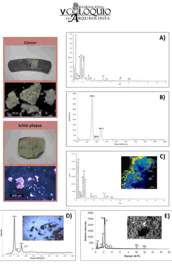
Similarly to what has already been reported in other sites [11-13,23], cinnabar was identified with red iron oxides in the same archaeological contexts. Cinnabar was found covering lithic objects, but a large amount of the red pigment could also be recovered from soil which had been collected in the previous excavation campaigns. Figure 9 presents a summary of red pigment analysis on several archaeological materials from the Zambujeiro Dolmen.
Figure 8.
Figure 9. Summary of red pigment analysis on selected objects from Zambujeiro Dolmen: A) and B) EDS and Raman spectra on a crozier, C) EDS spectra on a schist plaque, D) Raman spectra of the red pigment
covering some amber beads, E) EDS spectra of the red pigment found on the surface of some lithic arrowheads
Using SEM-EDS analysis, it was possible to determine the presence of iron oxide (Figure 9C) and cinnabar with small amounts of iron oxide (Figure 9A). Raman spectroscopy also allowed to identify cinnabar on a crozier and on the surface of some amber beads (Figure 9B and 9D). Stereomicroscopy revealed the presence of dark red particles on the surface of several arrowheads, generally encapsulated by the varnish used to cover their reference number (Figure 10). T hese particles are most likely cinnabar (HgS) as many other, smaller particles composed of Hg and S were found entrapped by terrigenous superficial deposits, located on the surface of the arrowheads, by SEM-EDS (Figure 9E).
Stereomicroscopy image showing the dark red particles found encapsulated by varnish in a lithic arrowhead
Contrary to what happens with the ochres, cinnabar is not frequently found in nature, and there are only four known places in the Iberian Peninsula where its mining would be possible: Las Alpujarras (Granada), Sierra de los Filabres (Almeria), Usagre (in the geological area of Ossa Morena, Badajoz), and the most relevant of all in Almadén (Ciudad Real).
The reasons that have led the communities of recent Prehistory to decide on the use of a rare pigment, which demanded greater effort tobe acquired, in detriment of other local pigments, will always remain a mystery, being the most plausible explanation that cinnabar was believed to have some magical-religious meaning. According to [24], there is also another possible explanation, more pragmatic, which is related with the ability to preserve bones in cinnabar.
The identification of cinnabar in funerary contexts [23], although not unheard of, is important to establish the existence of trade routes between the Zambujeiro dolmen and locations with cinnabar ores.
Fifty siliceous lithic arrowheads recovered from the Zambujeiro Dolmen were studied. Taking into account morphological and temporal criteria, the arrowheads were divided into nine typological groups: A – straight base or slightly convex-base arrowheads; B – convex-base, including leaf-shaped and rhomboidal
Figure 10.
arrowheads; C – concave-base with increasing degrees of concavity from A to B; C1; C2; D – protruding-base arrowheads; E – group that contains both straight base or convex-base arrowheads and concave-base arrowheads. T he arrowheads that incorporate this group were differentiated based on their small dimensions (9.1 to 15.3 mm); F – Alcalar arrowheads (concave-base arrowheads with greatly pronounced wings); G – Mitriform arrowheads (concave-base arrowheads with recurved lateral edges, taking a shape that greatly resembles bishop’s miter); H – Eiffel-tower arrowheads (named due to their resemblance to the Eiffel-tower; concave-base arrowheads with concave lateral edges).
Jorge de Oliveira attributes straight base or convex-base arrowheads, as well as concave-base arrowheads, with low degrees of concavity to the Middle Neolithic period; while concave-base arrowheads with high degrees of concavity and protruding-base arrowheads are attributed to Late Neolithic – Early C halcolithic. However, Oliveira admits the possibility that arrowheads of these typologies were used continuously through the Neolithic and Chalcolithic. Regarding the Mitriform and Eiffel-tower arrowheads, according to [17], their chronological position as C halcolithic is irrefutable. The Alcalar arrowheads are also attributed to the C halcolithic period. Nevertheless, it is important to keep in mind that although raw material availability and quality have been considered the main characteristics that influence the morphology of the tools produced by prehistoric men [18], recent studies [25] suggest that hominids’ ability to manipulate raw materials (i.e. technical competence) may have also influenced stone tool morphology.
The p-XRF results revealed that despite the fact that silicon was found to be a major element in all the samples, the type of fine-grained siliceous rock (including chert) used to manufacture the arrowheads is not always the same. Moreover, no correlation was found between chemical composition and typology, suggesting that a specific type of rock was not used to produce a certain type of arrowhead.
Thanks to the micro-XRD results, the samples were divided into three groups according to their mineralogical composition. T he minerals identified by this technique were quartz, moganite (a polymorph of quartz), alkali feldspars, calcite, micas and clays. T he micro-XRD results are congruous with the idea that arrowhead typology and rock type are not related.
The LA-ICP-MS results were used to produce chondrite-normalized rare earth element (REE) plots. A comparison between these REE patterns and rock databases might enable the identification of the stone sources in the future.
Zambujeiro Dolmen is one of the largest megalithic monuments in the Iberian Peninsula and the number and variety of archaeological materials deposited at the collection of the Évora Museum accounts for its major importance.
The use of appropriate analytical methodology and different analytical techiques applied to diverse types of materials provided a better knowledge of the Zambujeiro Dolmen objects. T his work points out for the importance of the chemical analysis for the unequivocal identification of archaeological materials.
The results have shown that while some materials travel hundreds or thousands of kilometres to arrive to the Zambujeiro Dolmen, local materials were also used in the items selected by the communities to honour their deceased.
[1] Soares J, Silva CT. 3 ( ) 83-129.
[2] Kramar S, Lux J, Mladenoviæ H, Pristacz M, Mirtiè M , Sagadin M , Rogan-Šmuc N. 57 ( ) 39-48.
[3] Maritan L, Mazzoli C , Nodari L, Russo U. 29 ( ) 31-44.
[4] Ravisankar R, Kiruba S, Shamira C, Naseerutheen A, Balaji PD, Seran M. 99 ( ) 370-375.
[5] Moropoulou A, Bakolas A, Bisbikou K. 269 ( ) 743-753. [6] Wentworth C K. 30 ( ) 377-392.
[7] Rice PM. . University of Chicago Press, Chicago and London, .
[8] Vazquez Varela JM. 22 ( ) 407-411.
[9] Schuster V. 4 ( ) 1-26.
[10] Evershed RP. 50 ( ) 895-924.
[11] Domínguez-Bella S, Morata-Céspedes D. XLVIII ( ) 129-142. [12] Ortiz M, Pérez V. ( ) 123-132.
[13] Borja PG, Sanz ID, Garcia C R. 38 ( ) 49-60.
[14] Martínez Fernández MJ, Gavilán Ceballos B, Barrios Neira J, Montealegre Contreras M. , 111-116.
[15] Domingo I, García-Borja P, Roldán C. 54 ( ) 868-892. References 2010 2012 2005 2011 1995 1922 1987 2003 2016 2008 1995 2009 2006 1999 2012 MUSA
Appl. Clay Sci.
Appl. Clay Sci.
Microchem. J.
Thermochim. Acta J. Geol.
Pottery analysis. A sourcebook Gallaecia Comechingonia Virtual Archaeometry Zephyrus VIII C IA Saguntum Archaeometry
[16] Hunt-Ortiz MA, Consuegra-Rodríguez S, del Río-Español PD, Hurtado-Pérez VM , Montero-Ruiz I. Neolithic and Chalcolithic – VI to III millennia BC – use of cinnabar (HgS) in the Iberian Peninsula: analytical identification and lead isotope data for an early mineral exploitation of the Almadén (Ciudad Real, Spain) mining district. In Ortiz JE, Puche O, Rábano I, Mazadiego LF (Eds.).
. Cuadernos del M useo Geominero, 13. Instituto Geológico y Minero de España, Madrid, , 3-13.
[17] Forenbaher, S.
. Doctoral T hesis. Southern Methodist University, Dallas, Texas. University Mycrofilms, Ann Arbor. .
[18] Andrefsky Jr W. . Cambridge University Press, Madrid,
.
[19] Mukherjee AJ, Gibson AM, Evershed RP. 35 ( ) 2059-2073. [20] Regert M. 27 ( ) 244-254.
[21] Izzo FC , Zendri E, Bernardi A, Balliana E, Sgobbi M. 40 ( ) 595-600. [22] Murillo-Barroso M, Martinón-Torres M. 15 ( ) 187-216.
[23] Lazarich González M, Fernández de la Gala JV, Jenkis V, Peralta P, Briceño E, Ramos A, Richarte MJ, Carreras AM , Núñez M, Versaci M, Stratton S, Sánchez M, Grillé JM. 39 ( ) 67-83. [24] Martín Gil J, Martín Gil F, Delibes de Castro G, Zapatero Magdaleno P, Sarabia FJ. 51 ( ) 759-761.
[25] Eren MI, Roos C I, Story BA, von Cramon-Taubadel N, Lycett SJ. 49 ( ) 472-487.
History of Research in Mineral Resources
Production and Exchange of Bifacial Flaked Stone Artifacts During the Portuguese Chalcolithic
Lithics - Macroscopic Approaches to Analysis
J. Archaeol. Sci. J. Sep. Sci. J. Archaeol. Sci. Eur. J. Archaeol. Almoraima Experientia J. Archaeol. Sci. 2011 1997 2005 2008 2004 2013 2012 2009 1995 2014