Drug-induced bone regeneration in a
diabetic model
José Carlos Osório Rodrigues da Silva
Orientador
Professor Doutor Pedro Sousa Gomes
Co-orientadora
Professora Doutora Maria Helena Raposo Fernandes
Porto Fevereiro de 2017
Dissertação submetida à Faculdade de Engenharia, U. do Porto para
obtenção do grau de Doutor em Engenharia Biomédica
This thesis was supervised by:
Prof. Doutor Pedro Sousa Gomes
Faculdade de Medicina Dentária, U. Porto
Prof. Doutora Maria Helena Raposo Fernandes Faculdade de Medicina Dentária, U. Porto
Advisor:
Prof. Doutor Bruno Jorge Antunes Colaço Universidade de Trás-os-Montes e Alto Douro
The host institutions in which the experimental work was conducted were:
Laboratório de Metabolismo e Regeneração Óssea Faculdade de Medicina Dentária, U. Porto
- Cell cultures establishment and characterization
Serviço de Biotério
Universidade de Trás-os-Montes e Alto Douro - Animal housing and experimental surgeries
The research was supported by:
“Nothing in life is to be feared, it is only to be understood.
Now is the time to understand more, so that we may fear less.”
i
Table of Contents
Acknowledgements ... vii Abstract ... xi Resumo ... xiii Abbreviations ... xvList of figures... xix
List of Tables ... xxv
Chapter 1 – Aim and Structure of the Thesis ... 1
1.1 – Aim and structure ... 3
Chapter 2 - Background and Literature overview ... 9
2.1 - Bone Tissue ... 11 2.1.1 - Macrostructure ... 12 2.1.2 - Microstructure ... 17 2.1.3 - Bone minerals ... 22 2.1.4 - Bone cells ... 23 2.1.5 - Bone remodelling ... 31 2.1.6 - Bone healing ... 36 2.2 - Diabetes mellitus ... 38
ii
2.2.1 - Diabetes classification ... 40
2.2.2 - Diabetes diagnosis ... 48
2.2.3 - Diabetes and Bone ... 50
2.2.4 - Diabetes and Biomaterials Implantation ... 57
2.3 - Tetracyclines ... 60
2.3.1 - Chemistry and anti-microbial properties ... 63
2.3.2 - Non-antibiotic properties ... 66
2.3.3 – Doxycycline ... 68
2.3.4 - Minocycline ... 71
Chapter 3 – The Osteogenic priming of mesenchymal stem cells is impaired in experimental diabetes ... 73
3.1 – Introduction ... 75
3.2 – Research hypothesis and objectives ... 77
3.3 – Materials and methods ... 78
3.3.1 – Animals... 78
3.3.2 – Diabetic bone alterations... 79
3.3.3 – Establishment of bone-marrow cell cultures ... 79
3.3.4 – Optical microscopy ... 80
3.3.5 – Cell proliferation and metabolic activity ... 81
3.3.6 – Cell morphology ... 81
iii
3.3.8 – Programmed cell death ... 84
3.3.9 – Collagen synthesis ... 84
3.3.10 – Gene expression ... 85
3.3.11 – Osteogenic induction and culture characterization ... 86
3.3.12 – Activation of specific signaling pathways in STZ-derived cultures .. 87
3.3.13 – Statistical analysis ... 88
3.4 – Results... 89
3.4.1 – Diabetic experimental model ... 89
3.4.2 – Diabetic bone alterations... 89
3.4.3 – Cell proliferation and metabolic activity ... 90
3.4.4 – Cell morphology ... 93
3.4.5 – Alkaline phosphatase activity ... 94
3.4.6 – Programmed cell death ... 95
3.4.7 – Collagen synthesis ... 96
3.4.8 – Gene expression in standard conditions and in osteogenic-inducing conditions ... 99
3.4.9 – Mineralization assessment in osteogenic- and STZ-induced conditions ... 101
3.4.10 – Evaluation of specific signaling pathways... 102
3.5 – Discussion ... 104
iv
Chapter 4 – Doxycycline enhances the osteogenic functionality of
diabetic-derived mesenchymal stem cells ... 113
4.1 – Introduction ... 115
4.2 – Research hypothesis and objectives ... 118
4.3 – Materials and Methods ... 119
4.3.1 – Animals... 119
4.3.2 – Characterization of the experimental groups ... 120
4.3.3 – Cell cultures ... 120
4.3.4 – Cell proliferation and metabolic activity ... 121
4.3.5 – Cell morphology ... 121
4.3.6 – Alkaline phosphatase activity ... 122
4.3.7 – Apoptotic behaviour ... 122
4.3.8 – Collagen synthesis ... 122
4.3.9 – Gene expression ... 123
4.3.10 – Neonatal calvaria defect ex vivo model... 124
4.3.11 – Statistical analysis ... 126
4.4 Results ... 127
4.4.1 – Establishment of a diabetes experimental model ... 127
4.4.2 – Evaluation of diabetes effects on bone ... 127
4.4.3 – Characterization of MSCs cultures grown in the presence of doxycycline ... 129
v
4.4.4 – Calvarial bone defect regeneration – Ex vivo model ... 139
4.5 – Discussion ... 146
4.6 Conclusion ... 151
Chapter 5 – Minocycline-loaded PMMA bone Cement as a delivery system to enhance bone healing – Biocompatibility evaluation in a diabetic model ... 153
5.1 – Introduction ... 155
5.2 – Research hypothesis and Objectives ... 158
5.3 – Materials and Methods ... 159
5.3.1 – PMMA and minocycline-loaded Pmma samples preparation ... 159
5.3.2 – PMMA and minocycline-loaded Pmma samples Characterization . 159 5.3.3 – Animals... 162
5.3.4 – Subcutaneous implantation of minocycline-loaded PMMA ... 163
5.3.5 – Sample gathering and fixation ... 165
5.3.6 – Histological analysis of inflammatory response ... 165
5.4 – Results... 166
5.4.1 – FTIR evaluation of PMMA and minocycline-loaded Pmma samples 166 5.4.2 – Surface analysis of PMMA and minocycline-loaded Pmma samples ... 166
5.4.3 – Minocycline release evaluation ... 168
vi
5.4.5 – Inflammatory response to PMMA and minocycline-impregnated PMMA ... 169
5.5 – Discussion ... 179 5.6 – Conclusions ... 189 Chapter 6 – Conclusions and future perspectives ... 191 6.1 – General conclusions ... 193 6.2 – Future perspectives ... 195 References ... 197
vii
ACKNOWLEDGEMENTS
The development of a work driven to obtain a Doctor degree is a hard and complex path and it can also be a very lonely walk. During this process many changes emerge, which can be either personal or professional and may happen to be major causes of difficulties and discouragement.
However, my walk was neither lonely, nor demotivating. On the other hand, I can state that this was one of the most precious journeys I have experienced and also the most enriching. All the goals and results were achieved with plenty of work, dedication, and the support of teams that helped me during these four years in the Doctoral Program of Biomedical Engineering. Thus, I wish to share this moment of great importance with those who have been crucial during this stage and in my personal development as well.
Therefore, I would like to express my gratitude to those without whom this victory would not have been possible:
To Professor Maria Helena Raposo Fernandes for all of the transmitted knowledge, constant encouragement and support. For all of the opportunities given to me, from the possibility of integrating her research team at the Laboratory for Bone Metabolism and Regeneration, to the possibility of participating in several works and for all the results of this collaboration. I am sincerely grateful for the friendship and for all the advices that helped me through the last years and that will still help me to grow both as a person and as a professional.
viii
To Professor Pedro de Sousa Gomes, for all of the knowledge I acquired during these six years we worked together. For the exemplar orientation and professional accuracy that allowed me to grow scientifically. For all of the support, encouragement and persistence. For all of the opportunities offered to me to acquire knowledge in many different areas from my initial training, namely Tissue Regeneration and Science in Laboratory Animals which became my main preference areas and greater scientific interest. I would also like to thank for his friendship and counseling during the most difficult times. For all the constant calmness and tolerance during my experience at the Laboratory for Bone Metabolism and Regeneration, as well as the availability he has shown to me under any circumstance. I also want to thank him for having accepted to supervise my work, since he represents an example for me to follow.
To Professor Bruno Jorge Antunes Colaço, I thank for all of the availability and cooperation during this work development. I am also grateful for the academic basis and encouragement he transmitted to me in what concerns the work with laboratory animals.
To my colleagues of the Laboratory for Bone Metabolism and Regeneration of the Faculty of Dental Medicine: Elisabete Gonçalves, Fábio Costa, Gabriel Fidelis, Liliana Grenho and Mónica Garciafor all of the excellent team work and cooperation, friendship and aid in the final phase of my PhD.
To my friends, Joana Venâncio, Jorge Cerdeira and Rita Carmona who supported me through this journey sharing with me moments of happiness and who always transmitted me the most positive strength and encouragement.
ix
To Hélder Martinho and Nuno Magalhães, my deepest and heartfelt gratitude for having been present in all of the occasions. As earning a PhD involves hard work and may have consequences in one’s personal life and relationships, I appreciate the understanding and care given to me during this more difficult period. I thank you for all of those days when I could not be with you because I had work to do and yet you came to meet me to support me and give strength. Above all, I appreciate the unwavering friendship.
To my grandparents, I thank the values they instilled in me and for having been present in every stages of my life until today and I wish that they always keep the same faith in me. For the strength that they always gave me and for teaching me that "willpower can move mountains”. I would also still like to thank for all of their trust in me and for the unconditional support throughout all these years always with all of the availability, affection and love.
To my parents, my deepest gratitude for the trust, for the transmitted values and education and for the support in my whole academic and personal life. For the unlimited love and for being role-models of humility, strength and perseverance for me. Also, for being the most solid pillars of my training and in both of my personal and professional development and finally for being by my side in all of the moments. Last but not the least, thank you for having taught me everyday, that together we are able of overcome any difficulty and to take down any obstacle.
To my brother, Pedro Gustavo, I acknowledge so much more than I can here describe. I am thankful for the friendship, the unconditional support in my personal and professional life, whatever the circumstances are. For all of the tight hugs during good and bad moments. For being an integral part of my life and for having been and still being my biggest inspiration and
x
example of loyalty and positivism under any circumstance.
To Leonardo, I thank how he converted all of the complications in a smile or a laugh. All the moments we spent together, for the unrestricted support during the development of this work. I appreciate the tolerance and understanding during the weakest moments throughout this path, either at a personal or professional level. For all the happy moments, for all of the positivity and energy. I also thank the affection, intimacy and love that have been the biggest sources of motivation to continue until here and hereafter.
xi
ABSTRACT
Diabetes mellitus (DM) is one of the most established systemic conditions, with increasing epidemiological importance. It is known to cause numerous complications and severe damage among a variety of tissues and organs, including bone. Accordingly, type 1 diabetes mellitus (T1D) has been associated decreased skeletal mass, higher risk of fractures and delayed healing. Despite the numerous studies on diabetes and its influence on bone, the molecular events affecting both osteoblasts and progenitor cells, within the diabetic milieu, are still not fully understood.
In this context, a detailed study was carried out in order to assess the functionality of diabetic-derived bone marrow-derived mesenchymal stem cells – i.e., osteoblastic precursor cells – regarding proliferation, functional activity, osteogenic priming, and relevant signalling pathways modulating these processes. Data showed that the diabetic environment affected both mesenchymal stem cells signalling and functionality, in a long-lasting way, contributing to a decreased osteogenic priming and increased adipogenic activation, which may converges to the verified bone alterations and weakening.
Given the verified hindrances in diabetes, a novel therapeutic strategy for the enhancement of osteogenic activation was developed, based on the delivery of minocycline and doxycycline, two semi-synthetic derivatives of tetracycline, known to enhance cell proliferation and the functional activity of osteoblasts. Initially, the osteogenic enhancement of bone marrow-derived mesenchymal stem cells and osteoblasts, developed under diabetic-induced conditions, was validated in vitro, in
xii
established cell culture models, and ex vivo, in a model of calvarial bone regeneration. Assayed regimens of low dosage tetracyclines, were able to normalize the impaired osteogenic commitment of osteoblastic precursor populations, enhancing the metabolic equilibrium, as assessed within the in vitro system. Furthermore, tetracycline administration were found to increase tissue healing and tissue mineralization in simulated diabetic conditions, as assayed ex vivo.
Subsequently, and envisaging the development of a translational therapeutic application with reliable effectiveness within the clinical scenario, a PMMA cement-based minocycline delivery system was developed and assayed for biocompatibility. Developed system was subcutaneously implanted in experimental animals, either in control or diabetic conditions. The controlled release of minocycline was able to enhance several inflammation-related factors, suggesting an enhanced tissue healing and biomaterial integration which may further be applied as a local therapy for bone regeneration.
Overall, a novel therapeutic approach – the application of a low dosage regimen of tetracyclines – for the management of the verified hindrances in the osteogenic activation within the diabetic milieu was assayed and validated within in vitro, ex vivo and in vivo models, exhibiting an improved biological outcome and a prospective clinical application.
xiii
RESUMO
A diabetes mellitus é uma patologia sistémica, com elevada incidência, e cuja relevância epidemiológica tem vindo a aumentar. A longo prazo, a diabetes está associada a inúmeras complicações e lesões graves nos tecidos e órgãos, incluindo o osso. Em particular, a diabetes tipo 1 está associada à perda de conteúdo mineral ósseo, aumento do risco de fraturas e atraso nos processos de regeneração. Apesar de terem sido realizados diversos estudos para compreender os efeitos da diabetes no osso, não estão claros quais os mecanismos moleculares envolvidos na afetação, tanto dos osteoblastos como das suas células progenitoras.
Neste sentido, foi realizado um estudo pormenorizado, focado na avaliação da funcionalidade das células estaminais mesenquimais provenientes da medula óssea – células percursoras dos osteoblastos – na condição diabética. Foram avaliados parâmetros como a proliferação, atividade funcional, expressão osteogénica e vias de sinalização relevantes, que modulam estres processos. Os resultados mostraram que a condição diabética afeta tanto vias de sinalização como a funcionalidade das células mesenquimais de forma duradoura, contribuindo para uma diminuída expressão osteogénica e aumento da expressão adipogénica, convergindo desta forma para alterações na estrutura óssea, e seu consequente enfraquecimento.
Tendo por base a identificação das alterações vigentes na condição diabética, foi desenvolvida uma nova abordagem terapêutica para melhorar a ativação osteogénica, baseada na libertação de minociclina e doxiciclina, dois derivados semissintéticos da tetraciclina, que demonstraram um efeito indutor na proliferação celular e na atividade
xiv
funcional de osteoblastos. Inicialmente, foi realizada uma validação in vitro dos efeitos promotores da expressão osteogénica de células estaminais mesenquimais da medula óssea, bem como os efeitos da doxiciclina num modelo ex vivo da regeneração da óssea da calote. As baixas doses de tetraciclinas que foram estudadas permitiram a normalização do comprometimento com a linhagem osteogénica, melhorando assim o equilíbrio metabólico no osso diabético.
Com o intuito de desenvolver uma aplicação terapêutica com uma efetividade clínica, foi desenvolvido um sistema de libertação controlada de minociclina, a partir de um cimento de polimetil metacrilato (PMMA). O sistema desenvolvido foi implantado subcutaneamente em animais controlo e animais diabéticos, para avaliação da sua biocompatibilidade. A libertação controlada de minociclina permitiu melhorar determinados fatores relacionados com a resposta inflamatória, sugerindo uma melhoria tanto nos processos de regeneração dos tecidos envolventes como na integração do biomaterial que, por sua vez, poderá ser posteriormente aplicado em terapias locais de regeneração óssea.
No geral, foi estudada uma nova abordagem terapêutica, baseada na aplicação de baixas doses de tetraciclinas, tendo em vista a indução osteogénica, que se encontra alterada na condição diabética. Esta abordagem foi validade com estudos in vitro, in vivo e ex vivo, e revelou resultados biológicos favoráveis para futuras aplicações clinicas.
xv
ABBREVIATIONS
αMEM Alpha-modification minimum essential medium ALP Alkaline phosphatase
AP2 Adipocyte protein 2 BMD Bone mineral density
BSP-1 Bone sialoprotein 1, osteopontin BSP-2 Bone sialoprotein 2
CAP Fibrotic capsule
CLSM Confocal laser scanning microscopy
DGAV Direção Geral de Alimentação e Veterinária
DM Diabetes mellitus
DO Diabetic osteopenia
ECM Extracellular matrix
EDS Energy-dispersive x-ray spectroscopy EDTA Ethylenediaminetetraacetic acid
FB Fibroblasts
FBS Foetal bovine serum
FTIR Fourier Transform Infrared (spectroscopy) GAPDH Glyceraldehyde 3-phosphate dehydrogenase
xvi
GC Giant cells
GDM Gestational diabetes mellitus
GH Growth hormone
IGF-1 Insulin-like growth factor-1 IP Intraperitoneal (injection) IR Inflammatory reaction IRS1 Insulin receptor substrate 1 IRS2 Insulin receptor substrate 2
LYM Lymphocytes
MAC Macrophages
MAP Mycobacterium avium subspecies paratuberculosis µCT Microcomputed tomography
M-CSF Macrophage colony-stimulating factor MMP Matrix metalloproteinase
MMP-9 Matrix metalloproteinase 9
mPMMA Minocycline-loaded poly(methyl methacrylate)
ND Neonatal diabetes
NEO Neovascularization
NO Nitric oxide
xvii
OPG Osteoprotegerin
OPN Osteopontin
ON Osteonectin
PBS Phosphate buffered saline PMMA Poly(methyl methacrylate) PMNs Polymorphonuclear neutrophils
PPAR𝛾 Peroxisome proliferator-activated receptor gamma
PTH Parathyroid hormone
RANKL Receptor activator of nuclear factor kappa-β ligand RUNX2 Runt-related transcription factor 2
SC Subcutaneous (injection)
SDD Subantibacterial dose doxycycline
STZ Streptozotocin
T1D Type 1 diabetes mellitus T2D Type 2 diabetes mellitus
TC Tetracycline
xix
LIST OF FIGURES
Chapter 2
Figure 2.1 Different types of bone morphology ………. 13
Figure 2.2 Microstructural organization of mature lamellar bone comprising areas of compact and cancellous bone ……….. 18
Figure 2.3 Scanning electron microscopy image of collagen fibers of human cancellous bone ……….… 20
Figure 2.4 Rat multinucleated osteoclast; Histological analysis of human bone ………..…. 24
Figure 2.5 Histological analysis of rat bone section stained with toluidine blue ………... 27
Figure 2.6 Bone lining cells ……….. 30
Figure 2.7 Schematic representation of the four stages of bone remodelling cycle ………... 34
Figure 2.8 Schematic representation of the four stages of bone fracture repair ……… 37
Figure 2.9 Chemical structure of naphthacene ring system, first-generation antibiotics, and second-generation antibiotics ………. 62
xx
Chapter 3
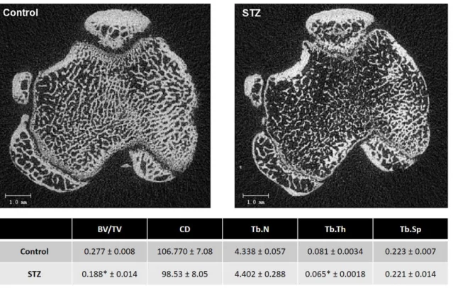
Figure 3.1 Representative 2D microtomographic images of the proximal tibia methaphysis, in control and STZ animals ………... 90
Figure 3.2 Cell proliferation (DNA assay) of STZ-derived bone marrow mesenchymal stem cell cultures ……….. 91
Figure 3.3 Cell viability/metabolic activity (MTT assay) of STZ-derived bone marrow mesenchymal stem cell cultures .……….. 92
Figure 3.4 Confocal laser scanning microscopy imaging of rat bone marrow-derived mesenchymal stem cell cultures ………. 94
Figure 3.5 Alkaline phosphatase activity (ALP per total protein content) of STZ-derived bone marrow mesenchymal stem cell cultures ……… 95
Figure 3.6 Apoptotic analysis (caspase-3 activity assay) of STZ-derived bone marrow mesenchymal cell cultures ……….. 96
Figure 3.7 Total collagen staining of STZ-derived bone marrow mesenchymal stem cell cultures ………..… 97
Figure 3.8 Colorimetric determination of the total collagen stained product within the established STZ-derived bone marrow mesenchymal stem cell cultures ………..…. 98
Figure 3.9 qPCR gene expression analysis of ALP, RUNX2, Col1α1, osteopontin, osteocalcin, and osteoprotegerin in MSCs cultures ……… 99
Figure 3.10 qPCR gene expression assessment of adipogenic genes PPARγ, IRS1, IRS2, and AP2, in undifferentiated STZ-derived MSCs cultures .…..… 100
xxi
Figure 3.11 Colorimetric determination of the mineralization nodules within the established control- and STZ-derived bone marrow mesenchymal stem cell cultures ………. 101
Figure 3.12 Metabolic activity and gene expression analysis (of ALP, RUNX2, and PPARγ) of MSCs cultures ………... 102
Chapter 4
Figure 4.1 Representative 2D microtomographic images of the proximal tibia methaphysis, in control and STZ animals ……… 128
Figure 4.2 Cell viability/metabolic activity (MTT assay) of rat bone marrow-derived cells cultured in presence and absence of doxycycline …….. 130
Figure 4.3 Confocal laser scanning microscopy imaging of rat bone marrow-derived cell cultures, from control and STZ-induced diabetic animals in presence and absence of doxycycline ………... 132
Figure 4.4 Alkaline phosphatase activity (ALP per total protein content) of rat bone marrow-derived cells cultured in presence and absence of doxycycline from both control and STZ-induced animals ……… 133
Figure 4.5 Alkaline phosphatase staining of rat bone marrow-derived cell cultures ………. 134
Figure 4.6 Apoptotic analysis (caspase-3 activity assay) of rat bone marrow-derived cells cultured in presence and in absence of doxycycline from both control and STZ-induced animals ………. 135
xxii
Figure 4.7 Total type 1 collagen staining of rat bone marrow-derived cell cultures, from control and STZ-induced diabetic animals in presence and in absence of doxycycline ……….. 136
Figure 4.8 Colorimetric determination of the total collagen stained product within the established MSCs cultures from both control and STZ-induced diabetic animals in presence and absence of doxycycline ……….... 137
Figure 4.9 qPCR gene expression analysis of ALP, BMP-2, Col 1, OPN, LRP5, OC, and OPG in MSCs cultures, established for days 5 and 8, from control and STZ-induced diabetic animals ……….. 139
Figure 4.10 Phase contrast optical microscopy images obtained at days 0, 10 and 20 for the established experimental conditions of new-born rat calvarial organ culture ……….……….. 141
Figure 4.11 SEM images of new-born rat parietal bone at days 8 and 15 ………. 143
Figure 4.12 SEM images of mineralization deposits after 15 days of culture of newborn rat parietal bones ………. 144
Figure 4.13 Percentage of regenerated area along the 15 days of new-born rat parietal bones’ culture, in both control and diabetic conditions, in presence and absence of doxycycline ………..… 145
Chapter 5
xxiii
Figure 5.2 FTIR spectra of PMMA, minocycline-loaded PMMA and free minocycline ……… 167
Figure 5.3 SEM micrographs of the control BC matrix (PMMA), and BC loaded with minocycline ………. 167
Figure 5.4 In vitro release profiles of minocycline, in both LOW and HIGH concentrations ………. 168
Figure 5.5 Histological analysis of the tissues surrounding PMMA constructs implantation in control and diabetic animals ……….. 171
Figure 5.6 Histological analysis of PMMA and mPMMA surrounding tissue, in control animals ……….………..… 173
Figure 5.7 Histological analysis of PMMA and mPMMA surrounding tissue, in diabetic animals ……….. 174
xxv
LIST OF TABLES
Chapter 2
Table 2.1 Differences between human cortical and cancellous bone …….………. 14
Table 2.2 Etiologic types and stages of glycemic disorders ……….…... 41
Table 2.3 Criteria for the diagnosis of diabetes mellitus ……….. 49
Chapter 3
Table 3.1 Forward and reverse sequences of the primers used for the qPCR analysis ………. 86
Chapter 4
Table 4.1 Forward and reverse sequences of the primers used for the qPCR analysis ……….. 124
Chapter 5
Table 5.1 Semi-qualitative analysis of overall inflammatory reaction and inflammatory response parameters around PMMA and mPMMA samples, in both control and diabetic conditions ………. 177
1
CHAPTER 1 – AIM AND STRUCTURE OF THE
THESIS
3
1.1 – AIM AND STRUCTURE
Of worldwide importance, diabetes mellitus represents one of most established systemic conditions with increasing epidemiological importance. It encompasses a group of metabolic disorders which are ultimately characterized by a hyperglycaemic state and consequent requirement for continuous medical care with multifactorial risk-reduction strategies (1). Diabetic hyperglycaemia is strongly associated with long-term tissue and organs failure due to severe damaged and unbalanced carbohydrate metabolism. The most well-known complications related with diabetes comprise neuropathy, nephropathy, retinopathy and osteopenia, among others (2).
In which concerns the bone tissue, uncontrolled hyperglycaemia was broadly described to affect the normal bone metabolism interfering with the natural bone renewal, as well as, increasing the risk of fractures due abnormal cellular function to produce new bone, impaired mineralization and consequent altered bone mineral density (3, 4). Despite the contradictory reports, regarding bone tissue affection in the different types of diabetic condition (5), it is stablished that type 1 diabetes, in particular, exerts severe effects in bone though a variety of mechanisms including impairment of osteoblastic recruitment, maturation and function to produce new bone; promoting a disorganized and defective deposition of collagen matrix; inhibiting the expression of growth factors, essential for bone mineralization to occur; among other which culminate in a weakened structure (6).
Recently, several studies reported that, tetracycline’s semi-synthetic derivatives, i.e., minocycline and doxycycline, were able to modulate osteoblastic cells and
4
improving the bone regeneration process in a mechanism independent of the antibacterial activity (7, 8). Furthermore, these non-antibacterial properties of minocycline and doxycycline were demonstrated to exert a significant beneficial effects in pathological conditions involving abnormal immune and inflammatory response and unbalanced apoptotic behaviour (such as rheumatoid arthritis, periodontitis, rosacea, among others) (9, 10). Accordingly, these semisynthetic tetracyclines may represent promising candidates to support bone healing therapies in both cases of fractures in physiological or pathological conditions.
The increased risk of fracture among diabetic patients, lead to an increased requirement for orthopaedic implants. However, diabetes systemic abnormalities include defective host response and impaired wound healing which greatly increases the risk of infection among the patients with the disease (11).
In this context, this work aims to achieve a deeper insight on the effects of minocycline and doxycycline as osteogenic agents, promotors of bone regeneration and tissue healing, in the tissues and cells of a well-established animal model of diabetes – the streptozotocin-induced diabetic rat.
5 The thesis is organized into six chapters:
Chapter 1
General considerations about the aim and structural organization of the present PhD thesis.
Chapter 2
The second chapter is divided in three parts. The first part consists of an overview of bone tissue including composition, structure and metabolic activity; bone remodelling and bone healing processes. Following, in the second part, a review of diabetes mellitus is made focusing the systemic unbalances, classifications and diagnosis; with especial relevance, the effects of diabetes in bone tissue; and biomaterial implants to support bone healing in diabetic-mediated pathological conditions. In the third part, a revision of tetracycline derivatives, minocycline and doxycycline, is carried out. In addition to their role as antibiotics, their properties as modulatory agents of bone metabolism and regeneration in physiological and pathological conditions are reviewed.
Chapter 3
The third chapter describes a detailed characterization of bone marrow-derived mesenchymal stem cells, harvested from both healthy and streptozotocin-induced diabetic Wistar rats, and cultured in control and osteogenic-induced conditions. In this module, in vitro assays leading to the characterizations of cell proliferation, metabolic
6
activity, as well as the assessment of gene expression and significant signalling pathways activated within the osteogenic priming and functionality, were critically detailed.
Chapter 4
In chapter 4, the effect of doxycycline is assessed on both in vitro and ex vivo models of control and diabetic conditions. In the first part, the effect of a low dosage regimen of doxycycline was assayed in the previously described cell culture model of bone marrow-derived mesenchymal stem cells, harvested from control and diabetic-induced Wistar rats. Culture functionality was critically detailed, regarding cell proliferation, metabolic activity, osteogenic priming and activation. In a second part, an
ex vivo experiment was conducted with new-born rats’ calvaria to assess the effect of
the low dosage regimen of doxycycline in the bone regeneration process, in both control and diabetic simulated conditions.
Chapter 5
In the chapter 5, a novel PMMA cement-based system for the controlled release of minocycline, aiming the development of a clinical relevant formulation for drug delivery in diabetic conditions, was characterized for solid state parameters and biocompatibility. The tissue response to the implanted minocycline-loaded constructs was detailed, in both control and diabetic animal models, by histological and histomorphometric analysis, at several time points following subcutaneous implantation.
7 Chapter 6
In the last chapter, a general conclusion is presented, based on the data gathered from the experimental studies presented on chapter 3, 4 and 5.
9
CHAPTER 2 - BACKGROUND AND LITERATURE
OVERVIEW
11
2.1 - BONE TISSUE
The bone is a highly complex and hard form of connective tissue which may be both coped as a tissue or as organ system, making up most of the skeleton. It combines the endurance and toughness of a mineralized matrix composed essentially by proteins and hydroxyapatite crystals with a dynamic organic/inorganic matrix. As a component of the skeleton, bone tissue plays crucial functions in the human organism including protection, support, motion of the entire organism and metabolic homeostasis. Bone unique and individual properties such as a high flexibility and elasticity promote the protection of vital organs. Yet its stiffness also underwrites to the conservation of the structural support and mechanical action allowing for the precise and controlled muscular movement. The organic component ensembles a variety of bony cells which are continuously involved in metabolic modulation of pH regulation, mineral storage, homeostasis, bone remodelling and other functions, causing bone to be a biologically active and dynamic entity (12). The adult human skeleton possesses around of 213 bones.
12 2.1.1 - MACROSTRUCTURE
Macroscopically, bones of the skeletal system are white coloured and usually classified individually according to their shape in long, short, flat, sesamoid and irregular bones (figure 2.1a). Long bones are characterized for being longer than wider and this category includes the majority of the bones of limbs such as femurs, tibiae and humeri. They are specifically designed for rigidity, possessing some elasticity and great hardness, and attachment of muscles and ligaments. The majority of long bones present morphological similarities (figure 2.1b) as they are constituted by three common components: (1) the diaphysis (or shaft), the body of long bones being mainly made up of compact bone organized in a long tubular structure; (2) the epiphyses, the long bones’ ends in which the joints with adjacent bones are established, being predominantly composed by cancellous bone; (3) the epiphyseal plate (or growth plate), a hyaline cartilage plate located in the metaphysis in the long bone’s ends. Short bones are those bones that are almost as wider as longer and commonly exhibit a cuboidal shape. These bones include smaller bones and they are found only in the ankle and wrist (e.g. tarsus and carpus). Flat bones are usually associated with protection functions due to their morphology thinner and flatted which provides broad section for protection or muscular attachment (e.g. ribs, sternum and cranial bones). Sesamoid bones are usually found in locations where tendons intersect the ends of long bones, being responsible for protection of these tendons from an excessive mechanical load (e.g. patella and knee cap). Just like the vertebrae of the spine or the facial bones, the irregular bones are characterized by their peculiar morphology, which cannot be included in none of previous classifications (12).
13
Figure 2.1 - (a) Different types of bone morphology: long, short, flat, sesamoid and irregular; (b) Adult long bone structure, adapted (13).
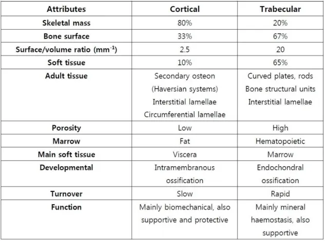
Despite these different morphologies, all bones are macrostructurally identical in that densely packed or compact bone – that is more external and the principal responsible for bone strength, whilst the inner bone tissue is commonly composed by a tridimensional structure of trabecular bone with metabolic and homeostasis functions. Although both compact and trabecular bone are made up of the same kind of extracellular matrix (ECM) and cell types, they differ in which regards to their structure and functions (14). Thereby, compact bone is mostly associated with biomechanical and
14
protective functions, whereas the majority of metabolic functions are due to the trabecular bone (table 2.1), such that it is more responsive to disturbances in metabolic homeostasis (12).
Table 2.1 - Differences between human cortical and cancellous bone. Adapted from (15).
15
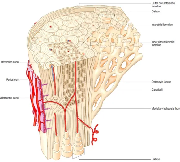
Compact bone, also known as cortical or dense bone, is found on the outer layer of all individual bones surrounding the trabecular structure and exhibit an ivory-like compact shape. It represents approximately 80% of the total bone mass and it is mainly composed of bony mineral and ECM providing the adequate strength to perform its functions. Structurally, cortical bone is made up of a collection of cylindrical units named Harvesian systems, or osteons, which run parallel to the bone axis. The Harvesian system represents the cortical bone fundamental unit and it combines a central canal – through which blood vessels, lymphatic nodes, connective tissue and poorly myelinated fibers pass – with its surrounding concentric bony tissue lamellae and respective osteocytes (14). The lamellae constitute a more external surface of the cortical bone and they are interspersed by small voids known as lacunae, which are interconnected by smaller channels – canaliculi (16). The osteocytes are found within the lacunae and they are interconnected with one another and with the osteoblasts on the surface of the bone by extending cytoplasmatic processes into the canaliculi. This communication with external osteoblasts allows the process of osteocytic osteolysis, which comprises the transference of Ca2+ from the interior of the bone to the surface (17). Additionally to
cell-to-cell interaction, canaliculi enable the nutritional support and oxygenation of the whole cellular constituents and also permit the removal of waste products resultant from metabolic activities. The gaps between the osteons are filled with a tissue similar to lamellar bone although with a less organized pattern. The interstitial lamellae separate each Harvesian system from its neighbouring structures by forming a strongly basophilic cement line constituted essentially by inorganic matrix content (16). The superficial layer of compact bone is mainly constituted by osteoblastic and osteoclastic
16
precursor cells which are continuously renewing the cortical bone by producing new osteoid (figure 2.2) (18, 19).
Trabecular bone, synonymous to cancellous or medullar bone, constitutes about 20% of the remaining total bone mass and it is located in the inner layer of individual bones. Structurally, cancellous bone presents a honeycombed shape and is considerably less dense and resistant than compact bone, being committed essentially to metabolic and mineral homeostatic functions. It has a higher surface area reaching about 67% of the total and its structure consists of an irregular network of calcified spicules extended from the cortex to the inner canal – trabeculae – made up of osteon fragments. The trabecular structure is classified as being rod-like or plate-like and the portion of each type depends on the magnitude and distribution of loading. Its thickness commonly ranges from 50 to 400 µm and it is formed by bone lamellae. Furthermore, trabeculae structural organization is not random, rather the structures run according to the mechanical stress guidance and they are continuously able to adapt to mechanical tension solicitations (20). Similarly to the cortical bone, the most external layer is mostly coated by osteoblasts and osteoclasts responsible for constant renewal of cancellous bone. The cavities between trabeculae are filled with hematopoietic marrow, in which the majority of metabolic supportive functions of bone occur (16).
17 2.1.2 - MICROSTRUCTURE
Independently from being cortical or cancellous, the microscopical structure of bone can be categorized in two major forms: woven bone or lamellar bone. Woven bone, also designated as primary bone, is broadly found during the embryonic stages, as well as in newborns and it is later resorbed and replaced by lamellar bone (21). Primary bone may also be found in locations where fast bone formation takes place, like in growth plates and during processes of bone healing of fractures, closure of cranial sutures or in pathological conditions, such as in Paget’s disease. Comparing to lamellar bone tissue, woven possesses a higher metabolic rate which substantiates an enhanced turnover during the remodelling phase. Woven bone is also characterized by an anisotropic distribution of cells and collagen fibers, which explains its diffused and irregular display, as well as its biomechanical fragility (21). After woven bone is laid down, it can undergo remodelling processes to become lamellar. Lamellar bone, or secondary bone (figure 2.2), commonly appears firstly during the third foetal trimester and it is produced much more slowly, being characterized by a highly and ordered arrangement whereas the cells are broadly uniform in size, shape and orientation, supporting a more resilient biomechanical performance than woven bone (21).
2.1.2.1 - EXTRACELLULAR MATRIX
Regarding its composition, the mature bone tissue may be considered a composite material constituted by a tough organic matrix (approximately 35%), which is greatly
18
strengthened by inorganic minerals (60 to 70% bone weight) and cells. The remainder ECM is filled with a homogenous gelatinous medium, named ground substance, that is composed of extracellular fluids and proteoglycans (e.g., chondroitin sulfate and hyaluronic acid), which are thought to assist on the regulation of calcium salts deposition. The proportions of these components vary with age, location and metabolic status (20).
Figure 2.2 - Microstructural organization of mature lamellar bone comprising areas of compact and cancellous bone (20).
19 2.1.2.2 - BONE COLLAGEN
The overall extracellular structure of the bone acts as an interconnected network for cells, in close interaction. The organic part of this matrix is 90 to 95 per cent constituted by collagen fibers, which closely resemble those of many other connective tissues and it is predominately composed by collagen type I fibers, combined with a very small amount of collagen type V, which is thought to regulate fibrillogenesis (20). Nevertheless, the molecular structure of collagen is unique in which regards its organization, which displays internal covalent cross-linkages, and larger transverse spacing, within its fibrils (figure 2.3). The cross-links make it stronger and chemically more inert while the internal gaps enable deposition of minerals. The adequate and ordered deposition of collagen in sufficient amounts is required to accomplish the formation of optimal bone mass and mineral density during the skeletal development, since bone mineral crystals deposition is aligned with the long axis parallel to the collagen axis. As so, collagen contributes largely to the mechanical strength of bone as well as to the tensile strength, compressive resistance and elasticity, which enhances the resistance to fractures during mechanical loading (20).
Collagen is produced by osteoblastic cells which synthesize tropocollagen, which is lately polymerized in the ECM and gradually increases its cross-links with maturation. Collagen fibres from the periosteum, also designated as extrinsic fibres, are incorporated in cortical bone and anchor the fibrocellular layer, at their surface. Terminal collagen fibres of tendons and ligaments are incorporated deep into the matrix of cortical bone. The fibres may be interrupted by new osteons during cortical bone
20
modelling and turnover or remodelling, and remain as islands of interstitial lamellae or trabeculae (20).
Figure 2.3 - Scanning electron microscopy image of collagen fibers of human cancellous bone (20).
2.1.2.3 - NON-COLLAGENOUS ORGANIC COMPONENTS
Several non-collagenous proteins can also be found among the extracellular organic component attached to collagen fibers or surrounding bone crystals. They are
21
essentially secreted by osteocytes and osteoblastic cells and they include among the several, osteopontin (or bone sialoprotein-1, BSP-1) (OPN), bone sialoprotein-2 (BSP-2), osteocalcin (OC) and osteonectin. Despite their exact biological role is far from being completely fulfilled these components play important role in bone metabolic functions as well as in osteoblastic and osteoclastic cell function and interaction (16).
Bone matrix also contains proteoglycans (e.g. decorin and biglycan, which seem to modulate cell activity, especially the collagen fibrillogenesis process), glycoproteins (e.g., fibronectin and vitronectin, with a distinctive role in cell signalling), many growth factors (e.g. transforming growth factor β (TGF-β), bone morphogenic protein (BMP), insulin-like growth factor (IGF-9 among other), proteases and proteases inhibitors (16, 20).
22 2.1.3 - BONE MINERALS
The inorganic constituents of the extracellular matrix play an important role, conferring strength, rigidity and allowing ion storing. This component is the main reason why the bone maybe recorded on radiographs since it needs to be at least 50% mineralized to be visible on radiographs produced by a standard X-ray unit. With the mineral removal with calcium chelators, such as ethylene diamine tetra-acetic acid (EDTA), the bone keeps its shape but becomes highly flexible (20).
The crystalline salts deposited in the organic matrix are constituted essentially by calcium and phosphate. The substance which largely mades up these crystals is commonly referred as hydroxyapatite, although with an important carbonate content and a lower Ca/P ratio than the pure hydroxyapatite, Ca10(PO4)6(OH)2. Bone crystals are
thin plates or leaf-like structures that possess a broad surface area despite the small size. These structures range in size up to 150 nm long, per 80 nm wide, per 5 nm thick; however most of them are broadly half this size (20).
Ionic substituents, such as magnesium, strontium, carbonate, citrate, and fluoride are broadly present among the bone salts. It is thought they are conjugated with hydroxyapatite rather than organized separately. Additionally, these ions seem to modulate the biological response in the local microenvironment (16).
23 2.1.4 - BONE CELLS
Bone tissue exhibits four main types of cells among its organic matrix including osteoclasts, osteoblasts, osteocytes and bone lining cells. Bony cells may be generally classified attending to their functional activity into bone forming and bone resorbing cells, or according to its differentiation or maturation state.
The cell-to-cell and cell-to-matrix interactions are critical in order to maintain bone mass balanced and bone tissue homeostasis. For instance, osteoblasts are known to bind bone matrix through integrins which recognize RGD among other bone-specific protein sequences, such as osteopontin, collagen and bone sialoproteins. Moreover, osteoclasts interaction with bone matrix and osteoblastic-secreted factors are essential for the osteoclastic function and bone renewal to occur correctly (18).
2.1.4.1 - OSTEOCLASTS
Osteoclasts are the only cells known to be able of absorb or breakdown bone tissue. These large (ranging 40 µm and higher) and multi-nucleated cells (normally 3 to 20 nuclei) are derived from circulating hematopoietic stem cells that differentiate along the monocyte/macrophage lineage (see figure 2.4a). Since their differentiation pathway is common to macrophages and dendritic cells, osteoclastic differentiation depends directly on precursor cell exposition to several specific factors such as the macrophage colony-stimulating factor (M-CSF) and the receptor activator of nuclear factor kappa-β ligand (RANKL), both secreted by osteoblastic cells (22). Both cytokines are crucial not
24
only for differentiation but are also required for proliferation, survival and differentiation of osteoclastic precursor cells (20). The M-CSF binds to its receptors in osteoclastic precursors, stimulating their proliferation and disrupting apoptosis, while RANKL binds to its ligand (RANK), triggering osteoclastogenesis (23, 24). Along with these two factors, osteoprotegerin (OPG) was found to bind RANK preventing RANK-to-RANKL interaction, leading to inhibition of osteoclastogenesis (24). This three-way system works as a key regulator of bone resorption.
Figure 2.4 - (a) Rat multinucleated osteoclast (OC) revealing some nucleus (N) and vacuoles (V) and the organization of a ruffle border (RB) next to bone surface (B) undergoing resorption, adapted from (18); (b) Histological analysis of human bone revealing multinucleated osteoclasts forming Howship’s lacunae (25).
Osteoclastic cells are commonly found in areas undergoing bone resorption (figure 2.4b) and reveal high mobility. These cells mediate the resorption process via the release
25
of powerful lysosomal enzymes and acids, which digest protein and mineral components of the bone matrix, respectively. The key family of proteinases involved in this degradation process of bony tissue are the cathepsins and matrix metalloproteinases (26).
2.1.4.2 - OSTEOBLASTS
Osteoblasts are derived from undifferentiated stem cells of mesenchymal origin, which can be found in bone marrow, endosteum, and periosteum, among other locations (see figure 2.5). These osteoblastic progenitor cells, also referred as “preosteoblasts”, can migrate from neighbouring tissues or through the vascular system to the target area. Commonly, osteoblasts comprise 4 to 6% of the total resident bone cells and are located in bone outer layers and in bone cavities undergoing bone remodelling events (i.e. trabeculae). These are the unique cells known to be able of promote new bone formation by deposition of extracellular matrix rich in collagenous and non-collagenous proteins, being further responsible for the subsequent mineralization process. Morphologically, mature and active osteoblasts are mononucleated cells with plump cuboidal-like shape, with a prominent Golgi apparatus and endoplasmic reticula, both related with their high secretory activity (18, 27). With the decreasing of the metabolic level, the osteoblasts shape becomes progressively flattened out with the decreasing of the metabolic index.
The synthesis of new bone tissue takes place in two main steps. In the first step, osteoblastic cells secrete the most of bone remodelling modulators and bone matrix
26
molecules such as type 1 collagen, non-collagenous proteins (such as osteonectin, bone sialoprotein 2, osteocalcin and osteopontin), proteoglycans (including decorin and biglycan) which promotes the matrix formation (16). The second step, focuses on the matrix mineralization and takes place into two phases namely, the vesicular (i) and the fibrillar phase (ii). In vesicular phase, numerous matrix vesicles (ranging from 30 to 200 nm) are released into the newly formed bone matrix binding to the previously secreted components (28). These vesicles were described to contain a phosphatase, active at neutral pH, which has strong specificity and promote further hydrolyse of a variety of nucleotide and metabolite. Additionally, matrix vesicles were found to up 45Ca, even in
the presence of low levels of Ca (29). Following, the fibrillary phase occurs with the rupture of the vesicles leading to the spreading of hydroxyapatite crystals on the surrounding matrix as the vesicles’ content contact with phosphate-containing substrates in the presence of ATP (29, 30).
Osteoblasts cytoplasmic membrane is rich in alkaline phosphatase (ALP), an important enzyme for ECM mineralization. The increasing of ALP expression is intimately bounded with a shift to a more differentiated state of the osteoblastic cells and generally determines the occurrence of the mineralization process, at least within in vitro systems (31). Furthermore, ALP is responsible for the degradation of matrix vesicles leading to the release of phosphate and calcium ions, allowing the subsequent formation of hydroxyapatite crystals (18).
Osteocalcin and osteonectin (ON) are both products of osteoblasts which play crucial roles in new bone mineralization. The first is a 6-kDa protein, synthetized at the locations undergoing bone remodelling and binds to hydroxyapatite, thus suggesting a
27
participation of OC in nucleation of the bone mineralization. The 35-kDa protein osteonectin also binds to hydroxyapatite and to collagen fibres, facilitating extracellular mineralization (17).
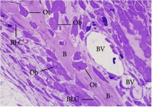
Figure 2.5 - Histological analysis of rat bone section stained with toluidine blue. The image reveals osteoblasts (Ob) and bone lining cells (BLC) present on bone surfaces and the osteocytes (Ot) trapped into the bone matrix along the bony trabecula (B). BV refers to blood vessels (18).
Osteoblastic activity continues in living bone through life time span, allowing a continuous renewal of bony tissue. Once the remodelling cycle is finished, osteoblasts cease the production of bone matrix and become committed to one of three pathways:
28
(1) become embedded by new mineralized bone matrix and differentiate in osteocytes, (2) shift their shape turning into flattened lining cells covering the bone surface, or (3) become relatively inactive or undergo apoptosis (18, 32).
2.1.4.3 - OSTEOCYTES
Osteocytes comprise around 90 to 95% of bone cells in the adult skeleton. When osteoblasts become entrapped in the newly formed bone matrix (figure 2.5), a subpopulation differentiate into osteocytes which are more mature cells and mainly responsible for bone matrix maintenance (33). This differentiation process comprises also conspicuous morphological and ultrastructural changes including osteoblast size reduction, decreasing number of organelles such as rough endoplasmic reticulum and Golgi apparatus, and decreasing nucleus-to-cytoplasm ratio resulting in a diminished protein synthesis (34). Despite that fully-matured osteocytes are relatively inactive when compared to active osteoblasts downregulating the expression of osteoblastic markers such as OC, BSP-2, collagen type 1 and ALP, they still can produce several factors, essential for bone matrix support, including dentine matrix protein 1 and sclerostin (18, 35). Osteocytes may be found in bone lacunae of 1 to 2 µm wide, surrounded by collagen fibrils, which support cytoplasmic process responsible for the intercellular communication through cannaliculi channels and gap junctions (20). Additionally to cell-mediated exchanges of minerals, this network also act as mechanosensor, as it has the capacity to detect mechanical deformation within bone, thus modulating processes such as bone formation or bone resorption (36). The specific
29
mechanical stimuli to which bone cells respond in vivo may be connected with strain changes itself, as well as strain-generated changes to their fluid environment, which consequently affects the release of signalling molecules and growth factors, that seem to regulate cell proliferation and differentiation (37).
2.1.4.4 - BONE LINING CELLS
Bone lining cells derive from osteoblastic cells which cease the production of new bone matrix, becoming partially inactive. They exhibit an elongated shape (see figure 2.5 and 2.6) with a flat nuclei and usually are located over bone surfaces where neither bone resorption nor bone formation occur (38). Due to their abridged metabolic activity, they possess fewer organelles than osteoblasts.
Bone lining cells are compactly associated and connect each other via gap junctions and communicate with internal bone cells, such as osteocytes, by cytoplasmic extensions or thigh junctions made through surface canaliculi (39). Since the activation of bone remodelling process takes place at inactive bone surfaces, bone lining cells are thought to interfere in bone metabolism in two possible ways: (1) building a microenvironment suitable for bone resorption mediated by osteoclasts, as well as by producing OPG and the receptor activator of nuclear factor kappa-β ligand; and (2) regulating new bone matrix deposition and mineralization, enrolling osteoblast-like functions (39).
30
Also, bone lining cells showed to play an important role in the maintenance of the bone fluids and the fluxes of ions between the bone fluid and interstitial fluid compartments, interfering deeply into mineral homeostasis (20).
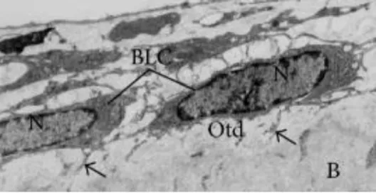
Figure 2.6 - Bone lining cells (BLC) exhibiting flat shape with scarce cytoplasm, located on the osteoid (Otd) surface. B refers to calcified bone surface and N to the nucleus. Adapted from (18).
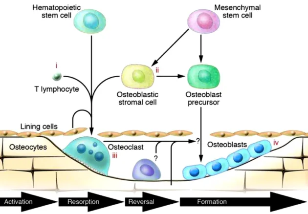
31 2.1.5 - BONE REMODELLING
Bone remodelling is a natural process wherewith bone tissue renew itself throughout lifespan. Due to its cellular components and functions, bone has been described as a highly dynamic and metabolic active specialized connective tissue. These cellular components are the main responsible for this remarkable ability and highly regulated process of bone renewal in which older bone is continually being absorbed by osteoclastic cells while new bone is being continually deposited by osteoblastic cells. Thus, the bone renewal process is dependent upon the stringent interaction between osteoclasts and osteoblasts, which are further modulated by specific bio-molecular factors. Generally, with the exception of growing bones, each cycle of bone remodelling is balanced such the bone resorption rate and the new bone deposition rate are equal to each other, preserving the loss of bone mass. Usually, in the adult bone, this cycle lasts about 90 to 130 days (40).
Bone remodelling process aims to preserve bone strength and mineral homeostasis, preventing skeleton weakening derived from the accumulation of fatigue and microdamage. Once under the specific environmental stimulus, bone remodelling occurs following four phases (see figure 2.7): osteoclast activation, old bone resorption, reversal phase, and new bone formation (41).
In the earliest phase, several factors induce osteoblasts to produce molecules that will give rise to osteoclastic precursor cells differentiation and osteoclast activation. Among the factors involved in this activation stage vitamin D and parathyroid hormone (PTH) play a critical role. Vitamin D is a known enhancer of calcium absorption in the intestinal tract and it is also an important agent, modulator of both bone resorption and
32
deposition. After being converted to 1,25-dihydroxycholecalciferol in liver and kidneys with PTH interference, vitamin D is able to modulate bone remodelling process. Accordingly, high doses of functional vitamin D increase bone resorption rate, nevertheless the reduction of vitamin D or its absence, interferes with PTH causing a sharp decrease of bone absorption and increased bone calcification (17). The cellular mechanisms behind these effects carried out by functional vitamin D are not completely cleared, although it is thought to be related with the ability of 1,25-dihydroxycholecalciferol to increase calcium ions transport through cell membranes.
The secretion of PTH is dependent on both ionized plasma calcium and vitamin D concentrations. The diminished blood calcium levels lead to an increased PTH production. This augmentation of PTH modulates osteoblasts and stromal cells inducing them to produce other important molecules for osteoclast activation: the macrophage colony-stimulating factor and the receptor activator of nuclear factor kappa-β ligand (17). As mentioned before, the M-CSF, also known as CSF-1, is an important factor for osteoclast development. Additionally, it was showed that cell-to-cell contacts between osteoblastic and osteoclastic cells were crucial for M-CSF signalling and osteoclast activation (18). With the increasing of PTH, osteoblasts initiate the expression of surface receptors RANKL, which are specific receptors for RANK, present in the membrane of osteoclast progenitor cells. Once this link occurs, osteoclastics progenitor cells are stimulated to differentiate in mature osteoclasts. RANKL together with M-CSF are able to differentiate osteoclasts and activate them for bone resorption, begging another bone remodelling cycle.
33
Once osteoclasts are activated, a resorption phase of limited duration (second phase) begins, in which osteoclasts give rise to bone resorption, following a two-step mechanism. Firstly, osteoclasts bind to bone matrix via integrin receptors and become polarized. Accordingly, four types of osteoclastic membrane domains can be observed: upon the contact with the bone matrix, the fibrillary actin cytoskeleton organizes into an actin ring promoting the formation of the sealing zone (i) - the bone resorption cavity. Then, osteoclast continues to rearrange its cell membrane forming a ruffled border (ii) that contacts with bone surface. The remaining domains of osteoclasts membrane are basolateral (iii) and functional secretory domain (iv) which are not in contact with the bone matrix (42, 43). Posteriorly to the establishment of ruffle border structure, two types of substances are produced and excreted to the microenvironment: proteases and acids. The acids secretion is mediated through an H+ pump at the ruffle border and aims to acidify the resorption lacuna and to enable dissolution of hydroxyapatite crystals (17, 42). The proteases digest the majority of bone matrix proteins by hydrolysis. Another important hormone playing an important role in bone remodelling is calcitonin. It is produced by the thyroid gland in response to a certain rising of calcium level and its role include the inhibition of osteoclastic activity by inducing loss of ruffle borders and cells’ dislocation from the underlying bone (17).
34
Figure 2.7 - Schematic representation of the four stages of bone remodelling cycle (41).
After the end of bone resorption, a reversal phase (third phase), representing the transition from bone resorption to bone formation, occurs. In this phase, several markers produced by osteoclasts and preosteoblasts are thought to induce osteoblastic maturation and activation. Then, osteoclasts leave the area undergoing remodelling and resorption cavities are filled with functional osteoblasts. The formation of new bone begins (fourth phase), with the synthesis of new collagenous matrix by osteoblasts, followed by the matrix mineralization (19). Once the new bone formation is completed, osteoblasts embedded in new matrix become osteocytes, and remaining functional
35
osteoblastic cells become flat lining cells or undergo apoptosis. Several matrix-derived markers were reported to be involved in bone turnover and homeostasis. Thus, osteoclast formation and activation is broadly regulated not only by RANKL and M-CSF but also by OPG, 1,25-dihydroxyvitamin D3, calcitonin and others (19). Factors such as growth hormone (GH) thyroxine, estrogens, androgens, glucocorticoids, as well as mechanical stress, have also influence in bone metabolism. These factors are particularly significant as they exert their effects on the bone by producing local growth factors. Further, bone metabolism is also directly mediated by several cytokines and growth factors, which are produced by bone cells, where other cells in the microenvironment act in an autocrine or paracrine way to regulate the proliferation and differentiation of bone cell precursors (44).
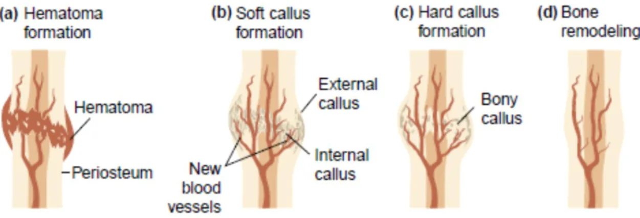
36 2.1.6 - BONE HEALING
Bone tissue is characterized by an adequate capacity to regenerate itself upon the establishment of a lesion or small defects. This healing process aims to generate new bone tissue to stabilize the damaged bone parts with minimal alterations of bone anatomy and function. Ordinarily, bone healing occurs following four main stages (see Figure 2.8). Primarily, after an injury such as a fracture, the damaged bone zone undergoes an inflammatory phase (a) in which the necrotic tissue is cleaned; following, a repair phase (b) is initiated with the formation of a cartilage tissue – soft callus – replacing the lost bone. Simultaneously, osteoclasts continue the resorption of remaining bone debris and new blood vessel formation occurs. Also during this stage, new collagen fibers are produced by fibroblasts leading to the formation of a denser fibrous net, which will consolidate new bone tissue; in a third phase, the newly formed cartilage is replaced by trabecular bone (c) in a process similar to endochondral ossification; finally, fracture healing is completed by a remodelling phase (d) that may last for more than a year and in which the morphology is mended to ensure a perfect bone recover (45).
This ability, however, is limited since it may prevent only low magnitude fractures or small defects. When bone defects or fractures exceed a critical size, additional therapeutic solutions are required in order to attain fully structural repair and normal functionality (46). Also, circumstances that impair the physiological tissue healing, such as local infections, may decrease the correct healing capability of the damaged bone. Several pathological conditions encompassing diabetes mellitus, osteoarthritis, osteopenia, osteoporosis, among other, are known to decrease cellular function and to
37
impair the formation of the bone mineralized matrix, leading to severe bone weakening and bone remodelling unbalancing (47).
These situations represent major complications in therapeutic strategies for bone tissue healing and require alternative therapeutic solutions.
Figure 2.8 - Schematic representation of the four stages of bone fracture repair (45).