Rita Aires
Dissertation presented to obtain the Ph.D degree in
Developmental Biology
Instituto de Tecnologia Química e Biológica António Xavier | Universidade Nova de Lisboa
Gdf11 signalling, Oct4 and the control
of vertebrate trunk length
Molecular interactions regulating the process of
axial extension during vertebrate development
Rita Aires
Dissertation presented to obtain the Ph.D degree in
Developmental Biology
Instituto de Tecnologia Química e Biológica António Xavier | Universidade Nova de Lisboa
Oeiras, July,2016
Gdf11signalling, Oct4 and the
control of vertebrate trunk length
Molecular interactions regulating the process of
axial extension during vertebrate development
iii “A sheep, a drum, and a snake fall off a cliff.”
v
The work presented in this thesis was supported by FCT and FSE,
grant SFRH/BD/51876/2012.
Apoio financeiro da FCT e do FSE no âmbito do Quadro comunitário
de apoio, BD n.º SFRH/BD/51876/2012.
Table of Contents
vii
Table of Contents
List of Abbreviations ... xi
List of Figures ... xv
List of Tables ... xvii
Acknowledgments ... xix
Abstract ... xxv
Sumário ... xxix
Chapter I: Introduction ... 33
I.1 The mouse as a model organism for development ...34
I.2 The early mouse embryo and the first cell fate decisions...35
I.2.1 First cell fate decision: Trophectoderm vs Inner Cell Mass ...35
I.2.2 Second cell fate decision: Primitive Endoderm vs Epiblast ...37
I.3 Postimplantation development and early axis specification ...39
I.3.1 The egg cylinder stage ...39
I.3.2 Proximo-distal axis and formation of the DVE ...40
I.3.3 Antero-posterior axis and formation of the AVE...42
I.4 Gastrulation and embryonic germ layer formation ...44
I.4.1 Primitive streak specification and positioning ...44
I.4.2 Primitive Streak morphogenesis and mechanisms of ingression ...46
I.4.3 Lineage allocation during gastrulation ...48
I.5 Axis extension in Vertebrates ...51
I.5.1 Neuromesodermal progenitors (NMPs) ...51
I.5.2 Node-Streak Border vs Chordoneural Hinge ...52
I.5.3 Molecular characterization of axial progenitors ...53
I.5.4 Cessation of body axis extension ...54
Table of Contents
I.6 Axial patterning ...57
I.6.1 Somitogenesis or segment formation ...57
I.6.2 Segment identity specification ...63
Thesis Aims ... 71
Chapter II: Gdf11 signalling and the control of axial progenitor population during the mouse trunk to tail transition ... 73
II.1 Summary ...75
II.2 Background ...75
II.3 Materials and Methods ...78
II.3.1 Embryos ...78
II.3.2 Genotyping ...79
II.3.3 Phenotypic analysis ...80
II.3.4 Lineage tracing analysis and β-galactosidase staining ...85
II.3.5 Gdf11-/- tail explant cultures ...85
II.3.6 RT-PCR analysis ...86
II.3.7 Chromatin immunoprecipitation and quantitative PCR (qPCR) ...86
II.3.8 Data analysis ...87
II.4 Results ...87
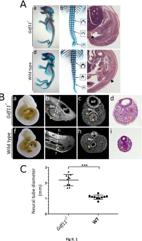
II.4.1 Gdf11-/- embryos present severe tail defects and enlarged neural tubes ...87
II.4.2 Gdf11 mutant tails show abnormal gene expression patterns ...88
II.4.3 Gdf11 mutants have an expanded axial progenitor population ...92
II.4.4 Ectopic Oct4 expression in Gdf11-/- tails at E10.5 ...95
II.4.5 Oct4 expression appears to be directly regulated by Gdf11 signalling ..99
II.5 Discussion ...102
III.6 Acknowledgements ...106
Chapter III:Oct4 is a key regulator of vertebrate trunk length ... 107
Table of Contents
ix
III.2 Background ...109
III.3 Materials and Methods ...112
II.3.1 Embryos ...112
II.3.2 Genotyping ...113
II.3.3 Phenotypic analysis ...113
II.3.4 β-galactosidase reporter analysis. ...116
II.3.5 BAC (Bacterial Artificial Chromosome) transgenics ...118
II.3.6 RT-PCR analysis ...118
II.3.7 Genomic analysis ...118
III.4 Results ...119
III.4.1 Sustained Oct4 activity in axial progenitors extends the trunk ...119
III.4.2 Oct4 expression delays activation of posterior Hox genes ...126
III.4.3 Oct4 expression is maintained for longer developmental times in snake embryos ...128
III.4.4 Genomic organization of the mouse and snake Oct4 loci ...128
III.4.5 Organization of the Oct4 locus in lizards ...134
III.4. 6 Gdf11 in squamates ...136
III.4.7 Regulation of Oct4 expression in snakes ...136
III.5 Discussion ...138
III.6 Acknowledgements ...144
Chapter IV: General Discussion ... 145
IV.1 Gdf11 controls the axial progenitor pool size during the trunk to tail transition ...147
IV.2 Gdf11 signalling vs Oct4 activity: a molecular tug-of-war in the control of vertebrate trunk length ...150
IV.3 Oct4, Gdf11 and the evolution of the snake body plan ...155
IV.5 Final considerations and future directions ...158
List of Figures
xi
List of Abbreviations
ActRIB/Acvr1b Activin Receptor IB
ActRIIA/Acvr2a Activin Receptor IIA
Alk4 Activin Receptor-like Kinase 4. Same as ActRIB/Acvr1b
AP Antero-Posterior
AVE Anterior Visceral Endoderm
BAC Bacterial Artificial Chromosome
Bapx1 Bagpipe Homeobox 1
BMP Bone Morphogenetic Protein
Bmpr1a Bone Morphogenetic Protein Receptor, type 1a
Bp Base pair
Cas9 CRISPR associated protein 9
Cchcr1 Coiled-Coil Alpha-Helical Rod Protein 1
Cdh1 E-cadherin
Cdx Caudal-type Homeobox
Cer1 Cerebrus-like protein 1
ChIP Chromatin Immunoprecipitation
CNH Chordoneural Hinge
CR Conserved Region
CreERT Tamoxifen inducible Cre recombinase
CRISPR Clustered Regularly Interspaced Short Palindromic Repeats
DE Distal Enhancer
DIG Digoxigenin
Dkk1 Dickkopf homologue 1
DNA Deoxyribonucleic acid
DVE Distal Visceral Endoderm
E Embryonic day
e.g. Exempli gratia
ECM Extracelular Matrix
List of Abbreviations
emVE Embryonic Visceral Endoderm
Eomes Eomesodermin homolog (Xenopus laevis)
EPC Ectoplacental Cone
EpiSCs Epiblast Stem Cells
ExEc Extraembryonic Ectoderm
exVE Extraembryonic Visceral endoderm
Fgf Fibroblast Growth Factor
FgfR Fibroblast Growth Factor Receptor
Foxa2 Forkhead Factor 2
Gata GATA binding protein
Gdf growth and differentiation factor
Hand2 Heart and neural crest derivatives expressed transcript 2
Hnf4 Hepatocyte nuclear factor 4
Hox Homeobox
ICM Inner Cell Mass
iPS Induced Pluripotent Stem Cells
kB Kilobase
Lefty1 Left-right determinant factor 1
Lsm2 LSM2 Homolog, U6 Small Nuclear RNA And mRNA Degradation Associated Mesp Mesoderm posterior
MET Mesenchymal-to-Epithelial Transition
MMP-1 Matrix Metalloproteinase-1
Myf Myogenic factor
Nkx1.2/Sax1 NK1 Homeobox 2/ Spastic ataxia 1
NMPs Neuromesodermal progenitors
No Notochord
Npdc1 Neural proliferation, differentiation and control 1
NSB Node-Streak Border
NT Neural Tube
OE Olfactory Epithelium
Otx2 Orthodenticle homolog 2 (Drosophila melanogaster)
List of Figures
xiii
PBS Phosphate Buffered Saline
PBT Phosphate Buffered Saline, containing 0.1% Tween-20
PCR Polymerase Chain Reaction
PD Proximo-Distal
PE Proximal Enhancer
PEΔSBE Deletion of Smad Binding Elements in Oct4 Proximal Enhancer PEΔTIE Deletion of TGF-β Inhibitor Element in Oct4 Proximal enhancer
PFA Paraformaldheyde
PG Paralogue Group
Pou5f1/Oct4 POU domain, class 5, transcription factor 1/ Octamer-binding transcription factor 4
PrE Primitive Endoderm
PS Primitive Streak
PSM Presomitic Mesoderm
pSmad2/3 Phosphorylated Smad2/3
qPCR Quantitative Polymerase Chain Reaction
RA Retinoic Acid
Raldh2 Retinaldheyde Dehydrogenase 2
RAR Retinoid Acid Receptor
RNA Ribonucleic Acid
SBEs Smad Binding Elements
Scx Scleraxis
Shh Sonic hedgehog
Snail Snail homolog 1 (Drosophila)
Sox2 SRY-related HMG box-containing 2
SV40 Simian vacuolating virus 40
T Thoracic
TBE Tris Borate EDTA Buffer
TBM Tail Bud Mesenchyme
TBST Tris Buffered Saline, containing 0.1% Tween-20
Tbx6 T-box 6
Tcf19 Transcription Factor 19
List of Abbreviations
TE buffer Tris EDTA buffer
TGCs Trophectoderm Giant Cells
TGF-β Transforming Growth Factor-β
TgfβRI TGF-β Receptor I
TIE TGF-β inhibiting element
TREEs Tissue Regeneration Enhancer Elements
Vars Valyl-TRNA Synthetase
VER Ventral Epidermal Ridge
Wnt Wingless-type MMTV Integration site family
List of Figures
xv
List of Figures
Fig I.1 Preimplantation mouse development and the first cell fate decisions...36
Fig I.2 Proximo-Distal (PD) and Antero-Posterior (AP) axis formation...42
Fig I.3 Ingression of epiblast cells through the primitive streak (PS) and mesodermal lineage allocation during mouse gastrulation...49
Fig I.4 Axial extension during early organogenesis and tail bud stages...55
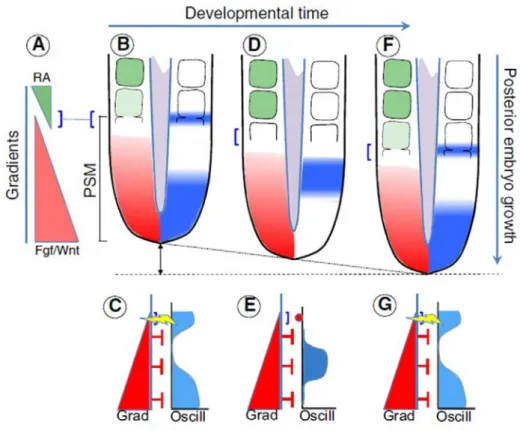
Fig I.5 Clock-and-Wavefront model of somitogenesis...60
Fig I.6 Hox gene expression and genomic organization in the mouse embryo...65
Fig II.1 Gdf11 mutants present severe tail defects and enlarged neural tubes...89
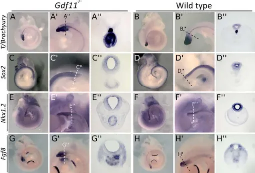
Fig II.2 Gdf11-/- tails show abnormal gene expression patterns (part I)...91
Fig II.2 Gdf11-/- tails show abnormal gene expression patterns (part II)...92
Fig II.3 Gdf11 mutants have an expanded axial progenitor population...93
Fig II.4 Gdf11-/- tails show ectopic Oct4 expression at E10.5...96
Fig II.5 Oct4 overlaps with Sox2 and T/Brachyury proteins in E10.5 Gdf11-/- tails...97
Fig II. 6 Gdf11-/- tails are not able to generate true EpiSCs...98
Fig II.7 Oct4 expression appears to be directly regulated by Gdf11 signalling...101
Fig III.1 Sustained Oct4 expression in axial progenitors extends the trunks in mouse embryos....121
Fig III.2 Additional phenotypes of Cdx2-Oct4 transgenics...122
Fig III.3 Molecular characterization of Cdx2-Oct4 transgenics...124
Fig III.4 Molecular characterization of Cdx2-Oct4 transgenics (cont)...125
Fig III.5 Hox gene expression in Cdx2-Oct4 transgenic embryos...127
Fig III.6 Oct4 expression and genomic environment in snake embryos...129
Fig III.7 Comparison of king cobra and python Npdc1 and Oct4 sequences...131
Fig III.8 Comparison of last Npdc1 exons and first Oct4 exons between python and mouse...133
Fig III.9 Comparison of Oct4-containing genomic regions of different species...135
Fig III.10 Gdf11 in squamates...137
List of Tables
xvii
List of Tables
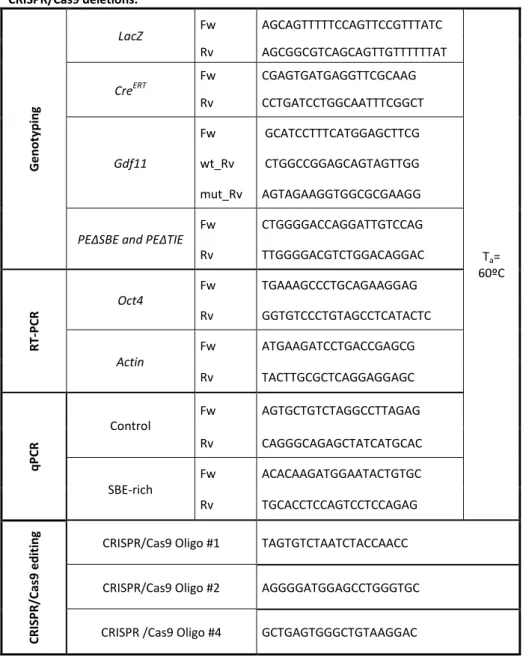
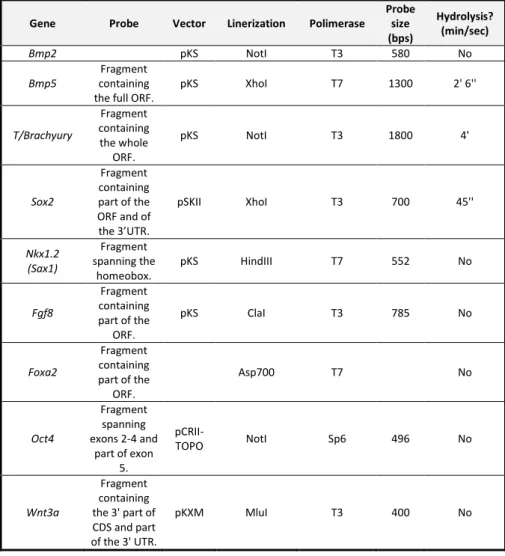
Table II. 1 Oligonucleotides used in this chapter for genotyping, RT-PCR, qPCR , and CRISPR/Cas9 deletions... 81 Table II. 2 In situ hybridization probes used in this chapter. ... 83 Table III. 1 Oligonucleotides used in this chapter for genotyping, RT-PCR, corn snake Oct4 in situ hybridization probe cloning and python Oct4 upstream region sequencing. ... 115 Table III. 2 In situ hybridization probes used in Chapter III. ... 117
Acknowlegdments/Agradecimentos
xix
Acknowledgments
Agradecimentos
“No duck is an island”
- In Tiny Toon Adventures (1990-1994)
Há crianças que, quando forem grandes, querem ser médicos, enfermeiros ou veterinários. Ou mesmo bombeiros, gestores ou artistas. E depois há aquelas que descobrem a Física sob a forma de chá a ferver em queda livre e, mesmo assim, querem ter um doutoramento. Este, caro leitor, foi o meu caso; só não sabia em quê na altura. Até que um dia vi um embrião de galinha pela primeira vez e o resto é história. E, realmente, porque nenhum pato é uma ilha e eu fui incrivelmente sortuda por ter tido pessoas tão boas, importantes, e que admiro sempre à minha volta durante este tempo todo….
… É tempo de agradecer à Academia!
À malta do Mallo Lab, a começar por Nosso Senhor O Chefe. Obrigada, Moisés, por teres aceitado ser o meu orientador. Sem a tua ajuda, supervisão e, especialmente, enorme entusiasmo sobre tudo o que é Ciência, e Desenvolvimento em particular, nada disto teria sido possível. Mas, sobretudo, gostei mesmo de ter partilhado este projecto contigo: como companheiro de bancada, de escrita e como editor extraordinaire. Muitas e muitas gracias!!
Ao Arnon Dias Jurberg (aka Arnonzinho), que é parte integrante da Dream Team
(Dream Team FTW©). Começámos isto juntos e, de certa forma, acabámos isto
Acknowlegdments/Agradecimentos mas prefiro recordar as vezes em que íamos à praia beber uma imperial ao final da tarde. Enquanto tínhamos importantes discussões científicas – sempre! À Ana Casaca (aka Casaquinha), o meu grande pilar no laboratório. Realmente não há palavras: sem ti, estes anos não teriam sido a mesma coisa. Obrigada não só por toda a ajuda (és a minha guru #2 para tudo o que é sequências e biologia molecular) mas também por, de vez em quando, ‘converteres para PCR’. E pelos nossas canções partilhadas, pelos cafés e jornal clubs de terça-feira. E falando nestes jornal clubs! Obrigada, Tiago Carneiro, por seres O membro honorário do laboratório. Nunca as conversas à hora do café/almoço/ lanche foram tão ecléticas – desde o Direito Português e a definição de personalidade jurídica, passando pela cozinha, até à vida numa certa casa na Venda do Pinheiro. E por compreenderes todas as minhas piadas parvas e por te deixares contaminar pela minha selecção musical do dia, sempre de qualidade!... duvidosa. Juntos, conseguimos anerdalhar o laboratório; a seguir: o Mundo! (Novo hastag: #anerdalharoMundo). À Ana Nóvoa, por ser a mais estupenda mestre de transgénicos e a pessoa ideal com quem ir a um workshop de ponto de cruz subversivo. A um Sábado à tarde. Numa tasca no Cais do Sodré (True story, caro
leitor!). À Luisinha, por estar connosco, por partilhar da loucura que é manter uma
colónia de Gdf11 (e infinitas variações) em heterozigotia e por toda a ajuda com a estatística. Ao André, por ter sido o meu primeiro aluno mais a sério, mesmo que só por três semanas. Mega super proud of you! À Irma, pelo bom companheirismo no laboratório e, especialmente, pela ajuda enorme nas genotipagens. E um obrigada muito especial aos velhos e novos membros do lab: Aybuke, Sofia, André... E à Inês Domingues, fonte inesgotável de alegria, entusiasmo e, de vez em quando, bolachinhas de limão com sementes de papoila.
To my thesis committee, Florence Janody e Domingos Henrique, for being available at all times when I needed you. Thank you so much!
Acknowlegdments/Agradecimentos
xxi Ao PhD Programme in Integrative Biomedical Sciences (PIBS) do IGC e aos seus directores: Thiago Carvalho e Élio Sucena. Em especial ao Élio, por organizares um primeiro ano do mestrado BED na FCUL que faz com que um doutoramento pareça (quase) um passeio no parque. E à Manuela Cordeiro, pela competência e ocasionais puxões de orelhas. Obrigada por tudo! To all members of PIBS 2011: Jarek, Rafal, Rômulo, Oz, Sandra, Sara, Marta, Ana Stankovic, Ana Martins, Inês, Irma, you guys rock! You were the best people to twilight with on the wee hours of some classes! Em especial à Sandra Tavares: a melhor DJ, companheira de AmeeGuS e de gabinete; mulher de gosto musical impecável, e de uma força, honestidade e integridade inabalável. Foi incrível! Ao Rómulo Areal, pelas sessões de discussão metafísicas nas tardes de segunda-feira – na realidade, pelas sessões de discussão metafísicas que aconteciam de cada vez que nos encontrávamos por aí. Nunca motivações tão reais de personagens fictícias foram dissecadas de forma tão apaixonante durante tanto tempo! Obrigada por me teres feito descobrir as alegrias do Kindle, do The name of the wind e pelas discussões de
Game of thrones e Black Sails. To Ana Stankovic: the best gym buddy, the sharpest
scientific mind, and the most amazing friend a gal can have. For telling me five years ago (and even now) that I didn’t have to go through stuff alone. You possess the uncanny ability to make me spill the beans and man up in a way I didn’t think it was possible. Ao IGC, e a toda a gente que faz deste instituto o melhor sítio do mundo para trabalhar. Às meninas da histologia, Joana Rodrigues e Marta Pinto, um agradecimento especial: não só são talentosas e competentes, como tornaram as minhas tardes passadas no criostato e no vibrótomo tão mais divertidas! Aos membros do meu laboratório de mestrado, o Organogenesis lab: Joaquín Rodríguez-Léon, Catarina Certal, Raquel Tomás, Nando, Diana Chapela. Mas especialmente à Joana Monteiro, que me ensinou as bases da vida no laboratório, desde a primeira in situ à última amputação de peixes.
Acknowlegdments/Agradecimentos À Isabel Guerreiro, a minha bestie for ever and ever and ever. Realmente, palavras não são mesmo suficientes para ti. Obrigada por toda a tua ajuda, no laboratório e fora dele. Por seres uma força motriz. E por seres a melhor condutora que uma copiloto pode desejar! E ao Tomás, por partilhar da minha nerdice a todos os níveis. It is really a great time to be alive and a nerd contigo! À minha querida Rita Félix, a pessoa com quem quero sempre ir viajar, comer gelados e ouvir música pimba. À Filipa Nunes, o ser mais determinado, focado, motivado e, de vez em quando, azarado à face da Terra. Mi casa es su casa, and I really mean it. À Catarina Dourado, parte crucial dos jantares de quinta-feira na Pizza Hut: és grande, moça!
À malta do secundário, por continuarmos a ser uma família. Em especial à Ari e à Ana, as criaturas mais livres e corajosas que tive a sorte de conhecer e de me aturarem. Ao Miguel Moutinho, o meu irmão de alma, par científico, e gémeo musical. Não imagino outra pessoa com quem conduzir sem destino só pelo prazer de ouvir a música, ir beber café ao aeroporto à 00h30, e fazer rotundas ao contrário à 1 da manhã (outra true story, caro leitor).
E finalmente, aos meus pais. A culpa disto da Ciência, penso, foi do meu pai, que um dia me mostrou o porquê da Lua ter fases usando uma laranja + luz da janela como modelo. E por me ensinar que é possível fazer um portão automático usando o motor de uma betoneira. Obrigada, pai e mãe (ou, em bom anerdalhanço de geneticista, à minha F0), por tanto amor e compreensão, fé e por estarem sempre lá. E por nunca terem parado essa criança estranha que lia sem parar de brincar com os cogumelos venenosos no quintal e de fazer misturas estranhas com eles (isto olhando em restrospectiva, foi um bocado arriscado, hein?). À minha irmã, que será sempre das pessoas mais importantes da minha vida e meu exemplo de vida (sim, leste bem, oh minha bimba). Ao resto da família por fazer parte da minha vida, em especial à Gorete, Guidinha, Belinha e Madrinha (que lindo que até rima). Vocês alimentam-me o corpo e a i’álma. À
Acknowlegdments/Agradecimentos
xxiii Dani, que fez de mim quem sou quando me deu a ler Mafalda, Calvin and Hobbes e Artemis Fowl. E às minhas meninas, por terem mantido a minha sanidade mental na maior parte dos últimos quinze anos.
Estes cinco anos passaram depressa demais, e realmente não é justo. But time does fly when you’re having fun, não é?
Abstract
xxv
Abstract
Axial elongation is a conserved, fundamental mechanism in vertebrate development. This process consists in the gradual addition of tissue to the posterior-most part of the embryo, resulting in a progressive assembling of the embryonic body in a rostro-caudal sequence. Axial elongation relies on the activity of a dedicated population of cells located in the caudal part of the embryo, the axial progenitors. These progenitors are a highly dynamic, self-renewing, multipotent pool of cells, whose properties vary with the progression of the axial elongation. During the initial stages of development, they reside in the epiblast and primitive streak and include precursors for all three embryonic germ layers – ectoderm, mesoderm and endoderm. These will subsequently differentiate and interact amongst themselves and with other tissues in order to produce the different organs and body structures of the neck and trunk regions. Later in development, the caudal part of the embryo undergoes profound reorganization involving the disappearance of the epiblast and PS and the emergence of the tailbud. This process is associated with major changes in the axial progenitors as well. In particular, the progenitors for the lateral and intermediate mesoderm that are involved in the formation of the trunk organs undergo a process of terminal differentiation resulting in the formation of the hindlimbs and the organization of the embryonic cloaca. Concurrently, the remaining major subset of axial progenitors - the neuromesodermal progenitors (NMPs), which contribute to the generation of the neural tube, axial skeleton and associated muscles - relocate from the epiblast to the tailbud, where they continue with the remaining body axis elongation. Thus, the specific control of axial progenitor types, numbers, and balance between precursors and their derivatives will determine a particular species’ final body length, as well as allocation of the post-cranial body into neck,
Abstract trunk and tail. As such, axial elongation is a key mechanism involved in the generation of the broad diversity of body shapes and sizes within vertebrates.
In the first part of this thesis, we performed an in-depth analysis of the tail
defects observed in Gdf11-/- embryos, which were associated with the previously
described delayed trunk to tail transition occurring in these mutants. We show that the tail abnormalities are already clear at mid-gestation stages shortly after this transition and seem to result from an expanded population of axial progenitors, combined with alterations in the tissue reorganization associated with the transition from primitive streak-driven to tailbud-dependent axial growth. In particular, morphological, lineage tracing and molecular analyses indicated that Gdf11 mutant tails contain an excess of axial progenitors, most of them residing in an ectopic ventral structure composed of an epithelium enclosing a mass of mesenchymal cells. Importantly, we discovered that a subset of cells in this ectopic structure expressed Oct4, which suggested that the epithelial component of the ectopic ventral tissue likely represents incomplete resolution of the epiblast. We thus demonstrate that Gdf11 signalling is an integral part of the mechanisms involved in epiblast extinction and regulation of the axial progenitor pool size during the trunk to tail transition. These most likely involve functional interactions with Oct4, which probably include direct transcriptional regulation of this gene’s expression by downstream components of the Gdf11 signalling pathway. As a result of all these complex processes, Gdf11 activity ensures a proper transition from trunk to tail-forming mechanisms, as well as an adequate, gradual axis termination.
The second part of this thesis aimed at the analysis of the contribution of Oct4’s ectopic expression for the Gdf11 mutant phenotype. Using a transgenic approach,
we proved that most axial phenotypes found in Gdf11-/- embryos derive from
abnormally extended Oct4 activity in axial progenitor regions during axis elongation. These studies also revealed that sustained Oct4 activity in these
Abstract
xxvii regions was able to keep axial progenitors in a trunk-forming configuration, delaying the trunk to tail transition. Interestingly, this delay was associated with a concomitant caudal shift in the axial level of activation of posterior Hox genes, thus linking global distribution of the vertebrate body into trunk or tail regions with the patterning of the axial structures associated with these main areas.
These findings led us to analyze Oct4 expression in snake embryos. Our research demonstrated that this gene likely suffered heterochronic shifts in its regulation, as its expression seems to be maintained for longer developmental periods in snakes relative to mouse embryos. Genomic analyses indicated that these temporal changes in Oct4 expression seem to have originated from dramatic genomic rearrangements during the evolutionary trajectories of mammals, lizards and snakes, which could have altered the Oct4 regulatory landscape in these different vertebrate clades. Indeed, transgenic reporter analyses in mice identified the existence of potential regulatory sequences upstream of the Oct4 gene in squamates that are not shared by its mammalian counterpart. Together, our observations suggest that these genomic and regulatory changes involving Oct4 might have been essential components of the mechanisms originating vertebrate body diversity and the emergence of the snake body plan.
Overall, we show a new role for the pluripotency factor Oct4 as a key regulator of vertebrate trunk length, and establish the balance between Oct4 and Gdf11 activities as a major regulator of the body allocation into trunk and tail regions during axial extension in vertebrate embryos. Our results not only provide important insights into the different developmental events involved in the making of these two body regions, but also hint at possible ways of generating evolutionary novelty, ultimately contributing to the generation of the wide diversity of body plans observed among members of the vertebrate clade.
Sumário
xxix
Sumário
A extensão axial ou alongamento do eixo corporal é um mecanismo fundamental e evolutivamente bem conservado no desenvolvimento embrionário dos vertebrados. Este processo consiste na adição progressiva de tecido à zona mais posterior do embrião, numa sucessão rostro-caudal que resulta da actividade de um conjunto especializado de células - os progenitores axiais. Os progenitores axiais são uma população celular altamente dinâmica, multipotente e com capacidade de auto-renovação, cujas propriedades se modificam à medida que a extensão axial ocorre. Nas fases iniciais do desenvolvimento embrionário, estas células residem no epiblasto e linha primitiva do embrião e incluem precursores para os três folhetos germinativos embrionários – ectoderme, mesoderme e endoderme. Estes percursores, por sua vez, experimentam processos de diferenciação e interacção com os tecidos circundantes, dando origem a todos os órgãos e restantes estruturas pertencentes às regiões do pescoço e tronco. Contudo, em fases mais tardias do desenvolvimento, a parte posterior do embrião sofre uma complexa reorganização que envolve o desaparecimento do epiblasto e linha primitiva e o início da formação da cauda. A população de progenitores axiais também é restruturada durante este processo. Enquanto os progenitores de mesoderme lateral e intermédia são submetidos a um processo de diferenciação terminal, que resulta na formação dos membros inferiores e na organização da cloaca do embrião, uma outra parte significativa da população – os progenitores neuro-mesodérmicos, que contribuem para a formação do tubo neural, esqueleto axial e músculos associados – é realojada no recém-formado botão da cauda e aí continua o processo de extensão axial do corpo até ao seu final. Deste modo, o controlo adequado do número e tipo de percursores axiais, assim como o balanço entre a quantidade de progenitores e
Sumário respectivas formas diferenciadas, é fundamental para a determinação do comprimento final do corpo de cada espécie e distribuição deste pelas regiões do pescoço, tronco e cauda. A extensão axial é, portanto, um processo chave na geração da grande diversidade de formas e tamanhos corporais observados entre vertebrados.
Na primeira parte desta tese foi realizada uma análise profunda dos defeitos
observados nas caudas de embriões Gdf11-/-. Estas anormalidades desde logo
aparentaram estar relacionadas com o atraso na transição entre o tronco e a cauda já previamente descrito nestes mutantes. Neste estudo, mostrou-se não só que os defeitos observados nas caudas mutantes surgem logo após o período de transição entre o tronco e a cauda, como também que estas malformações parecem resultar simultaneamente de uma expansão da população de progenitores axiais e de alterações na reorganização dos tecidos associadas à passagem da extensão axial dependente da linha primitiva para a dependente dos progenitores residentes na cauda. As análises morfológicas e moleculares realizadas, assim como os dados obtidos em experiências de seguimento de linhagens celulares, indicaram que a cauda dos embriões mutantes para Gdf11 apresenta, de facto, um excesso de progenitores axiais. A maioria destes aparenta estar concentrada numa estrutura ectópica, localizada na parte mais ventral da cauda, constituída por uma massa de células mesenquimatosas que se encontram envolvidas por um epitélio. O facto de algumas destas células inesperadamente também expressarem Oct4 sugeriu que a componente epitelial desta estrutura ectópica poderia ter tido origem em fragmentos residuais do epiblasto. Assim, neste capítulo demonstrou-se que a sinalização Gdf11 é parte integrante do conjunto de mecanismos que regula tanto a extinção do epiblasto como a regulação do número de progenitores axiais durante a transição entre o tronco e a cauda, possivelmente através de interacções funcionais com Oct4. Estas
Sumário
xxxi parecem incluir a regulação directa da expressão de Oct4 durante a transição, provavelmente através de factores resultantes da via de sinalização Gdf11. Ao coordenar todos estes processos intrincados, a actividade de Gdf11 assegura assim uma correcta transição entre os mecanismos de formação do tronco e os de formação da cauda, tal como uma terminação progressiva do eixo corporal.
A segunda parte desta tese teve como objectivo inicial a análise da contribuição
da expressão ectópica de Oct4 para o fenótipo observado nos embriões Gdf11-/-.
Fazendo uso de uma abordagem experimental baseada em ratinhos transgénicos, verificou-se que grande parte das alterações axiais encontradas em embriões mutantes para Gdf11 resulta da persistência anormal da expressão de Oct4 em regiões que incluem progenitores axiais durante o processo de extensão axial. Estes ensaios também revelaram que a manutenção da actividade de Oct4 nestas regiões possibilita a retenção dos progenitores axiais numa configuração favorável à formação do tronco, provocando, dessa forma, um atraso na transição entre o tronco e a cauda. O atraso no início da transição revelou estar associado a uma concomitante posteriorização no nível axial da activação da expressão dos genes
Hox, sugerindo que a distribuição do corpo nas regiões do tronco e cauda em
vertebrados está estreitamente coordenada com a padronização das estruturas axiais associadas a estas zonas.
O facto destes embriões transgénicos apresentarem troncos longos, uma característica específica das cobras, instigou o estudo da expressão de Oct4 em embriões deste grupo de organismos. Efectivamente, a expressão de Oct4 em embriões de cobra pareceu ser mantida durante um maior período no desenvolvimento relativamente a embriões de ratinho, o que sugeriu que a regulação da expressão de Oct4 possivelmente teria estado sujeita a alterações heterocrónicas durante a evolução da linhagem das cobras. Análises genómicas indicaram que as alterações na expressão deste gene podem ter tido origem em rearranjos genómicos dramáticos nas regiões a 5’ de Oct4 durante as trajectórias
Sumário evolutivas de mamíferos, lagartos, e cobras. Estas modificações terão resultado numa alteração do ambiente genómico responsável pela regulação da expressão de Oct4 nos diferentes grupos de vertebrados. De facto, a análise de embriões de ratinho transgénicos contendo genes repórteres sob a influência destas potenciais sequências regulatórias de cobra, permitiu identificar elementos reguladores de
Oct4 partilhados exclusivamente por cobras e lagartos. No geral, estas
observações sugerem que as alterações genómicas nas regiões contíguas a Oct4, nas quais provavelmente se inseriam as zonas regulatórias deste gene, podem ter sido elementos essenciais nos mecanismos que originaram a diversidade das formas corporais em vertebrados, particularmente no aparecimento do plano corporal das cobras.
Em suma, os resultados descritos nesta tese atribuem uma nova função para o factor de pluripotência Oct4, que actua como um gene chave na regulação do tamanho do tronco em vertebrados. Para além disso, estabeleceu-se que as actividades de Oct4 e de Gdf11 constituem os principais componentes envolvidos no controlo da distribuição do corpo em tronco e cauda durante a extensão axial em vertebrados. As descobertas aqui descritas, para além de contribuem para uma maior compreensão dos fenómenos envolvidos na formação destas duas regiões corporais, também sugerem possíveis formas de gerar novidades evolutivas capazes de assegurar a vasta diversidade de planos corporais observados em vertebrados.
Chapter I
33
Chapter I:
Introduction
“I’m a leaf on the wind – watch how I soar.”
Chapter I: Introduction I.1 The mouse as a model organism for development Development of a whole multicellular complex organism from a single cell is not only an evolutionary triumph, but also the most daunting and formidable of tasks. The organism’s entire body plan has to be laid down in a series of intricate and interconnected events that comprise various levels of organization, from intracellular processes to vast morphogenetic tissue movements. This means that the embryo’s early symmetries must be gradually broken and that most of the initial cell potency needs to be progressively surrendered so that the body can increase in complexity and, ultimately, achieve its final form. Yet, the minutest of mistakes can be either fatal or represent a huge evolutionary opportunity.
This introductory chapter tells the story of this progression, mainly focusing in the mouse embryo. It is intended as a “crash course” on early mouse development, describing its most important events and the major players that take part during this process.
I.1 The mouse as a model organism for development
From all model organisms, the mouse stands out as one of the most popular. As a fellow mammalian vertebrate, the mouse shares many physiological and pathological features with humans (Rosenthal and Brown, 2007). Also, mouse and human genomes show a high degree of evolutionary conservation, even if the two species have diverged more than 96 million years ago (Nei et al., 2001; Nguyen and Xu, 2008).
Small size, ready availability, easy maintenance and husbandry, as well as docility are some of the advantages that mice have compared with other mammalian models (Nguyen and Xu, 2008; Rosenthal and Brown, 2007; Wolpert et al., 1998). For developmental biologists in particular, their relatively short generation time and high fertility are also very convenient characteristics. Even considering its limitations regarding embryo accessibility for grafting and other direct surgical manipulations during embryonic development, the mouse presents
Chapter I: Introduction I.2 The early mouse embryo and the first cell decisions
35 unparalleled opportunities for research. In fact, the sheer amount of available phenotypic data, genomic resources and genetic tools developed for more than 80 years – many of them used in the present work – makes the mouse one of the most powerful model organisms in the pursuing of fundamental questions in mammalian biology and disease (Schofield et al., 2012).
I.2 The early mouse embryo and the first cell fate decisions
Mouse development can be broadly divided into pre- and postimplantation (Lawson and Wilson, 2016; Wilson and Lawson, 2016; Wolpert et al., 1998). Preimplantation development takes approximately four and a half days and comprises all stages between oocyte fertilization and embryonic implantation in the uterine wall. Preimplantation mouse development is highly regulative, which means that the embryo can adapt and compensate for perturbations either in number and/or position of cells (Ziomek et al., 1982). This shows that cells in the early embryo have a high developmental potential and are still quite flexible in terms of cell fate. Yet, cell labelling and lineage tracing experiments show that there might be differences in blastomere developmental properties and a bias towards particular fates as early as the 2-cell stage (Piotrowska-Nitsche et al., 2005; Tabansky et al., 2013).
I.2.1 First cell fate decision: Trophectoderm vs Inner Cell Mass
After fertilization the zygote undergoes successive rounds of cell division without growth. This process of cleavage generates small cells with little cytoplasm – the blastomeres (Wolpert et al., 1998). As the embryo reaches the 8-cell stage, it undergoes a process of compaction and becomes a morula (Fig I.1) (Ducibella and Anderson, 1975; Wolpert et al., 1998). Compaction involves changes in blastomere shape, assembly of intercellular adhesion complexes and development of strong apical-basal polarization (Johnson and Ziomek, 1981b).
Chapter I: Introduction I.2 The early mouse embryo and the first cell decisions
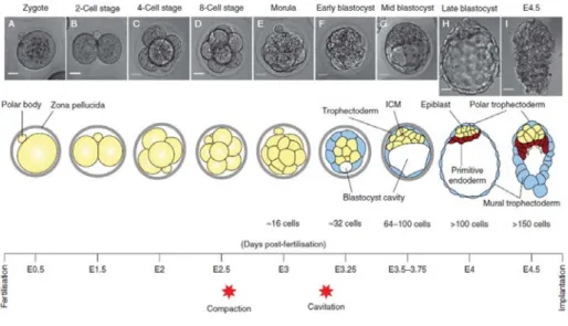
Fig I.1 Preimplantation mouse development and the first cell fate decisions. At the 8-cell stage the
embryo undergoes compaction, which generates differences both in position and intercellular contacts among morula cells. This triggers the first cell fate decision whereby cells in the periphery become trophectoderm (TE), whereas internal cells become inner cell mass (ICM). The second lineage decision occurs within cells of the ICM after the blastocyst cavity (or blastocoel) is formed by cavitation. Asymmetries in cell division generate asymmetries in cell signalling, which ultimately result in the acquisition of primitive endoderm (PrE) or epiblast fate. Arrows in the E4.5 scheme denote PrE migration over TE cells. Timeline indicates time elapsed since fertilization in embryonic days (E). Adapted from Saiz and Plusa, 2013.
This polarization allocates specific proteins to apical and basal domains, which will eventually give rise to asymmetries after cell division (reviewed in Bedzhov et al., 2014; Saiz and Plusa, 2013).
The first clear cell fate decision in the mouse embryo, which occurs around embryonic day (E)2.5, has its origins precisely on these subtle molecular differences. In particular, two rounds of asymmetric cell divisions generate an embryo composed of small, non-polarized, inside cells – the Inner Cell Mass (ICM) – that are enclosed within larger, highly polarized, outer cells that make up the trophectoderm (TE) (Johnson and Ziomek, 1981a; Wilson and Lawson, 2016; Wolpert et al., 1998). The TE constitutes the first epithelium formed during embryo development. As such, its cells are highly polarized and strongly connected by intercellular junctions that provide extensive cell-to-cell
Chapter I: Introduction I.2 The early mouse embryo and the first cell decisions
37 communication and cohesion. In contrast, the ICM is mainly composed of cells that have lost polarity and which are exposed to uniform cell-to-cell contacts. By this time (around E3.5) the embryo starts to develop a fluid-filled cavity, the blastocoel, which causes the TE to expand and the ICM to become confined to one side of this vesicle (Wolpert et al., 1998). The resulting embryo is the blastocyst (Fig I.1). TE cells up-regulate caudal-type homeobox-2 (Cdx2) (Beck et al., 1995; Dietrich and Hiiragi, 2007; Niwa et al., 2005; Ralston and Rossant, 2008; Strumpf et al., 2005). Even though not necessary for TE specification per se, Cdx2 is crucial for epithelial integrity and tissue maturation, in such a way that Cdx2 mutant embryos fail to implant (Strumpf et al., 2005). On the other hand, ICM cells express Oct4 - also known as Pou5f1 and a member of the POU domain family of octamer-binding transcription factors - which is absolutely required for the formation and maintenance of the ICM (Nichols et al., 1998; Ryan and Rosenfeld, 1997). Interestingly, both Cdx2 and Oct4 are expressed in every blastomere at early cleavage stages. However, as development progresses, these genes become gradually restricted to the TE and ICM, respectively, in part as a consequence of mutual repressive activities (Dietrich and Hiiragi, 2007; Downs, 2008; Niwa et al., 2005; Palmieri et al., 1994; Schöler et al., 1989). Thus, TE and ICM seem to be mutually exclusive identities characterized by complementary
Cdx2 and Oct4 expression (Niwa et al., 2000; Niwa et al., 2005). In fact, in the
absence of Oct4 all morula cells are diverted into TE fate and lose pluripotency (Nichols et al., 1998). Oct4 later becomes a crucial factor in the specification and maintenance of the epiblast and primordial germ cell survival (Kehler et al., 2004; Nichols et al., 1998; Niwa et al., 2000).
I.2.2 Second cell fate decision: Primitive Endoderm vs Epiblast
The second lineage fate decision occurs within the ICM at approximately E3.5. It results in the formation of the epiblast, which will give rise to the embryo proper,
Chapter I: Introduction I.2 The early mouse embryo and the first cell decisions and the primitive endoderm (PrE), an epithelium that covers the epiblast and separates it from the blastocyst cavity (Fig I.1 and Fig I.2A). Once again, subtle heterogeneities between cells seem to trigger different genetic programs. Biases in the internalization of cells into the deep ICM layers during asymmetric divisions generate variation in expression levels of Fibroblast Growth Factor 4 (Fgf4) and its receptor, Fibroblast Growth Factor Receptor 2 (FgfR2), among ICM cells (Krupa et al., 2014; Morris et al., 2013). Cells that express high levels of Fgf4 down-regulate
FgfR2 and will become Nanog-expressing epiblast cells, whereas cells expressing
high levels of FgfR2 down-regulate Fgf4, activate Gata6 and other PrE markers and enter a PrE cell fate (Chambers et al., 2003; Chazaud et al., 2006; Guo et al., 2010; Mitsui et al., 2003; Yamanaka et al., 2010). This results in a “salt-and-pepper” pattern of epiblast and PrE progenitors that will be subsequently sorted into their final positions by mechanisms such as active cell migration, differential adhesion and selective apoptosis (Chazaud et al., 2006; Meilhac et al., 2009; Plusa et al., 2008). The epiblast and PrE cell lineages will then become stabilized through cross-regulatory processes. Nanog directly represses Gata6 and promotes Fgf4 secretion that, in turn, activates signalling through FgfR2 in adjacent cells and stabilizes Gata6 expression to keep their PrE identity (Frankenberg et al., 2011; Schrode et al., 2014; Singh et al., 2007). Interestingly, Oct4, Cdx2, Nanog and
Gata6 are all coexpressed in every blastomere until the 64-cell blastocyst stage
(Guo et al., 2010). This phenomenon might explain why preimplantation mouse development is so characteristically regulative, since strict lineage specifications are delayed until implantation and cells maintain a high potential to change fate due the presence of all these fundamental lineage markers.
Another important epiblast marker is the SRY-related HMG box-containing transcription factor Sox2. This gene is up-regulated specifically in epiblast cells and is crucial for epiblast maintenance. In fact, Sox2 mutant embryos completely fail
Chapter I: Introduction I.3 Postimplantation development and early axis specification
39 to develop while still being able to implant and form extraembryonic structures (Avilion et al., 2003). Nanog, Oct4 and Sox2 therefore make up the core of the pluripotency network in the epiblast as the absence of any of these factors is enough to disrupt its formation and maintenance. Oct4 and Sox2 are also two of the four factors that compose the genetic cocktail able to reprogram somatic cells back into an embryonic stem cell-like state, generating the so-called induced pluripotent stem cells (iPS) (Takahashi and Yamanaka, 2006).
As a result of all these events, at the end of preimplantation development the embryo consists of three different cell lineages: epiblast, TE and PrE. The embryo derives exclusively from epiblast cells, whereas the TE and PrE will give rise to extra-embryonic structures such as the placenta and the yolk sac. These lineages are not only crucial for intra-uterine development, but also play important roles as signalling centres essential for patterning of the embryo proper. By E4.5 the embryo is ready to implant.
I.3 Postimplantation development and early axis specification
I.3.1 The egg cylinder stage
The first terminally differentiated cell type arises upon implantation, when the mural TE cells – TE cells surrounding the blastocoel – go through rounds of endo-reduplication and differentiate into TE giant cells (TGCs). These cells invade the uterine tissues and induce extensive vasculature remodeling and angiogenesis, important to mediate the embryo´s nutrient uptake, waste removal and gas exchanges (Bedzhov et al., 2014). On the other hand, TE cells closer to the epiblast (designated polar TE) proliferate due to the presence of TE progenitors and generate the ectoplacental cone (EPC) and the extraembryonic ectoderm (ExEc) that will become part of the placenta (Bedzhov et al., 2014; Wolpert et al., 1998). The PrE also expands, diversifying into parietal endoderm, embryonic visceral
Chapter I: Introduction I.3 Postimplantation development and early axis specification endoderm (emVE) and extraembryonic visceral endoderm (exVE) (Fig I.2A) (Rivera-Pérez and Hadjantonakis, 2015; Wolpert et al., 1998).
Rapid cell division with growth by both epiblast and extraembryonary tissues causes the embryo to expand, elongating into the blastocyst cavity. At the same time, a second lumen – the proamniotic cavity – is created amidst the epiblast by a process of hollowing. From a ball of non-polarized cells, the epiblast turns into a highly organized rosette-like structure by the establishment of a strong apical-basal polarization, epithelialization and consequent changes in cell shape. This self-organization of epiblast cells seems to be mediated by deposition of extracellular matrix components (ECM) and by activation of β1-integrin receptors (Bedzhov and Zernicka-Goetz, 2014). A similar process is thought to occur at the level of ExEc so that, ultimately, epiblast and ExEc become two contiguous, although distinct, epithelia (Arnold and Robertson, 2009; Bedzhov and Zernicka-Goetz, 2014; Rivera-Pérez and Hadjantonakis, 2015). The embryo thus enters the egg cylinder stage, acquiring a hollow, cylindrical conformation, with a proximal-distal (PD) axis. The site of connection with the uterine tissue becomes the proximal pole, whereas the tip of the cup shaped epiblast represents the distal-most part of the axis (Bedzhov et al., 2014; Lawson and Wilson, 2016).
I.3.2 Proximo-distal axis and formation of the DVE
The establishment of the PD axis in the conceptus (embryo and supporting structures) at E5.0 is the first step towards the generation of the antero-posterior (AP) embryonic axis. The key to a correct PD patterning in the egg cylinder is the establishment of a robust PD gradient of Nodal (Brennan et al., 2001; Kumar et al., 2014). Nodal is a member of the transforming growth factor beta (TGF-β) superfamily of growth factors and, as such, requires two types of serine-threonine kinase receptors – normally known as type I and type II receptors - for signalling transduction. Nodal binds to the type I receptor Alk4 (ActRIB/Acvr1b), which
Chapter I: Introduction I.3 Postimplantation development and early axis specification
41 promotes recruitment of the type II receptors ActRII (ActRIIA/Acvr2a) or ActIIB (Acvr2b) that, in turn, trans-phosphorylate and thus fully activate the type I receptor (Kumar et al., 2001; Shen, 2007). Activated Alk4 phosphorylates cytoplasmatic Smad2 and/or Smad3 that then form a complex with Smad4, enters the nucleus and ultimately regulates expression of target genes (Morikawa et al., 2013). Nodal activity also requires the presence of EGF-CFC co-receptors such as Cripto that confer binding specificity for Alk4 (Yeo and Whitman, 2001). Nodal is initially expressed as an immature ligand (proNodal) by epiblast cells (Conlon et al., 1994; Varlet et al., 1997). ProNodal activates expression of Furin and PACE4 convertases in ExEc cells, which will cleave the propeptide, thus producing mature Nodal in the proximal epiblast (Beck et al., 2002; Ben-Haim et al., 2006). Mature Nodal is then able to activate a positive autoregulatory loop, stimulating its own expression and therefore creating a high Nodal concentration in the proximal epiblast region. Nodal is also capable of inducing Bmp4 production by ExEc cells, which enhances Wnt3 expression in the proximal egg cylinder that, in turn, promotes Nodal production as well (Ben-Haim et al., 2006; Winnier et al., 1995). Both mature Nodal and Bmp4 can likewise induce expression of the co-receptor Cripto, which is essential for Nodal signalling transduction and, thus, for a correct PD and AP specification (Beck et al., 2002; Ding et al., 1998). Mature Nodal protein is essential for correct visceral endoderm specification as well, since it represses ExVE genes (Gata4, Hnf4, etc) while maintaining expression of emVE related genes like Fgf8, Fgf5, Bmp2, Otx2 and Foxa2 (Mesnard et al., 2006).
Nodal activity is also involved in promoting expression of its inhibitors in the distal-most emVE, which becomes a local epithelial thickening known as the Distal Visceral Endoderm (DVE) (Fig I.2B) (Brennan et al., 2001; Meno et al., 1999; Rivera-Pérez et al., 2003; Takaoka et al., 2006). The DVE constitutes an important signalling center in the embryo that secretes inhibitors of the Nodal [like Cerebrus-like protein 1 (Cer1) and Left-right determinant factor 1 (Lefty1)] and
Chapter I: Introduction I.3 Postimplantation development and early axis specification
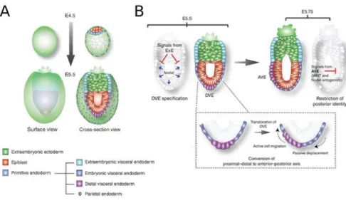
Fig I.2 Proximo-Distal (PD) and Antero-Posterior (AP) axis formation. A. Schematic representation
of cell lineages and their spatial relationship in an implanting blastocyst (E4.5) and early egg cylinder stage embryo (E5.5). B. AP axis formation. The egg cylinder’s radial symmetry is broken when cells belonging to the distal visceral endoderm (DVE) start migrating towards the embryo’s future anterior pole. Establishment of the anterior visceral endoderm (AVE) results in a repositioning of the Nodal and Wnt antagonists source, confining the activity of these pathways to the embryo’s posterior side. Adapted from Rivera-Pérez and Hadjantonakis, 2015.
Wnt [such as Dickkopf homologue 1 (Dkk1)] signalling pathways (Pfister et al., 2007; Takaoka et al., 2006). The overall result of all these processes is generation of high Nodal/Wnt activities in the proximal epiblast (Richardson et al., 2006; Rodriguez et al., 2005), and their attenuation at the distal epiblast, which eventually lead to correct patterning of the embryo proper.
I.3.3 Antero-posterior axis and formation of the AVE
At E5.75, soon after the PD axis is established, the egg cylinder’s radial symmetry is broken when DVE cells start migrating towards the prospective anterior side of the embryo. This process leads to the formation of the Anterior Visceral Endoderm (AVE) near the now anterior ExEc/epiblast boundary (Fig I.2B). The AVE is a distinct visceral endoderm population composed by cells with a tall columnar morphology that express the Nodal and Wnt signalling antagonists Lefty1, Cer1
Chapter I: Introduction I.3 Postimplantation development and early axis specification
43 and Dkk1, in addition to a number of transcription factors (Pfister et al., 2007; Rivera-Pérez et al., 2003). Similarly to the DVE, the AVE’s primary role is to maintain a Nodal and Wnt signalling gradient throughout the tissues, this time as an AP gradient. This structure also constitutes an important source of signals for the correct specification of anterior structures while inhibiting the activity of posterior genes in the anterior epiblast (Kimura et al., 2000; Perea-Gomez et al., 2001; Thomas and Beddington, 1996; Yamamoto et al., 2004).
Despite sharing many morphological and molecular features, clonal analysis studies have shown that the AVE does not entirely derive from DVE cells, but that a significant part of it is formed de novo from emVE cells that acquire AVE expression markers (Srinivas et al., 2004; Takaoka et al., 2011; Torres-Padilla et al., 2007). Yet, ablation experiments indicate that the DVE is indeed crucial for proper AVE positioning. In fact, DVE displacement seems to be the triggering event leading to the overall movement of emVE towards the future anterior pole of the embryo, which will include the cells that will be part of the future AVE (Miura and Mishina, 2007; Takaoka et al., 2011). This means that the AVE is not static or homogeneous and that its cellular composition varies as it moves anteriorly.
The repositioning of Nodal and Wnt antagonist sources, following the establishment of the AVE, results in a conversion of the PD axis into the AP axis. Hence, posteriorizing factors stay confined to the pole opposite to the AVE, which will play an essential role in gastrulation. At this stage (~E6.0) the cross section of the egg cylinder is not perfectly circular but ellipsoid instead. When first specified, the AP axis is aligned with the short axis of the oval embryo. However, within few hours after AVE settlement, AP axis is shifted towards the long axis by progressive tissue remodelling (Mesnard et al., 2004; Perea-Gomez et al., 2004). This process seems to be dependent on Wnt3 and Fgf8 activity as mutant embryos for these two factors fail to undergo reshaping (Barrow et al., 2007; Guo and Li, 2007). Yet, gastrulation can be induced even in the absence of reshaping, indicating that axis
Chapter I: Introduction I.4 Gastrulation and embryonic germ layer formation realignment might serve as a way to maximize the distance between both poles, thus minimizing possible interferences amidst anterior and posterior signals.
I.4 Gastrulation and embryonic germ layer formation
Once the AP axis is settled, the embryo is ready to go through gastrulation. This is a process whereby concerted cell proliferation, migration, differentiation and changes in cell shape and adhesion properties, among other morphogenetic events, creates the organism’s basic body plan (Wolpert et al., 1998). As such, gastrulation encompasses dramatic changes in the global structure of the embryo. The most important – and striking – rearrangement is the conversion of the two-layered embryo into a more complex structure composed by three embryonic germ layers: ectoderm, mesoderm and definitive endoderm (Wolpert et al., 1998). Each of these layers gives rise to specific types of tissues. The ectoderm or the “outer layer” will generate the animal’s epidermis and nervous system, whereas the mesoderm (or the “middle layer”) will provide the skeleto-muscular system, connective tissues and contribute to different extents to the formation of internal organs such as the heart, the kidneys or the muscular layers of the intestine. Finally, the inner-most tissue, the endoderm, will produce the epithelial lining of the gut and respiratory system, besides playing a major part in the formation of digestive organs such as the liver and the pancreas.
I.4.1 Primitive streak specification and positioning
In the mouse, gastrulation begins around E6.5 with the formation of a transient, specialized structure in the proximal posterior epiblast designated as the Primitive Streak (PS). Once formed, the PS is a stable structure and is present throughout gastrulation. However, shortly after it reaches its maximum length at the distal tip of the embryo, it begins to regress progressively, disappearing completely by early organogenesis (around E9.5) (Fig I.3A).
Chapter I: Introduction I.4 Gastrulation and embryonic germ layer formation
45 The PS begins as a local epithelial deformation and becomes a cellular discontinuity generated by the progressive initiation of an epithelial-to-mesenchymal transition (EMT) in epiblast cells (Perea-Gomez et al., 2004; Williams et al., 2012; Wolpert et al., 1998). Ingression through the PS, followed by a change from an epithelial to a mesenchymal state, allows cells to insert themselves and migrate between the epiblast and the visceral endoderm, or to intercalate among the visceral endoderm becoming epithelial again (Acloque et al., 2009). This way, transiting cells can become mesoderm or definitive endoderm, respectively, whereas cells that do not ingress through the PS and remain in the epiblast will be part of the neurectoderm and surface ectoderm (Lawson et al., 1991) (Fig I.3A).
Signalling activity from the AVE restricts the posteriorizing activity of the Nodal, BMP and Wnt pathways to the proximal posterior epiblast, eventually leading to PS formation. In fact, Nodal inhibitors are essential for a correct PS positioning since embryos lacking both Cer1 and Lefty1 develop multiple, ectopic, primitive streaks throughout the epiblast (Ben-Haim et al., 2006; Brennan et al., 2001; Mishina et al., 1995; Perea-Gomez et al., 2002; Rodriguez et al., 2005). Proper PS formation requires high levels of Smad2- and Smad3-dependent Nodal activity (Ben-Haim et al., 2006; Chu et al., 2005; Conlon et al., 1994; Ding et al., 1998; Dunn et al., 2004; Vincent et al., 2003), as well as ExEc-secreted Bmp4 acting on its receptor Bmpr1a in epiblast cells (Mishina et al., 1995; Winnier et al., 1995). Besides Nodal and Bmp4, several studies have shown that Wnt3 signalling through β-catenin and its co-receptors LRP5 and LRP6 is also indispensable for PS initiation and maintenance (Huelsken et al., 2000; Kelly et al., 2004; Liu et al., 1999; Mohamed et al., 2004; Tortelote et al., 2013; Yoon et al., 2015). In fact, absence of any of the aforementioned factors results in severe PS abnormalities and deficient or non-existing mesoderm production. The resulting PS is thus characterized by a gene expression profile composed of genes belonging or
Chapter I: Introduction I.4 Gastrulation and embryonic germ layer formation responding to these signalling pathways, including Nodal, Wnt3, Wnt3a, Axin2,
Lefty2, Fgf8 and T/Brachyury, and various others like Snail and Sp5 (Ben-Haim et
al., 2006; Pfister et al., 2007; Robb and Tam, 2004; Tam and Loebel, 2007; Tortelote et al., 2013).
I.4.2 Primitive Streak morphogenesis and mechanisms of ingression Contrary to what has been described in other vertebrates like chicken or rabbit, PS morphogenesis in the mouse does not involve large-scale cellular movements to position PS precursor cells or convergence and extension mechanisms for its elongation (Halacheva et al., 2011; Lawson and Schoenwolf, 2001; Viebahn et al., 2002; Voiculescu et al., 2007). Instead, PS morphogenesis in the mouse seems to be a positional phenomenon that occurs by in situ EMT in three consecutive phases: basement membrane loss, cell ingression and streak elongation (Williams et al., 2012). Basement membrane loss is an important first step required to break the major physical barrier to cell movement represented by extracellular matrix (ECM) constituents. Localized disaggregation of this structure most likely occurs by down-regulation of genes such as laminin and type IV collagen in PS-forming cells or by active degradation of its components. The next step involves major changes in epiblast cells, so they can leave the epithelial sheet and ingress through the PS. This requires not only cell shape changes (like apical constriction), but also the disassembling of intercellular adhesion complexes and down-regulation of epithelial junctional and polarity proteins (such as E-cadherin, Occludin and β-catenin) so that cells can delaminate from the epithelial sheet and migrate away. Finally, cells in the vicinity of the PS are gradually recruited in a posterior to anterior direction, resulting in an elongation of the PS all the way to the distal tip of the embryo that is complete by E7.0 (Williams et al., 2012). The anterior-most tip of the PS is occupied by the node, which is populated by a specialized group of ciliated, columnar cells that function as an important
Chapter I: Introduction I.4 Gastrulation and embryonic germ layer formation
47 signalling centre for patterning (Balmer et al., 2016; Lawson and Wilson, 2016; Yamanaka et al., 2007).
Gastrulation is a continuous process that occurs simultaneously with PS formation. It encompasses the inactivation of epiblast genes, such as Oct4 and
Sox2, and the acquisition of mesoderm and endoderm-specific factors (Pfister et
al., 2007). The gradual and sequential loss of cells from the epiblast sheet as cells ingress through the PS is compensated by the high proliferation rates observed during these stages. In the end, the overall net result is the generation of a force that passively pulls lateral epiblast cells towards the PS, while its overall epithelial integrity is maintained (Williams et al., 2012).
Several genes and signalling pathways have been identified as having an important role in ingression and EMT through the PS (reviewed in Acloque et al., 2009). FGF signalling is among the best-studied cases in mouse gastrulation and seems to be particularly relevant in cell movement and mesoderm layer formation. In the absence of Fgf8 or its receptor, FgfR1, cells fail to migrate away from the PS as they are unable to down-regulate E-cadherin (also known as Cdh1) (Ciruna and Rossant, 2001; Ciruna et al., 1997; Sun et al., 1999). E-cadherin is an integral part of intercellular adherent junctions and is essential to maintain epithelial integrity; thus, its down-regulation is absolutely crucial for EMT. In fact, perturbing E-cadherin function with a blocking antibody is sufficient to trigger the conversion of epiblast into mesenchymal cells (Burdsal et al., 1993). E-cadherin regulation by FGF signalling is mediated by the zinc-finger transcription factor Snail, which has the ability to bind directly to the Cdh1 gene promoter region and repress its expression (Batlle et al., 2000; Cano et al., 2000; Ciruna and Rossant, 2001). Snail mutant embryos are actually capable of forming a “mesoderm” expressing the right set of mesodermal markers; however these cells retain epithelial characteristics such as apical-basal polarity and adherent junctions, as well as maintaining a robust E-cadherin expression (Carver et al., 2001). The T-box
Chapter I: Introduction I.4 Gastrulation and embryonic germ layer formation transcription factor Eomesodermin (Eomes) appears to have a role in E-cadherin regulation as well. Conditional abrogation of Eomes in epiblast cells leads to a failure in efficient down-regulation of both E-cadherin transcripts and protein. These embryos also have a thickened PS that likely result from impaired cell delamination and mesoderm migration. However, E-cadherin regulation by Eomes seems to be indirect (Arnold et al., 2008). Other transcription factors like Mesp1 and Mesp2 have likewise been shown to be essential for nascent mesoderm migration, but whether or not these proteins have any influence upon E-cadherin regulation is unknown (Kitajima et al., 2000; Saga et al., 1999). Besides Snail, T-box transcription factors Tbx6 and T/Brachyury were found to be important for mesoderm formation and are regulated by both FGF and Wnt signalling (Chapman and Papaioannou, 1998; Ciruna and Rossant, 2001; Galceran et al., 2001; Wilson et al., 1995; Yamaguchi et al., 1999). In particular, Tbx6 seems to repress Sox2 expression, ensuring the complete suppression of the neural transcription program in mesoderm-fated cells (Chapman and Papaioannou, 1998; Takemoto et al., 2011). Brachyury/T, in turn, is absolutely required for mesoderm specification and notochord morphogenesis (Herrmann, 1991; Stott et al., 1993; Wilson and Beddington, 1997).
I.4.3 Lineage allocation during gastrulation
During gastrulation cells are continuously recruited from the epiblast to undergo ingression and EMT through the PS (Williams et al., 2012). Fate mapping studies have revealed that not only all epiblast cells are competent to be part of any of the three germ layers, but that distinct mesodermal lineages are specified depending on the time and site of ingression through the PS (Fig I.3B) (Kinder et al., 1999; Lawson et al., 1991; Smith et al., 1994; Wymeersch et al., 2016).
At early stages of gastrulation or early streak stages, the most caudal part of the PS will give rise to the extraembryonic mesoderm. This tissue will provide the
Chapter I: Introduction I.4 Gastrulation and embryonic germ layer formation
49
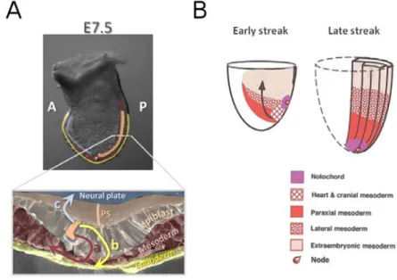
Fig I.3 Ingression of epiblast cells through the primitive streak (PS) and mesodermal lineage allocation during mouse gastrulation. A. Gastrulation begins with the formation of a specialized
transient structure, the primitive streak (PS), located in the embryo’s posterior side. Epiblast cells (orange) approach the PS region and undergo an epithelial to mesenchymal transition (EMT). These cells can then migrate away from the PS and insert themselves between the epiblast layer and the visceral endoderm (VE), becoming mesoderm (a). Alternatively, ingressing cells can become epithelial once more by a process of mesenchymal to epithelial transistion (MET) and intercalate between cells of the VE, becoming definitive endoderm (b). Cells that do not ingress become part of the ectoderm, giving rise to the neuroctoderm and surface ectoderm (c). B. Different mesodermal lineages are specified according to their time and place of ingression within the PS. Extraembryonic mesoderm ingresses through the most posterior part of the PS, whereas the cardiac mesoderm and head mesoderm enter the PS through increasingly more anterior regions. Finally, the anterior-most end of the PS and the node will generate mainly midline axial mesendodermal tissues. By the late streak stage, heart and head mesoderm have ingressed completely, whereas the remaining lateral and paraxial mesoderm continue to be produced. B is adapted from Tam and Behringer, 1997.
mesodermal component of extraembryonic supportive tissues (such as the chorion and visceral yolk sac) and is the origin of the first hematopoietic tissue, the blood islands. Intermediate levels of the PS will be the ingression site of cardiac mesoderm, whereas its anterior part will generate the paraxial mesoderm of the head. Finally, the rostral-most region of the PS and the node will supply the first midline axial mesendodermal tissues: the prechordal plate (the head mesoderm) and cells for the anterior definitive endoderm. By late streak stages,