UNIVERSIDADE DE LISBOA
Faculdade de Farmácia
3-H
YDROXY
-
QUINOLIN
-2(1H)-
ONES
,
A USEFUL
SCAFFOLD
:
S
YNTHESIS AND BIOLOGICAL EVALUATION
Roberta Paterna
Orientadores: Doutor Pedro Miguel Pimenta Góis
Professor Doutor Rui Ferreira Alves Moreira
Tese especialmente elaborada para obtenção do grau de Doutor em Farmácia, especialidade em Química Farmacêutica
Júri:
Presidente: Doutora Matilde da Luz dos Santos Duque da Fonseca e Castro, Professora Catedrática e Diretora da Faculdade de Farmácia da universidade de Lisboa
Vogais:
Doutor Nuno Filipe Candeias, University Lecture
Tampere University of Technology, Finland;
Doutora Maria Miguéns Pereira, Professora Associada com Agregação Faculdade de Ciência e Tecnologia da Universidade de Coimbra;
Doutora Maria Manuel Martinho Sequeira Barata Marques, Investigadora Auxiliar com Agregação
Faculdade de Ciência e Tecnologia da Universidade Nova de Lisboa, Doutora Maria Matilde Soares Duarte Marques, Professora Catedrática
Instituto Superior Técnico da Universidade de Lisboa;
Doutora Ana Paula Costa dos Santos Peralta Leandro, Professora Auxiliar Faculdade de Farmácia da Universidade de Lisboa;
Doutor Pedro Miguel Pimenta Góis, Professor Auxiliar Faculdade de Farmácia da Universidade de Lisboa, orientador.
Fundação para a Ciência e Tecnologia 2017
Statement
O presente trabalho foi desenvolvido sob orientação do Doutor Pedro Gois e co-orientaçao do Professor Doutor Rui Moreira do iMed.ULisboa (Instituto de Investigação do Medicamento), amos da Faculdade de Farmácia da Universidade de Lisboa. Este trabalho foi financiado pela Fundação para a Ciência e Tecnologia através da bolsa de doutoramento SFRH/BD/78301/2011.
This work was developed under scientific supervision of Dr. Pedro Góis and Professor Dr Rui Moreira from iMed.LULisboa (Research Institute for Medicines), faculty of pharmacy, University of Lisbon. The work was financially supported by Fundação para a Ciência e Tecnologia, through the doctoral grant SFRH/BD/78301/2011.
Ackonwledgement
First, I would like to express my gratitude to my supervisor Dr. Pedro Gois for the continuous support of my Ph. D studies and related research, for his patience, motivation, and immense knowledge. His guidance helped me in all the time of research and writing of this thesis.
I also acknowledge my co-supervisor Prof. Rui Moreira for his sage advice, insightful criticism, and patient encouragement aided my Ph.D. progress in innumerable ways.
My sincere thanks also go to Prof. Ana Paula Leandro from Metabolism and Genetics Group of iMed.UL who provided me the opportunity to join her team, and who gave access to the laboratory and research facilities. I have also to acknowledge the member of her group Dr. João B. Vicente, Dr. João Leandro, Mariana P.
Amaro, Raquel Lopes, Ana Carolina Costa which helped me to understand
techniques that had no previous knowledge to develop specific aspects of this project and also performed the biochemical assay.
I would like to Dr. Raquel Frade and Dr. Pedro Borralho which performed the cells assays described in this thesis.
I would also like to express my indebtedness gratitude to Fundação para a Ciência e Tecnologia for their financial support (SFRH/BD/78301/2011), and to the Faculdade de Farmácia, Universidade de Lisboa, Portugal
During this time spend in the Biorganic Chemistry Group at iMed.UL, I had the fortune to be surrounded by people that made my day-to-day routine so much happier and pleasant. Expetially Dr. Nuno Candeias, Dr. Carlos Monteiro, Dr. Jaime
Coelho, Dr. Alexandre Trindade, Drª. Filipa Siopa, Drª. Andreia Rosatella, Drª.Catarina Rodrigues, M.S. Fábio Santos, Dr. Pedro Cal, Dr Rudi Oliveira, Dr Svilen Simeonov, Dr Sudarshan Reddy, M.S. Joao Antonio for all the advices
and scientific support of my project and also for being true friends that i can belive and I will be able to keep for the rest of my life. Futhermore, I can not dismiss the opportuyty of thanking all the new present members of Biorganic Chemistry Group and Medicinal Chemistry.
Special regards also to Dr. João Rosa that took the effort of doing an extensive review of theis thesis. Also, I would like to thanks Dr Helio Faustino for helped me in the last stage of my work.
I would like to thank all undergraduate students that I guided during the course of my PhD, since they helped me to grow as a scientist and person: M.S Marcio Santos and M.S. Roberto Russo.
I would like to thank all of my friends that helped me to be myself and to be happy during this period far from home in particular Angela Paterna, Joao Lavrado
Francesco Montalbano, Simone Tulumello. Also, I would like to thanks my
roommates and friends Damla Uyar, Julia Plötz who endured everyday my stress and complaints at home with a smile on their face.
Finally, I would like to thank my Mother who support was crucial all these years far from home and for all the pride that she has demostated in my little achievements that gave me the required confidence to belive in myself and capabilities. Also, I would like to thank my sister and my little nephews for their support.
Furthermore, I would like to acknowledge my deceased father and dedicate this thesis in his lovely memory.
List of Pubblications
Papers in International Scientific Periodicals with Referees
o “A Sustainable Protocol for the Aqueous Multicomponent Petasis Borono– Mannich Reaction”, Candeias, N.R., Paterna R., Cal P.M.S.D., Góis, P.M.P.,. Journal of Chemical Education (2012), 89 (6), 799-802 (DOI: 10.1021/ed200509q)
o “Ring-Expansion Reaction of Isatins with Ethyl Diazoacetate Catalyzed by Dirhodium(II)/DBU Metal-Organic System: En Route to Viridicatin Alkaloids” Paterna, R., André, V., Duarte, M.T., Veiros, L.F., Candeias, N.R., Gois, P.M.P. European Journal of Organic Chemistry (2013), 28, 6280-6290. (DOI: 10.1002/ejoc.201300796)
o “Homologation Reaction of Ketones with Diazo Compounds” Candeias, N.R., Paterna R., Gois, P.M.P., (2016). Chemical Reviews, 116 (5), 2937– 2981. (DOI:10.1021/acs.chemrev.5b00381)
Oral Communications in Scientific Conferences
o 7th iMed.ULisboa Post-graduate Students Meeting organized by iMed.ULisboa (Instituto de Investigação do Medicamento) held in Lisbon, in 2015.
o 11ºPortuguese National Meeting of Organic Chemistry and 4º Portuguese National Meeting of Medicinal Chemistry, 1-3 December 2015, Porto, Portugal.
Poster communications in Scientific Conferences
o XII Encontro Nacional da Sociedade Portuguesa de Quimica, 2011, Braga o Challenges in organic chemistry and chemical Biology (ISACS7) organized
by RCS (Royal society of Chemistry) held in Edinburgh, 2012.
o 4th iMed.ULisboa Post-graduate Students Meeting organized by iMed.ULisboa (Instituto de Investigação do Medicamento) held in Lisbon, in 2012.
o 10º Portuguese National Meeting of Organic Chemistry and the 1st Portuguese-Brazilian Organic Chemistry Symposium, Lisbon, September 2013.
o 5th iMed.ULisboa Post-graduate Students Meeting organized by iMed.ULisboa (Instituto de Investigação do Medicamento) held in Lisbon, in 2013.
o 50th International Conference on Medicinal Chemistry RICT 2014, Rouen, July 2014.
o 6th iMed.ULisboa Post-graduate Students Meeting organized by iMed.ULisboa (Instituto de Investigação do Medicamento) held in Lisbon, in 2014.
o 6th European Conference Chemistry in the Life Sciences organized by EuCheMS (European Association for Chemical and Molecular Sciences) held in Lisbon, 2015.
o Ciência 2016 - Encontro com a Ciência e Tecnologia em Portugal, organized by Fundação para a Ciência e a Tecnologia em colaboração com a Ciência Viva held in Lisbon, 2016
Abstract
The quinolin-2(1H)-ones ring establish the core structure of many natural and synthetic molecules and a broad spectrum of biological properties like, antimicrobial, enzymatic and neuro protective activities, have been attributed to these molecules. Additionally, 4-hydroxyquinolin-2-ones (4HQs) and 3-hydroxyquinolin-2-ones (3HQs), derivatives of quinolin-2-one, have also been reported with promising biological properties, and have attracted much attention from the medicinal chemist community. The 3HQ core is present in the structure of naturally occurring products viridicatin, viridicatol and 3-O-methyl viridicatin first isolated from the mycelium of
Penicillium viridicatum. Although, due to the reduced knowledge about 3HQs, from a
synthetic and biological perspective, in the last years, the development of new methodologies for their synthesis has been stimulated and strategies based on condensations, intramolecular cyclization and ring expansions have been applied. Recently reported has nonclassical bioisosteres of α-glycine, 3HQs derivatives are potent inhibitors of the Human D-amino acid oxidase (DAAO) and due to their ability to chelates metal centres, 3HQs are counted as inhibitors of HIV-1 reserve transcriptase associated RNase H activity and as inhibitors of influenza A endonuclease.
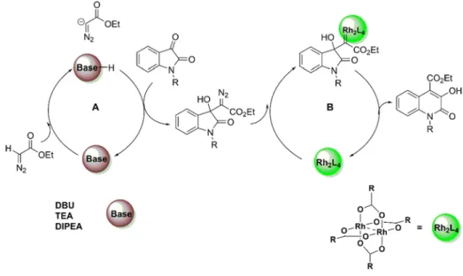
In view of the present stat-of-the-art, 3HQs new derivatives were synthetized using a new efficient methodology centered on the emergent metal-organo-catalysed (MOC) concept. A one-pot protocol using the MOC system NHC-dirhodium(II)/DBU catalyzed Eistert ring expansion reaction of isatins with ethyl diazoacetate to afford the 3-hydroxy-4-ethylesterquinolin-2(1H)-ones core. The reaction provides the final products regioselectively and with yields ranging from good to excellent. Furthermore, DFT calculations were performed on this system and support a mechanism in which the key step is the metallocarbene formation between the 3-hydroxyindole-diazo intermediate and the dirhodium(II) complex.
After the above mentioned optimized methodology the second part of this work is dedicated to the biological activity of 3HQ and its derivatives. Various synthetic modifications have been made to introduce specific chemical group keeping the 3HQs core structure. Several compounds with different properties were synthesized and important biological studies were performed on 4-carboxamide-3HQ derivatives showed interesting biological activity as a potential anticancer lead molecule. Additionally, based on the that 3HQs can complex metallic centers and been an isoster of glycine, we hypothesized that 3HQ derivatives could be a useful platform to design new modulators of human phenylalanine hydroxylase (hPAH), the enzyme responsible by the genetic disease phenylketonuria. The new hPAH modulators were simply prepared based on ring-expansion reaction of isatins with NHS-diazoacetate catalysed by di-rhodium(II) complexes yielding 4-Carboxamide-3HQs in good-to-excellent yields. The 7-trifluoromethyl-4-carboxamide-3HQs 134, was identified as the most efficient hPAH modulator, with an apparent binding affinity nearly identical to the natural allosteric activator L-Phenylalanine.
Therefore, as 3-hydroxyquinolies have demonstrated to be good scaffolds for the design and development of compounds with activity over phenylalanine hydroxylase and an excellent starting point for the development of novel therapeutics for a phenylketonuria.
Keywords: 3-Hydroxyquinoline-2(1H)-ones; Eistert Ring Expansion; anticancer
Resumo
O anel de quinolin-2(1H)-ona é a estrutura central de muitas moléculas tanto de origem natural, como sintética, e ao qual tem sido atribuído um amplo espectro de propriedades biológicas, como antimicrobianas, enzimáticas e neuro-protetoras. Para além das quinolin-2(1H)-onas, os seus derivados 4-hidroxiquinolin-2-onas (4HQs) e as 3-hidroxiquinolin-2-onas (3HQs) apresentam também propriedades biológicas promissoras e têm atraído muita atenção da comunidade de químicos medicinais. Recentemente relatados como bioisósteres não clásicos da α-glicina, os derivados da 3HQs são potentes inibidores da D-aminoácido oxidase humana (DAO) e devido à sua capacidade para quelar centros metálicos, as 3HQs são também apontados como inibidores da RNase H associada à transcriptase reserva do HIV-1 e como inibidores da endonuclease do vírus Influenza A. Assim, as 3HQs têm sido reconhecidas como uma estrutura química com interesse farmacológico. O núcleo 3HQ está presente na estrutura dos produtos naturais viridicatina, viridicatol e 3-O-metil viridicatina, isolados primeiramente do micélio do Penicillium viridicatum. No entanto, devido a reduzido numero de métodos de síntese e falta de conhecimento das propriedades biológicas, têm-se assistido nos últimos anos a um crescente interesse da comunidade científica e um estimulo no desenvolvimento de novas metodologias de síntese. Estratégias baseadas em condensações, ciclizações intramoleculares e expansões de anel, têm sido descritas com o objetivo de obter a viridicatina e seus derivados com maior eficiência.
Com base no atual estado da arte, neste trabalho foram sintetizados novos derivados da 3HQ utilizando uma nova metodologia centrada no conceito emergente metal-organo-catalise (MOC). Esta metodologia, usa o sistema NHC-diródio(II)/DBU para catalisar a reação de expansão do anel de Eistert entre a isatina e o diazoacetato de etilo para se obter o anel de 3-hidroxi-4-etilesterquinolin-2(1H)-ona. Assim, uma nova e eficiente metodologia de 4 passos foi desenvolvida para a síntese dos alcalóides de viridicatina através da reação de acoplamento de Suzuki-Miyaura entre os ácidos aril-borónicos e a 3-hidroxi-4-bromoquinolin-2 (1H)-ona,
preparada a partir da 3-hidroxi-4-etilesterquinolina-2(1H)-ona. A reação ocorre com boa regioseletividade e com rendimentos que variam de moderados a excelentes, e em que os alcaloides da vidicatina foram sintetizados com rendimentos superiores a 80%. Finalmente, cálculos de DFT foram realizados neste sistema, e suportam um mecanismo no qual o passo determinante é a formação de metalocarbeno entre o intermediário 3-hidroxi-indole-diazo e o complexo di-ródio (II).
Após a otimização da metodologia, a segunda fase do trabalho desenvolvido, foi dedicada à atividade biológica da 3HQ e seus derivados em linhas celulares tumorais. Assim, várias modificações sintéticas foram efetuadas para introduzir grupos químicos específicos, mantendo a estrutura base do núcleo de 3HQ. Com base na reação de expansão do anel de isatinas com diazoésteres catalisados por complexos di-ródio (II), sintetizou-se a 4-carboxilato-3HQ, com rendimentos até 92%. Utilizando o NHS-diazoacetato, as 4-carboxamida-3HQ foram preparadas de forma eficiente e esta metodologia inovadora permitiu a construção de 3HQs "semelhantes a peptídeos" com rendimentos até 88%. Entre as séries sintetizadas, a L-leucina-4-carboxamida-3HQ induziu a morte em linhas celulares tumorais MCF-7 (IC50 = 15,12
μM), NCI-H460 (IC50 = 2,69 μM) sem causar qualquer citotoxicidade apreciável em
linhas celulares não tumorais (CHOK1). Assim, os estudos biológicos realizados em derivados de 4-carboxamida-3HQ mostraram atividades biológicas apreciáveis e demonstraram o seu potencial anti-tumoral.
Sendo as 3HQs agentes quelantes de centros metálicos e isosteros do amino acido glicina, neste trabalho foi colocada a hipótese das 3HQs poderem ser uma interessante plataforma para o desenvolvimento de modeladores da enzima fenilalanina hidroxilase humana (hPAH). A hPAH pertence a uma família de enzimas de hidroxilases de aminoácidos aromáticos, que inclui a hPAH, a tirosina hidroxilase (TH) e o triptofano hidroxilase (TPHs). Estas enzimas são mono-oxigenases que usam tetraidropterina (BH4) como cofator, um ião Fe(II) não-heme e o oxigénio como substrato para a catalise da hidroxilação da fenilalanina (Phe) a tirosina (Try). Durante a reação, o oxigénio molecular é clivado heteroliticamente com incorporação
sequencial de um átomo de oxigénio em BH4 e no substrato de fenilalanina. Este é o primeiro passo na degradação catabólica da Phe, e cerca de 75% da Phe obtida através da dieta, é degradada desta forma em condições fisiológicas. A fenilcetonúria, uma doença autossómica recessiva que afeta 1 em cada 10000 nados-vivos na Europa, é caracterizada por elevadas concentrações fisiológicas de Phe, devido à atividade deficiente da fenilalanina hidroxilase. Quando não tratada, a fenilcetonúria pode gerar retardo mental progressivo, dano cerebral, epilepsia e problemas neurológicos e comportamentais causados por efeitos neurotóxicos. Assim, uma vez que L-Phe é o substrato natural da PAH, foi idealizado a incorporação do aminoácido L-Phe na posição C-4 do núcleo 3HQs, o que conjuntamente com as propriedades quelantes de centros metálicos, teve como objetivo modelar a atividade da PAH. Uma pequena biblioteca de derivados L-Phe-3HQs foi sintetizada de modo a avaliar a capacidade de modulação da atividade da enzima PAH, por efeito de estabilização em seu domínio regulador e centro ativo. Dos compostos avaliados, a 3HQ 141, demostram estabilizar o domínio regulador e, além disso, o menor efeito de inibição da atividade da PAH. Assim, a com base nos resultados obtidos, a 3HQ 141 foi escolhida como ponto de partida para o desenvolvimento de novos derivados através da introdução de diferentes aminas na posição C-4 do núcleo de 3HQ. Uma nova biblioteca de derivados de 4-carboxamida-F3CO-3HQs foi sintetizada e avaliada quanto ao seu efeito na estabilidade térmica de hPAH e na atividade enzimática. Dos compostos avaliados, o derivado 134, contendo carboxamida com um grupo fenetilamina, foi identificado como o composto mais eficaz, capaz de aumentar diretamente a atividade de hPAH por um mecanismo de pré-ativação semelhante ao induzido pelo substrato L-Phe.
Assim, as 3-hidroxiquinolinas demonstraram assim serem bons esqueletos para o desenho e desenvolvimento de compostos com atividade sobre a fenilalanina hidroxilase e um excelente ponto de partida para o desenvolvimento de novos agentes terapêuticos para a fenilcetonúria.
Palavras-chave: 3-hidroxiquinolin-2-onas; expansão do anel de Eistert; atividade
Table of Contents Statement ... i Ackonwledgement ... iii List of Pubblications ... v Abstract ... vii Resumo ... ix
Table of Contents ... xiii
List of Figures ... xvii
List of Schemes ... xxi
List of Tables ... xxv
Abbreviations and Definitions ... xxvii
General Introduction ... 1
Rational and Aims ... 1
Outline of the thesis ... 2
I. Hydroxy-quinolin-2(1H)-ones: A synthetic and biological overview ... 1
The Quinolin-2(1H)-one scaffold ... 3
Overview of 4-hydroxyquinolin-2-ones ... 6 4-Hydroxyquinolin-2-ones biology ... 6 4-Hydroxyquinolin-2-ones chemistry ... 8 Overview on 3-Hydroxyquinolin-2-ones ... 10 3-Hydroxyquinolin-2-ones biology ... 10 3-Hydroxyquinolin-2-ones chemistry ... 19
2 II. Synthesis of Viridicatin Alkaloids ... 31
Combining transition metal catalysis and organocatalysis: A new
emerging concept ... 33
Generation of metallocarbenes from diazo compounds using di-rhodium dimers 35 Exploring metal organo catalytic systems based on di-Rhodium complexes 37 Preliminary Results ... 42
Rh(II) recycling ... 44
Implementation of MOC system. ... 45
Eistert Ring expansion of isatins with EDA using a sequential DBU/Rh2(OAc)4 system ... 48
Eistert ring expansion of isatins with EDA using a one-pot relay DBU/Rh2(OAc)4 system. ... 50
Computational Study ... 53
Synthesis of Viridicatin alkaloids ... 59
Conclusion ... 63
3 III. Synthesis of 4-Substituted-3-Hydroxyquinolin-2(1H)-ones and Anticancer Activity Evaluation ... 65
Cancer hallmarks ... 67
Anti-proliferative activity and chemical modifications of the 3-hydroxyquinolin-2-ones lead core ... 68
Preliminary anti-proliferative screening ... 68
Structural modifications on the 3-hydroxyquinolin-2-one lead core 87. ... 70
Conclusion ... 82
Phenylketonuria: an introduction. ... 85
Phenylalanine Hydroxylase ... 86
Regulation of phenylalanine hydroxylase ... 87
Treatment and emerging PKU therapies ... 89
Phenylanlanine Hydroxylase Activation ... 94
Conclusion ... 105
5 V. General Discussion and Conclusions... 107
Introduction ... 109
Synthesis of 3HQs derivatives ... 110
Biological evaluation of 3-HQs ... 112
In vitro Anticancer Activity SAR ... 112
Biochemical studies of PAH modulators ... 115
Conclusions ... 116
6 VI. Material and Methods ... 119
General ... 121
Chemicals ... 121
Instrumentation ... 121
Methods ... 122
General method for the tandem synthesis of 3-hydroxy-2(1H)-oxoquinoline-4-ethylesters : ... 123
General method for the sequential synthesis of 3-hydroxy-4-ethylesterquinolin-2-(1H)-ones ... 123
Synthesis of 3-hydroxyquinolin-2(1H)-one 9. ... 129
Synthesis of 4-bromo-3-hydroxyquinolin-2(1H)-one ... 129
Preparation of α-Diazo carbonyl compounds ... 135
Preparation of succinimidyl diazoacetate 124 ... 137
General synthesis of 4-carboxylate-3HQs: ... 138
Synthesis of 4-NHS-3HQs 125-126, 130-131 ... 141
Synthesis of 4-carboxamide-3HQ 127-129 and 132-143: ... 144
7 VII. References... 153
8 VIII. Appendix ... 171
A. Computational details ... 173
B. Biological Evaluation ... 187
B1. Cell viability assays ... 187
B2. Cell death assays ... 187
B3. Enzymatic activity assays ... 188
B4. Differential Scanning Fluorimetry ... 189
C. NMR chemical shift assignment ... 191
List of Figures
Figure 1.1 – Number of publications referring quinolin -2(1H)-ones since 1982,
according to Web of Science and using “Quinolin -2(1H)-ones” as keyword. ... 3
Figure 1.2 – Putative binding mode of amide 42 in the RT RNase H catalytic site.72 ... 13
Figure 1.3 – Binding of compound 43 at the endonuclease active site.73 ... 14
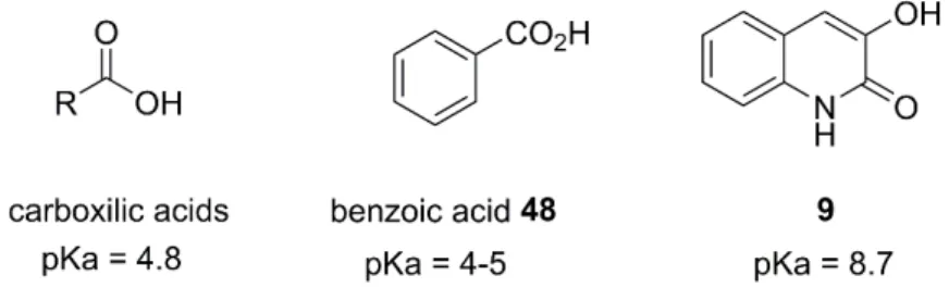
Figure 1.4 – Characteristic pKa values of carboxylic acid, benzoic acid 48 and 3HQ 9. ... 17
Figure 1.5 – Compound 9 at DAAO enzyme active side. a) Schematic representation of residues in DAAO-compound 9; b) Compound 9 (carbon atoms in magenta and oxygen in red) at h-DAAO enzyme active site. Side chains of key interacting residues are shown with carbon coloured in green and nitrogen in blue. Hydrogen bonding interactions are shown in dash. FAD is shown with carbons in cyan; b) structure of compound 9.9 ... 18
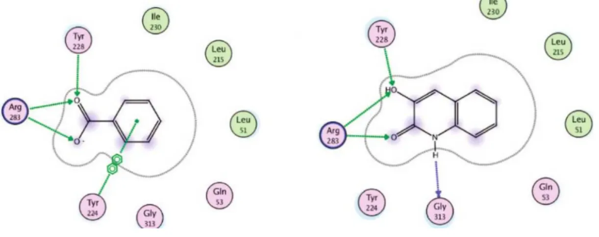
Figure 1.6 – Schematic representation of bonding interactions of compound 9 with those of carboxylic acids inhibitors (benzoic acid).9 ... 18
Figure 1.7 – a) Schematic representation of 3HQ as a bioisoster of aminoacid Glycine; b) compound 9 and cofactor FAD (PDB code: 3G3E). All atoms in type code except ligand carbon atoms in orange and FAD carbons in green.8 ... 19
Figure 1.8 – General strategies for the synthesis of the 3-Hydroxyquinolin-2(1H)-one skeleton. ... 20
Figure 2.1- (a) The concept of cooperative catalysis. (b) The concept of synergistic catalysis. (c) The concept of sequential or relay catalysis. ... 34
Figure 2.2 - General structure and reactivity of Di-rhodium(II) complexes. ... 36
Figure 2.3 – X-ray crystallography of 69. ... 43
Figure 2.5 – X-ray crystallography of 93... 50
Figure 2.6 - Energy profiles calculated for the metallocarbene formation between the
3-substituted 3-hydroxy-oxindole (70). The relevant bond distances (Ǻ) are indicated, as well as the respective as well as the respective Wiberg indices (WI, italics) ... 54
Figure 2.7 - Energy profiles calculated for ethyl diazoacetate (eda) and dirhodium(II)
tetraacetate. The relevant bond distances (Ǻ) are indicated, as well as the respective as well as the respective Wiberg indices. ... 55
Figure 2.8 – Energy profiles calculated for the 1,2-aryl migration of the
metallocarbene formed between the 3-substituted 3-hydroxy-oxindole and dirhodium(II) tetraacetate. The relevant bond distances (Ǻ) are indicated, as well as the respective Wiberg indices. ... 56
Figure 2.9 – Mechanistic representation of the dirhodium catalyzed ring expansion
reaction of 3-hydroxy oxindole. ... 57
Figure 2.10 - General catalytic cycle for Suzuki-Miyaura couplings. ... 60
Figure 3.1 - Incidence (blue) and mortality (red) of cancer worldwide.
GLOBALCAN 2012 (IARC). ... 67
Figure 4.1 - conversion of L-Phe to L-Trr is via a pathway involving the
para-hydroxylation of the benzene by PAH, the cofactor BH4 snf molecular oxygen. .. 86
Figure 4.2 - The domain structure of hPAH. Each hPAH subunit is classified into
three structural and functional domains which are involved in regulation, catalytic activity, and oligomerization. Regulatory domain (yellow), catalytic domain (green) and tetramerization domain (blue) of the hPAH. ... 88
Figure 4.3 - Proposed model of PAH activation by Phenylalanine... 89
Figure 4.4 - Rational for the design of new PAH modulators ... 94
Figure 4.5 – Differential scanning fluorimetry (DSF) assay ... 96
Figure 4.6 – Thermal denaturation of hPAH followed by differential scanning
hPAH (2.5 SYPRO Orange) CFX96 Touch Real-Time system (Bio-Rad); FRET channel Melting curve: 20 to 70 ºC with increasing steps of 0.2 ºC with 1 s incubation time, using the for fluorescence acquisition ... 97
Figure 4.7 - DSF analysis of compounds 141 and 165-167 on the mid-point
denaturation temperature of the a) regulatory domain (Tm1) and b) catalytic domain
(Tm2) of hPAH. ... 98 Figure 4.8 - Activity of compound 20-22 and 2 in hPAH enzyme assay. ... 99
Figure 4.9 - a) DSF analysis of compounds 129 and 169 on the mid-point
denaturation temperature of the regulatory (Tm1) and catalytic domain (Tm2) of
WT-hPAH. b) Results of the activity assay for compounds 129 and 169. ... 101
Figure 4.10 – Results of 7-trifluoromethyl-4-carboxamide-3HQs in DSF assay. .. 103
Figure 4.11 – Activity assay of compounds on tetrameric wild-type hPAH enzyme.
... 104
Figure A1 - Metallocarbene conformations with and without intramolecular
hydrogen bond determined at PBE1PBE/b1//PBE1PBE/b2level of theory. The energy corresponds to Gibbs Free Energy in ethanol, after thermal correction and the energy values are referred to the 70 +Rh2(OAc)4 pair of reactants. The relevant bond distances (Å) are indicated, as well as the respective Wiberg indices (WI, italics) ... 175
Figure A2 - Energy profiles calculated for the dirhodium catalyzed quinolone
formation. The minima and the transition states were optimized and the energy values (kcal/mol) are referred to pair of starting materials (70+Rh2(OAc)4) after thermal correction to Gibbs Free Energy in ethanol (in black) or to Gibbs free energy in vacuum at the PBE1PBE/b1 level of theory (in red). ... 175
Figure A3 - Energy profiles calculated for rhodium free quinolone formation, via a
concerted pathway. The minima and the transition states were optimized with at the pbe1pbe/6-31G** level of theory. The energy values (kcal/mol) are referred to the Gibbs Free Energyof the 3-hydroxy-oxindole (70) in the A conformation represented. ... 176
Figure A4 - Energy profiles calculated for rhodium free quinolone formation, via a
free carbene pathway. The minima and the transition states were optimized at the pbe1pbe/6-31G** level of theory. The energy values (kcal/mol) are referred to the Gibbs Free Energyof the 3-hydroxy-oxindole (70) in the A conformation represented. ... 176
Figure A5 - Energy profiles calculated for the rhodium catalyzed quinolone
formation, via coordination to the carbonylic ester of the 3-hydroxy-oxindole (5). The minima and the transition states were optimized at the PBE1PBE/b1level of theory. The energy values (kcal/mol) are referred to the Gibbs Free Energyof the pair of starting materials represented (J). ... 177
Figure A6 - Energy profiles calculated for the dirhodium catalyzed quinolone
formation, via coordination to the carbonyl of the oxindole ring. The minima and the transition states were optimized at the PBE1PBE/b1level of theory. The energy values (kcal/mol) are referred to the Gibbs Free Energyof the pair of starting materials represented (M). ... 177
Figure B3.1 - Depiction of the enzymatic reactions used in this study for evaluation
of competition between substrate and compound (I - Substrate-activated condition), and activation by the compound (II - Non-activated versus III – Compound-activated condition). A blank reaction without the substrate was included and subtracted for each condition in order to rule out contribution of the compound to tyrosine formation. ... 189
Figure C1 – Assignment of 1H and 13C NMR spectra of 69. ... 191
Figure C2 – Assigment of 1H and 13C NMR spectra of 31. ... 191
Figure C3 – Assignment of the 1H and 13C NMR spectra of a) 125 and b) 126. ... 192
List of Schemes Scheme 1.1 – Tautomers of quinolin-2(1H)-one. ... 4
Scheme 1.2 – Biologically active Quinolin-2-ones and Medicinal Agents. ... 4
Scheme 1.3 – Structure and atom numbering of 4HQs and 3HQs. ... 5
Scheme 1.4 – Tautomers of 4-Hydroxyquinolin-2-ones ... 6
Scheme 1.5 – Representative 4-hydroxyquinolin-2-one natural products and
pharmaceutical agents. ... 6
Scheme 1.6 – Antitumor 3-carboxamide-hydroquinolin-4-ones. ... 7
Scheme 1.7 – General Methodology for the synthesis of
4-hydroxyquinolin-2(1H)-one skeleton ... 9
Scheme 1.8 Synthesis of 4HQ esters from isatonic Anhydride 32 ... 10
Scheme 1.9 – The 3-hydroxyquinolin-2(1H)-one core present in the structure of
naturally occurring products. ... 11
Scheme 1.10-Structure of 2,3-dihydroxypyridine 34 and tautomeric form of 3HQs.
... 11
Scheme 1.11 – Schematically representation of coordination cores. ... 12
Scheme 1.12 – Tautomeric form of quinoxaline. ... 15
Scheme 1.13 – Most active compound towards to [H-3]-glycine binding to the site
associated with the NMDA receptor. in the 3HQs series. ... 16
Scheme 1.12 – Preparation of 2,3-dihydroxyquinoline 35. ... 21
Scheme 1.13 – Synthesis of 7-nitroquinolin-2-one 55 from
2-methyl-N-(2-methyl-5-nitrophenyl)formamide 51. ... 21
Scheme 1.14 –Mechanism of cyclopenin conversion into viridicatin 31. ... 22
Scheme 1.16 Pd-catalyzed formation of 62 using 3-bromo-4-phenylquinolinone 61.
... 24
Scheme 1.17 – Eister Ring Expantion ... 27
Scheme 1.18 XXX ... 28
Scheme 2.1 Schematic representation of the catalytic cycle of the diRhodium
-catalyzed C-H bond activation/ C-C bond forming reaction of an α-diazoacetate with an Alkane. ... 37
Scheme 2.2- Rhodium catalysed three component reaction using chiral Brønsted
acids. ... 38
Scheme 2.3 - [2+2] cycloaddition reaction between ethyl glyoxylate and
trimethylsilylketene catalyzed by a di-Rhodium carboxamide ... 39
Scheme 2.4 - Cooperative metal-organo-catalysed reactions based on di-Rh(II) and
Lewis base organo-catalysts ... 39
Scheme 2.5 - Potential di-Rh(II) inhibition when used in combination with Lewis
base organocatalysts. ... 40
Scheme 2.6 - Molecules that can be prepared via a ring expansion strategy. ... 41
Scheme 2.7 - Unwanted reaction in the Metal-Organo-Catalysed ring expansion
using EDA. ... 42
Scheme 2.8 –EDA addition to isatin followed by Eistert ring expansion reaction
catalysed by Rh2(OAc)2 ... 43
Scheme 2.9 - Recycling dirhodium(II) complex ring expansion-reaction. ... 45
Schema 2.10 - A metal-organo-catalytic system for the synthesis of 3HQs ... 46
Scheme 2.11 – One-pot reaction using N-methyl-isatin with EDA in the presence of
DBU 15 mol% and Rh2(OAc)4 inDCM ... 50
Scheme 2.12 – Ligands influence on the electrophilicity of dirhodium(II) complexes
Scheme 2.13 – Dirhodium(II) catalysts evaluated in the one-pot Eistert ring
expansion of isatins with EDA. ... 52
Scheme 2.14- Synthesis of 3-hydroxy-4-bromoquinolin-2(1H)-one 106 ... 60
Scheme 2.15 – Synthesis of viridicatin alkaloid derivatives based on the
Suzuki-Miyaura coupling reaction of aryl-boronic acids with 3-hydroxy-4-bromoquinolin-2(1H)-ones. ... 62
Scheme 3.1 Synthesis of 4-carboxylate substituted 3HQs 69, 84-96 based on an
Eistert ring expantion reaction of isatins with diazo acetate (EDA) catalysed by di-rhodium complexes. ... 69
Scheme 3.2 – Synthesis of compound 111 ... 71
Scheme 3.3 Synthesis of compound 112 based on an Eistert ring expansion reaction
of isatins with EDA catalysed by di-rhodium complexes. ... 71
Scheme 3.4 – Synthesis of diazo acetates 113-115 ... 72
Scheme 3.5 Synthesis of 4-carboxylate substituted 3HQs 116-120 based on an Eistert
ring expansion reaction of isatins with different diazo compounds, catalysed by di-rhodium complexes. ... 73
Scheme 3.6- Synthesis of 4-carboxamides-3HQs. ... 75
Scheme 3.7 Alternative synthetic route for synthesis of 4-carboxamides-3HQs ... 75
Scheme 3.8 Preparation of succinimidyl diazoacetate 124. ... 76
Scheme 3.9 Synthesis of 4-NHS -3HQs based on Eistert ring expansion reaction of
protected isatins with NHS-diazo acetate, followed by an amidation step. ... 78
Scheme 3.10 Synthesis of 4-carboxamide-3HQs 127-129 ... 78
Scheme 3.11 Synthesis of 6-F3CO-4-NHS -3HQs based on Eistert ring expansion
reaction of protected isatins with NHS-diazo acetate, followed by an amidation step. ... 79
Scheme 3.12 Synthesis of 4-carboxamide-3HQs based on Eistert ring expansion
reaction of 6-trifluoromethoxy-isatin with NHS-diazo acetate, followed by an amidation step. ... 80
Scheme 4.1 - Catalytic mechanism of catalytic mechanism of PAH and its
intervenients: Fe, O2 and BH4.155 ... 87
Scheme 4.2 - Chemical structure of compound with potential pharmacological
chaperone ability hits from Pey at al. 161 ... 91
Scheme 4.3 - Compounds with potential pharmacological chaperone ability. Hits
from Santos-Sierra et al. 163 ... 92
Scheme 4.4 - Structure of compound 161, a Phe-like modulator with affinity to the
active site of hPAH. ... 93
Scheme 4.5 - Synthesis of 4-L-Phe-3HQs 141 and 165-167. ... 95
Scheme 4.6 - Synthesis of 4-L-Phe-3HQs 129 and 166. ... 100
Scheme 4.7 - Compounds 7-trifluoromethyl-4-carboxamide-3HQs. ... 102
Scheme 5.1 - The 3-hydroxyquinolin-2(1H)-one (3HQ) core present in the structure
of natural occurring compounds, as a carboxylic acid bioisoster and as an enzyme inhibitor. ... 109
Scheme 5.2 - Synthesis of 4-Ester-3HQs, 4-Carboxamide-3HQs and viridicatin
derivatives 31. a) DBU, dirhodium complex (1 mol%), absolute EtOH, r.t., 3h; b) (i) NaOH, H2O, reflux, 7h; (ii) aq HCl; (iii) NBS, DMF; c) 10 mol% Pd(PPh3)4,
Na2CO3/H2O, DME:H2O 3:1, MW, 150ºC, 2h; f) TEA, Rh2(OAc)4 (1 mol%),
DCM, r.t.; g)HNRR’, Na2CO3, DMF, r.t., overnight. ... 111
Scheme 5.3 – Structure-activity-relationship of compound 143 towards cancer cells.
... 113
Scheme 5.4 – Structure-activity relationships (SAR) of 6-F3OC-carboxamide-3HQ
List of Tables
Table 1.1 – 3HQs preparation through one-pot Knoevenagel
condensation/epoxidation of cyanoacetanilides followed by decyanative epoxide-arene cyclization – substrate scope ... 25
Table 1.2 –Synthesis of 3HQs derivatives... 26
Table 1.3 – Lewis Acids promoted ring expansion of α-diazo-β-hydroxy ester 70. 29
Table 2.3- Effect of the amount of DBU in the ring expansion reaction in ethanola
... 48
Table 2.4 – Eistert ring expansion of isatins with EDA using a sequential
DBU/Rh2(OAc)4 system. ... 49
Table 2.4 – Eistert ring expansion of isatins with EDA using a one-pot
DBU/Rh2(OAc)4 system.a ... 52
Table 2.5 - Eistert Ring expansion of isatins with EDA using a one-pot relay
DBU/Rh2(OAc)4 system. ... 53
Table 2.5 - Catalyst screening for Suzuki-Miyaura coupling of 106 and phenylboronic
acid ... 61
Table 3.1 – Anti-proliferative evaluation of compounds 69 and 84-96 against
MCF-7, NCI-H460 and HT-29 cancer cell lines. ... 70
Table 3.2. Anti-proliferative evaluation of compounds 111-112 against MCF-7,
NCI-H460 and HT-29 cancer cell lines. ... 72
Table 3.3 – Anti-proliferative evaluation of compounds 116-120 against MCF-7,
NCI-H460 and HT-29 cancer cell lines ... 74
Table 3.4 – Optimization of reaction conditions of NHS-diazo addition on
N-benzyl-isatin. ... 77
Table 3.3 Anti-proliferative evaluation of compounds 132-143 against MCF-7, NCI-H460,HT-29 AND CHOK1cell lines. ... 81
Table 5.1 – Anti-proliferative activity of 7-OCF3-3HQ series against MCF-7,
Abbreviations and Definitions 3HQ: 3-hydroxyquinolin-2(1H)-one 4HQ: 4-hydroxyquinolin-2(1H)-one
ACT: aspartate kinase, Chorismate mutase and TyrA BH4: tetrahydropterin
BMS: Bristol-Myers Squibb CNS: Central nevous system
CHOK1: Chinese hamster ovary cells
DAOO: D-amino acid oxidase
DBU: 1,8-Diazabicyclo[5.4.0]undec-7-ene
DCM: dichloromethane DMO: dimethyl oxalate
DIPEA: diisopropyl ethyl amine DSF: differential scanning fluorimetry EDA: ethyl diazoacetate
FAD: flavin adenine dinucleotide HIV: Human Immunodeficiency Virus hPAH: human Phenylalanine Hydroxylase HRNase H: Ribonuclease H
HT-29: human colorectal adenocarcinoma cells HPA: hyperphenylalaninemia
LNAA: large neutral amino acid
L-Phe: L-phenylalanine
L-Tyr: L-tyrosine
MCF-7: breast cancer cells MS: multiple
MOC: Metal-organocatalysed system
NCI-H460: human non-small lung cancer cells NHCs: N-heterocyclic carbenes
NMDA: N-methyl-D-aspartate
NMRAr: N-methyl-D-aspartate receptors PAH: Phenylalanine Hydroxylas
PCs: pharmacolo
PEG-PAL Polyethylene glycol phenylalanine ammonia lyase PKU: Phenylketonuria
RE: ring expansion RD: regulatory domain RT: Reverse transcriptase
SAR: structure-activity relationship SMC: Suzuki-Miyaura reaction
SMOLs: chemically manufactured molecules
TH: tyrosine hydroxylase THF: Tetrahydrofuran TNF: tumor necrosis factor
WHO: World Health Organization
General Introduction
Rational and Aims
Humankind has been enroled in the discovering new drugs for thousands of years.1
A 90 % percent of the drugs on the market are small, chemically manufactured molecules (or SMOLs for short).2Furthermore, small-molecule drugs still account for
approximately two-thirds of the candidates in the current robust pharmaceutical industry pipeline. 2 Computer technology coupled with emerging protein structures
for identification and validation of biological target gave a great emphasis on inventive design of biologically active small molecules to generate high quality drug candidate. To keep up with the demand of new entities, development of new chemical tools for the synthesis of these molecules has been equally swift.
The 3-hydroxyquinolin-2(1H)-one (3HQ) core is an important motif that is present in the structure of viridicatin, viridicatol and 3-O-methyl viridicatin naturally occurring products.3, 4 These metabolites, isolated from penicillium species, have been
shown to inhibit the replication of human immunodeficiency virus and to be promising lead compounds for the development of new anti-inflammatory agents.5, 6
Furthermore, this unique heterocycle was recognized to be a valuable bioisoster for the carboxylic acid function of α-amino acids. Although less acidic (pKa of 8.7) then a carboxylic acid,7, 8 a series of 3HQs were prepared at Pfizer and shown to be potent
inhibitors of the D-amino acid oxidase activity, eliciting similar binding interactions with the enzyme active site as the carboxylic acid containing inhibitors.9 These
discoveries were not left unnoticed, and recently this pharmacophore was found to bind to metal cofactors, by this way inhibiting the influenza A endonuclease. Prompted by these results, the main objective of this project is to synthesize novel derivatives of 3HQ scaffold and evaluated its biological activity.
The specific objectives of this PhD project are:
o to develop robust synthetic methods for the target 3HQ derivatives based on
Ring Expansion with ethyl diazo acetate and cooperative Metal-Organo-Catalysed (MOC)- using di-rhodium(II) complexes in combination with Lewis base organo catalysts. These method can allow the creation of small library of 3HQs and the preparation of Viridicatin alkaloids;
o to use advanced two-dimensional NMR techniques (COSY, HMQC and
HMBC) as well as elementar analisis mass and UV to confirm the chemical structure of all synthesized derivatives:
o to understand and get further insight into the reaction mechanism of the ring expansion reaction catalyzed by dirhodium complexes by Density Functional Theory (DFT)( performed by Dr Nuno Candeias).
o to evaluate 3-hydroxyquinolin-2(1H)-one derivatives as antiproliferaqtive agents and as modulators of PAH
Outline of the thesis
Chapter 1 aims to present an overview of biological activities of compounds
derived from the quinolin-2(1H)-one. An overview on synthetic methodology and biological activity of the 3-Hydroxyquinolin-2(1H)-one scaffold and its isomer 4 -Hydroxyquinolin-2(1H)-one are going to be discussed. Moreover this chapter aims to point out the important properties of 3HQ scaffolds as bioisoster of a-aminoacid8
and chelator of metallic centers.10
Chapter 2 will discuss the design and synthetic strategy for the preparation of 3-hydroxyquinolin-2(1H)-one derivatives via ring expansion protocols based on ethyl diazo acetate and MOC system - di-Rhodium (II)/organic bases-. Characterization of obtained compounds by several methodologies, like NMR, will be discussed, as well as the synthesis and characterization of several intermediates used to achieve these derivatives. Also investigation of the mechanism of reaction by DFT calculation is
going to be discussed. Finally, the methodologies developed will be used to prepare viridicatin alkaloids and its derivatives.
Chapter 3 will present the evaluation of 3HQs library as anti-cancer agents, against
a panel of cancer lines: breast cancer cells, human non-small lung cancer cells and human colorectal adenocarcinoma cells. Also the library was evaluated for toxicity using the non-cancer Chinese hamster ovary cells.
Chapter 4 aims at designing molecules based on the L-Phenylalanine and 3HQ
structure to target Phenylalanine hydroxylase enzyme responsible for Phenilkenonuria disease. The objective in this progect is to stabilize the active site and the regulator domain without inhibiting severely the enzyme and restore its activity. By exploring this double stabilization mechanism we hope to develop a method that can be used to rescue the stability of a broad panel of PAH mutations.
Chapter 5 will integrate all studies described in the previous four chapters and
provide a global overview of the synthesized 3HQ derivatives. Also biological activity of these compounds will be discussed
Chapter 6 will presente all the experimental procedures used to development in
the present study. In particular synthetic methodologies, physical-chemical properties, biochemical studies and in vitro studies will be described.
1
Chapter
I
I. Hydroxy-quinolin-2(1
H
)-ones: A synthetic and
biological overview
Abstract
3HQs heterocycle is an aromatic ring system fused to a lactam ring that present an enol hydroxyl moieties as bioisoster of carboxylic acid. Although, less acidic than carboxilic acids (pka =8) series of 3HQs were shown to be potent inhibitors of the D-amino acid oxidase activity, eliciting similar binding interactions with the enzyme active site as the carboxylic acid containing inhibitors. Moreover, this acidic feature together with the lactamic nitrogen can mimic the a-aminoacid glycine, registering 3HQs as one of the limited examples of bioisoster of aminoacid.
The Quinolin-2(1H)-one scaffold
n the pharmaceutical industry nitrogen heterocycle compounds have paved the way for exceptional achievements in the fight against many life threatening diseases.11 Quinolin-2(1H)-ones establish the basic structure of many natural and
synthetic biologically active molecules and their literature has been extensively reviewed each year since 1989 in Progress in Heterocyclic Chemistry.12 In figure 1.1 is
displayed a chart of published papers about quinolin-2(1H)-ones in each year since 1982. The large number of publications depicted in the chart, suggest with no surprise, that the development of new methodologies to synthetize biologically active quinolin-2(1H)-ones compounds still remains as a very important goal in organic chemistry.
Figure 1.1 – Number of publications referring quinolin -2(1H)-ones since 1982, according to Web
of Science and using “Quinolin -2(1H)-ones” as keyword.
Generally, quinolin-2(1H)-ones are mentioned also as carbostyrils, 2-hydroxyquinolines, 2-quinolonols, or 2-oxaquinolines. Quinolin-2(1H)-ones are an important class of compounds since they are a coumarin isoster. They are isomeric to quinolin-4-ones and have two tautomeric forms, the lactam form 1 and the phenolic form 2 (Scheme 1). However, in the solid state, the compound exists exclusively as the lactam form 1.13, 14
As a “privileged” scaffold, the quinolin-2(1H)-one shows interesting biological properties and it is found in many natural products15-18 and medicinal agents. In
particular, the quinolin-2(1H)-one core is found in rebamipide 3, a medicinal antiulcer agent19, used in a number of Asian countries, or repirinast 4, an antiallergenic
compound useful in the treatment of allergic asthma (Scheme 1.2).20
Scheme 1.1 – Tautomers of quinolin-2(1H)-one.
Noteworthy, a broad range of biological activities of quinolin-2-one compounds were disclosed in recent years. Members of this class of compound have been reported to show potent antimicrobial activity21, possess neuro protective properties22
and have also proved their potential as excellent inhibitors of acyl co-enzyme A and cholesterol acyltransferase.23, 24
Furthermore, a group from Bristol-Myers Squibb (BMS) identified compound 5 and the related reduced allylic alcohol, as novel and potent maxi-K channel openers useful for the treatment of male erectile dysfunction.25 A class of potent KDR (kinase
insert domain-containing receptor) inhibitors, a primary mediator of tumour-induced angiogenesis containing the 1H-indole-2-yl-quinolin-2(1H)-one core structure 6 was reported by Merck and show great interest as potential therapeutic agents.26 A
clinically important quinolin-2(1H)-one was discovered by Johnson & Johnson Pharmaceutical Research & Development, with registration number R115777 (Zanestra) 7 and is currently under phase II clinical trials as a novel orally active antitumor agent. Zanestra is a 4-arylquinolin-2(1H)-one that emerged as a selective nonpeptide farnesyl protein antitumor inhibitor of Ras , an oncoprotein involved in the intracellular signalling pathway leading to cell proliferation.
A number of derivatives of quinolin-2-one have been reported to show different and promising biological properties and have attracted much attention from the medicinal chemist community.18, 21, 23, 24, 26-42 In particular, 4-hydroxyquinolin-2-ones
(4HQs) and 3-hydroxyquinolin-2-ones (3HQs) have demonstrated to be a very appealing class of small-size heterocycle molecules. 4HQs and 3HQs are isomeric compounds, the OH group changes in the lactam ring position from C-3 to C-4 and despite this small variation, the physicochemical properties and the biological activity of both compounds change considerably. These properties and the synthetic methods used to prepare 4HQs and 3HQs compounds will be discussed in the next section.
Overview of 4-hydroxyquinolin-2-ones
4-Hydroxyquinolin-2-ones biology
4-Hydroxyquinolin-2-ones (10) derivatives are important as biologically active compounds and synthons in organic synthesis. The main feature of this hydroxyquinoline is that it can exist in three tautomeric forms, namely 2-hydroxyquinolin-4-ones (11) and 2, 4-dihydroxyquinolines (12). Normally, they exist as 10 but solvents can affect the equilibrium.
Scheme 1.4 – Tautomers of 4-Hydroxyquinolin-2-ones
This ring core is the base of a large number of alkaloids present in many medicinal plants, microbial sources and animals. Usually in nature these alkaloids are found prenilated (13) in the 4-hydroxy or with a methoxy groups (14) or substituted with anellated pyrano rings (15) (e.g. flinderisine, oricine, orixalone D and huajiaosimuline) that also display a wide range of biological activities.
Scheme 1.5 – Representative 4-hydroxyquinolin-2-one natural products and pharmaceutical agents. Recently a directed bioassay of the CH2Cl2-MeOH extract of Euodia roxburghiana
infectious HIV-1 in human lymphoblastoid host cells (EC50=1.64 µM, IC50 26.9 µM)
and to inhibit the activity of the HIV-1 reverse transcriptase assay (IC50 = 8 mM).43
4-Hydroxyquinolinones have attracted considerable attention for various therapeutic areas including applications as antimicrobial agents,44 antimalarial
agents,45 aldose reductase inhibitors,46 anticonvulsants,47 and RNA polymerase
inhibitors for the treatment of Hepatitis C.48
Recently, carboxamide derivatives of 4HQs have been investigated for their important activities against auto-immune diseases such as rheumatoid arthritis, systemic lupus erythematosis and multiple sclerosis. 49-51 A remarkable representative
of this derivatives is linomide 16 (Scheme 1.6), an orally active agent that consistently inhibits growth of a large series of both rodent and human prostate cancer xenografts tested in vivo. The anti-tumour ability of this compound is related to the capacity to inhibit tumour angiogenesis. It was demonstrated by a study in rats bearing linomide treated tumour, that the agent decrease the number of tumour blood vessels with a consequently reduction in the tumour bloodflowin.52-54 A second generation of
3-carboxamide-hydroquinolin-4-ones such as ABR-215050 (tasquinimod) 17 (Scheme 1.6) inhibit the growth of a series of four additional human and rodental protate cancer model in mice.55 The mechanism for linomide’s therapeutic activities is not
fully understood.
However considerable amount of data attribute its therapeutic activities to its ability to regulate cytokine production.56-58 Furthermore, the production of
proinflamatory cytokines involved in tumor angiogenesis by macrophages is also involved in the auto-destruction and demyelination in multiple sclerosis (MS).
Therefore, linomide was tested in a series of phase II and III trials in MS patients, although phase III trial had to be discontinued because of undesirable toxicity.59, 60 In
order to obtain more efficient compounds for the treatment of MS, an optimization of the lead compound 16 was performed. Chemical modifications and structure-activity relationship (SAR) give raise to a new series of 3-quinolinecarboxamide derivatives and compound laquinimod 18 gave a similar immune response and cytokine balance as the lead compound 16. Currently there is an ongoing study of laquinimod in phase II to access the efficacy, safety and tolerability of the oral dose in subjects with primary progressive MS. A discontinuation of higher doses 1.2mg/day of laquinimod has been done, after the occurrence of cardiovascular events, none of which was fatal, in eight patients.61 Nevertheless the study for lower
doses 0.6mg/day is still ongoing.
4-Hydroxyquinolin-2-ones chemistry
One of the requirements of any synthetic strategy for drug development is that the synthetic pathway must be amenable to provide chemical diversity in order to obtain a large number of structural motifs.62, 63 From the chemical point of view, 4HQs,
possessing this enolic β-dicarbonyl moiety, have attracted chemists not only with the aim to develop simple and efficient routes to achieve highly functionalized 4-hydroxyquinolin-2-ones but also using this 4HQs as synthon for the preparation of other natural products such as dimeric quinoline alkaloids and other polycyclic heterocycles.64 There are well documented different synthetic methods for the
synthesis of 4HQs. Generally, a common route to these compounds is the intramolecular Claisen-type condensation of acylated anthranilate esters. The N-acylated anthranilate esters 19 can be N-acylated with malonyl chlorides 20 and cyclized
to 4HQ ester 22 under acidic conditions (Scheme 1.7 method A).65 An alternative
approach to synthetize new 4HQ derivatives has been proposed by Jonsson et al. Starting from aromatic 2,6-difluorobenzonitrile 23, a double nucleophilic aromatic substitution was performed to introduce at position 5C of 4HQs core, substituents such as methoxy, dimethylamino, and thiol 27 (Scheme 1.7 method B).61
Scheme 1.7 – General Methodology for the synthesis of 4-hydroxyquinolin-2(1H)-one skeleton Also, reaction of indoline 28 heated with an excess of methanetricarboxylates 29 yielded derivatives of 4HQs in good yield (Scheme 1.8 method C).66Although this
transformation presents advantages for the synthesis of 4HQs, high reaction temperatures (>200 °C), limited availability of a broad range of suitably substituted starting materials, and the need to isolate the acylated intermediate prior to cyclization, limit the widespread application of the method. The most common employed method was develop by Coppola and co-workers which have synthesized 4HQs compounds using isatoic anhydrides 32 as precursors and involves an N-alkylation, followed by malonate addition-intramolecular cyclization sequence (Scheme 1.8). 67, 68,69
Scheme 1.8 Synthesis of 4HQ esters from isatonic Anhydride 32
Overview on 3-Hydroxyquinolin-2-ones 3-Hydroxyquinolin-2-ones biology
Despite being an isomer of 4HQs, very little is known about 3-hydroxyquinolin-2(1H)-one (3HQs) from biological and synthetic perspectives. The 3HQ core is an important motif that is present in the structure of naturally occurring products viridicatin 31, viridicatol 32 and 3-O-methyl viridicatin 33. These metabolites, were first isolated from the mycelium of Penicillium viridicatum Westling and later on various strains of Penicillium cyclopium Westling4 with the production of a strong, penetrating
earthy odour. The earliest biological assay of 31 were done against Escherichia coli,
Bacillus subtilis, and Staphylococcus aureus (Micrococcus pyogenes, var. aureus) but no
antibiotic activity was found. Although some activity was observed on in vitro tests against Mycobacterium tuberculosis at a dilution of 1:15 000, while no activity against
Entamoeba histolytica was detected.3 The viridicatin metabolite 33 methylated in the
3-OH was isolated in 1964 by Austin and Myers from the fungus Penicillium puberulum.70
It remained unexplored until 1998 when Heguy and co-worker reported its effect as inhibitor of replication of the HIV virus induced by tumor necrosis factor (TNF). Having an IC50 of 2.5 µM, this compound was recorded as a promising lead for the
Scheme 1.9 – The 3-hydroxyquinolin-2(1H)-one core present in the structure of naturally
occurring products.
These discoveries were not left unnoticed, and recently a group from the University of Lille, developed a series of 3HQs with potent activity against HIV-1 reserve transcriptase associated RNase H activity. Ester and amide groups were introduced at C-4 position of the 3HQs scaffold and also some modulation was performed in the benzenic moiety, which allowed the construction of a library of 19 compounds. The rational for choosing 3HQs as pharmacophore was made on the bases of its ability to complex some bivalent metals, as showed by the work of Strashnova and co-workers.10 In their previous studies on the complexation of 2,3-dihydroxypyridine 34
with metals, they shown that this compound participates in the coordination as mono – or dicantonionic species and acts as a bridging ligand.
Scheme 1.10-Structure of 2,3-dihydroxypyridine 34 and tautomeric form of 3HQs.
3HQ 9 and its tautomeric form, 2,3-dihydroxyquinoline 35, are structural analogues of 2,3-dihydropyridine 34 (Scheme 1.10). The main common characteristics of these two molecules are: a slight tautomerization, presence of several potential coordinative centres, which can yield the cationic, neutral and anionic complexes. The complex formation with 2,3- dihydroxyquinoline (HL)2 show that the coordination
core structure depends mainly on the characteristics of the central metal atom and on the most stable tautomeric form of the ligand under the synthec conditions. The authors identified complexes with different metals and summarized the result as shown in the Scheme 1.11. HL2 participates in the coordination in the monoanionic
or neutral forms with the formation of chelate cycles. Two main class of coordination species are depicted (Scheme 1.11), first one represented by the formula M(HL)2·2H2O containing metal such Mn, Ni, Cu, where two molecules of 3HQs are
coordinated with the metal and two molecules of water giving chelate cycles. The second type of coordination involved the 1:1 coordination of the 3HQ compound and the metal Fe(HL)OH· 2H2O 39, Co(HL)OH·H2O 40, while Cadmium participate
as Cd(H2L)Cl2 41.
Scheme 1.11 – Schematically representation of coordination cores.
In a recent review entitled “Viral enzymes containing magnesium: Metal binding as a successful strategy in Drug design”71 is shown that metal-activated enzymes are
important targets in drug discovery and in particular for antivirals discovery. Such proteins contain one or more metal ion cofactors, prevalently located in the active site, which are essential to perform biological functions. The common features of possible efficient inhibitors of metal enzymes are resumed in: highly polar pharmacophore motives, ionisable moieties, and coplanar pre-organized structure capable of simultaneous binding two Mg2+ ions.71 Based on this rational, the work of
Cotelle and co-workers72 aimed at developing derivatives of 3HQs to target the
catalytic site of the ribonuclease H (HRNase H) function, associated to the viral coded reverse transcriptase (RT). In order to complex the bivalent metals in the catalytic site of the enzyme, the author introduced a carbonyl function at position C-4 of the 3HQ scaffold. By this introduction the 3HQs comprises three oxygens, which is the ideal topology to bind two divalent cations, separated by 4–5 Å in the case of an enzyme– metals–ligand ternary complex. Such a pharmacophore can be observed in the structure of most recently discovered RNase H inhibitors.72 The most active
compounds were the 4-amido series able to inhibit the RT RNase H with an IC50
between 16 and 22 µM, comparable with a reference compound. The authors also performed in silico docking studies in order to determinate the possible binding mode. The magnesium chelation was examined in the study and the authors confirmed the ability of this three oxygen pharmacophore to chelate both metal cofactors within the active site of the enzyme. Compound 42 is an inhibitor of the enzyme with an activity of 19 μM. As shown in Figure 1.2, the quinolone scaffold is positioned in such a way that the two oxygen atoms of the carbonyl and the enol functions in positions 2 and 3 target the magnesium cations.72
The metal-chelating properties of 3HQs inspired also the work of La Voie to develop a series of these compounds as inhibitors of Influenza A Endonuclease. The most active molecule was found to be compound 7-(p-fluorophenyl)-3-hydroxyquinolin-2(1H)-one 43 with and IC50 of 0.5 µM. An X-ray crystal structure
of 43 complexed with influenza A endonuclease nicely disclosed that it binds through bimetal chelation at the active site as shown in Figure 1.3.73
Figure 1.3 – Binding of compound 43 at the endonuclease active site.73
Chelation of enzyme metal cofactors is not the only property of this interesting core. Recently, with the purpose to discover ligands for N-methyl-D-aspartate
(NMDA) associated glycine binding, a series of 3HQs have been synthetized. NMDA receptors (NMDARs) are glutamate-gated cation channels with high calcium permeability that play important roles in many aspects of the biology of higher organisms. They are critical for the development of the central nervous system (CNS), generation of rhythms for breathing and locomotion, and the processes underlying learning, memory, and neuroplasticity. Consequently, abnormal expression levels and altered NMDA receptor function have been implicated in numerous neurological disorders and pathological conditions (including stroke, hypoxia, ischemia, head trauma, Huntington’s, Parkinson’s, and Alzheimer’s diseases, epilepsy, neuropathic pain, alcoholism, schizophrenia, and mood disorders).74-76
Based on the tautomer 45 of quinoxaline derivatives 44, in which the amide and the enol hydroxyl moieties mimic a protonated glycine responsible for bonding with NMDA receptors, Sing-Yuen Sit and co-workers77 synthesized twenty-four 3HQs
derivatives 9, formally isoster of quinoxoxaline tautomer 45 .
Scheme 1.12 – Tautomeric form of quinoxaline.
3HQs derivatives were studied and their ability to displace radio ligand (3
H)-glycine from rat cortical membranes was evaluated. All compounds demonstrated a 60% displacement of the radio ligand at 10 µM and from the results of the assay a structure-activity relationship was elucidated, supporting the 3-hydroxyquinolin-2-one heterocycles as effective structural elements for glycine ligands. Some modification on the central core was done leading to improved activity of these compounds, namely introduction of an electron withdrawing group in position C-4 and modification of the benzyl moiety resulted in more affinity for the glycine binding site on NMDA, hypothesised to be due to the increase acidity of 3-hydroxyl group. However, no activity was detected in the assay where 3-hydroxy group was methylated, identifying the free OH group as essential pharmacophore of the molecule
Scheme 1.13 – Most active compound towards to [H-3]-glycine binding to the site associated with
the NMDA receptor in the 3HQs series.
Ultimately, introduction of pyruvate ester moiety at C-4 and a 5,7-dichloro pattern of substitution in the aromatic ring resulted in a substantial increase in affinity. The most active compound in this series was compound 47 with an IC50 of 29 nM.77
To better understand the biological properties of these compounds it would be important to shed light on the main essential features of this important heterocycle. This unique molecule, was recently recognized to be a valuable carboxylic acid bioisoster .7 The carboxylic acid is an important functional group that often takes part
of the pharmacophore of different therapeutic agents.7 Furthermore, the aptitude of
this group to create strong electrostatic interactions and hydrogen bonds, in association with its acidity, classify carboxylic acid as a key function in the interaction between drug and target. Despite the importance of the carboxylic acid group, it exhibit when this moiety is present in a drug, significant drawbacks namely metabolic instability, toxicity and limited diffusion across biological barriers are shown. To avoid this limitation, the replacement of carboxylic group with a surrogate or bioisoster can overcome these problems and can represent an effective strategy in drug development. Recently Ballatore and co-workers7 provided an overview of the most
commonly employed carboxylic acid (bio)isosteres and present some examples to show the use and utility of isosteres in drug design. In this review 3HQs are classified as bioisosteres of carboxylic acids, despite their lower acidity (pka = 8.7), and the authors refer to the work done by Duplantier et al. to exemplify this bioisosterism.
Figure 1.4 – Characteristic pKa values of carboxylic acid, benzoic acid 48 and 3HQ 9. The described inhibitors in literature of D-amino acid oxidase (DAAO) are small aryl carboxylic acids or acid-isosters, such as benzoic acid 48 that are ionized at the peroxisomal pH (c.a. 8). In a high-throughput screening in a functional assay to find potential inhibitors of DAAO, 3HQ 9 was identified as a potent one (IC50 = 4nM).
Co-crystallization of 9 with the human DAAO enzyme showed that the 3-hydroxyl group of the molecule is involved in two hydrogen bonds, one with the Tyr228-OH and the other with the Arg283-NH (Figure 1.5 a). Furthermore, the 2-carbonyl group is also involved in a strong hydrogen bonding with the same Arg residue, while the lactam-NH donates a hydrogen bond to the backbone carbonyl of Gly313. Also a fundamental π-π interaction with the re-face of the flavin ring of flavin adenine dinucleotide (FAD) and Tyr224 is provided by the aromatic moiety of compound 8 consistent with similar structures of aryl acid bounded to DAAO.78 Figure 1.6 shows
a schematic diagram comparing the bonding interactions of compound 9 with those of carboxylic acids inhibitors (benzoic acid). The hydrogen-bonding interaction of the carboxylic moiety of benzoic acid with the enzyme active site and hydroxyquinolin-2-one behave in a very similar fashion.9 Another important characteristic of these
compounds is that 3HQs are also classified as non-classical bioisosteres of α-amino acids. Whereas classical bioisosteres include replacement of similar atoms (e.g. hydrogen with fluorine, carbon with silicon)79 or ring-to-ring transformations (e.g.
replacement of phenyl group with thiophene) nonclassic bioisosterism includes all other forms such as ring-to-chain, chain to ring transformations, functional group replacement, as well as regioisosterism.80, 81