R E S E A R C H
Open Access
Bacterial and protozoal agents of feline
vector-borne diseases in domestic and stray cats
from southern Portugal
Carla Maia
1,2,3*, Cláudia Ramos
1, Mónica Coimbra
4, Filipa Bastos
3, Ângela Martins
5, Pedro Pinto
3, Mónica Nunes
6,7,
Maria Luísa Vieira
6,7, Luís Cardoso
8,9and Lenea Campino
1,10Abstract
Background: Feline vector-borne diseases (FVBD) have emerged in recent years, showing a wider geographic distribution and increased global prevalence. In addition to their veterinary importance, domestic cats play a central role in the transmission cycles of some FVBD agents by acting as reservoirs and sentinels, a circumstance that requires a One Health approach. The aim of the present work was to molecularly detect feline vector-borne bacteria and protozoa with veterinary and zoonotic importance, and to assess associated risk factors in cats from southern Portugal.
Methods: Six hundred and forty-nine cats (320 domestic and 329 stray), from veterinary medical centres and animal shelters in southern Portugal, were studied. Anaplasma spp./Ehrlichia spp., Babesia spp., Bartonella spp., Borrelia burgdorferi sensu lato, Hepatozoon spp. and Leishmania spp. infections were evaluated by polymerase chain reaction (PCR) in blood samples.
Results: One hundred and ninety-four (29.9%) cats were PCR-positive to at least one of the tested genera or complex of FVBD agents. Sixty-four (9.9%) cats were positive to Leishmania spp., 56 (8.6%) to Hepatozoon spp., 43 (6.6%) to Babesia spp., 35 (5.4%) to Anaplasma spp./Ehrlichia spp., 19 (2.9%) to Bartonella spp. and 14 (2.2%) to B. burgdorferi s.l. Thirty-three (5.1%) cats were positive to two (n = 29) or three (n = 4) genera/complex. Babesia vogeli, Bartonella clarridgeiae, Bartonella henselae, Ehrlichia canis, Hepatozoon felis and Leishmania infantum were identified by DNA sequencing.
Conclusions: The occurrence of FVBD agents in southern Portugal, some of them with zoonotic character, emphasizes the need to alert the veterinary community, owners and public health authorities for the risk of infection. Control measures should be implemented to prevent the infection of cats, other vertebrate hosts and people.
Keywords: Cats, Feline vector-borne diseases, Bacteria, Protozoa, Portugal
Background
Vector-borne diseases comprise a group of globally dis-tributed and rapidly spreading illnesses that are caused by a range of pathogens transmitted by arthropods, in-cluding ticks, fleas, mosquitoes and phlebotomine sand flies [1-3]. In addition to their veterinary importance,
cats and dogs play a central role in the transmission cycles of some agents of vector borne diseases (e.g. anaplasmosis, bartonellosis, borreliosis and leishmaniosis) by acting as reservoirs, amplifying hosts or sentinels, with such cir-cumstances requiring a One Health approach [4,5].
Feline vector borne diseases (FVBD) have emerged in recent years, showing a wider geographic distribution and increased global prevalence. Environmental, demographic and human behavioral factors (e.g. travelling with pets, changes in social and leisure activities), together with the direct impact of climate changes on the abundance,
* Correspondence:[email protected]
1
Unidade de Parasitologia Médica, Instituto de Higiene e Medicina Tropical (IHMT), Universidade Nova de Lisboa (UNL), Lisbon, Portugal
2
Centro de Malária e outras Doenças Tropicais, IHMT-UNL, Lisbon, Portugal Full list of author information is available at the end of the article
© 2014 Maia et al.; licensee BioMed Central Ltd. This is an Open Access article distributed under the terms of the Creative Commons Attribution License (http://creativecommons.org/licenses/by/2.0), which permits unrestricted use, distribution, and reproduction in any medium, provided the original work is properly credited. The Creative Commons Public Domain Dedication waiver (http://creativecommons.org/publicdomain/zero/1.0/) applies to the data made available in this article, unless otherwise stated.
Maia et al. Parasites & Vectors 2014, 7:115
geographical distribution and vectorial capacity of vector arthropods have contributed to the changing epidemiology of these arthropod-borne diseases [2,6].
The detection of FVBD agents can be challenging as some of them occur in healthy cats, and the clinical signs, whenever present, are normally unspecific of those dis-eases. PCR-based methods applied to vector-borne patho-gens are very effective to detect and characterize infecting organisms, for monitoring cure after chemotherapy and to evaluate the role that subclinically-infected cats can play in the transmission of infections [7].
A recent polymerase chain reaction (PCR) study re-ported positivity to Anaplasma/Ehrlichia, Babesia,
Hepa-tozoon, Leishmania and Rickettsia in client-owned cats
from the north and centre regions of Portugal [8]. Mo-lecular and serological studies on Leishmania infantum [9,10], Anaplasma phagocytophilum, Bartonella spp. and Rickettsia conorii [11] have been performed in domestic and stray cats from southern Portugal. Nevertheless, infor-mation about FVBD agents circulating countrywide is still limited, especially in the south, and therefore the aim of the present study was to assess the presence of bacteria and protozoa with veterinary and zoonotic importance in cats from southern of Portugal, and to assess positivity-associated risk factors.
Methods Cats and samples
From January 2012 to August 2013, a total of 649 cats (320 domestic and 329 stray), from veterinary medical centres and animal shelters in southern Portugal, were studied. Cats were from the districts of Lisbon (n = 282) and Setúbal (n = 104), in the region of Lisbon, and from the district of Faro (n = 263), which overlaps the region of the Algarve. In the Lisbon region most of the domestic cats lived in apartments or in semi-detached houses, while cats from the Algarve region lived in rural areas and used to spend most of their time exclusively outdoors.
Out of the stray cats, 294 were collected to be neutered for birth-rate control or to be housed in a shelter for adoption, and 35 were captured and euthanized in the scope of official animal control programs. Domestic cats were randomly included after obtaining the owners in-formed consent. In the case of stray cats, written consent for enrolment was also obtained from the person in charge of each shelter.
Whole blood samples were collected by cephalic or jugu-lar venipuncture and spotted onto filter paper (Whatman no. 3) for DNA extraction. Samples were dried at room temperature and kept at 4°C until tested. Whenever avail-able, data on gender, breed, living conditions, age, use of acaricides/insecticides, clinical status (presence or absence of clinical signs compatible with a FVBD), and serological status regarding feline immunodeficiency virus (FIV) and
feline leukaemia virus (FeLV) infections were registered for each cat (Table 1). Clinical signs compatible with FVBD comprised anorexia, muscular atrophy, dermato-logical manifestations, exercise intolerance, fever, gastro-intestinal alterations, lameness, lymphadenopathy, muscular lethargy, ocular manifestations, pale mucous membranes or weight loss.
This study was ethically approved by the boards of the Institute of Hygiene and Tropical Medicine (IHMT-UNL) and of the Faculty of Veterinary Medicine (ULHT) as complying with the Portuguese legislation for the pro-tection of animals (Law 92/1995).
PCR amplification and sequencing
A commercial kit (Kit Citogene®, Citomed) was used to extract DNA from blood on filter paper. Four discs of filter paper (4 mm in diameter each) were incubated
with lysis buffer (150 μl) and 1.5 μl of proteinase K
(20 mg/ml). Further DNA extraction followed the kit manufacturer’s instructions.
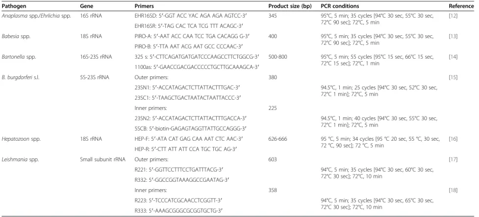
Positivity to Anaplasma spp./Ehrlichia spp., Babesia spp., Bartonella spp., B. burgdorferi s.l., Hepatozoon spp. and Leishmania spp. DNA in blood samples was tested by PCR according to previously described protocols (Table 2). PCR amplifications were performed in a 25μl final volume
containing 2 mM MgCl2, 1 unit of Taq DNA polymerase
(GoTaq DNA Polymerase®, Promega), 10 pmol of each pri-mer (15 pmol in the case of L. infantum), 0.2μM each of dATP, dTTP, dCTP and dGTP (Dntps set®, Bionline, Citomed), and 3μl of DNA template (5 to 200 ng). In all amplifications a positive control containing genomic target DNA and a negative control without DNA were included. PCR products were visualized under UV illumination after electrophoresis migration on a 1.5% gel agarose stained with 0.2 mg/ml ethidium bromide, using a 100 bp DNA ladder as a marker.
Twenty per cent of the PCR products (30% in the case of Bartonella spp.) were purified with a High Pure PCR Product Purification Kit (Roche® Mannheim) according to the manufacturer’s instructions and directly sequenced (one direction) (Stabvida®), using the same primers as those used for the DNA amplification. Species identity of the obtained sequences was determined according to the closest BLAST match (identity≥97%) to a GenBank acces-sion and deposited in DNA Data Bank of Japan (DDBJ) (http://www.DDBJ.nig.ac.jp).
Statistical analysis
Percentages of positivity to FVBD agents relative to the independent variables and categories (Table 1) were compared by the Chi-square or Fisher’s exact tests. A p value <0.05 was considered as statistically significant. Analyses were performed with SPSS® 21 software for Windows.
Results
One hundred and ninety-four (29.9%) cats were PCR-positive to at least one of the tested genera or complex of FVBD agents (Table 2). Sixty-four (9.9%) cats were posi-tive to Leishmania spp., 56 (8.6%) to Hepatozoon spp., 43 (6.6%) to Babesia spp., 35 (5.4%) to Anaplasma spp./
Ehrlichia spp., 19 (2.9%) to Bartonella spp. and 14 (2.2%) to B. burgdorferi s.l. Thirty-three (5.1%) cats were positive to two (n = 29) or three (n = 4) genera/complex (Table 3).
As shown in Table 1, the non-use of acaricides/insecti-cides and living in rural areas were associated with Babe-siaspp. and Hepatozoon spp.. Furthermore, the prevalence Table 1 Comparison of prevalence of FVBD pathogens in different groups of cats from southern Portugal
Variable/ category
Nº of
tested cats (%)
N° of positive cats (%)
Anaplasma/Ehrlichia Babesia Bartonella B. burgdorferi s.l. Hepatozoon Leishmania ≥ 1 positive PCR Gender 649 Female 372 (57.3) 18 (4.8) 31 (8.3) 11 (3.0) 9 (2.4) 32 (8.6) 44 (11.8) 122 (32.8) Male 277 (42.7) 17 (6.1) 12 (4.3) 8 (2.9) 5 (1.8) 24 (8.7) 20 (7.2) 72 (26.0) Breed 484 DSH 432 (89.3) 22 (5.1) 22 (5.1)* 10 (2.3) 4 (0.9) 31 (7.2) 33 (7.6) 109 (25.2) Other breed 52 (10.7) 1 (1.9) 10 (19.2)* 2 (3.8) 1 (1.9) 3 (5.8) 7 (13.5) 19 (36.5) Lifestyle 649 Domestic 320 (49.3) 9 (2.8)* 28 (8.8)* 5 (1.6) 3 (0.9) 24 (7.5) 33 (10.3) 91 (28.4) Stray 329 (50.7) 26 (7.9)* 15 (4.6)* 14 (4.3) 11 (3.3) 32 (9.7) 31 (9.4) 103 (31.3) Age (months) 462 [3-11] 129 (27.9) 5 (3.9) 13 (10.1) 4 (3.1) 1 (0.8) 3 (2.3)* 6 (4.7)a 27 (20.9) [12-59] 216 (46.8) 10 (4.6) 13 (6.0) 6 (2.8) 0 (0.0) 17 (7.9) 14 (6.5)b 54 (25.0) [60-228] 117 (25.3) 5 (4.3) 5 (4.3) 0 (0.0) 2 (1.7) 11 (9.4)* 16 (13.7)a,b 35 (29.9) Acaricides-insecticides 568 Yes 204 (35.9) 5 (2.5) 4 (2.0)* 3 (1.5) 3 (1.5) 11 (5.4)* 27 (13.2)* 51 (25.0)* No 364 (64.1) 22 (6.0) 37 (10.2)* 16 (4.4) 10 (2.7) 42 (11.5)* 28 (7.7)* 126 (34.6)* Region 649 Lisbon 386 (59.5) 18 (4.7) 9 (2.3)* 8 (2.1) 3 (0.8)* 25 (6.5)* 31 (8.0) 85 (22.0)* Algarve 263 (40.5) 17 (6.5) 34 (12.9)* 11 (4.2) 11 (4.2)* 31 (11.8)* 33 (12.5) 109 (41.4)* Habitat 649 Urban 282 (43.5) 18 (6.4) 8 (2.8)* 4 (1.4) 0 (0.0) 14 (5.0)* 9 (3.2)* 47 (16.7)* Rural 367 (56.5) 17 (4.6) 35 (9.5)* 15 (4.1) 14 (3.8) 42 (11.4)* 55 (15.0)* 147 (40.1)* Housing 589 Totally indoors 124 (21.1) 3 (2.4) 6 (4.8) 1 (0.8) 0 (0.0) 3 (2.4)* 13 (10.5) 24 (19.4)* Access to outdoors 465 (78.9) 28 (6.0) 36 (7.7) 17 (3.7) 8 (1.7) 51 (11.0)* 39 (8.4) 151 (32.5)* FeLV 242 Negative 231 (95.5) 12 (5.2) 15 (6.5) 6 (2.6) 6 (2.6) 13 (5.6) 29 (12.6) 70 (30.3) Positive 11 (4.5) 1 (9.1) 1 (9.1) 0 (0.0) 1 (9.1) 1 (9.1) 0 (0.0) 3 (27.3) FIV 247 Negative 226 (91.5) 11 (4.9) 16 (7.1) 6 (2.7) 6 (2.7) 14 (6.2) 28 (12.4) 69 (30.5) Positive 21 (8.5) 3 (14.3) 1 (4.8) 0 (0.0) 1 (4.8) 1 (4.8) 1 (4.8) 6 (28.6) Clinical status 222 Non-suspect 197 (88.7) 8 (4.1) 26 (13.2) 1 (0.5) 0 (0.0) 12 (6.1) 8 (4.1) 49 (24.9) Suspect 25 (11.3) 3 (12.0) 1 (4.0) 0 (0.0) 0 (0.0) 1 (4.0) 3 (12.0) 6 (24.0) Total 649 (100) 35 (5.4) 43 (6.6) 19 (2.9) 14 (2.2) 56 (8.6) 64 (9.9) 194 (29.9) *,a,b
Statistically significant difference (p <0.05); DSH: domestic short-haired; FeLV: feline leukaemia virus; FIV: feline immunodeficiency virus.
Maia et al. Parasites & Vectors 2014, 7:115 Page 3 of 8
Table 2 Primer sets for PCR amplification of FVBD agents
Pathogen Gene Primers Product size (bp) PCR conditions Reference
Anaplasma spp./Ehrlichia spp. 16S rRNA EHR16SD: 5′-GGT ACC YAC AGA AGA AGTCC-3′ 345 95°C, 5 min; 35 cycles [94°C 30 sec, 55°C 30 sec, 72°C 90 sec]; 72°C, 5 min
[12] EHR16SR: 5′-TAG CAC TCA TCG TTT ACAGC-3′
Babesia spp. 18S rRNA PIRO-A: 5′-AAT ACC CAA TCC TGA CACAGG G-3′ 400 95°C, 5 min; 35 cycles [94°C 30 sec, 55°C 30 sec, 72°C 90 sec]; 72°C, 5 min
[13] PIRO-B: 5′-TTA AAT ACG AAT GCC CCCAAC-3′
Bartonella spp. 16S-23S rRNA 325 s: 5′-CTTCAGATGATGATCCCAAGCCTTCTGGCG-3′ 500-800 95°C, 5 min; 55 cycles [95°C 15 sec, 66°C 15 sec, 72°C 15 sec]; 72°C, 1 min
[14] 1100as: 5′-GAACCGACGACCCCCTGCTTGCAAAGCA-3′
B. burgdorferi s.l. 5S-23S rRNA Outer primers: 380 [15]
23SN1: 5′-ACCATAGACTCTTATTACTTTGAC-3′ 94.5°C, 1 min; 25 cycles [94°C 30 sec, 52°C 30 sec, 72°C 1 min]; 72°C, 5 min
23SC1: 5′-TAAGCTGACTAATACTAATTACCC-3′
Inner primers: 225
23SN2: 5′-ACCATAGACTCTTATTACTTTGACCA-3′ 94.5°C, 1 min; 40 cycles [94°C 30 sec, 55°C 30 sec, 72°C 1 min]; 72°C, 5 min
5SCB: 5′-biotin-GAGAGTAGGTTATTGCCAGGG-3′
Hepatozoon spp. 18S rRNA HEP-F: 5′-ATA CAT GAG CAA AAT CTC AAC-3′ 626-666 95 °C, 5 min; 34 cycles [95 °C 20 sec, 55 °C, 30 sec, 72 °C, 90 sec]; 72 °C, 5 min
[16] HEP-R: 5′-CTT ATT ATT CCA TGC TGC AG-3′
Leishmania spp. Small subunit rRNA Outer primers: 603 [17]
R221: 5′-GGTTCCTTTCCTGATTTACG-3′ 94°C, 5 min; 35 cycles [94°C 30 sec, 60°C 30 sec, 72°C 30 sec]; 72°C, 10 min
R332: 5′-GGCCGGTAAAGGCCGAATAG-3′
Inner primers: 358 [18]
R223: 5′-TCCCATCGCAACCTCGGTT-3′ 94°C, 5 min; 35 cycles [94°C 30 sec, 65°C 30 sec, 72°C 30 sec]; 72°C, 10 min R333: 5′-AAAGCGGGCGCGGTGCTG-3′ & Vectors 2014, 7 :115 Page 4 o f 8 vectors.co m/content/7/1 /115
of Babesia spp. and Anaplasma spp/Ehrlichia spp. was statistically higher in domestic and stray cats, respectively. Cats from exotic breeds (including their crosses) had higher positivity to Babesia spp. than domestic short-haired cats. Prevalence of Hepatozoon spp. was higher in cats with access to outdoors and in cats older than 60 months (5 years). Leishmania spp. was more prevalent in cats aged 12-59 months and in cats aged 60-228 months than in cats younger than 12 months, in cats living in rural habitats and in those protected against ectoparasites. Posi-tivity to Babesia spp., to Hepatozoon spp. and to Leish-maniaspp. was higher in cats living in the Algarve region. Statistically significant differences were also found for PCR positivity to at least one of the studied agents in cats living in the Algarve region, in cats from rural areas, in cats with access to outdoors and in cats without protec-tion against ectoparasites.
Sequencing confirmed Hepatozoon felis in 13 cats [DDBJ accession numbers: AB872992 to AB872995; AB896686 to AB896694], Babesia vogeli in eight [DDBJ accession num-bers: AB896788 to AB896795], Leishmania infantum in five cats [DDBJ accession numbers: AB896681 to AB896685], Bartonella clarridgeiae in four [DDBJ accession numbers: AB896695 to AB896698], Bartonella henselae in two [DDBJ accession numbers: AB872991 and AB896699] and Ehrli-chia canisin one cat [DDBJ accession number: AB896787]. Although sequencing results were not obtained for all the products of PCR positive reactions, mainly due to small quantities of amplified DNA, all the obtained sequences re-vealed an agent species consistent with the PCR result. Discussion
The present study represents the first survey on FVBD agents performed in cats from southern Portugal. The overall prevalence of Leishmania spp. infection in the present study (9.9%) was higher than the one obtained in domestic cats from the north and centre of the coun-try (0.3%) [8], but lower than the prevalence obtained in domestic (20.3%) and stray (30.4%) cats from Lisbon [9,10], suggesting that the rate of Leishmania infection might be dynamic over time, depending on the abundance and distribution of proven vector species in conjunction with the number of infected vertebrate hosts. The signifi-cant differences of Leishmania spp. prevalence between juvenile and adult or old cats corroborated the results ob-tained in cats from the north of the country [19] and match the situation previously found in a national serosur-vey of Leishmania canine infection [20]. Seropositivity to L. infantumwas significantly higher in dogs and cats older than 2 years of age [19,20], which may probably be ex-plained by a cumulative exposure of older animals to the protozoan parasite. The increased contact with the vectors might also be the reason for a significantly higher preva-lence of Leishmania infection in the surveyed cats living in a rural environment [20].
L. infantum has been reported in cats co-infected with immunosuppressive viruses [21]. However, in this study only one cat was co-infected with FIV, corroborating other studies where no association was observed between the presence of Leishmania and of FeLV or FIV infections [10].
The use of topical insecticides on dogs has been shown to be effective in reducing the incidence of canine and hu-man visceral infections. However, in the present work cats treated with acaricides/insecticides presented a higher prevalence of positivity to Leishmania. Although the com-pliance of ectoparasiticide application was not evaluated, this result is not entirely surprising because, even if owners had regularly administered insecticides/acaricides, the only repellents effective against sand flies, the pyre-throids, are toxic to cats. A trend to consider cats as a do-mestic reservoir of L. infantum exists, as infection in Table 3 Single and mixed PCR-positivity to genera
(Anaplasma/Ehrlichia, Babesia, Bartonella, Hepatozoon and Leishmania) and/or complex (B. burgdorferi s.l.) of FVBD agents in 649 cats from southern Portugal
Agent(s) No. of cats (%)
Single positivity 161 (24.8) Anaplasma spp./Ehrlichia spp. 24 (3.7) Babesia spp. 28 (4.3) Bartonella spp. 10 (1.5) B. burgdorferi s.l. 8 (1.2) Hepatozoon spp. 37 (5.7) Leishmania spp. 54 (8.3) Mixed positivity 33 (5.1)
Anaplasma spp./Ehrlichia spp. + Bartonella spp. 2 (0.3) Anaplasma spp./Ehrlichia spp. + B. burgdorferi s.l. 1 (0.2) Anaplasma spp./Ehrlichia spp. + Hepatozoon spp. 2 (0.3) Anaplasma spp./Ehrlichia spp. + Leishmania spp. 2 (0.3) Babesia spp. + Bartonella spp. 3 (0.5) Babesia spp. + Hepatozoon spp. 8 (1.2) Babesia spp. + Leishmania spp. 2 (0.3) Bartonella spp. + B. burgdorferi s.l. 1 (0.2) Bartonella spp. + Hepatozoon spp. 2 (0.3) B. burgdorferi s.l. + Hepatozoon spp. 1 (0.2) B. burgdorferi s.l. + Leishmania spp. 3 (0.5) Hepatozoon spp. + Leishmania spp. 2 (0.3) Anaplasma spp./Ehrlichia spp. + Babesia spp. + Hepatozoon spp. 2 (0.3) Anaplasma spp./Ehrlichia spp. + Bartonella spp. + Hepatozoon spp. 1 (0.2) Anaplasma spp./Ehrlichia spp. + Hepatozoon spp. + Leishmania spp. 1 (0.2) Total 194 (29.9)
Maia et al. Parasites & Vectors 2014, 7:115 Page 5 of 8
domestic and stray cats has been increasingly reported in endemic areas [22]. The potential role of cats in zoonotic leishmaniosis, together with the inexistence of suitable re-pellents that can be used on cats against sand flies, is a critical issue that should be addressed to prevent feline Leishmaniainfection [22].
The detection of Hepatozoon spp. and H. felis in cats from southern Portugal reported in the present study, together with the sequenced genetic variants of H. felis from cats living in the north and centre [8] suggest that the protozoan is widespread throughout the country. The vectors and route of infection of H. felis remain un-known [23], although it was recently amplified from
Rhi-picephalus sanguineus collected from cats and dogs
living in the centre and south of Portugal [24].
Sporadic cases of Babesia canis and the Babesia microti-like piroplasm (syn. Theileria annae) infections were reported in three immunocompromised domestic cats from Portugal [25,26]. The overall prevalence of Ba-besia infection (6.9%) obtained in the present study was similar to the 9.1% obtained by Vilhena et al. [8], corrob-orating that piroplasmid infections in cats are frequent and that B. vogeli is probably the most common species circulating in felines in Portugal. Cats from the Algarve re-gion, those living in rural habitats or not treated with acar-icides/insecticides had a significantly higher prevalence of Babesia spp. infection in comparison with cats living in the Lisbon region or in urban areas or chemically pro-tected against ectoparasites, probably due to a higher ex-position of the former to the vectors. Differences in the genetic/immune background could be the reason why exotic breeds (including their crosses) presented a higher predisposition of positivity to piroplasms. As the clinical importance of infection with most Babesia species in cats remains unknown, as well as the vectors responsible for their transmission [26], further studies are needed to understand the epidemiological relevance of piroplasm in-fection in the feline population.
Several pathogens belonging to the genera Anaplasma and Ehrlichia are shared by man and companion animals [1], and there is serological and molecular evidence that cats can be infected with species of these intracellular bac-teria [7,21,27]. In fact, antibodies to A. phagocytophilum and E. canis and DNA of Anaplasma/Ehrlichia were pre-viously detected in cats from Portugal [8,11,28]. Nonethe-less, and to the best of our knowledge, this is the first time in the country that E. canis has been molecularly con-firmed to infect cats.
The prevalence of positivity to Anaplasma/Ehrlichia in this work (5.4%) was higher than the 1.0% obtained in Spain [27] and than the 0.6% obtained in cats from the centre and north of Portugal [8]. These differences can be related to the targeted population, as only client-owned cats were evaluated in the two above-mentioned
studies. In fact, the prevalence of Anaplasma/Ehrlichia infection was significantly higher in stray cats in the present study. On the other hand, the seroprevalence of Ehrlichiainfection in stray cats from the Madrid region was lower than in owned cats [21], thus highlighting that other factors favoring vector-host interactions, such as vector density and geographic distribution, and host im-munological status, might play a role in the prevalence of feline ehrlichiosis. Interestingly, our results in com-bination with those from Vilhena et al. [8] seem to fol-low the trend of significantly higher prevalences of antibodies to Anaplasma spp. and E. canis in dogs from southern Portugal than in dogs from the northern and central regions of the country [3].
Subclinical infection with B. clarridgeiae or B. henselae, agents of the cat scratch disease, is frequently reported in cats, which are therefore regarded as a major reservoir for human infection [27,29,30]. Recognised risk factors for bacteraemia in cats are young age (<12 months), infest-ation with fleas, an outdoor lifestyle and a multicat envir-onment [11,29,30]. Data obtained in the present study corroborates these findings, as most of the cats PCR-positive to Bartonella spp. were stray cats and/or with access to outdoors and were not protected against ecto-parasites. We report the first molecular evidence of B. clarridgeiae infection in cats from Portugal. So far, B. clarridgeiaehad only been previously detected in
Cteno-cephalides felis fleas from Lisbon and Évora districts
[11]. The prevalence of Bartonella spp. obtained in the present study (2.9%) was higher than the prevalence (0.3%) described in cats from Madrid, Spain [21], but considerably lower than the one previously reported in cats from Portugal (67.6%) [11]. Prevalence of B. hense-lae(0.3%) was also much lower than the ones previously obtained in Portugal (8.1%) [11] and in Barcelona, Spain (17.5%) [27], while prevalence of B. clarridgiae infection (0.6%) was similar to a study conducted in Barcelona (1.0%) [7]. Differences in prevalence could be due to cli-matic and environmental differences among study areas, which result in more frequent flea infestation or a higher level of Bartonella spp. infection among both cats and fleas [21,27].
Borreliosis (or Lyme disease) due to the spirochete B. burgdorferi continues to receive intense attention in the milieu of companion animals. Domestic cats are exposed to B. burgdorferi, with reported seroprevalence rates of 47-71% in cats from endemic areas of the northeastern USA [31]. Regarding Europe, and to the best of our knowledge, only Shaw et al. [32] reported B. burgdorferi s.l. infection by PCR, in two clinically suspected cats from the United Kingdom. In the present work, B. burgdorferi s.l. DNA was amplified from 2.2% of the screened cats, providing the first molecular evidence of naturally occurring B. burg-dorferi s. l. infection in cats from Portugal. Nevertheless,
the situation of Borrelia infection transmission and clinical signs in cats remains a subject for further investigation in Portugal.
The contact with arthropod-borne pathogens varies with the season and depends on the activity and abundance of competent vectors. For instance, feline seropositivities to
A. phagocytophilum and E. canis antigens were shown to
be higher during autumn, and in May and November, re-spectively [27]. As most of the blood samples analysed in the present work were collected from October to May, the effect of the different seasons in the prevalence of infec-tion by the different pathogens was not evaluated. A rural habitat, an outdoor housing or access to outdoors, and the non-use of ectoparasiticides were found to be associated with PCR-positivity to one or more genera/complex of FVBD agents, which is related to a higher exposure of cats to arthropod vectors and the agents they might transmit. As documented for dogs, certain organisms (e.g. B. vogeli, E. canis, H. canis and L. infantum) might be associated with long-term subclinical infections [1] and, in spite of remaining apparently healthy for months or even years, infected cats might serve as reservoirs to other hosts in-cluding humans.
Co-infections with different canine vector-borne patho-gens are frequent in dogs living in geographic areas where the presence of competent vectors for the different patho-gens overlap [1]. In previous entomological surveys made in the south of Portugal, L. infantum was amplified in phle-botomine sand flies [33], Bartonella was molecularly de-tected in C. felis [11], while R. sanguineus specimens were found to harbour Anaplasma/Ehrlichia, Babesia, Borrelia or Hepatozoon DNA [24]. Thus, the detection in the present study of 33 cats co-infected with two or three agents/complex of FVBD is not surprising. Nevertheless, it is important to keep in mind that the occurrence of differ-ent combinations of vector-borne pathogens, with a pos-sible dysregulation of the immune system, may lead to a severe and non-characteristic clinical outcome which will further complicate the diagnosis, treatment and prognosis. Conclusion
In conclusion, the wide spectrum of FVBD agents identified in southern Portugal, some of them of zoonotic concern, reinforces the importance to alert the veterinary commu-nity, owners and public health authorities for the risk of transmission of vector-borne pathogens. Therefore, effect-ive prophylactic measures, such as the use of ectoparasiti-cides against arthropods, and education and awareness, must be put in place, in order to prevent infection and avoid the dissemination of these pathogens among cats and to other vertebrate hosts including human beings.
Competing interests
The authors declare that they have no competing interests.
Authors’ contributions
CM planned, designed and supervised the study, and wrote the manuscript; CR, FB and PP collected samples and clinical data, and performed DNA extraction and molecular analyses; AM and MC collected samples and clinical data; MN performed B. burgdorferi s.l. nested-PCR; LuC performed data analysis and revised the manuscript; MLV and LeC reviewed the manuscript. All authors read and approved the final manuscript.
Acknowledgements
This work was supported by Centro de Malária e outras Doenças Tropicais, IHMT-UNL, Portugal, and EU grant FP7-261504 EDENext, and is catalogued by the EDENext Steering Committee as EDENext201 (http://www.edenext.eu). The contents of this publication are the sole responsibility of the authors and do not necessarily reflect the views of the European Commission. The authors thank the cooperation of veterinarians, auxiliary staff, cat owners and shelters that contributed with collection of samples, and also acknowledge Prof. G. Baneth for providing DNA of Anaplasma spp./Ehrlichia spp., Babesia spp. and Hepatozoon spp. CM (SFRH/BPD/44082/2008) and MN (SFRH/BD/78325/ 2011) hold scholarships from Fundação para a Ciência e a Tecnologia, Ministério da Educação e Ciência, Portugal.
Publication of the CVBD9 thematic series has been sponsored by Bayer HealthCare–Animal Health division.
Author details
1Unidade de Parasitologia Médica, Instituto de Higiene e Medicina Tropical
(IHMT), Universidade Nova de Lisboa (UNL), Lisbon, Portugal.2Centro de
Malária e outras Doenças Tropicais, IHMT-UNL, Lisbon, Portugal.3Faculdade
de Medicina Veterinária, Universidade Lusófona de Humanidades e Tecnologias, Lisbon, Portugal.4Clínica Veterinária Porto Seguro, Olhão,
Portugal.5Hospital Veterinário da Arrábida, Azeitão, Portugal.6Unidade de
Microbiologia Médica, IHMT-UNL, Lisbon, Portugal.7Centro de Recursos
Microbiológicos, Faculdade de Ciências e Tecnologia, UNL, Caparica, Portugal.8Department of Veterinary Sciences, School of Agrarian and
Veterinary Sciences, University of Trás-os-Montes e Alto Douro, Vila Real, Portugal.9Parasite Disease Group, Instituto de Biologia Molecular e Celular,
Universidade do Porto, Oporto, Portugal.10Departamento de Ciências
Biomédicas e Medicina, Universidade do Algarve, Faro, Portugal.
Received: 19 December 2013 Accepted: 17 February 2014 Published: 24 March 2014
References
1. Otranto D, Dantas-Torres F, Breitschwerdt EB: Managing canine vector borne diseases of zoonotic concern: part one. Trends Parasitol 2009, 25:157–163.
2. Baneth G, Bourdeau P, Bourdoiseau G, Bowman D, Breitschwerdt E, Capelli G, Cardoso L, Dantas-Torres F, Day M, Dedet JP, Dobler G, Ferrer L, Irwin P, Kempf V, Kohn B, Lappin M, Little S, Maggi R, Miró G, Naucke T, Oliva G, Otranto D, Penzhorn B, Pfeffer M, Roura X, Sainz A, Shaw S, Shin S, Solano-Gallego L, Straubinger R, et al: Vector-borne diseases–constant challenge for practicing veterinarians: recommendations from the CVBD World Forum. Parasit Vectors 2012, 5:55.
3. Cardoso L, Mendão C, Madeira de Carvalho L: Prevalence of Dirofilaria immitis, Ehrlichia canis, Borrelia burgdorferi sensu lato, Anaplasma spp. and Leishmania infantum in apparently healthy and CVBD-suspect dogs in Portugal–a national serological stu. Parasit Vectors 2012, 5:62. 4. Day MJ: One health: the importance of companion animal vector-borne
diseases. Parasit Vectors 2011, 4:49.
5. Mencke N: Future challenges for parasitology: vector control and‘One health’ in Europe: the veterinary medicinal view on CVBDs such as tick borreliosis, rickettsiosis and canine leishmaniosis. Vet Parasitol 2013, 195:256–271.
6. Beugnet F, Marié J: Emerging arthropod-borne diseases of companion an-imals in Europe. Vet Parasitol 2009, 163:298–305.
7. Tabar MD, Altet L, Francino O, Sánchez A, Ferrer L, Roura X: Vector-borne infections in cats: molecular study in Barcelona area (Spain). Vet Parasitol 2008, 151:332–336.
8. Vilhena H, Martinez-Díaz VL, Cardoso L, Vieira L, Altet L, Francino O, Pastor J, Silvestre-Ferreira AC: Feline vector-borne pathogens in the north and centre of Portugal. Parasit Vectors 2013, 6:99.
Maia et al. Parasites & Vectors 2014, 7:115 Page 7 of 8
9. Maia C, Nunes M, Campino L: Importance of cats in zoonotic leishmaniasis in Portugal. Vector Borne Zoonotic Dis 2008, 8:555–559. 10. Maia C, Gomes J, Cristóvão J, Nunes M, Martins A, Rebêlo E, Campino L:
Feline Leishmania infection in a canine leishmaniasis endemic region, Portugal. Vet Parasitol 2010, 174:336–340.
11. Alves AS, Milhano N, Santos-Silva M, Santos AS, Vilhena M, de Sousa R: Evi-dence of Bartonella spp., Rickettsia spp. and Anaplasma phagocytophilum in domestic, shelter and stray cat blood and fleas, Portugal. Clin Microbiol Infect 2009, 15:1–3.
12. Harrus S, Perlman-Avrahami A, Mumcuoglu K, Morick D, Eyal O, Baneth G: Molecular detection of Ehrlichia canis, Anaplasma bovis, Anaplasma platys, Candidatus Midichloria mitochondrii and Babesia canis vogeli in ticks from Israel. Clin Microbiol Infect 2011, 17:459–463.
13. Olmeda AS, Armstrong PM, Rosenthal BM, Valladares B, del Castillo A, de Armas F, Miguelez M, Gonzalez A, Rodriguez JA, Spielman A, Telford SR: A subtropical case of human babesiosis. Acta Trop 1997, 67:229–234. 14. Diniz P, Maggi R, Schwartz D, Cadenas M, Bradley J, Hegarty B, Breitschwerdt E:
Canine bartonellosis: serological and molecular prevalence in Brazil and evidence of co-infection with Bartonella henselae and Bartonella vinsonii subsp. berkhoffii. Vet Res 2007, 38:697–710.
15. Schwartz J, Gazumyan A, Schwartz I: rRNA gene organization in the Lyme disease spirochete, Borrelia burgdorferi. J Bacteriol 1992, 174:3757–3765. 16. Inokuma H, Okuda M, Ohno K, Shimoda K, Onishi T: Analysis of the 18S
rRNA gene sequence of a Hepatozoon detected in two Japanese dogs. Vet Parasitol 2002, 106:265–271.
17. Van Eys M, Schoone G, Kroon M, Ebeling B: Sequence analysis of small subunit ribosomal RNA genes and its use for detection and identification of Leishmania parasites. Mol Biochem Parasitol 1992, 51:133–142.
18. Cruz I, Cañavate C, Rubio JM, Morales MA, Chicharro C, Laguna F, Jiménez-Mejías M, Sirera G, Videla S, Alvar J, Spanish HIV-Leishmania Study Group: A nested polymerase chain reaction (Ln-PCR) for diagnosing and monitor-ing Leishmania infantum infection in patients co-infected with human immunodeficiency virus. Trans R Soc Trop Med Hyg 2002, 96:1–5. 19. Cardoso L, Lopes AP, Sherry K, Schallig H, Solano-Gallego L: Low
seropreva-lence of Leishmania infantum infection in cats from northern Portugal based on DAT and ELISA. Vet Parasitol 2010, 174:37–42.
20. Cortes S, Vaz Y, Neves R, Maia C, Cardoso L, Campino L: Risk factors for canine leishmaniasis in an endemic Mediterranean region. Vet Parasitol 2012, 189:189–196.
21. Ayllón T, Diniz PP, Breitschwerdt EB, Villaescusa A, Rodríguez-Franco F, Sainz A: Vector-borne diseases in client-owned and stray cats from Madrid, Spain. Vector Borne Zoonotic Dis 2012, 12:143–150.
22. Maia C, Campino L: Can domestic cats be considered reservoir hosts of zoonotic leishmaniasis? Trends Parasitol 2011, 27:341–344.
23. Baneth G, Sheiner A, Eyal O, Hahn S, Beaufils JP, Anug Y, Talmi-Frank D: Re-description of Hepatozoon felis (Apicomplexa: Hepatozoidae) based on phylogenetic analysis, tissue and blood form morphology, and possible transplacental transmission. Parasit Vectors 2013, 6:102.
24. Maia C, Ferreira A, Nunes M, Vieira L, Campino L, Cardoso L: Molecular detection of bacterial and parasitic pathogens in hard ticks from Portugal. Ticks Tick Borne Dis, : . accepted for publication.
25. Criado-Fornelio A, Martinez-Marcos A, Buling-Saraña A, Barba-Carretero J: Presence of Mycoplasma haemofelis, Mycoplasma haemominutum and piroplasmids in cats from southern Europe: a molecular study. Vet Micro-biol 2003, 93:307–317.
26. Solano-Gallego L, Baneth G: Babesiosis in dogs and cats–expanding parasitological and clinical spectra. Vet Parasitol 2011, 181:48–60. 27. Solano-Gallego L, Hegarty B, Espada Y, Llull J, Breitschwerdt E: Serological
and molecular evidence of exposure to arthropod borne organisms in cats from northeastern Spain. Vet Microbiol 2006, 118:274–277. 28. Breu D, Menn B, Guthard J, Lorentz S, Naucke T: Hepatozoon canis may be
considered a co-infecting pathogen in dogs and cats from Portugal and Sardinia. In Proceedings of the 21st ECVIM-CA Annual Congress: 8-10 September 2011; Seville. Edited by Mandigers PJJ, German AJ. Seville: European College of Veterinary Internal Medicine–Companion Animals; 2011:255. 29. Chomel B, Boulouis H, Maruyama S, Breitschwerdt E: Bartonella spp. in pets
and effect on human health. Emerg Infect Dis 2006, 12:389–394. 30. Pennisi MG, Marsilio F, Hartmann K, Lloret A, Addie D, Belák S,
Boucraut-Baralon C, Egberink H, Frymus T, Gruffydd-Jones T, Hosie MJ, Lutz H, Möstl K, Radford AD, Thiry E, Truyen U, Horzinek MC: Bartonella species infection
in cats: ABCD guidelines on prevention and management. J Feline Med Surg 2013, 15:563–569.
31. Berrada ZL, Telford SR: Burden of tick-borne infections on American com-panion animals. Top Comcom-panion Anim Med 2009, 24:175–181.
32. Shaw SE, Binns SH, Birtles RJ, Day MJ, Smithson R, Kenny MJ: Molecular evidence of tick transmitted infections in dogs and cats in the United Kingdom. Vet Rec 2005, 157:645–648.
33. Maia C, Dionísio L, Afonso MO, Neto L, Cristóvão JM, Campino L: Leishmania infection and host-blood feeding preferences of phleboto-mine sandflies and canine leishmaniasis in an endemic European area, the Algarve Region in Portugal. Mem Inst Oswaldo Cruz 2013, 108:481–487.
doi:10.1186/1756-3305-7-115
Cite this article as: Maia et al.: Bacterial and protozoal agents of feline vector-borne diseases in domestic and stray cats from southern Portugal. Parasites & Vectors 2014 7:115.
Submit your next manuscript to BioMed Central and take full advantage of:
• Convenient online submission
• Thorough peer review
• No space constraints or color figure charges
• Immediate publication on acceptance
• Inclusion in PubMed, CAS, Scopus and Google Scholar
• Research which is freely available for redistribution
Submit your manuscript at www.biomedcentral.com/submit