Development assessment of natural latex membranes : a new proposal for the treatment of amblyopia
Texto
Imagem


Documentos relacionados
The characterization and chemical analysis of the synthesized biomimetic apatite powders were performed by scanning electron microscopy (SEM), powder X ray diffraction
The structural properties of particles were investigated by X-ray diffraction (XRD), atomic force microscopy (AFM), differential thermal analysis (DTA), and N 2
The grown crystals were characterized by single crystal and powder X-ray diffraction analysis, UV analysis, microhardness, dielectric, SHG, photoconductivity measurements and
Further, we present a detailed characterization of the solid form by X-ray powder diffraction, elemental analysis, FT-IR spectroscopy, thermal analysis and absorption and
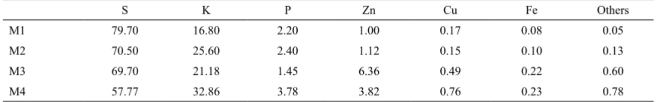
The prepared latex membranes M1, M2, M3, and M4 were characterized by X-ray difraction, SEM, thermal gravimetric analysis (TGA), DSC, X-ray luorescence analysis (EDX), water
Starting from the scanning electronic microscopy characterizations, the chemical analysis for energy dispersive spectroscopy, and phase analysis by X-ray diffraction, the
Samples were produced by different routes and characterized by scanning electron microscopy, differential thermal analysis, thermogravimetric analysis and X-ray diffraction, whereas
Figure 1 shows the X-ray diffraction patterns of the samples prepared from refluxing (R1) and sonochemical (S1) methods. All the diffraction peaks in the X-ray diffraction
