*Supported by Fundação de Amparo à Pesquisa do Estado de São Paulo (FAPESP), Brazil, grant #2016/17829-6. 1 Universidade Federal de São Paulo, Escola Paulista de Enfermagem, São Paulo, SP, Brazil.
2 Universidade Federal de São Paulo, Escola Paulista de Medicina, São Paulo, SP, Brazil.
Identification of warning signs for prevention of in-hospital
cardiorespiratory arrest*
Objective: to identify the occurrence of warning signs and changes in vital signs in individuals who experienced in-hospital cardiorespiratory arrest and correlate them with the occurrence of this event. Method: this is a retrospective, analytical and quantitative study that included 218 medical records of patients who suffered in-hospital cardiorespiratory arrest and identified warning signs and alterations in vital signs. Mean, standard deviation, median, minimum and maximum values were calculated for the continuous variables, and frequency and percentage for the categorical variables. We compared the age and occurrence of cardiorespiratory arrest with the occurrence of warning signs using the Chi-Square Test and the Mann Whitney non-parametric test (p-value < 0.05). Results: 62.1% of the patients presented signs and symptoms of shock, 44.9% of neurological alteration, 40.4% of malaise, 15.2% presented signs suggestive of acute coronary syndrome, and 25.9% presented mental confusion. In the last measurement of vital signs before cardiorespiratory arrest, the majority of patients had altered abnormal (32.6%) and severely abnormal (23.9%) heart rate, and abnormal (37.1%) and severely abnormal (27.0%) respiratory rate. Conclusion: the warning signs identified were: shock, neurological signs, malaise and acute coronary syndrome. The prevalent changes in vital signs were: heart rate, respiratory rate and O2 saturation. Patients with severely abnormal systolic blood pressure were not discharged and those with abnormal respiratory rate did not survive 6 months after cardiorespiratory arrest.
Descriptors: Emergency Nursing; Cardiopulmonary Arrest; Vital Signs; Hospital Care; Secondary Prevention; Quantitative Research.
How to cite this article
Souza BT, Lopes MCBT, Okuno MFP, Batista REA, Goís AFT, Campanharo CRV. Identification of warning signs for prevention of in-hospital cardiorespiratory arrest. Rev. Latino-Am. Enfermagem. 2019;27:e3072. [Access ___ __ ____]; Available in: ___________________. DOI: http://dx.doi.org/10.1590/1518-8345.2853.3072
year day
month URL
2019;27:e3072
DOI: 10.1590/1518-8345.2853.3072
www.eerp.usp.br/rlae
Beatriz Tessorolo Souza
1Maria Carolina Barbosa Teixeira Lopes
1Meiry Fernanda Pinto Okuno
1Introduction
The nursing team is often the first to identify clinical changes in patients. These changes can be easily detected by monitoring vital signs (VS) and careful observation of the patient’s facial expressions and neuro-emotional behavior. The identification of changes in deviant values is accompanied by an increased risk of adverse clinical events such as cardiorespiratory arrest (CRA). Early identification of abnormalities offers the opportunity for timely intervention and increased survival with better quality of life of patients(1).
CRA is characterized by sudden interruption of the heart rate, respiratory movements and immediate loss of consciousness, leading to irreversible brain damage and death if adequate measures to stabilize the patient are not taken immediately(2).
Annually, more than 200,000 adults undergo in-hospital CRA in the United States(3-4), and many of
these events could have been prevented(5-6) through
early identification of signs and initiation of appropriate therapy(5).
CRA is rarely a sudden event. It is the result of progressive deterioration of respiratory and circulatory function(7). CRA in hospitalized patients is often
preceded by signs of clinical worsening. Early detection and intervention in situations of clinical instability is an opportunity to prevent CRA in these patients and increase the safety of hospitalized patients(8).
Studies have demonstrated the relationship between abnormalities in routine measures of VS and poor outcomes, including death and in-hospital CRA(9-10).
An American study found a high prevalence of abnormal VS preceding CRA. Patients with three abnormal VS had 20% higher mortality than those without alterations, showing the direct relationship between these changes and the increase in in-hospital mortality rate(11). A
study in Japan implemented an Early Warning Score (EWS) system in which each change in a vital signal (systolic blood pressure, heart rate, respiratory rate, temperature, level of consciousness) was given a value of 0-3; the total score corresponded to the sum of these values. Scores greater than or equal to 7 corresponded to “danger zone”, that is, a greater possibility of acute deterioration. Patients in the “danger zone” received early interventions. After the implantation of the EWS, the rate of in-hospital CRA per 1000 admissions decreased from 5.21 to 2.05(12).
The measurement of VS, which is usually the responsibility of the nursing team, is a routine and extremely important hospital activity, because it
determines the health status of the individuals, the evolution of the clinical picture, and can predict clinical deterioration(1).
The first link in the chain of survival for in-hospital CRA is surveillance of patients and identification of warning signs. The literature cites changes in VS as risk factors for CRA. The performance of the nursing team in the periodic verification of VS is relevant in this scenario, to the identification of early changes that may precede CRA and other cardiovascular emergencies, thus increasing patient safety.
The objective of this study was to identify the occurrence of warning signs and changes in VS in individuals who presented in-hospital CRA and correlate the presence of warning signs and changes in VS with the occurrence of CRA.
Method
This is a retrospective, analytical and quantitative study performed at the Emergency Unit of the Hospital of São Paulo (HSP). The HSP is a large, high complexity, university hospital that offers multiprofessional health care in ambulatory, hospitalization and urgency and emergency modalities(13).
In the case of patients who died, the cause of death was investigated.
Descriptive and inferential analyses of data were carried out in the SPSS software (Statistical Package for the Social Sciences, version 11.5 for Windows). Statistical analyses included the calculation of mean, median, minimum and maximum values of the continuous variables. In the case of the categorical variables, frequencies and percentages were calculated. The QUI-Square test was used to calculate the occurrence of death with variables of interest, and, when necessary, the Fisher’s exact test or the Likelihood Ratio Test were used. Cox Regression was used to identify factors related to patient survival. A significance level of 5% (p-value < 0.05) was adopted. The Fisher’s exact test was used to compare the evolution of Cerebral Performance Category (CPC) with variables of interest (categorical). The Mann-Whitney test was used to compare the occurrence of death with variables of interest (continuous); a significance level of 5% (p-value < 0.05) was adopted. The results were presented through tables and graphs. For statistical analysis of VS, the following values were considered abnormal: heart rate (HR) ≤ 60 or ≥ 100bpm, respiratory rate (RR) ≤ 10 or > 20 rpm and systolic blood pressure (SBP) ≤ 90mmHg. A subgroup of severely abnormal VS was also considered: HR ≤ 50 or ≥ 130bpm, RR ≤ 8 or ≥ 30rpm and SBP ≤ 80 mmHg(11).
This study is part of a doctoral thesis approved by the Research Ethics Committee of the Federal University of São Paulo (protocol 0030/2011). Considering that this study is observational and that data collection was done by means of medical records, not causing any type of interference in the sector or in patient care, the study was exempt from informed consent term.
Results
The mean age of the study population was 66.8 years, with 52.3% of males and 47.7% of females. Regarding color, 71.1% declared to be white, 15.1% yellow, 10.6% black and 3.2% brown. Most were independent in activities of daily living (53.9%) and had not had a previous CRA (97.7%).
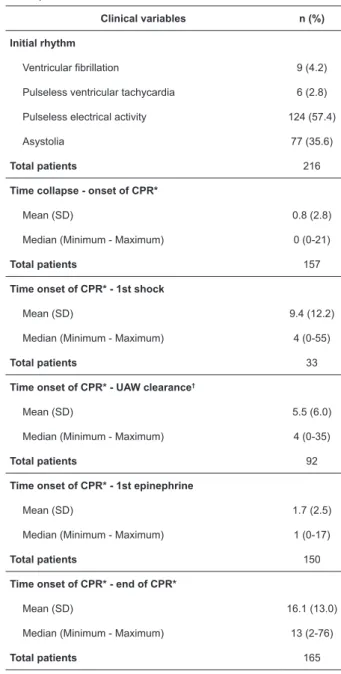
According to Table 1, the most frequent initial rhythm of CRA was pulseless electrical activity (57.4%), and the mean time between collapse and onset of CPR maneuvers was 0.8 minutes. The mean time between onset of CPR and the 1st shock was 9.4 minutes; between onset of CPR and airway clearance was 5.5 minutes; and between onset of CPR and administration
of the 1st dose of epinephrine was 1.7 minutes. The total duration of CPR was 16.1 minutes, on average.
Table 1 – Initial cardiac arrest rhythm and time intervals during care in the studied population. São Paulo, SP, Brazil, 2017
Clinical variables n (%)
Initial rhythm
Ventricular fibrillation 9 (4.2)
Pulseless ventricular tachycardia 6 (2.8)
Pulseless electrical activity 124 (57.4)
Asystolia 77 (35.6)
Total patients 216
Time collapse - onset of CPR*
Mean (SD) 0.8 (2.8)
Median (Minimum - Maximum) 0 (0-21)
Total patients 157
Time onset of CPR* - 1st shock
Mean (SD) 9.4 (12.2)
Median (Minimum - Maximum) 4 (0-55)
Total patients 33
Time onset of CPR* - UAW clearance†
Mean (SD) 5.5 (6.0)
Median (Minimum - Maximum) 4 (0-35)
Total patients 92
Time onset of CPR* - 1st epinephrine
Mean (SD) 1.7 (2.5)
Median (Minimum - Maximum) 1 (0-17)
Total patients 150
Time onset of CPR* - end of CPR*
Mean (SD) 16.1 (13.0)
Median (Minimum - Maximum) 13 (2-76)
Total patients 165
*CPR - Cardiopulmonary Resuscitation; †UAW - upper airways;
Concerning the warning signs for the occurrence of CRA (n = 198), 62.1% of the patients presented signs and symptoms of shock; 44.9% presented neurological signs; 40.4% presented malaise; and 15.2% had signs and symptoms suggestive of acute coronary syndrome.
Table 2 – Vital signs, oxygen saturation and level of consciousness in the 24 hours preceding the cardiorespiratory arrest in the studied population. São Paulo, SP, Brazil, 2017
Clinical variables n (%)
Respiratory frequency
Mean (Standard deviation) 25.5 (8.7)
Median (Minimum-Maximum) 24 (8-48)
Total patients 116
Systolic Blood Pressure
Mean (SD) 98.3 (33.2)
Median (Minimum - Maximum) 97.5 (30-200)
Total patients 180
Diastolic Blood Pressure
Mean (SD) 60 (22.2)
Median (Minimum - Maximum) 60 (20-131)
Total patients 180
Mean Blood Pressure
Mean (SD) 60.1 (35.8)
Median (Minimum - Maximum) 63.7 (0-145)
Total patients 218
Heart rate
Mean (SD) 84.7 (30.2)
Median (Minimum - Maximum) 84 (8-148)
Total patients 184
Temperature
Mean (SD) 36.3 (1.6)
Median (Minimum - Maximum) 36 (32-41.6)
Total patients 117
O2 saturation
Mean (SD) 90.4 (8.5)
Median (Minimum - Maximum) 93 (35-100)
Total patients 144
Level of consciousness n (%)
Alert 23 (20.5)
Confuse 29 (25.9)
Responds to pain 7 (6.3)
Unconscious 21 (18.8)
Sedated 24 (21.4)
Responds to verbal stimulation 8 (7.1)
Total patients 112
one year after the discharge. Regarding the reasons of death, 92 (42.6%) died from infection, 52 (24.1%) from cancer, 39 (18.1%) from cardiovascular diseases, 3 (1.4%) from trauma, 95 (44%) due to other causes.
According to Table 3, most survivors at discharge were independent in daily life activities 6 months and 1 year after the CRA, with CPC 1 or 2.
Regarding outcomes, 48.6% had return of spontaneous circulation, with 16.8% surviving the first 24 hours, 6.3% surviving at hospital discharge, 5.3% surviving six months after discharge, and 4.9% surviving
Table 3 – Neurological state after cardiorespiratory arrest in the study population. São Paulo, SP, Brazil, 2017
Clinical variables n (%)
CPC* at discharge
1 3 (27.3)
2 6 (54.5)
3 1 (9.1)
4 1 (9.1)
Total patients 11
CPC* 6 months after CRA
1 5 (55.6)
2 3 (33.3)
3 1 (11.1%)
Total patients 9
CPC* 1 year after CRA
1 5 (62.5)
2 3 (37.5)
Total patients 8
*CPC - Cerebral Performance Category
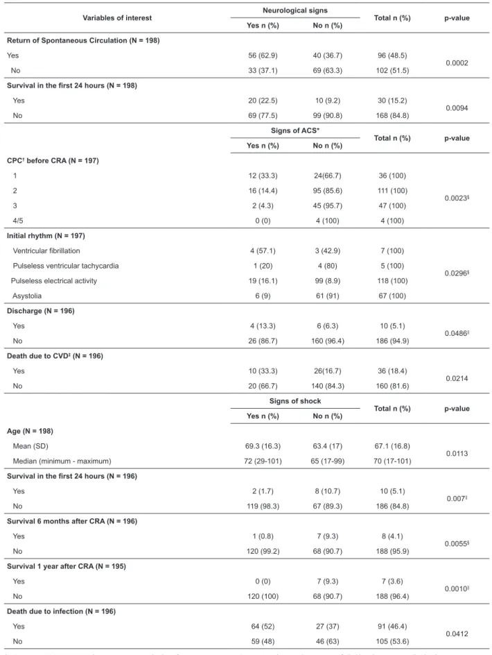
Table 4 shows the association of socio-demographic variables, neurological state before CRA, the initial CRA rhythm, and the outcomes with the warning signs presented by the patients in this study.
A higher mean age (69.3) and rate of death due to infections (52%) were observed in patients who presented signs of shock when compared to patients who did not presented such signs (63.4 years and 37%,
respectively). Patients without signs of shock had a higher percentage of survival, survival in the first 24 hours after CRA (10.7%), 6 months after CRA (9.3%) and 1 year after CRA (9.3%).
Table 4 - Association warning signs with socio-demographic variables, neurological state before cardiorespiratory arrest, initial cardiac arrest rhythm and outcomes. São Paulo, SP, Brazil, 2017
Variables of interest Neurological signs Total n (%) p-value
Yes n (%) No n (%)
Return of Spontaneous Circulation (N = 198)
Yes 56 (62.9) 40 (36.7) 96 (48.5)
0.0002
No 33 (37.1) 69 (63.3) 102 (51.5)
Survival in the first 24 hours (N = 198)
Yes 20 (22.5) 10 (9.2) 30 (15.2)
0.0094
No 69 (77.5) 99 (90.8) 168 (84.8)
Signs of ACS*
Total n (%) p-value Yes n (%) No n (%)
CPC† before CRA (N = 197)
1 12 (33.3) 24(66.7) 36 (100)
0.0023§
2 16 (14.4) 95 (85.6) 111 (100)
3 2 (4.3) 45 (95.7) 47 (100)
4/5 0 (0) 4 (100) 4 (100)
Initial rhythm (N = 197)
Ventricular fibrillation 4 (57.1) 3 (42.9) 7 (100)
0.0296§
Pulseless ventricular tachycardia 1 (20) 4 (80) 5 (100)
Pulseless electrical activity 19 (16.1) 99 (8.9) 118 (100)
Asystolia 6 (9) 61 (91) 67 (100)
Discharge (N = 196)
Yes 4 (13.3) 6 (6.3) 10 (5.1)
0.0486||
No 26 (86.7) 160 (96.4) 186 (94.9)
Death due to CVD‡ (N = 196)
Yes 10 (33.3) 26(16.7) 36 (18.4)
0.0214
No 20 (66.7) 140 (84.3) 160 (81.6)
Signs of shock
Total n (%) p-value
Yes n (%) No n (%)
Age (N = 198)
Mean (SD) 69.3 (16.3) 63.4 (17) 67.1 (16.8)
0.0113 Median (minimum - maximum) 72 (29-101) 65 (17-99) 70 (17-101)
Survival in the first 24 hours (N = 196)
Yes 2 (1.7) 8 (10.7) 10 (5.1)
0.007||
No 119 (98.3) 67 (89.3) 186 (84.8)
Survival 6 months after CRA (N = 196)
Yes 1 (0.8) 7 (9.3) 8 (4.1)
0.0055§
No 120 (99.2) 68 (90.7) 188 (95.9)
Survival 1 year after CRA (N = 195)
Yes 0 (0) 7 (9.3) 7 (3.6)
0.0010||
No 120 (100) 68 (90.7) 188 (96.4)
Death due to infection (N = 196)
Yes 64 (52) 27 (37) 91 (46.4)
0.0412
No 59 (48) 46 (63) 105 (53.6)
Regarding the last measurement of vital signs before CRA, the majority of patients had abnormal and severely abnormal heart rate (32.6% and 23.9%, respectively) and systolic blood pressure (54.4%) within the normality. Regarding respiratory rate, the majority of patients had abnormal and severely abnormal parameters (37.1% and 27.0%, respectively), as presented in Table 5.
more than 80 years(15) and in another study aimed at
determining whether early administration of epinephrine in patients with non-shockable initial CRA rhythm is associated with better neurological prognoses(16).
In this study, the mean time interval in minutes between collapse and onset of CPR (0.8’), between onset of CPR and defibrillation (9.4’), and between onset of CPR and clearance of UAW (5.5’) were higher than those observed in a prospective study performed in an ICU in Minas Gerais, which presented means of 0.7’; 7.1’; and 4.8’, respectively(18). This may be related to the fact
that this study was performed in an Emergency Service, which may imply a delay in the monitoring of patients presenting CRA, different from the intensive care setting where patients are already monitored. Another aspect that may be related is the fact that this study was retrospective, and the information of interest was taken from hospital records, with possibility of incomplete information.
The mean time interval between onset of CPR and administration of the first dose of epinephrine was 1.7 min in this study, lower than that observed in another study (2.5’). This can be attributed to the fact that the present study was carried out in a university hospital with a large contingent of professionals. Regarding the time between onset and end of CPR, this interval in the studied sample (16.1 ‘) was similar to that of another study performed in an ICU (16.3’)(18).
The monitoring of VS of patients allows the detection of changes that increase the risk of adverse clinical events such as CRA. A study conducted in the United Kingdom used the National Early Warning Score, a system based on HR, RR, blood pressure, O2 saturation,
temperature, level of consciousness, and supplemental oxygen use, ranging from 0 to 20, to detect patients at risk of CRA(10). This study found that mean HR (81 bpm),
RR (17 rpm), SBP (126 mmHg), diastolic blood pressure (70 mmHg), temperature (36.7 °C) and O2 saturation
(96%) were normal(10), differently from the findings of
the present study. Another parameter analyzed in the research was the level of consciousness, in which 91.7% of the patients were alert(10), different from the patients
in the present sample in which the majority had altered level of consciousness (79.5%), being mental confusion the most frequent state (25.9%). This difference can be explained by the fact that all patients in this study progressed to CRA.
Similar studies investigated the prevalence of abnormal vital signs and their association with the risk of cardiorespiratory arrest. Among the VS changes described in the literature associated with the risk of CRA Table 5 – Last measurement of heart rate, respiratory
rate and systolic blood pressure before cardiorespiratory arrest, classified as normal/abnormal. São Paulo, SP, Brazil, 2017
Vital signs n(%)
Heart rate (bpm*)
Normal 80 (43.5)
Abnormal 60 (32.6)
Severely abnormal 44 (23.9)
Total patients 184
Respiratory Rate (ripm†)
Normal 41 (35.3)
Abnormal 43 (37.1)
Severely abnormal 32 (27.0)
Total patients 116
Systolic Blood Pressure (mmHg‡)
Normal 98 (54.4)
Abnormal 20 (11.1)
Severely abnormal 62 (34.4)
Total patients 180
*bpm - Beats per minute; †ripm - Respiratory incursions per minute; ‡mmHg - Millimeters of Mercury;
Discussion
In the present study, some socio-demographic characteristics of the patients were similar to those reported in the literature, such as higher proportion of white skin and males. The mean age of the patients in this sample was higher than in other national studies conducted in coronary care units and intensive care units. However, international studies on hospitalization and intensive care units (ICUs) found a higher mean age than that found in the present study(14-19). The most
are abnormal HR, abnormal RR and decreased SBP(11,20).
In the study, all patients with severely abnormal SBP (≤ 80 mmHg) died. Similar results were observed in another study, where mortality increased as SBP values decreased, and patients with SBP ≤ 80 mmHg had a mortality rate above 90%(11).
In the current study, the majority of patients (37.1%) presented abnormal respiratory rate, and all patients who presented such change died 6 months after the CRA. However, a study conducted with the objective of examining the association between critical changes in VS and mortality reports that patients with RR < 10 rpm at admission presented 10% mortality(9), values lower
than those found in the present study, and in another study that revealed mortality rates ranging from 80% to 90% for RR values > 20 rpm(11).
Regarding pre-CRA CPC, the majority of patients in this sample were classified as CPC 2 (53.9%). Other national studies also presented higher percentage of patients with CPC 2 (50%(21) and 96.7%(19)). The high
value of the second study(19) may be associated with
the fact that the study only included patients who were discharged after the event.
The CPC of the patients in this study was reassessed at the moment of hospital discharge and at two other times: six months and one year after the CRA. An improvement of the neurological condition was observed over time. At hospital discharge, most patients had CPC 2, whereas in the following assessments, the percentages of CPC 1 were higher. Similar results were obtained in another national study, with progressive improvement of the neurological condition in these time intervals(21).
The association of variables in this study with warning signs presented by the patients showed that the occurrence of neurological signs was associated with higher percentages of return to spontaneous circulation. These results may be related to the state of low cerebral perfusion that can precede CRA, evidenced by lowering of consciousness level, occurrence of seizures, and changes in movement, sensitivity and speech, commonly detected by health professionals(22).
Most patients with signs of ACS presented CPC 1 and 2 before CRA, and as expected, the most frequent CRA rhythm was ventricular fibrillation. Shockable rhythms are common in the first few hours after the onset of ACS symptoms and high-quality immediate CPR and early defibrillation are associated with higher survival and better neurologic outcome in these individuals(22).
Coronary reperfusion is recommended in patients with suspected or confirmed diagnosis of ACS
post-CRA(22). The highest percentages of patients who showed
signs of ACS in this study may be related to the early and specialized care given at the studied hospital, which has a cardiologist in the emergency department and an interventional cardiology sector.
In this study, the presence of signs of shock was associated with patients with a higher mean age and higher mortality due to infectious diseases. On the other hand, patients who did not show signs of shock had a higher percentage of discharge and survival in the first 24 hours, six months after CRA and one year after CRA.
Patients who experience a CRA, in most cases, require critical and long-term care after the event and undergo invasive procedures, factors that make them vulnerable to local and systemic infections. A high incidence of sepsis is reported in the world according to literature(23-25) and the increasing number of elderly and
patients with chronic diseases are factors, among others, that may contribute to increased mortality in these situations(23). A post-CRA care plan has the potential to
avoid early mortality caused by hemodynamic instability and multiple organ failure, as well as late morbidity and mortality resulting from persistent neurological damage. Thus, such measures should be encouraged and disseminated(26).
Among the limitations of this study we can highlight the fact that secondary data were used, obtained from medical records, which sometimes have incomplete information. Furthermore, although the study site was a high-complexity university hospital, it has limited resources and this may have influenced the findings.
This study is of extreme relevance to the practice because the warning signs preceding a CRA are common and may be manifested through changes in vital signs and occurrence of signs and symptoms. The periodical monitoring, according to the needs of the patients, of vital signs and provision of full and uninterrupted care are fundamental activities of the nursing team. By doing so, the nursing team is able to identify signs and symptoms preceding cardiocirculatory collapse in a timely manner.
Conclusion
References
1. Jorge VC, Barreto M da S, Ferrer ALM, Santos EAQ, Rickli HC, Marcon SS. Nursing team and detection of indicators of worsening condition in emergency room patients. Esc Anna Nery. 2012 Oct/Dec; 16(4):767–74. doi: http://dx.doi.org/10.1590/S1414-81452012000400018
2. Souza SFM, Silva GNS. Parada cardiorrespiratória cerebral: assistência de enfermagem após a reanimação. Rev Ciênc Saúde Nova Esperança. [Internet]. 2013 Sep [Acesso 12 nov 2016]; 11(2):143-57. Disponível e m : h t t p : / / w w w. f a c e n e . c o m . b r / w p - c o n t e n t / uploads/2010/11/Parada-cardiorrespiratória-cerebral.pdf 3. Go AS, Mozaffarian D, Roger VL, Benjamin EJ, Berry JD, Blaha MJ, et al. Heart disease and stroke statistics--2014 update: a report from the American Heart Association. Circulation. 2014 Jan 21; 129(3):e28-292. doi: https:// doi.org/10.1161/CIR.0000000000000558
4. Merchant RM, Yang L, Becker LB, Berg RA, Nadkarni V, Nichol G, et al. Incidence of treated cardiac arrest in hospitalized patients in the United States. Crit Care Med. 2011 Nov; 39(11):2401–6. doi: 10.1097/ CCM.0b013e3182257459
5. Hodgetts TJ, Kenward G, Vlackonikolis I, Payne S, Castle N, Crouch R, et al. Incidence, location and reasons for avoidable in-hospital cardiac arrest in a district general hospital. Resuscitation. 2002 Aug; 54(2):115–23. doi: https://doi.org/10.1016/S0300-9572(02)00098-9
6. Galhotra S, DeVita MA, Simmons RL, Dew MA, Members of the Medical Emergency Response Improvement Team (MERIT) Committee. Mature rapid response system and potentially avoidable cardiopulmonary arrests in hospital. Qual Saf Health Care. 2007 Aug; 16(4):260–5. doi: http://dx.doi. org/10.1136/qshc.2007.022210
7. Stub D, Smith K, Bernard S, Nehme Z, Stephenson M, Bray JE, et al. Air Versus Oxygen in ST-Segment-Elevation Myocardial Infarction. Circulation. 2015 Jun 16; 131(24):2143–50. doi: https://doi.org/10.1161/ CIRCULATIONAHA.114.014494
8. Taguti P da S, Dotti AZ, Araujo KP de, Pariz PS de, Dias GF, Kauss IAM, et al. The performance of a rapid response team in the management of code yellow events at a university hospital. Rev Bras Ter Intensiva. 2013 Jun; 25(2):99–105. doi: http://dx.doi.org/10.5935/0103-507X.20130020.
9. Bleyer AJ, Vidya S, Russell GB, Jones CM, Sujata L, Daeihagh P, et al. Longitudinal analysis of one million vital signs in patients in an academic medical center.
Resuscitation. 2011 Nov; 82(11):1387–92. doi: https:// doi.org/10.1016/j.resuscitation.2011.06.033
10. Smith GB, Prytherch DR, Meredith P, Schmidt PE, Featherstone PI. The ability of the National Early Warning Score (NEWS) to discriminate patients at risk of early cardiac arrest, unanticipated intensive care unit admission, and death. Resuscitation. 2013 Apr; 84(4):465–70. doi: https://doi.org/10.1016/j. resuscitation.2012.12.016
11. Andersen LW, Kim WY, Chase M, Berg KM, Mortensen SJ, Moskowitz A, et al. The prevalence and significance of abnormal vital signs prior to in-hospital cardiac arrest. Resuscitation. 2016 Jan; 98:112–7. doi: https://doi. org/10.1016/j.resuscitation.2015.08.016
12. Nishijima I, Oyadomari S, Maedomari S, Toma R, Igei C, Kobata S, et al. Use of a modified early warning score system to reduce the rate of in-hospital cardiac arrest. J Intensive Care. 2016; 4:12. doi: https://doi. org/10.1186/s40560-016-0134-7
13. Hospital São Paulo - Atendimento Hospitalar. [Internet]. 2012 [cited Apr 14, 2018]. Available from: http://www.hospitalsaopaulo.org.br/atendimento-hospitalar
14. Nolan JP, Soar J, Smith GB, Gwinnutt C, Parrott F, Power S, et al. Incidence and outcome of in-hospital cardiac arrest in the United Kingdom National Cardiac Arrest Audit. Resuscitation. 2014 Aug; 85(8):987–92. doi: https://doi.org/10.1016/j. resuscitation.2014.04.002
15. Terman SW, Shields TA, Hume B, Silbergleit R. The influence of age and chronic medical conditions on neurological outcomes in out of hospital cardiac arrest. Resuscitation. 2015 Apr; 89:169–76. doi: https://doi. org/10.1016/j.resuscitation.2015.01.006
16. Donnino MW, Salciccioli JD, Howell MD, Cocchi MN, Giberson B, Berg K, et al. Time to administration of epinephrine and outcome after in-hospital cardiac arrest with non-shockable rhythms: retrospective analysis of large in-hospital data registry. BMJ. 2014 May 20; 348:g3028. doi: https://doi.org/10.1136/bmj.g3028 17. Morrison LJ, Schmicker RH, Weisfeldt ML, Bigham BL, Berg RA, Topjian AA, et al. Effect of gender on outcome of out of hospital cardiac arrest in the Resuscitation Outcomes Consortium. Resuscitation. 2016 Mar; 100:76–81. doi: https://doi.org/10.1016/j. resuscitation.2015.12.002
Received: May 21st 2018
Accepted: Ago 13th 2018
Copyright © 2019 Revista Latino-Americana de Enfermagem This is an Open Access article distributed under the terms of the Creative Commons (CC BY).
This license lets others distribute, remix, tweak, and build upon your work, even commercially, as long as they credit you for the original creation. This is the most accommodating of licenses
offered. Recommended for maximum dissemination and use of
licensed materials. Corresponding Author:
Maria Carolina Barbosa Teixeira Lopes E-mail: [email protected]
https://orcid.org/0000-0002-8989-4404
19. Nacer DT. Sobrevivência a parada cardiorrespiratória: avaliação da performance cerebral. Campo Grande. [Internet]. [Dissertação – Universidade Federal de Mato Grosso do Sul. 2016 [Acesso 15 mar 2017]. Disponível em: https://posgraduacao.ufms.br/portal/trabalho-arquivos/download/2421
20. Hodgetts TJ, Kenward G, Vlachonikolis IG, Payne S, Castle N. The identification of risk factors for cardiac arrest and formulation of activation criteria to alert a medical emergency team. Resuscitation. 2002 Aug; 54(2):125–31. doi: https://doi.org/10.1016/S0300-9572(02)00100-4
21. Vancini-Campanharo CR, Vancini RL, Lira CAB de, Lopes MCBT, Okuno MFP, Batista REA, et al. One-year follow-up of neurological status of patients after cardiac arrest seen at the emergency room of a teaching hospital. Einstein. 2015; 13(2):183–8. doi: 10.1590/ S1679-45082015AO3286
22. Piegas L, Timerman A, Feitosa G, Nicolau J, Mattos L, Andrade M, et al. V Diretriz da sociedade brasileira de cardiologia sobre tratamento do infarto agudo do miocárdio com supradesnível do segmento ST. Arq Bras Cardiol. [Internet]. 2015 [Acesso 14 abril 2018];105(2). Disponível em: http://www.gnresearch.org/doi/10.5935/ abc.20150107
23. Rhodes A, Evans LA, Alhazzani W, Levy MM, Antonelli M, Ferrer R, et al. Surviving Sepsis Campaign: International Guidelines for Management of Sepsis and Septic Shock: 2016. Intensive Care Med. 2017; 43:304-77. doi: 10.1007/s00134-017-4683-6
24. Tillmann B, Wunsch H. Epidemiology and Outcomes. Crit Care Clin. 2018; 34(1):15–27. doi: https://doi. org/10.1016/j.ccc.2017.08.001
25. Mayr FB, Yende S, Angus DC. Epidemiology of severe sepsis. Virulence. 2014; 5(1):4–11. doi: https:// doi.org/10.4161/viru.27372