Clara Lúcia Gonçalves Dias
Licenciada em Genética e Biotecnologia
HDL in HIV
-
1 infection: a quality perspective
through paraoxonase
-
1 activities
Dissertação para obtenção do Grau de Mestre em
Genética Molecular e Biomedicina
Orientador: Sofia de Azeredo Pereira, Professora Auxiliar
NOVA Medical School, UNL
Co-orientador: Alexandra Moita Antunes,
Investigadora Auxiliar, Instituto Superior Técnico, UL
Júri:
Presidente: Prof. Doutora Margarida Casal Ribeiro Castro Caldas Braga
Arguente: Prof. Doutora Maria Gabriela Machado de Almeida
Vogal: Prof. Doutora Sofia de Azeredo Pereira
iii HDL in HIV-1 infection: a quality perspective through paraoxonase-1 activities
Copyright Clara Lúcia Gonçalves Dias, FCT/UNL, UNL
v The current work was funded by the Portuguese Foundation for Science and Technology (FCT)
(EXPL/DTP-FTO/0204/2012)
TeachIng and olD drug nEw tricks: re-profiling An anti-HIV drug as an HDL modulator
vii The results discussed in this thesis originated:
Publications in international scientific journals:
Dias CG, Batuca JR, Marinho AT, Caixas U, Marques MM, Monteiro EC, Antunes AMM, Pereira SA. Quantification of the arylesterase activity of paraoxonase-1 in human blood. Anal Methods DOI: 10.1039/C3AY41527A.
Oral communications in national meetings:
Dias CG, Antunes AM, Pereira SA. HDL quantity and quality in HIV-infected patients.
Jornadas Intercalares das Dissertações Anuais dos Mestrados. Faculdade de Ciências e Tecnologia – Universidade Nova de Lisboa, Lisboa, Portugal, 2013.
Oral communications in international meetings:
Dias CG, Marinho AT, Antunes AMM, Caixas U, Branco T, Marques MM, Monteiro EC, Batuca JR, Pereira SA. Effect of chronic exposure to the antiretroviral drug nevirapine on Paraoxonase-1 activities in HIV-infected patients. 2nd International Conference on
Occupational & Environmental Toxicology, Porto, Portugal, 2013
Awards:
Best Oral Presentation award on the 2nd International Conference on Occupational & Environmental Toxicology, for presenting Effect of chronic exposure to the antiretroviral drug nevirapine on Paraoxonase-1 activities in HIV-infected patients, granted by the
Journals of Toxicology and Environmental Health and Taylor & Francis Group.
Participation as team member in the following grant applications:
Paraoxonase-1 (PON1) as a player in brain tumorigenesis. Application to the 3rd Annual Scholarship on Oncology sponsored by Liga Portuguesa Contra o Cancro and Pfizer Lda., led by Pereira SA, Serpa J and Antunes AMM (not funded).
ix
Acknowledgments
A realização deste trabalho teria sido impossível sem a contribuição de inúmeras pessoas a quem aqui deixo o meu sincero agradecimento.
Em primeiro lugar, gostaria de agradecer à Professora Doutora Sofia de Azeredo Pereira e à Doutora Alexandra Antunes por me terem apresentado ao grandioso mundo da investigação e pela motivação e voto de confiança que sempre depositaram em mim. À Professora Doutora Sofia de Azeredo Pereira agradeço ainda por me ter acolhido de braços abertos no Laboratório de Farmacologia, pela paciência e transmissão de ensinamentos e experiências. À Doutora Alexandra Antunes agradeço também a simpatia e disponibilidade com que sempre me recebeu.
Gostaria também de agradecer à Professora Doutora Emília Monteiro por ter permitido a minha integração no Departamento de Farmacologia e também pela disponibilidade e confiança.
Agradeço também à Doutora Joana Batuca, que muito me ajudou e sempre se disponibilizou a partilhar o seu conhecimento comigo.
Não posso deixar de agradecer ao grupo do Laboratório de Farmacologia que ao longo deste ano conseguiram animar-me/aturar-me todos os dias (mesmo quando eu não estava lá!). À Patrícia (Patrrrice!) e à Raquel (a mais linda!), pela instituição da hora da pausa! À Nádia, pela sua contagiante boa disposição e à Aline, por aparecer sempre com um lindo sorriso na cara! À Joana, pelas nossas animadas conversas e à Maria João, pela sua cultura musical e
“fosforilações”! À Inês, pelo auxílio e “faltas de ar” características dela! Por último, gostaria
também de agradecer à Professora Doutora Sílvia Conde e à Doutora Judit Morello pela simpatia, conselhos e boa disposição.
Às minhas mais que amigas e (ex-) colegas de casa, Graça e Juliana, obrigada pelo
“bulling” e pela “rainha na barriga”, respectivamente, que me ofereceram neste último ano! A
sério, obrigada mesmo! Agradeço também às minhas restantes amigas, Sara, Andreia e Marta pelas fofoquices e desabafos!
Não posso deixar de agradecer a quem sempre me deu tudo: aos meus pais, Florbela e Joaquim. Sem eles, não era a pessoa que sou hoje nem certamente teria tido esta oportunidade. Agradeço também à minha irmã, Eva, pela paciência (por vezes) necessária para me aturar. Aos meus avós, António e Maria, agradeço todo o carinho e preocupação sempre demonstrados e ainda por nunca se esquecerem de quem está longe. Ao meu cão, Bóris, fiel companheiro sempre pronto para me animar e chatear!
xi
Abstract
Cardiovascular disease is highly prevalent on human immunodeficiency virus (HIV)-infected young adults, with serious implications on the choice of the most cardiovascular friendly antiretroviral regimen and its management. Nevirapine (NVP) is an antiretroviral drug that, although associated with hepatotoxicity and skin rash, is currently recognized as a high-density lipoprotein (HDL) booster in HIV-infected patients. This HDL booster effect is even more pronounced than the current available drugs for this purpose. However, besides HDL quantity, its quality is also essential. On this regard, the present study aims to give new insights into the effect of NVP on HDL quality, namely its antioxidant potential, which can be measured through the activities of its associated enzyme, paraoxonase-1 (PON1): paraoxonase (POase), arylesterase (AREase) and lactonase (LACase) activities. The role of PON1 as a protective player against the toxicity inherited to the use of NVP was also explored. Additionally, new methods were developed for the assessment of PON1 AREase and LACase activities.
The study protocol received prior approval from the ethics committees of the hospitals and a total of 54 HIV-infected patients were included.
The methods herein developed are reliable and suitable for monitoring PON1 AREase and LACase activities in human blood.
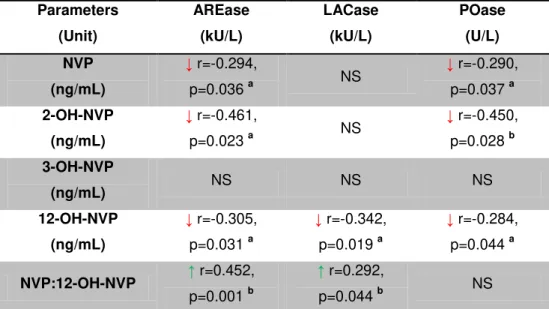
The negative effect of NVP on PON1 activities is dependent from the formation its phase-I metabolites, especially 12-OH-NVP. The 3-OH-NVP seemed to be the safest metabolite.
The current study gives new insights into the players on the mechanism of NVP-toxicity. Moreover, it provides evidence for the development of new NVP or its metabolites analogues, less toxic than NVP, which might in the future have a place in the border of HDL boosters.
Keywords: cardiovascular disease and human immunodeficiency virus infection,
xiii
Resumo
A doença cardiovascular é altamente prevalente em jovens adultos infectados pelo vírus da imunodeficiência humana (VIH), com implicações sérias na escolha e gestão da terapêutica antiretroviral combinada mais adequada para a comorbilidade cardiovascular. A nevirapina (NVP) é um antiretroviral que, apesar de estar associado a toxicidade hepática e cutânea, é actualmente reconhecido por aumentar os níveis da lipoproteína de elevada densidade (HDL). Este efeito é mais pronunciado que o conseguido com os fármacos actualmente disponíveis para esse fim. No entanto, além da sua quantidade, a qualidade da HDL é também essencial. Desta forma, este estudo tem como objectivo demonstrar o efeito da NVP na qualidade da HDL, nomeadamente no seu potencial antioxidante, que pode ser medido através das três actividades do enzima paraoxonase-1 (PON1) que circula associado à HDL: paraoxonase (POase), arilesterase (AREase) e lactonase (LACase). O papel protector do PON1 contra a toxicidade associada à toma da NVP também foi explorado. Adicionalmente, foram desenvolvidos e validados novos métodos para a monitorização das actividades AREase e LACase do PON1.
O protocolo do estudo foi previamente aprovado pela comissão de ética dos hospitais e foram incluídos um total de 54 doentes infectados pelo VIH positivo.
Os métodos desenvolvidos são válidos e adequados para a monitorização das actividades AREase e LACase do PON1 em sangue humano.
O efeito negativo da NVP nas actividades do PON1 é dependente da formação dos seus metabolitos de fase I, especialmente o 12-OH-NVP. O metabolito 3-OH-NVP foi o que menos influenciou a actividades do PON1.
Este estudo permitiu identificar novos protagonistas no mecanismo da toxicidade induzida pela NVP. Além disso, abre portas ao desenvolvimento de novos análogos da NVP ou dos seus metabolitos, que sejam menos tóxicos que a própria NVP e que possam futuramente ser usados como moduladores da HDL.
xv
Table of contents
Acknowledgments ... ix
Abstract ... xi
Resumo ... xiii
Table of contents ... xv
Index of figures ... xvii
Index of tables ... xix
Abbreviations... xxi
1. Introduction ... 1
1.1
Cardiovascular disease in human immunodeficiency virus infection: the virus and
the antiretroviral drugs ... 3
1.2
High-density lipoprotein in HIV-infection: the influence of combined antiretroviral
therapy
……….
... 6
1.3
The paraoxonase family in HIV-infection. The effect of combined antiretroviral
therapy on PON1 activities ... 7
1.4
Nevirapine two-faces: an high-density lipoprotein booster VS an hepatotoxic
drug
…………..
... 11
1.5
Objectives ... 13
2. Materials and Methods ... 17
2.1
Inclusion of patients, clinical data gathering and blood sampling ... 19
2.2
PON1 activities assessment ... 19
2.2.1
Arylesterase activity of PON1 ... 19
2.2.2
Lactonase activity of PON1... 22
2.2.3
Paraoxonase activity of PON1 ... 24
2.2.4
Blood sampling conditions definition ... 25
2.3
Effect of chronic exposure of nevirapine on PON1 activities in HIV-infected
patients
……..
... 25
2.3.1
Quantification of nevirapine and its phase-I metabolites in plasma of HIV-infected
patients
………
... 25
2.3.2
Quantification of PON1 activities ... 25
xvi
3. Results ... 27
3.1
Development and validation of PON1 activities methods ... 29
3.1.1
Arylesterase activity of PON1 ... 29
3.1.2
Lactonase activity of PON1... 30
3.2
Blood sampling conditions definition ... 31
3.2.1
Quantification of the PON1 activities ... 31
3.2.2
Evaluation of the association among the different PON1 activities in the different
conditions tested ... 32
3.3
Effect of chronic exposure of nevirapine on PON1 activities in HIV-infected
patients
……….
... 33
3.3.1
Anthropometric and clinical data of the included patients ... 33
3.3.2
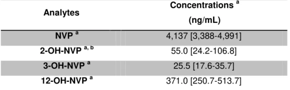
Quantification of nevirapine and its phase-I metabolites ... 33
3.3.3
Influence of anthropometric and clinical data of patients on nevirapine and its
metabolites concentrations ... 34
3.3.4
Assessment of the possible relations among the three activities of PON1 in
HIV-infected patients ... 35
3.3.5
Association between the anthropometric and clinical data from the included
patients and PON1 activities ... 36
3.3.6
Relationship between the assessed analytes and PON1 activities ... 37
4. Discussion ... 39
xvii
Index of figures
Figure 1.1 Schematic representation of the HIV-life cycle main steps and the targets of the several classes of antiretroviral drugs (Chen et al., 2007) ... 4
Figure 1.2 Biological effects of PON1 and its modulation (Macharia et al., 2012). ... 9
Figure 1.3 Nevirapine biotransformation, disposition and proposed bioactivation pathways (Marinho et al., 2013) (adapted).. ... 14
Figure 1.4 Work hypothesis. ... 15 Figure 1.5 Graphic summary of the work plan. ... 16 Figure 2.1 Method rational: hydrolysis of phenyl acetate by paraoxonase-1 and its monitoring for the assessment of the arylesterase activity. ... 20
Figure 2.2 Method rational: hydrolysis of dihydrocoumarin by paraoxonase-1 and its monitoring for the assessment of the lactonase activity... 23
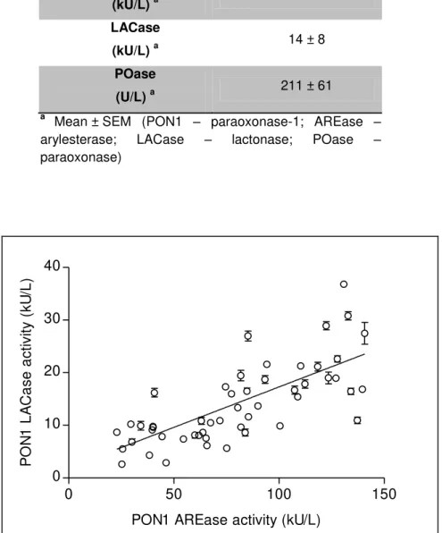
Figure 3.1 Correlation between PON1 AREase and LACase activities in serum samples of HIV-infected patients. ... 35
xix
Index of tables
Table 3.1 Accuracy, intra-assay precision and inter-assay precision of the method for the
arylesterase activity quantification... 30
Table 3.2 Accuracy, intra-assay precision and inter-assay precision of the method for the lactonase activity quantification. ... 31
Table 3.3 Blood sampling conditions and arylesterase activity. ... 32
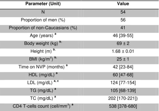
Table 3.4 Anthropometric and clinical data from the included patients. ... 33
Table 3.5 Nevirapine and its phase-I metabolites concentrations. ... 34
Table 3.6 Influence of body weight, body mass index and time on nevirapine-containing combined antiretroviral therapy on nevirapine and its phase-I metabolites. ... 34
Table 3.7 PON1 activities in serum samples. ... 35
Table 3.8 Relations between the three activities of PON1. ... 36
Table 3.9 Possible relations between the anthropometric and clinical data and PON1 activities ... 37
xxi
Abbreviations
2-OH-NVP 2-hydroxy-nevirapine
3-OH-NVP 3-hydroxy-nevirapine
4-COOH-NVP 4-carboxy-nevirapine
8-OH-NVP 8-hydroxy-nevirapine
12-OH-NVP 12-hydroxy-nevirapine
12-sulfoxi-NVP 12-sulfoxi-nevirapine
ABC Abacavir
ABCA1 ATP-binding cassette transporter
AIDS Acquired immunodeficiency syndrome
Apo A-1 Apolipoprotein A-1
AREase Arylesterase
ARV Antiretroviral
BMI Body mass index
BSA Albumin from bovine serum
CAD Coronary artery disease
cART Combined antiretroviral therapy
CD4 Cluster differentiation 4
CETP Cholesterylester transfer protein
CRI Co-receptors inhibitor
CV Variation coefficient
CVD Cardiovascular disease
CYP450 Cytochrome P450
DAD The Data Collection on Anti-HIV Drugs
DHC Dihydrocoumarin
DNA Deoxyribonucleic acid
EDTA Ethylenediaminetetra-acetic acid
EFV Efavirenz
FDA US Food and Drug Administration
FI Fusion inhibitor
GSH Glutathione
Hcy Homocysteine
HcyTL Homocysteine thiolactone
HDL High-density lipoprotein
HIV Human immunodeficiency virus
HLOQ Higher limit of quantification
HPLC High-performance liquid chromatography
INI Integrase inhibitor
IQR Interquartile range
xxii
LCAT Lecithin-cholesterol acyltransferase
LDL Low-density lipoprotein
LLOQ Lower limit of quantification
MI Myocardial infarction
Nef Negative regulatory factor
NNRTI Non-nucleoside reverse transcriptase inhibitor
NRTI Nucleoside reverse transcriptase inhibitor
NtRTI Nucleotide reverse transcriptase inhibitor
NVP Nevirapine
o-HPPA 3-(o-hydroxyphenyl) proprionic acid
ox-LDL Oxidized LDL
PI Protease inhibitor
POase Paraoxonase
PON1 Paraoxonase-1
PON2 Paraoxonase-2
PON3 Paraoxonase-3
PROCAM Prospective Cardiovascular Münster study
QC1 Quality control 1
QC2 Quality control 2
RCT Reverse cholesterol transport
SEM Standard error of the mean
TC Total cholesterol
TG Triglycerides
UGT UDP-glucoronosyltranferase
UV Ultraviolet
1
3
1.1
Cardiovascular disease in human immunodeficiency virus
infection: the virus and the antiretroviral drugs
Nearly 34 million people are currently living with the human immunodeficiency virus (HIV)-infection, the cause of acquired immunodeficiency syndrome (AIDS) (WHO, 2013). HIV-infection is a chronic condition characterized by persistent infection and inflammation. The virus infects and depletes cluster of differentiation 4 (CD4) lymphocyte cells, resulting in immunodeficiency and on a slowly progressive disease (Shor-Posner et al., 1993; Riddler et al., 2003) that, if left untreated, has a high rate of
morbidity and mortality (Tohyama et al., 2009).
Though HIV-infection continues to spread, the survival of these patients is considerably extended by the combined antiretroviral therapy (cART). As so, the HIV population is getting older and it is likely that long-term consequences of both HIV-treatment and infection will become increasingly common (Parra et al., 2010a). The persistent infection/inflammation, residual viremia, compromised immune
system, co-infections (e.g. tuberculosis and hepatitis), polimedication and drug-drug interactions,
potentially along with antiretroviral (ARV)-toxicity, could place these patients at a particular increased risk of developing several complications, many of which are commonly associated with ageing. For instance, liver disease is often present in these patients, partly due to the high rates of chronic viral hepatitis and alcohol misuse, as well as long-term exposure to potentially hepatotoxic ARVs (Deeks and Phillips, 2009). Compelling data suggest that other diseases are more prevalent in HIV-infected patients than in age-matched uninfected people, such as cancer (Kirk et al., 2007) and bone disease
(Arnsten et al., 2007). Also, neurological disease persists or even progresses during long-term
treatment (McCutchan et al., 2007). Currently, cardiovascular disease (CVD) is turning into a
preponderant condition in HIV-infected patients, as it has become a leading cause of morbidity and mortality in these patients (Triant et al., 2007; Hsue et al., 2008; Knudsen et al., 2012).
With the implementation of cART in 1996, HIV-infection has changed from a lethal to a chronic disease in properly medicated patients (Deeks, 2009). The number of people receiving cART has tripled in the last five years and has reached in 2012 to 9.7 million in low- and middle-income countries. That total represents 65% of the global target of 15 million people set for 2015 (WHO, 2013).
4 Figure 1.1 Schematic representation of the HIV-life cycle main steps and the targets of the several classes of antiretroviral drugs (Chen et al., 2007). Mechanism of action, through the different phases of HIV-life cycle, of the different antiretroviral drugs classes: nucleoside reverse transcriptase inhibitors (NRTIs), nucleotide reverse transcriptase inhibitors (NtRTIs), non-nucleoside reverse transcriptase inhibitors (NNRTIs), protease inhibitors (PI), fusion inhibitors (FI), co-receptors inhibitors (CRIs) and integrase inhibitors (INIs) (De Clercq, 2009).
The ARV drugs are a very dynamic therapeutic group since, drug availability, drug combination and also the criteria for the beginning and managing of cART has widely varied throughout the last two decades. Currently, the national guidelines state that the first-line cART should include a first generation NNRTI, nevirapine (NVP) or efavirenz (EFV) (DGS, 2012). Successful cART is associated with dramatic decreases in AIDS-defining conditions (Thompson et al., 2010) and an increase in
5 Increased evidence supports the association between CVD and cART in HIV-infected patients. The
Data Collection on Anti-HIV Drugs (DAD) study showed that overall, cART was associated with a 26%
relative increase in the rate of myocardial infarction (MI), per year of exposure during the first four to
six years of use (The DAD Study Group, 2003). In HIV-infected patients, lipid abnormalities are very common (Grunfeld et al., 1992), which as consequence, can lead to further development of CVD.
Before treatment, HIV-infection results in substantial decreases in serum total cholesterol (TC), high-density lipoprotein (HDL), low-density lipoprotein (LDL) levels (Grunfeld et al., 1992; Riddler et al., 2003), and an increase in triglycerides (TG) levels (Fernández-Miranda et al., 1998). Though not
always is possible to see hypertriglyceridemia in early stages of the infection (Grunfeld et al., 1992;
Zangerle et al., 1994), low levels of HDL are present in all its stages (Grunfeld et al., 1992; Zangerle et al., 1994; Fernández-Miranda et al., 1998). The Multicenter AIDS Cohort Study (MACS) is one of the
best reports that show that the reduction of HDL and LDL levels is associated with the infection (Riddler et al., 2003), as it is the only study that has access to the patient’s lipidic profile before the
infection, post-infection without cART and after cART initiation. The first two stages were characterized by low levels of TC (-30 mg/dL), LDL (-22 mg/dL) and HDL (-12 mg/dL). Subsequent cART initiation was associated with increases in TG, TC and LDL but little change in HDL levels (Riddler et al., 2003).
Both controlled and uncontrolled HIV-infection are associated with the risk of developing CVD. In treated HIV-infected patients, several metabolic complications have been associated with cART (Grinspoon and Carr, 2005), including dyslipidemia, insulin resistance and overt diabetes mellitus, all well-known risk factors for the development of CVD (Carr et al., 1999; Vergis et al., 2001; The DAD
Study Group, 2003).
The introduction of cART has improved both morbidity and mortality rates of HIV-infected patients (Palella Jr et al., 1998). Hence, it was expected that the successful control of HIV-infection would also
reduce the risk of coronary artery disease (CAD) associated with the condition. However, the reported data suggests the opposite: despite treatment or possibly because of it, HIV-infection is associated with an increased risk of development of atherosclerosis (Hsue et al., 2004) and at least a threefold of
the risk of CAD (Hsue et al., 2004; de Saint Martin et al., 2006). Moreover, both MI (Guaraldi et al.,
2010) and asymptomatic ischaemic disease (Calza et al., 2005) are also common conditions in
HIV-infected patients. As so, cardiovascular complications are rapidly becoming one of the prevalent causes of morbidity and mortality in HIV-infected patients (Varriale et al., 2004), and though the
relative contribution of HIV-infection itself and adverse effects of cART is not quite clear, two consistent trends have emerged. Firstly, the risk of CVD is higher in untreated than treated HIV infection, probably because inflammation is increased on the first case (Kuller et al., 2008). Secondly,
some ARVs have direct effects that can potentiate the development of CVD. For instance, prolonged exposure to PIs is associated with hyperlipidaemia, insulin resistance and a higher rate of CVD events (The DAD Study Group, 2007). Moreover, abacavir (ABC), a commonly used nucleoside reverse transcriptase inhibitor (NRTI), seems to increase the risk of heart disease (Sabin et al., 2008), perhaps
6 Nevertheless, HIV-infection per se is connected with dyslipidaemia and, particularly in advanced
states, it has been shown to be an independent risk factor for adverse lipid parameters (El-Sadr et al.,
2005; Batuca et al., 2012).
1.2
High-density lipoprotein in HIV-infection: the influence of combined
antiretroviral therapy
Among the several factors influencing CVD, HDL is the most powerful independent predictor. The HDL protective effects have first been attributed to the ability to promote cellular cholesterol efflux from peripheral cells to the liver for excretion, a process known as reverse cholesterol transport (RCT) (Précourt et al., 2011), which is essential for preventing foam cell formation and consequent
atherosclerosis (Tohyama et al., 2009). HDL is also involved in the inhibition of oxidation,
inflammation, activation of the endothelium and coagulation or platelet aggregation (van Leuven et al.,
2007).
Additionally, HDL has also several other features that able it to be used for the assessment of the risk for developing CVD. For instance, on a methodological view, HDL can be an effective biomarker due to its easy quantification. Moreover, from an epidemiologic perspective, this lipoprotein has also been shown to be crucial for the development of many cardiovascular events in the non-HIV population. Large prospective studies such as the Framingham Heart study, the Prospective Cardiovascular Münster (PROCAM) study, the Helsinki Heart study and the Lipid Research Clinics Prevalence Mortality study have shown that the risk of CAD is reduced by 2-5% for every 0.025
mmol/L increase in plasma HDL levels (Abbott et al., 1988; Wilson et al., 1988; Frick et al., 1990;
Jacobs et al., 1990; Assmann and Schulte, 1992; Assmann, 2001; Clotet et al., 2003). From a
pharmacological point of view, till this time, few drugs have the capacity to increase HDL levels, all of them showing practically no clinical benefits, as the increase in HDL concentration is really low (Birjmohun et al., 2005; Filippatos and Elisaf, 2013). All the attempts to develop new drugs with HDL
boosting effect have failed. Intensive efforts are in progress to develop new drugs that not only increases HDL quantity but also its quality (Tall et al., 2007; Filippatos and Elisaf, 2013).
In HIV-patients, HDL metabolism is affected (Shor-Posner et al., 1993; The DAD Study Group,
2007). However, in contrast to the atherogenic lipoprotein profile observed in patients treated with other HIV drugs such as PIs (Riddler et al., 2003), several studies have demonstrated that treatment
with NNRTIs such as NVP and EFV, increase plasma levels of HDL cholesterol by up 49% (van der Valk et al., 2001; Clotet et al., 2003; Tebas et al., 2004; van Leth et al., 2004; Fisac et al., 2005; Young et al., 2005; Pereira et al., 2006), through an unknown mechanism. Moreover, NVP increases
HDL and improves TC/HDL ratio apparently more than does EFV (van Leth et al., 2004; Fisac et al.,
7 Nowadays, the information given by HDL quantity has to be complemented with quality assessment (Tall et al., 2007; Filippatos and Elisaf, 2013). In this context, the enzyme paraoxonase-1
(PON1) has been implicated to play a pivotal role in the antioxidant protective functions of HDL, as it is found closely bound to this lipoprotein particle (Lusis, 2000; Gaidukov et al., 2006).
1.3
The paraoxonase family in HIV-infection. The effect of combined
antiretroviral therapy on PON1 activities
The PON enzyme family comprises three members, namely PON1, PON2 and PON3, that exhibits antioxidative properties mainly in the blood circulation. Their corresponding genes appears to have arisen from a duplication event of a common evolutionary ancestral, as they share considerable structural homology and are located adjacent to each other on chromosome 7q21-22 (Primo-Parmo et al., 1996).
The three PON enzymes have different cell and tissues distributions, as well as different regulatory mechanisms. In the human body, PON1 and PON3 are found associated to HDL in circulation (Furlong, 2008). Regarding PON2, it is an intracellular enzyme, which is not detectable in serum, but is expressed in many tissues including brain, liver, kidney and testis (Ng et al., 2001).
The different tissue distributions suggest distinct physiological roles for each of them, though they remain largely unknown (Draganov, 2007). Nevertheless, all three enzymes are able to reduce LDL oxidation (Aviram and Rosenblat, 2004). Moreover, PON2 is also able to reduce cellular oxidative stress and prevents apoptosis in vascular endothelial cells (Horke et al., 2007). Among the three
enzymes, PON1 is the best well known and characterized family member, and much of our understanding of PON enzymes is derived from the studies involving PON1 proteins.
In 1946, Abraham Mazur, who reported the presence of an enzyme in animal tissue able to hydrolyze organophosphate compounds (Mazur, 1946), led to the initial identification of PON1 (Aldridge, 1953a, b). PON1 is a calcium-dependent serum A-esterase enzyme, which protects LDL from oxidative modification by hydrolyzing lipid peroxides, thus exerting antioxidant and antiatherogenic effects (Mackness et al., 1993). This glycoprotein of 354 amino acids, with a molecular
mass of 43-45 kDa, is expressed in a variety of tissues (Marsillach et al., 2008), but it seems probably
that the liver is the main source of serum PON1. This organ is reported to have the highest PON1 gene expression and is also where a great part of HDL is synthesized and secreted into the circulation (Camps et al., 2009).
In 1991, Mackness and co-authors did the first approximation for the identification of the physiological role of PON1, by showing that the enzyme prevents the generation of lipoperoxides during the process of LDL oxidation (Mackness et al., 1991a). Subsequent studies reached the
8 Aviram and co-authors in an in vitro incubation of PON1 with oxidized palmitoyl arachidonoyl
phosphatidylcholine, lysophosphatidylcholine and oxidized cholesteryl arachidonate (Aviram et al.,
1999). PON1 was also shown to be able to hydrolyze hydrogen peroxide, a potentially important oxidative stress mediator in atherosclerosis (Aviram et al., 1998). The effects of PON1 overexpression
were also investigated in mice. Oda and co-authors (2002) generated a mouse model of a 5-fold increase in PON1 expression specifically in the liver. They found that PON1 was redistributed to HDL vehicles in the circulation. However, increased PON1 content in HDL did not alter HDL composition or properties, except that they were more protected from lipid peroxidation (Oda et al., 2002).
Numerous factors are known to influence PON1 status (Fig 1.2), including diet and life style habits (Deakin and James, 2004; Aviram et al., 2005; Costa et al., 2005b). Also, PON1 polymorphisms and
its association with lipid metabolism, CVD and ischemic stroke have been documented in a considerable number of studies (McElveen et al., 1986; Mackness et al., 1991b; Garin et al., 1997;
Voetsch et al., 2002). Epidemiological and molecular studies have identified that there are two major
common functional genetic polymorphisms in the coding region of the PON1 gene due to glutamine or arginine at position 192 (Q192R) and leucine or methionine at position 55 (L55M) (Adkins et al., 1993;
Humbert et al., 1993). The L55M polymorphism is located in the N-terminal side of PON1 gene, which
plays a role in the binding of PON1 to HDL. Moreover, it also affects the enzyme concentration (Adkins et al., 1993) with the M allele causing a decrease in protein stability (Leviev et al., 2001). On
the other hand, the Q192R polymorphism does not affect PON1 protein concentration (Leviev and James, 2000), although it is responsible for a striking substrate specific difference in the hydrolytic activities of the enzyme (Adkins et al., 1993; Humbert et al., 1993; Leviev et al., 2001), especially in
what regards the Q allele (Davies et al., 1996; Mutch et al., 2007). Furthermore, four additional
polymorphisms in the promoter region of the PON1 gene has also been reported, namely C-107T, A-162G, G-824A and G-907C. These polymorphisms are reported to affect the expression and thus the serum concentration of the enzyme (Leviev and James, 2000). The C-107T polymorphism has been the most important genetic determinant of PON levels (Brophy et al., 2001; Deakin et al., 2003),
influencing the gene expression, with the T allele reducing PON1 levels. This polymorphism contributes around 22-25% of variation in PON1 expression in caucasian adults (Leviev and James, 2000; Brophy et al., 2001). However, PON1 allele frequencies show great variations between different
9 Figure 1.2 Biological effects of PON1 and its modulation (Macharia et al., 2012). (PON1 - paraoxonase-1; Apo A1 – Apolipoprotein A-1; HDL – high-density lipoprotein)
The PON1 enzyme is considered as a human body endogenous free-radical scavenging system and has three main activities identified that could possibly explain its antioxidant and anti-inflammatory potential, which are paraoxonase (POase), arylesterase (AREase) and lactonase (LACase) activities.
Paraoxonase Activity
The enzyme was firstly found to have POase activity, reflecting its ability to catalyze the hydrolysis of paraoxon, an insecticide that gave rise to the family name, hence protecting against xenobiotic toxicity (Costa et al., 2005a). The POase activity is not shared by the three enzymes of the PON
family, as it is only confined to PON1.
Several reports using paraoxon as substrate demonstrated that there is a high inter-individual variability in PON1 POase activity (Humbert et al., 1993), considering part of this variability due to the
polymorphisms found for the PON1 gene. Regarding the Q192R polymorphism, the Q allele has been reported to have lower POase activity than the R allele (Humbert et al., 1993; Davies et al., 1996;
Mackness et al., 1997; Li et al., 2000). For the L55M polymorphism, the L allele is correlated with
higher POase activity and mRNA levels than the M allele (Leviev et al., 1997; Li et al., 2000).
Moreover, PON1 C-107T polymorphism also has influence on the POase activity. For instance, the homozygotes for the T allele have, on average, 33-45 % lower POase activity as adults (Leviev and James, 2000; Brophy et al., 2001) and 63 % lower as neonates (Chen et al., 2003), relatively to the C
homozygotes. Overall, genetic factors, including polymorphisms, were found to explain more than 60 % of phenotypic variance in PON1 POase activity, while demographic environmental factors accounted for only 1-6 % of changes and metabolic covariates for 4-19 % (Rainwater et al., 2009).
10 disease used the POase activity as a biomarker for the enzyme status (Pereira et al., 2009; Soyoral et al., 2011), this activity does not reflect the real physiological role of PON1. Thus, it is critical to start
looking at its remaining activities, AREase and LACase, which have been proved to be more physiological (Rosenblat et al., 2006).
Arylesterase Activity
The detoxification of lipid peroxides by PON1 is possible via its AREase activity, being phenyl acetate one of its best substrates. Due to this feature, this activity is thought to be the one which best reflects the antioxidant capacity of PON1 (Rosenblat et al., 2006). Hence, PON1 AREase activity seems to be involved on the reduction of the magnitude of LDL oxidation. It was found that components of oxidized LDL (ox-LDL) displayed the potency to strongly inactivate the activity. However, the presence of antioxidants, such as flavonoids, quercetin and glabridin during LDL oxidation, attenuated the loss of PON1 AREase activity (Aviram et al., 1999). On the other hand, the
activity declined rapidly in HDL following treatment with 3-morpholinosydnonimie, which generates peroxynitrite, a powerful oxidant (Ahmed et al., 2001). PON2 does not have this activity and although
PON3 also has it, it is very limited.
Regarding PON1 polymorphisms influence in the enzyme AREase activity, there has been some controversy. Initially, it was proposed that, in contrast with the POase activity, the measurement of the AREase activity of PON1 was not influenced by genetic polymorphisms (Eckerson et al., 1983).
Nevertheless, in 2001, Brophy and co-authors reported that the highest AREase activity was reported in individuals with the 192QQ genotype (Brophy et al., 2001). Additionally, in a more recent study by
Rainwater and co-authors (2009), the highest AREase activity was found in 192RR and 55LL individuals (Rainwater et al., 2009).
Lactonase Activity
PON1 LACase activity was the latest to be discovered and protects against homocysteine thiolactone (HcyTL) toxicity (Jakubowski, 2000). Moreover, this activity is also involved in the metabolism of certain drugs, including the activation of the aquinolone antibiotic NM394, by the hydrolysis of the unsaturated cyclic carbonate prodrug prulifloxacin (Tougou et al., 1998). The LACase
activity of PON1 has also been used in the development of locally acting glucocorticoid drugs, which undergo rapid hydrolysis and inactivation when they reach the circulation (Biggadike et al., 2000).
More recently, PON1 LACase activity has been linked to the efficacy of clopidogrel (Camps et al.,
2011), an antithrombotic drug used to prevent CAD. The prevailing notion is that the hydrolytic activity towards lactones is the native activity of PON1 – a view supported by structure-activity studies indicating that lactones are the preferred substrate of PON1 (Harel et al., 2004). Moreover, as the
enzyme play a role in anti-inflammatory and antioxidant response, many oxidized metabolites of polyunsaturated fatty acids are structurally similar to lactones (Draganov et al., 2005). The LACase
11 by all three members of the PON family. In fact, the term lactonase has been suggested as more fitting for the PON family as PON2 and PON3 lack any notable POase activity (Ng et al., 2005).
Although The PON1 natural-substrates are uncertain, HcyTL is hydrolyzed to homocysteine (Hcy) by PON1 LACase activity (Mackness et al., 1996; Draganov, 2007), and is a well-known risk factor for
the development of CVD (Jakubowski, 2000; Clarke et al., 2007). HcyTL is formed in all cell types,
resulting from an error-editing of the met-tRNA synthetase when there is excess of Hcy. The interaction of HcyTL with proteins leads to protein homocysteinylation and loss of function (Jakubowski et al., 2000). Therefore, detoxification of HcyTL is crucial, which in turn, is possible by the
LACase activity of PON1 (Domagala et al., 2006), contributing to its cardioprotective role. Regarding
the polymorphisms modulation, the highest LACase activity was detected in 192QQ and also in 55LL individuals (Brophy et al., 2001; Rainwater et al., 2009).
Concerning HIV-infection, it is likely that PON1 contributes to the beneficial effects of higher HDL levels in HIV-infected patients. However, few or no data is available about the influence of HIV-infection and cART on PON1 activities, which can further reflect the quality of HDL on these patients. Nevertheless, the majority of the studies reporting PON1 activities in HIV-infected patients concern the POase activity. Usually, the patients have lower serum PON1 POase activity and higher PON1 concentration than the general population, and the activity is inversely correlated with the concentration of ox-LDL (Parra et al., 2007). Both activity and concentration is thought to be
influenced by HIV-infection, the alterations in HDL composition and the immunological state of the patients. Furthermore, a positive association was found between serum PON1 concentration and active viral replication (Parra et al., 2007), which further suggests that PON1 could play a beneficial
role in protecting patients from HIV-infection. This activity was also found to be significantly higher in patients treated with EFV than in patients without cART (naïve) (Pereira et al., 2006). Another study
through the POase activity reported that the enzyme appeared to be a marker for metabolic syndrome in HIV-population (Bobin-Dubigeon et al., 2013). For the LACase activity, no differences were found
between healthy and HIV-infected subjects regardless of cART use (Djeghader et al., 2012).
1.4
Nevirapine two-faces: an high-density lipoprotein booster VS an
hepatotoxic drug
Several studies reported that NVP raises HDL levels, resulting in an improvement of the atherogenic index of the HIV-infected patients (van der Valk et al., 2001; van Leth et al., 2004). The
drug has been associated to an HDL increase up to 49%, which represents a more pronounced effect than the obtained with the currently available HDL-raising drugs, and is thought to be due to the stimulation of the apolipoprotein A-1 (Apo A-1) production (van Leth et al., 2004; Sankatsing et al.,
12 NVP was the first NNRTI approved by the US Food and Drug Administration (FDA), in 1996, for the treatment of HIV type-1 infection, as part of cART (FDA, 1996). The drug acts by non-competitive inhibition of HIV-1 reverse transcriptase (Sweetman, 2008), and still is the most prescribed drug of its class, partly due to its low cost (Ades et al., 2000; Lockman et al., 2007). Furthermore, NVP has many
features that can make it prone for its use in several conditions. The favorable metabolic profile conferred by the drug (Ruiz et al., 2001; Clotet et al., 2003; Srivanich et al., 2010) turns it suitable for
use in patients with diabetes, dyslipidemia or metabolic syndrome comorbidities. Moreover, the low incidence of adverse drug reactions in the central nervous system (Medrano et al., 2008), in
opposition to the other first-line NNRTI EFV, also allows its use in the context of psychiatric disorders or addiction to narcotic drugs. NVP is also highly efficient on the prevention of mother-to-child transmission of the HIV-1 infection, with the drug being commonly prescribed to pregnant women and their children (Ades et al., 2000; Medrano et al., 2008; Sweetman, 2008). Several studies have shown
that NVP-based cART is capable of increasing plasma levels of HDL and that this effect is greater than the one expected from simply suppressing HIV alone. Hence, a NVP-based regimen could potentially reduce the cardiovascular risk for HIV-infected patients (van der Valk et al., 2001; van Leth et al., 2004; Fisac et al., 2005).
While NVP is being monitored safely for long-term use in patients who are able to tolerate the initial regimen, the FDA has issued a black box warning to include information on hepatotoxicity associated with long-term use, and recommends against starting female patients on NVP if their CD4+ count is >250 cells/µL and for male patients if their CD4+ cell count is >400 cells/µL (Clotet, 2008).
Hence, the use of NVP has been associated with adverse toxicity reactions such as idiosyncratic hepatotoxicity and cutaneous hypersensitivity (Taiwo, 2006; De Lazzari et al., 2008; Medrano et al.,
2008). These concerns arose following case reports of liver failure in individuals on post-exposure prophylaxis (Johnson and Baraboutis, 2000) and in asymptomatic HIV-infected patients with well-preserved immunity, administered NVP-containing first line cART (Cattelan et al., 1999; Stern et al., 2003). Clinically, most patients on NVP present with erythematous rashes often accompanied by
fever, and in some cases with internal organ involvement (Pollard et al., 1998). Nevertheless, a two
week period of low dose treatment reduces the risk of rash (Montaner et al., 2003), which is currently
being applied as guideline.
However the mechanism that underlays this drug toxicity is still uncertain, several in vitro (Antunes et al., 2008; Antunes et al., 2010a; Antunes et al., 2010b), in animal models (Shenton et al., 2003;
Chen et al., 2008) and in man (Caixas et al., 2012; Marinho et al., 2013; Meng et al., 2013; Sharma et al., 2013) approaches have suggested that the bioactivation of the phase-I NVP metabolite
12-hydroxy-Nevirapine (12-OH-NVP), to reactive electrophiles, such as 12-sulfoxi-nevirapine (12-sulfoxy-NVP) is involved (Antunes et al., 2008; Chen et al., 2008; Antunes et al., 2010a; Antunes et al., 2010b; Caixas et al., 2012; Pereira et al., 2012).
13 could be substituted for NVP in order to improve PI-initiated lipid abnormalities (Gil et al., 2004; Fisac et al., 2005).
1.5
Objectives
What is known?
1. CVD is currently the number one cause of mortality and morbidity in HIV-infected patients. 2. Low HDL levels are considered the most negative predictor of CVD.
3. NVP surely works as an HDL-modulator and its positive effect on HDL is greater than the available drugs with HDL booster properties.
4. Beyond HDL quantity, its quality is also relevant.
5. PON1 enzyme is responsible for the HDL antioxidant function.
6. PON1 is an enzyme synthesized by the liver and has three main functions: paraoxonase, arylesterase and lactonase.
7. NVP upon biotransformation gives rise to several phase-I metabolites.
8. The bioactivation of the phase-I metabolite 12-OH-NVP is a plausible mechanism underlying NVP hepatotoxicity.
What is needed to know?
1. What is the individual contribution of NVP and each one of its phase-I metabolites on PON1 activities?
2. Is PON1 a protective player against NVP toxicity?
Working hypothesis:
NVP is known to increase HDL levels (Sankatsing et al., 2007), and can be considered a good
alternative against the development of CVD or even in patients already with the condition. The current work aims at explore this HDL-booster effect provided by NVP as well as analyzing its quality, where PON1 is a major player.
However, NVP is associated with hepatotoxicity and skin rash (Yuan et al., 2011), and the
14 Figure 1.3 Nevirapine biotransformation, disposition and proposed bioactivation pathways (Marinho et al., 2013) (adapted). NVP is metabolized into its phase-I metabolites,
by several isoforms of cytochrome P450 (CYP450), which are 2-hydroxy-nevirapine (2-OH-NVP), 3-hydroxy-nevirapine (3-OH-NVP), 8-hydroxy-nevirapine (8-OH-NVP) and 12-OH-NVP. This last metabolite is further oxidized by CYP450, giving rise to 4-carboxy-nevirapine (4-COOH-NVP). The UDP-glucoronosyltranferase (UGT) also represents a major pathway of NVP elimination. The bioactivation of 12-OH-NVP by SULTs can generate 12-sulfoxi-nevirapine (12-sulfoxy-NVP), which is a reactive metabolite that can bind to proteins and deoxyribonucleic acid
(
DNA).PON1 is part of the endogenous detoxification system (Fig 1.4). On one hand the enzyme is capable of detoxifying several endogenous toxic compounds (e.g. oxidized lipids and HcyTL) that can
15 Figure 1.4 Work hypothesis. (PON1 - paraoxonase-1; HcyTL - homocysteine thiolactone; Hcy - homocysteine;; Cys – cysteine; Cys-Gly – cysteine-glycine; GSH – glutathione)
On this regard, the present study is aimed at (Fig. 1.5):
1. Develop and validate new methods for the quantification of the AREase and LACase activities, suitable for application in human blood.
2. Quantify the AREase, LACase and POase activities in HIV-infected patients under NVP-containing cART.
3. To explore the effect of chronic exposure of NVP and its phase-I metabolites on the three PON1 activities.
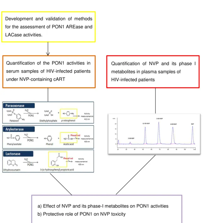
16 Development and validation of methods
for the assessment of PON1 AREase and LACase activities.
Quantification of the PON1 activities in serum samples of HIV-infected patients under NVP-containing cART
Quantification of NVP and its phase I metabolites in plasma samples of HIV-infected patients
a) Effect of NVP and its phase-I metaboliteson PON1 activities b) Protective role of PON1 on NVP toxicity
17
19
2.1
Inclusion of patients, clinical data gathering and blood sampling
A group of healthy volunteers were included for the definition in method validation of the blood sampling conditions for the PON1 activities assessment. Blood samples were collected by venipuncture, at the Centro Hospitalar de Lisboa Central, EPE.
The current work was conducted in accordance with the Declaration of Helsinki. The study protocol received prior approval from the Ethics Committees of Centro Hospitalar de Lisboa Central, EPE
(process number 32-CHLC) and Hospital Prof. Doutor Fernando Fonseca, EPE (process number CA
21/2011), and was also approved by the National Committees for Data Protection (process number 6567/2009). All patients gave their written informed consent and adherence was controlled by the clinicians. All patients were adults with documented HIV-infection, who had received continuous treatment with NVP-containing cART regimens (400 mg once daily) for more than 1 month, regardless of the past therapeutic history. Exclusion criteria were defined as being less than 18 years of age, having AIDS-defining conditions, and compliance issues. The following clinical data were gathered for each patient: age, sex, ethnicity, weight and height, time on NVP, viral load, CD4 T-cells count, and lipid parameters (e.g. HDL, LDL, TG and TC). Blood samples were collected by venipuncture.
2.2
PON1 activities assessment
2.2.1 Arylesterase activity of PON1
In order to assess PON1 AREase activity, a new method was developed and validated.
i.
Rational
Although being considered a non-physiological activity, the measurement of the POase activity of PON1 enzyme using paraoxon as substrate has been the most widely used standardized method for the assessment of its status (Pereira et al., 2009; Soyoral et al., 2011). Therefore, the study of the
AREase activity of PON1, which best reflects its antioxidative role, can potentially give additional information.
Despite the methods already available for this purpose (Beltowski et al., 2002; Naderi et al., 2011),
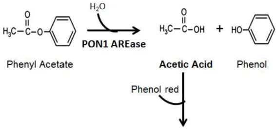
20 to the substrate hydrolysis. This reaction can be monitored spectrophotometrically, at 405 nm, by the color change of the phenol red reagent.
Figure 2.1 Method rational: hydrolysis of phenyl acetate by paraoxonase-1 and its monitoring for the assessment of the arylesterase activity. (AREase – Arylesterase; PON1 - paraoxonase-1)
ii.
Standards preparation for the calibration curve
Stock solutions
The stock solutions were prepared by adding the appropriate amount of acetic acid (M&B Laboratory Reagents) and phenol (Fluka) in freshly prepared HEPES (Roth) buffer (2.0 mM, at pH
8.0), containing CaCl2 (BDH Chemicals Ltd Pool England)(1.0 mM) and albumin from bovine serum
(BSA) (Roth) (0.005%). Ideally, these solutions should be used right after its preparation and should
be prepared in tubes covered in aluminum, since phenol is a photoreactive compound.
Standards preparation
21
iii.
Standard operating procedure
The AREase activity was obtained by measuring the extent of the hydrolysis of phenyl acetate using a spectrophotometric method adapted for a 96-well microplate. Serum or plasma samples were diluted in the proportion of 1:5 in physiological serum and 10 µL were added per well. The samples
and the previous prepared standards were incubated at 37 °C, during 10 minutes. Subsequently, 190 µL of freshly prepared HEPES buffer (2.0 mM, at pH 8.0) containing CaCl2 (1.0 mM), BSA
(0.005%), phenol red (Fluka) (106 µM) and 5.0 mM phenyl acetate (Fluka) (5.0 mM) were added to
each well. The absorbance at 405 nm was measured on a microplate reader (Biotrack II plate reader, Amersham Biosciences). The activity was directly obtained from the calibration curve and expressed
as kU/L, defined as the amount of enzyme producing 1 mM of acetic acid per minute. All samples/standards were analyzed in triplicate.
iv.
Method validation
The validation criteria were defined according to guidebooks, regarding the validation of bioanalytical methods (Shah et al., 1992; González and Herrador, 2007; EMA, 2011). For all validation purposes, each sample was analyzed in triplicate.
Linearity
Three calibration curves were prepared from different stock solutions and using six standards within the concentration range: 5.50 mM (lower limit of quantification, LLOQ) to 26.21 mM (higher limit of quantification, HLOQ). The calibration curves were constructed to explore the linearity of the method. Also, the slopes and Y-intercept (Y0) of the curves where compared in order to access reproducibility.
Lower limit of quantification
In order to validate the LLOQ, six samples with a concentration of 5.50 mM were analyzed for the accuracy and the intra-assay and the inter-assay precisions.
Accuracy
To study the accuracy of the method, six samples from the LLOQ and the HLOQ as well as 2 quality control samples (QC1 and QC2) in between the concentration range (12.58 mM and 16.78 mM) were analyzed. The accuracy was calculated according with the equation (1) and expressed in percentage (%)
Accuracy (%) = ___________________obtained concentration
22
Precision
Intra-assay precision
The intra-assay precision was evaluated by analyzing six aliquots of the LLOQ, QC1, QC2 and HLOQ. These aliquots were analyzed on the same run.
The intra-assay precision was obtained by subtracting the variation coefficients (CV) of the analyzed aliquots, according with the equation (2). The calculation of the intra-assay precision was performed assuming that its value would ideally be 100%. Samples obtained from a healthy volunteer were also analyzed.
Inter-assay precision
For the study of this parameter, the same samples described in the previous sub-section were analyzed, albeit these analyses were performed in different runs. The inter-assay precision was calculated using the equation (3). Samples obtained from a healthy volunteer were also analyzed.
2.2.2 Lactonase activity of PON1
In the subsequent sections it is described the steps used to develop and validate a method to assess PON1 LACase activity.
i.
Rational
The study of the LACase activity of PON1 also gives additional and relevant information than only using the POase activity for the assessment of PON1 status. Moreover, as the LACase activity is thought to be the primary activity of PON1, the interest is increased (Harel et al., 2004). The development of new techniques for the quantification of this activity is of crucial importance, as this is the activity known to detoxify innumerous toxic lactones, including HcyTL, a major player in CVD (Jakubowski, 2000).
Several methods have already been proposed for the assessment of the LACase activity of PON1 (Billecke et al., 2000; Gaidukov and Tawfik, 2005; Rock et al., 2008), though all of them have inherited
disadvantages in what concerns its clinical application. The method herein proposed is based on the production of 3-(o-hydroxyphenyl) proprionic acid (o-HPPA) when the lactone, dihydrocoumarin (DHC), is hydrolyzed by PON1 LACase activity (Fig. 2.2). This reaction can be monitored by the color change from red to yellow of the titration with phenol red reagent. Briefly, a molecule of DHC is
Intra-assay precision (%) = 100 - CV (2)
23 hydrolyzed into a molecule of o-HPPA. Hence, the acid is produced in stoichiometric amounts to the amount of substrate hydrolysis. This reaction can be monitored spectrophotometrically, at 405 nm, by the color change of the phenol red reagent.
Figure 2.2 Method rational: hydrolysis of dihydrocoumarin by paraoxonase-1 and its monitoring for the assessment of the lactonase activity. (LACase – lactonase; PON1 - paraoxonase-1)
ii.
Standards preparation for the calibration curve
Stock solutions
The stock solutions were prepared, by adding the appropriate amount of o-HPPA (Sigma-Aldrich)
in freshly prepared HEPES buffer (2.0 mM, pH 8.0), containing CaCl2 (1.0 mM) and BSA (0.005%).
Standards preparation
The standards were prepared with the same procedure used for the method for the AREase activity assessment.
iii.
Standards operating procedure
24 (0.005%), phenol red (106 µM) and DHC (1.0 mM) (Sigma-Aldrich) were added per well. Then, after 1
minute of incubation at room temperature, the absorbance at 405 nm was measured on a microplate reader. The activity was directly obtained from the calibration curve and expressed as kU/L, which is defined as the amount of enzyme producing 1 mM of o-HPPA per minute. All samples/standards were analyzed in triplicate.
iv.
Method validation
For the validation of this method, the same guidelines were used as for the method for the AREase activity assessment.
Linearity
The same procedure was applied, as it was for the method developed to quantify the AREase activity of PON1. The concentrations from the calibration curve ranged from 1.29 mM (LLOQ) to 10.24 (HLOQ).
Lower limit of quantification
The same procedure was applied, as it was for the method for the AREase activity assessment method, using the concentration of 1.29 mM as the LLOQ.
Accuracy
The same procedure was applied, as it was for the AREase activity quantification method. The QC1 and QC2 concentrations were 4.92 mM and 6.55 mM, respectively.
Precision
Intra-assay and inter-assay precisions
The same procedure was applied, as it was for the method to monitor the AREase activity.
2.2.3 Paraoxonase activity of PON1
The POase activity was assessed through the quantification of p-nitrophenol formation, as
previously described by Batuca et al. (Batuca et al., 2007). Briefly, paraoxon (1.0 mM) (Sigma-Aldrich)
25 monitored at 412 nm and the activity was expressed as µmol p-nitrophenol, per mL of serum, per
minute.
2.2.4 Blood sampling conditions definition
A group of five healthy volunteers were included for the definition of blood sampling conditions for the development and validation of the PON1 activities methods. Blood samples were collected from five healthy volunteers by venipuncture. Three types of samples were obtained: a) blood collected without anticoagulants, b) blood collected with lithium heparin and c) blood collected with ethylenediaminetetra-acetic acid (EDTA). Serum or plasma were aliquoted after centrifugation and then stored at -80 °C until analysis.
2.3
Effect of chronic exposure of nevirapine on PON1 activities in
HIV-infected patients
2.3.1 Quantification of nevirapine and its phase-I metabolites in plasma
of HIV-infected patients
The extraction and quantification of NVP and its phase-I metabolites was performed as previously described (Marinho et al., 2013). Briefly, the analytes were extracted from plasma previously heated at
60 ºC for 60 minutes for viral inactivation, with dichloromethane (VWR, Radnor, PA). NVP was
obtained from Cipla (Maharashtra, India) and the 2-OH, 3-OH, 8-OH, and 12-OH-NVP metabolites
were synthesized at Instituto Superior Técnico, as already described (Grozinger et al., 2000; Antunes et al., 2011).
High-performance liquid chromatography (HPLC) analyses was performed on an Agilent 1100 Series system (Agilent Technologies, Santa Clara, CA) using a reversed-phase Luna C18 (2) column
(250 mm × 4.6 mm; 5 μm; Phenomenex, Torrance, CA). The column temperature was 40 ºC, the
injection volume was 100 µL, and ultraviolet (UV) absorbance was monitored at 254 nm. For each analyte, the LLOQ of the method was 10 ng/mL.
2.3.2 Quantification of PON1 activities
26
2.4
Statistical analysis
Statistical analysis was performed using GraphPad Prism® version 5.0 (Motulsky, 2007). Data was
expressed as mean ± standard error of the mean (SEM), median [interquartile range, IQR] or frequencies (%), whenever applicable. The test of Pearson or the test of Spearman were used to
explore correlations. Comparisons among groups were performed using One-way ANOVA, Student t-test or Mann Whitney U test, whenever applicable. The F-test was used to explore differences
27
29
3.1
Development and validation of PON1 activities methods
3.1.1 Arylesterase activity of PON1
i.
Linearity
The r2 of the 3 calibration curves was 0.997 ± 0.003. There were no differences between the slopes and the elevations of the calibration curves.
ii.
Lower limit of quantification
The accuracy and precision of the LLOQ were calculated in the next sub-sections and are presented in Table 3.1.
iii.
Accuracy
The accuracy values obtained for the QC1, QC2 and HLOQ were between 90% and 103% (Table 3.1).
iv.
Precision
Intra-assay precision
The values obtained for the intra-assay precision were higher than 94% (Table 3.1).
The AREase activity of the serum sample from the healthy volunteer was 115 kU/L, and the intra-assay precision was 97%.
Inter-assay precision
30 Table 3.1 Accuracy, intra-assay precision and inter-assay precision of the method for the arylesterase activity quantification.
Standard (mM)
Accuracy (%)
Intra-assay precision (%)
Inter-assay precision (%)
LLOQ (5.50) 90 94 92
QC1(12.58) 103 96 96
QC2 (16.78) 102 95 96
HLOQ (26.21) 100 98 97
LLOQ – lower limit of quantification; QC1 – quality control 1; QC2 – quality control 2; HLOQ - higher limit of quantification.
3.1.2 Lactonase activity of PON1
i.
Linearity
The r2 of the 3 calibration curves was 0.999 ± 0.0002 and there were no differences between
the slopes and the elevations of the calibration curves.
ii.
Lower limit of quantification
The accuracy and precision of the LLOQ are presented in Table 3.2.
iii.
Accuracy
The accuracy values obtained for the QC1, QC2 and HLOQ were between 94% and 102% (Table 3.2).
iv.
Precision
Intra-assay precision
The values obtained for the intra-assay precision were higher than 93% (Table 3.2). The serum LACase activity of the healthy volunteer was 17 kU/L, and the intra-assay precision was 94%.