2017
UNIVERSIDADE DE LISBOA
FACULDADE DE CIÊNCIAS
DEPARTAMENTO DE QUÍMICA E BIOQUÍMICA
Characterization of Voltage-Gated Potassium Channels from
Dorsal Root Ganglia Neurons in Neuropathic and Inflammatory
Chronic Pain
Beatriz Szwarc dos Santos
Mestrado em Bioquímica
Especialização em Bioquímica Médica
Dissertação orientada por:
ACKNOWLEDGEMENTS
Ao meu orientador, Doutor Pedro Lima, por, no meu último ano de licenciatura, ter enaltecido o poder das ideias e de correr atrás delas. Desde então, vontade e entusiasmo são palavras de ordem, e por isso também, obrigada por dar espaço para abrir as asas e questionar acerca de tudo e de nada, até surgir uma ideia. Um grande obrigada me ter oferecido um lugar no seu laboratório, por me ter alimentado o bichinho da neurociência e por ter acompanhado todos os passos de perto, sempre com uma mão estendida pronta a ajudar. Obrigada pela descoberta da electrofisiologia e pela descoberta de um grupo de investigação tão porreiro como o nosso.
À minha co-orientadora não-oficial e amiga, Joana. Obrigada, Joana! Pela paciência, pelos ensinamentos, pelos rolares de olhos e pelas gargalhadas. Obrigada pelos brainstorms e obrigada pelos “TPCs”. Obrigada por saberes quando apertar comigo e quando deixar andar. Obrigada por tudo. Esta tese é muito graças a ti.
Ao André, a pessoa que me deu a conhecer o amplificador, as pipetas e os eléctrodos. Mais que isso, a pessoa que todos os dias trouxe um sorriso consigo para o laboratório. E que nunca deixou de dar uma palavra amiga, um abraço e uma ajuda. Obrigada por toda a ajuda, por toda a alegria e pelo bom apetxitxi. À Clara um grande, grande obrigada. Seja pelo ananás no dia do empurrão final, seja pelo sorriso animador, pronto a sair on demand. À Marisa, por sempre se disponibilizar a ajudar, por pegar numa cadeira e discutir resultados. Finalmente, mas não por fim, um grande obrigada Mãe e Pai. Obrigada por apoiarem todos os meus estudos, por apoiarem todos os meus sonhos, por me ampararem quando caio e por me desafiarem quando estagno. Obrigada pelo vosso amor e carinho. Ao meu irmão, Pedro. Obrigada por seres sempre capaz pôr tudo em perspectiva, obrigada pelos cimocerróides e camarões pistola. Obrigada pelo teu amor e carinho. Aos meus avós, à Ósa, à Bel e ao Vô, que sempre celebraram todas as minhas vitórias como deles. Obrigada pelo vosso amor e carinho.
Obrigada Zé, obrigada por nunca me deixares baixar os braços nem desanimar. Obrigada pelo teu amor e carinho.
Não poderia deixar de agradecer à Tia Maria. Obrigada pela voz doce que sempre me encorajou a seguir o coração e a ciência.
TABLE OF CONTENTS
Resumo ... xii Abstract ... xvi 1. Introduction ... 3 1.1. Perception of Pain and Nociception ... 3 1.1.1. Noxious Stimuli, Receptors and Pain Perception Ascending Pathway ... 4 1.1.2. Rapid Signal Transmission ... 7 1.1.3. Ascending Spinothalamic Pain Pathway ... 9 1.2. When Pain Becomes a Dysfunction ... 10 1.2.1. Chronic Pain Syndrome ... 10 1.2.2. Cellular Mechanisms of Neuropathic Pain ... 11 1.2.3. Cellular Mechanisms of Inflammatory Pain ... 11 1.2.4. Cellular Mechanisms of Painful Diabetic Neuropathy ... 11 1.3. Neuroexcitability ... 12 1.3.1. Resting Membrane Potential ... 12 1.3.2. VOLTAGE-GATED ION CHANNELS ... 12 1.3.3. WHY K+ CHANNELS? ... 13 1.3.6. Kv Channels Conformations ... 17 1.3.7. Activation of Kv channels ... 18 1.3.8. Inactivation of Kv channels ... 19 1.3.9. Kv currents in DRG ... 19 1.4. Animal Pain Models ... 22 1.4.1. Neuropathic Pain: Chronic Constriction Injury (CCI) ... 22 1.4.2. Inflammatory Pain: Complete Freund’s Adjuvant (CFA) ... 23 1.4.3. Neuropathic Pain: Painful diabetic Neuropathy (PDN) ... 24 1.4.4. How to Evaluate Pain in Animals ... 242. Goals ... 25
3. Methods ... 27
3.1. Animal Pain Models ... 27
3.1.1. Neuropathic Pain Model: Chronic Constriction Injury (CCI) of the Sciatic Nerve – CCI Pain Model ... 27
3.1.2. Inflammatory Pain Model: Complete Freund’s adjuvant (CFA) induced monoarthritis – CFA Pain Model ... 28 3.1.3. Diabetic Pain Model: Painful Diabetic Neuropathy (PDN) – PDN Pain Model 28 3.2. Animal Behaviour ... 29 3.2.1. Spontaneous Pain ... 29 3.2.2. Mechanical Sensitivity ... 30 3.3. DRG neurons extraction and maintenance ... 31 3.4. Electrophysiology ... 32 3.4.1. Voltage Protocols ... 32 3.4.2. Data Analysis ... 33 3.4.3. Statistical Analysis ... 35 3.5. Protein Expression ... 36 3.5.1. Western-Blot ... 36 4. Results ... 39 4.1. Animal Behaviour ... 39 4.1.1. Spontaneous Pain ... 39 4.1.1.1. CCI Rats Spontaneous Pain ... 40 4.1.1.2. CFA Rats Spontaneous Pain ... 42 4.1.2. Mechanical Sensitivity ... 43 4.1.2.1. Mechanical Sensitivity in CCI Rats ... 43 4.1.2.2. Mechanical Sensitivity IN CFA Rats ... 45
4.2. Electrophysiology ... 47 4.2.1. Whole-cell K+ currents in small diameter DRG neurons ... 47 4.2.2. Currents kinetics in small DRG neurons from naïve, CCI and CFA rats ... 49 4.2.3. Current Density ... 51 4.2.3.1. Current Density in small diameter DRG neurons from CCI rats ... 52 4.2.3.2. Current Density in small diameter DRG neurons from CFA rats ... 54 4.2.4. Voltage-Dependance of Activation: Conductance ... 55 4.2.4.1. Voltage Dependence of Activation in cci ratS ... 56 4.2.4.2. Voltage Dependence of Activation in cFA ratS ... 57 4.2.5. Voltage-Dependance of Steady-State InActivation ... 59 4.3. Western-blot ... 63 4.3.1. Kv1.3 and Kv1.4 expression in naïve and CCI from L4-L6 DRG ... 63 4.3.2. Kv1.3 and Kv1.4 expression in naïve and CCI from the sciatic nerve ... 65 5. Discussion ... 67
5.1.1. Did the Neuropathic and Inflammatory Pain Models evolve to a Chronic condition? ... 67 5.1.2. What are the functional consequences at the neuronal level of the observed hyperalgesic behaviour? ... 70 5.1.2.1. Is the voltage dependence of activation impared in Neuropathic Chronic Pain? 70 5.1.2.2. Is the Voltage-Dependence of Activation Impaired in Inflammatory Trasient Pain? 73 5.1.2.3. Is the voltage dependence of Steady-State Inactivation impared in Neuropathic Chronic Pain? ... 75
5.1.2.4. Is the voltage dependence of Steady-State Inactivation impared in Inflammatory Transient Pain? ... 77
5.1.3. Are Kv channels being recruited from DRG to sciatic nerve as consequence of nerve hyperexcitability? ... 77
Conclusions ... 79 Future Directions ... 80 References ... 81
ABBREVIATIONS
ATP Adenosine triphosphate BDNF Brain-derived Neurotrophic Factor BSA Bovine Serum Albumin Ca2+ Calcium ion CCI Chronic Constriction Injury CFA Complete Freund’s adjuvant Cm Whole-cell membrane Capacitance CNS Central Nervous System DRG Dorsal Root Ganglion DTT Dithiothreitol FBS Fetal Bovine Serum G Conductance HB Homogeneization Buffer I Current amplitude IASP International Association for the Study of Pain IB4 Isolectin 4 I-V Current-Voltage relationship J Current Density K+ Potassium ion Kv Voltage-gated Potassium Channel L Lumbar LB Lysis Buffer mRNA Messenger Ribonucleic Acid
Na+ Sodium ion Nav Voltage-gated Sodium Channel NGF Neurotrophic Growth Factor PBS Phosphate Buffered Saline PIC Protease Inhibitor Cocktail PNS Peripheral Nervous System RMP Rest Membrane Potential Rpm Rotations per minute S Sacral SDS Sodium dodecyl sulphate SEM Standard Error of the Mean SNL Spared Nerve Ligation TrkA Tropomyosin receptor kinase A TRP Transient Receptor Potential Channel TRPA Transient Receptor Potential Channel, sub-family A - Ankyrin TRPM Transient Receptor Potential Channel, sub-family M -Melastin TRPV Transient Receptor Potential Channel, sub-family V -Vanilloid TTX Tetrodotoxin VH Voltage of half activation or inactivation vFF von Frey Filament Vm Voltage pulsed current VS Slop constant τ Time constant
RESUMO
Dor crónica afeta 21% da população humana e, até à data, os tratamentos disponíveis são apenas parcialmente eficazes. Esta condição resulta, muitas vezes, em perturbação de sono, desenvolvimento de depressão e ansiedade que prejudicam a qualidade de vida dos pacientes. Uma das formas mais incapacitantes da dor crónica é a dor neuropática, resultante de lesões nos nervos sensoriais. Existem muitas outras etiologias de dor crónica como por exemplo a inflamatória (artrite reumática) e a neuropática diabética. Em dor crónica, os doentes sofrem de hiperalgesia (reação exagerada a um estímulo doloroso), alodínia (sensação de dor a um estímulo não doloroso), dor espontânea, parestesia (sensação anormal) e disestesia (parestesia desconfortável). A dor crónica é especialmente difícil de tratar muito porque não se compreende totalmente os mecanismos moleculares que a causam. Atualmente, o tratamento da dor crónica continua a ser um desafio pois a maioria dos alvos das atuais abordagens terapêuticas coexistem no sistema nervoso central e, por isso, promovem vários efeitos colaterais tais como dependência e habituação. As fibras sensoriais que são responsáveis pela transmissão de estímulos de dor (fibras C e Aδ) encontram-se num estado mais elevado de hiperexcitabilidade numa situação de dor crónica. Tal excitação é controlada por um conjunto de canais iónicos ativados por voltagem (entre outros) que produzem um grau de excitação que é, numa situação normal, proporcional à intensidade do estímulo externo. No contexto de nervo sensorial danificado, este torna-se hiperativo e produz atividade elétrica espontânea que o cérebro interpreta como sinais de dor. Tal pode se traduzir numa situação de dor crónica, onde a coordenação é interrompida, resultando em excitabilidade periférica desregulada. A velocidade de transmissão nas fibras C é mais lenta relativamente à velocidade de transmissão das fibras Aδ devido à ausência de um revestimento de mielina. Desta forma, a perceção dos estímulos transmitidos pelas fibras C é mais difusa que das fibras Aδ. As fibras sensoriais primárias ou nocicetivas são constituídas pelos neurónios do gânglio da raiz dorsal (DRG, do inglês Dorsal Root Ganglia). Os neurónios DRG de pequeno diâmetro apresentam uma atividade aumentada em pacientes com dor crónica, e por isso serão o foco desta dissertação.
Na presença de estímulos capazes de produzir dor (ou seja, cuja magnitude é superior ao limiar de ativação das fibras nervosas) os recetores nas terminações nervosas dos neurónios primários são ativados, despolarizando a membrana e produzindo um potencial
de ação. Esta despolarização propaga-se pela fibra nervosa, sendo o sinal transmitido para os neurónios secundários e sistema nervoso central onde, no córtex sensorial do cérebro, a informação a relativa à quantificação e localização da dor é integrada.
O papel dos canais de K+ no controlo do potencial de repouso da membrana, no limiar de disparo, frequência e forma do potencial de ação é crucial. Estudos indicam que a inibição dos canais de K+ induz a atividade espontânea em fibras sensoriais periféricas, cuja hiperexcitabilidade em estados de dor crónica coincidiu com a sua regulação negativa. Assim, a supressão da condutância de K+ pode representar uma condição geral de um nervo 'doloroso'. A diminuição da atividade dos canais de K+ ativados por voltagem faz parte de um mecanismo geral para a hiperneuroexcitabilidade periférica em dor crónica, e que potenciadores destes canais podem significar um melhoramento desta doença.
O objetivo do trabalho experimental foi a elucidação dos mecanismos comuns e específicos de dor crónica com origem neuropática e inflamatória em modelos de ratos. A compreensão da base mecanística do mau funcionamento de canais de K+ ativados por voltagem (Kv) em termos de localização, biofísica, e consequências para a neurotransmissão é uma potencial de novas terapias para a dor. Especificamente, foi estudada a biofísica de canais Kv em neurónios de DRG de pequeno diâmetro.
Sabendo que a dor crónica pode ter várias etiologias, este projeto focou-se na dor crónica de origem inflamatória e neuropática, baseando-se em modelos animais de dor em ratos. A dor crónica neuropática foi estudada com base no modelo Chronic Constriction
Injury (CCI) que assenta na constrição crónica do nervo isquiático do rato através de quatro nós soltos. A dor crónica inflamatória foi estudada através da indução de monoartrite no joelho do rato, pela injeção de uma substância que ativa o sistema imunitário: Complete Freund’s Adjuvant (CFA). O desenvolvimento de dor crónica foi acompanhado por testes comportamentais que incluem a medição de sensibilidade mecânica e de atividade vertical. Ao fim de 4 semanas (para CCI) e de 2 semanas (para CFA), os ratos foram sacrificados por decapitação após anestesia com pentobarbital para análise dos neurónios DRG de pequeno diâmetro por eletrofisiologia.
No modelo neuropático CCI, os ratos modelo desenvolveram hiperalgesia durante o curso da experiência mantendo-a sempre abaixo dos respetivos controlos. No modelo inflamatório CFA foi observada inicialmente uma maior sensibilidade após a indução do
modelo, evoluindo para uma recuperação total da sensibilidade no final do tempo do modelo.
Resultados de whole-cell voltage-clamp demonstram que a dor crónica altera as propriedades biofísicas das correntes dos canais de K+ ativados por voltagem. Especificamente, ambos os modelos CCI e CFA partilham um mecanismo relativo à redução da componente rápida das correntes de K+.
O modelo de dor crónica neuropática mostra ainda um mecanismo específico subjacente de aumento da componente lenta das correntes de K+. Os ensaios proteicos revelaram uma expressão aumentada de Kv1.3 (uma isoforma de canais de K+ ativados por voltagem), a qual aparenta ser responsável por este aumento da componente lenta.
O modelo de dor crónica inflamatória mostra uma alteração específica relativa a uma despolarização do perfil de ativação da componente rápida das correntes de K+, sugerindo uma alteração de sensibilidade à voltagem para valores mais despolarizados.
No seu conjunto, os dados apontam para a identificação de componentes da corrente de K+ que estão diferencialmente expressas nos modelos de dor crónica. Esta informação em combinação com os ensaios de expressão proteica sugerem a identificação molecular das componentes da corrente de K+.
Os resultados conseguidos durante esta tese de mestrado trouxeram novos conhecimentos para a validação de alvos dor moleculares terapêuticos e para a identificação de novas abordagens experimentais / terapêuticas para dor neuropática e inflamatória, bem como o seu mecanismo de ação, que continuará a ser explorado pelo laboratório.
Palavras-chave: Dor crónica; canais de potássio dependentes de voltagem; gânglios da raiz dorsal.
ABSTRACT
Chronic pain affects 21% of the human population and, to date, standard treatments are only partially effective. This condition often results in poor sleep, depression and anxiety, between other, which highly impair patients’ quality of life. One of the most disabling forms of chronic pain is called neuropathic pain, which results from injuries to sensory nerves. Pain or discomfort is felt in response to non-painful stimuli. Chronic pain is difficult to treat as currently we do not fully understand the associated molecular mechanisms.
Stimulating a nerve above the threshold of activation causes it to produce action potentials. Neurotransmission follows action potentials which are generated by ions moving into and out of the neuronal-membrane through voltage-gated ionic channel. Voltage-gated sodium and potassium channels (Nav and Kv, respectively) are critical to the generation of these action potentials that convey nociceptor signals to synapses in the dorsal horn. In this context, nociceptive fibers that are responsible for the transmission of pain stimuli are in a higher state of excitability with pain. Such excitation is controlled by an intricate set of ion channels that are coordinated to produce a degree of excitation that, in a naïve situation, is proportional to the strength of the external stimulation. Once a sensory nerve is damaged or injured it becomes hyperactive and produces spontaneous electrical activity that the brain interprets as pain signals. In chronic pain coordination by ion channels is disrupted, resulting in deregulated peripheral excitability. Of those sensory fibers, small diameter dorsal root ganglia (DRG) neurons are found to have an augmented activity in chronic pain patients. There is strong evidence that modulation of specific Na+, Calcium (Ca2+) or K+ channel sub-types can be effective in treating chronic pain, although there is still
no modulator on the clinical setting specifically targeting those channels expressed in pain-sensing neurons from DRG. Moreover, it is not fully understood how chronic pain affects the ion channel dynamics in a way that generates this hyperactivity.
The goal of the present work was to elucidate shared and specific mechanisms of neuropathic and inflammatory chronic pain involving K+ channels, using pain rat models. Understanding the mechanistic basis of ion channel malfunction in terms of trafficking, localization, biophysics, and consequences for neurotransmission is a potential route to new pain therapies. Specifically, we studied the biophysics of voltage activated K+ currents and
underlying Kv channels in small diameter DRG neurons and determine routes of pharmacological modulation of these currents as a strategy for new analgesic drug design.
Knowing that chronic pain can have several etiologies, this project focused on chronic pain of neuropathic and inflammatory origin, based on rat models of pain. Chronic neuropathic pain was studied based on Chronic Constriction Injury (CCI) , which is based on the chronic constriction of the rat sciatic nerve through four loose ligation Chronic inflammatory pain was studied through the induction of monoarthritis in the knee of the rat by the injection of a substance that activates the immune system: Complete Freund's
Adjuvant (CFA).
The pain models were followed with behavioral tests that included the measurement of mechanical sensitivity and vertical activity. After 4 weeks (for the CCI) and 2 weeks (for the CFA model), rats were sacrificed and their DRG removed aiming the electrophysiological analysis from small diameter DRG neurons.
In the neuropathic CCI model, the model rats developed hyperalgesia during the experiment, keeping their threshold for pain below the respective controls. In the inflammatory CFA model, a greater sensitivity was observed by the third day after model induction, progressing to a full recovery of sensitivity at the end of the model time.
Whole-cell voltage-clamp results demonstrated that chronic pain alters the biophysical properties of voltage-activated K+ channel currents. Importantly, both CCI and CFA models share a mechanism, a reduction of the fast component of K+ current (Ifast
, an A-type current).
The neurons from the CCI pain model also showed a specific current output: an increase of current density of the slow K+ current component (I slow). Western-blot protein assays revealed over expression of Kv1.3, which may be responsible for this increase in Islow. In terms of channel gating phenomena, neurons from CFA rats showed a specific biophysical alteration – in relation to neurons from naïve rats, the voltage dependence of activation showed more depolarized curves. Finally, in terms of the voltage dependence of steady-state inactivation, neurons from both pain rat models exhibited more depolarized voltage profiles. Such impairment of the inactivation is concordant with the alleged increase excitability in damaged neurons.
Taken together, the data reveals mechanisms underlying increases in neuroexcitability which lead to neuronal hyperactivity and hyperalgesia. The results also points to the identification of specific and shared phenomena occurring in K+ current components that are differentially expressed in the pain models. This information, together
with the protein expression assays, suggest the molecular identification of the K+
current-component.
The results obtained during this master's thesis bring new knowledge about the therapeutic molecular pain targets and for the identification of new experimental / therapeutic approaches for neuropathic chronic pain and inflammatory pain, as well as its underlying mechanisms, which will continue to be explored by the host laboratory.
Keywords: Chronic Pain; voltage-gated potassium channels; dorsal root ganglia.
2
3
1.
INTRODUCTION
1.1.
Perception of Pain and Nociception
Pain is the most frequent cause of medical consultation, accounting for 25-50% of all consultations of General Practice. Of these consultations, 20% are motivated by chronic pain (Finnerup et al. 2007), which may affect 22% of the population that uses the primary health care (Lepine & Briley 2004). Pain is an unpleasant feeling of utter most importance for the body’s defense system, as it provides a rapid warning to the nervous system to initiate a motor response to minimize potential physical harm. Pain is defined by the International Association for the Study of Pain (IASP) as "an unpleasant sensory and emotional experience associated with actual or potential tissue damage, or described in terms of such damage" (International Association for the Study of Pain). This setting recognizes pain as a subjective sensation, even in the absence of a demonstrable tissue injury, sustaining a well-known aphorism: "Pain is when the patient says it hurts" (McCaffery & Beebe 1989). Lack of the ability to experience pain as, for instance, in the rare congenital insensitivity to pain with anhidrosis (Axelrod & Hilz 2003) is very dangerous. This can cause very serious health problems such as self-mutilation, auto-amputation, and corneal scarring.
Nociception refers to the molecular mechanisms that underlie the detection of pain-producing stimuli by activation of specialized sensory receptors (nocipetors). Nociception is defined by IASP (International Association for the Study of Pain) as "nerve noxious stimuli
process of coding", which are in turn defined as "harmful stimuli or which threaten to damage the normal tissues". These stimuli are responsible for the activation of nociceptors, defined by the same organization as receptors of “high threshold, capable of transducing and encode noxious stimuli." Thus, according to the IASP, nociceptive pain can be defined as an "effective pain arising from a damage or damage of a threat, a non-nervous tissue, due to the activation of nociceptors". Thus, stimuli perceived as painful are those likely to cause tissue damage. The nociceptive pain is usually transient, disappearing with tissue repair. In some conditions, excitation of pain fibers becomes greater as the pain stimulus continues, leading to a condition called hyperalgesia, which stands for an increased sensitivity to noxious stimuli (Hart 1988).
In short, nociception provides information about tissue damage, pain is the unpleasant emotional experience that usually accompanies nociception (Julius 2001).
4
1.1.1. NOXIOUS STIMULI, RECEPTORS AND PAIN PERCEPTION
ASCENDING PATHWAY
Nearly a century ago, Sherrington proposed the existence of the nociceptor, a primary sensory neuron that is activated by stimuli capable of causing tissue damage and is able to provide information about that stimuli (Sherrington 1906). Nociceptors do not fire spontaneously at rest (Purves D, Augustine GJ, Fitzpatrick D 2001). Their electrical action potential is triggered by transduction, which occurs when a noxious stimulus of sufficient strength depolarizes the nociceptor membrane. The specific receptive properties of nociceptors are determined by their expression of transducing ion-channel receptors (Fein 2012). These ion channels are nonselective potassium or sodium channels gated by temperature, chemical stimuli, or mechanical shearing forces rather than by voltage. Activation of the channels by an appropriate stimulus leads to an inward current that depolarizes the receptor membrane (Julius & Basbaum 2001). If this depolarizing current is sufficient to activate voltage-gated sodium channels, further depolarization of the membrane will occur, and a burst of action potentials will be initiated. The duration and frequency of this burst are primarily determined by the duration and intensity of the noxious stimulus. Many, but not all, of these transducing receptors have been identified (Julius & Basbaum 2001).
Potentially damaging mechanical, thermal, and chemical stimuli, our noxious stimuli, are detected by nociceptors (found in different concentration the skin, on internal surfaces such as the periosteum, joint surfaces, and in some internal organs). Nociceptors are unspecialized free nerve endings that have their cell bodies outside the spinal column in the dorsal root ganglia (Purves D, Augustine GJ, Fitzpatrick D 2001). The dorsal root ganglia (DRG) is the segmental sensory ganglia of the spinal cord that contains the first-order neurons of the dorsal column and spinothalamic pathways (Julius & Basbaum 2001).
Chemical substances that modulate the transmission of painful stimuli are released into the extracellular tissue when tissue damage occurs. They activate the pain receptors by irritating nerve endings. These chemical mediators include histamine, substance P, bradykinin, acetylcholine, leukotrienes, and prostaglandins. These mediators can produce other reactions at the site of injury, such as vasoconstriction, vasodilatation, or altered capillary permeability. For example, prostaglandins induce inflammation and potentiate
5 other inflammatory mediators (Matsuka et al. 2001; Waxman & Zamponi 2014; Helms & Barone 2008). In this context, pain has a protective role as it motivates a response to damaging situations to protect a damaged body part while it heals and to avoid similar experiences in the future through the formation of memory (Holden & Winlow 1984). In this context, specificity is not imperative (Schaible 2007). Most high-threshold receptors respond to a variety of thermal, chemical and mechanical stimuli and are defined as polymodal nociceptors.
Recently, cell ablation studies using the Cre-loxP system (Sauer 1987) have demonstrated that distinct sensory subpopulations underlie distinct pain modalities, distinguishing mechanical and thermal pain (Abrahamsen et al. 2008; Mishra et al. 2010).
Nociceptors can be distinguished according to their differential expression of channels that confer sensitivity to heat (TRPV1), cold (TRPM8), acidic milieu (ASICs), and a host of chemical irritants (TRPA1), see Figure 1 below (Julius & Basbaum 2001).
Nociceptors have a certain threshold of activation (minimum intensity of stimulation) they require to transduce a given signal. If the stimuli is strong enough to reach the threshold, the resulting depolarization activates voltage-gated calcium (Ca2+) and sodium (Na+) channels present at the terminals of nerve fibers to initiate an action potential. This is conducted along the axon of the neuron into the spinal cord, following an afferent pathway from periphery into the central nervous system, as represented in Figure 2. (Purves D, Augustine GJ, Fitzpatrick D 2001). Voltage-gated sodium and potassium channels (Nav
Figure 1 Nociceptor Diversity There are a variety of
nociceptor subtypes that express unique repertoires of transduction molecules and detect one or more stimulus modalities. For example, heat-sensitive afferents express TRPV1 and possibly other, as yet unidentified heat sensors; the majority of cold-sensitive afferents express TRPM8, whereas a small subset likely express an unidentified cold sensor. Polymodal nociceptors also express chemoreceptors (e.g. TRPA1) and one or more as yet unidentified mechanotransduction channels. These fibers also express a host of sodium channels (such as NaV 1.8 and 1.9) and potassium channels (such as TRAAK and TREK-1) that modulate nociceptor excitability and/or contribute to action potential propagation. Reproduced directly from (Basbaum et al. 2009)
6 and Kv, respectively) are critical to the generation of these action potentials that convey nociceptor signals to synapses in the dorsal horn. Figure 2 The ascending spinothalamic pathway. The anterolateral pathway conveys nerve impulses for pain, cold, warmth, itch, and tickle from the limbs, trunk, neck, and posterior head to the cerebral cortex.
7
1.1.2. RAPID SIGNAL TRANSMISSION
Neurons are the cellular units of the nervous system which are highly differentiated while interconnected and bioelectrically driven (Margrie & Urban 2007). There are mainly three types of neurons: afferent, interneurons and efferent. The first are responsive neurons that transduce external stimuli. The interneurons transmit the signal from the afferent neurons to the central nervous system, and are the link between afferent and efferent neurons. Finally, there are efferent neurons whose function is to transmit the signal from the central nervous system to the effector organ (Ludwig & Pittman 2003).Communication between neurons is largely aided by the existence of clusters – nerve cords – of axons linked together. This way, the cells are in high proximity of each other, which enables faster and efficient communication: this is denominated centralization (Margrie & Urban 2007). In this context, DRG are clusters of cell bodies of neurons that give rise to the first-order neurons that innervate regions of the body, which aids fast communication.
The nerve fibers within a nerve include both afferent nerves and efferent (motor and autonomic) nerves (von Kitzing et al. 1994). The afferent nerve fibers innervate and communicate with the dorsal side of the spinal cord i.e., the dorsal horn and thus responsible for conducting information into the spinal cord (ascending pathway into the brain – see Figure 1); the efferent nerve fibers innervate and communicate with the ventral side of the spinal cord and are responsible for the descending pathway of the brain and so conduct the information out of the spinal cord (Nashmi & Fehlings 2001). The speed at which an individual nerve fiber conducts action potentials is related to the diameter of the fiber and on the existence of a myelin sheath (Campbell & Meyer 2006). The myelin sheath has a great amount of cytoplasm compacted which accelerates the propagation of the signal through saltatory conduction, without the enlargement of the axon. In the areas of the axon unmyelinated, the membrane is relatively thin, so the attraction force between cations outside the membrane and the anions inside the membrane is very high, high enough to overpower the force of repulsion between same charge ions in each side of the membrane (Kendall 1999). By augmenting the membrane thickness through the myelin sheath there is a decrease in the compaction of charges in the myelinated areas which facilitates the membrane depolarization and the signal propagation (Campbell & LaMotte 1983).
8
These fibers can be categorized into three main groups as seen below on Figure 3a: Aβ-, Aδ- and C-fibers (Fein 2012). Cell bodies with the largest diameters give rise to myelinated, rapidly conducting Aβ-primary sensory fibers. Most, but not all (Djouhri et al. 1998), Aβ fibers detect innocuous stimuli applied to skin, muscle and joints and thus do not contribute to pain – prioperception (see Figure 3) (Julius 2001). The other two, the small- and medium-diameter fibers and thus cell bodies, are the fibers that contribute to nociception (Fein 2012; Djouhri & Lawson 2004). There are many differences between the two types of fibers, but the one that stands out the most is the presence/absence of a myelin sheath. As stated before, the myelin sheath is the main protagonist in action potential conduction velocity. C-fibers are slowly conducting fibers since they are unmyelinated and therefore responsible for a delayed, more diffuse, dull pain evoked by noxious stimuli while Aδ-fibers are thinly myelinated, relatively more rapidly conducting, and mediates a rapid, acute, sharp pain (Figure 3) (Djouhri & Lawson 2004). Figure 3 Different nociceptive fibers and characteristics. Different nociceptors detect different types of pain. a.
Peripheral nerves include medium-diameter (Aδ) and large-diameter (Aβ) myelinated afferent fibers, as well as small-diameter unmyelinated afferent fibers (C). b. Most nociceptors are either Aδ or C fibers, and their different conduction velocities account for the first (fast) and second (slow) pain responses to injury. Adapted from Fields, 1987.
9
1.1.3. ASCENDING SPINOTHALAMIC PAIN PATHWAY
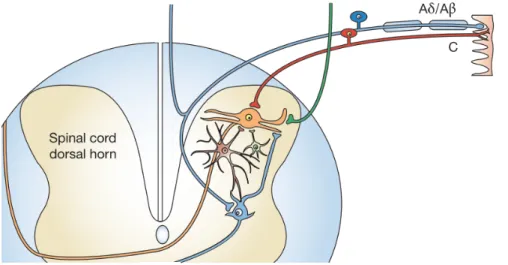
The signal travels from the free nerve endings to the cell bodies of the nociceptors that are located in the dorsal root ganglia (DRG) for the body and in the trigeminal ganglion for the face (Basbaum et al. 2009). Noxious signals originated from the periphery are transmitted by C-fibers and Aδ-fibers to the secondary neurons in the dorsal horn of the spinal cord. C-fibers project to the nociceptive upper laminae of the dorsal horn, whilst the Aδ-fibers projects to the deeper laminaeof the dorsal horn (Dib-Hajj et al., 2012).
Figure 4 Primary afferent pathways and their connections in the spinal cord dorsal horn. Nociceptive C-fibers
(red) terminate at spinothalamic projection neurons in upper laminae (orange neuron), whereas myelinated A-fibers (blue) project to deeper laminae. Adapted from (Baron 2006).
The first-order neurons from C fibers synapse with the secondary sensory neurons at the dorsal horn of the spinal cord, releasing neurotransmitters such as glutamate or substance P (Basbaum et al. 2009). As described by Purves D, Augustine GJ, Fitzpatrick D, 2001, once within the dorsal horn, the C-fibers send branches to innervate neurons in Rexed's lamina I and lamina II, while the Aδ-fibers terminates in the lamina II and V (Dib-Hajj et al. 2012; Fukuoka & Noguchi 2011). Finally, the large mechanosensor fibers sprout to the laminae III and V of the dorsal horn (Belkouch et al. 2014). The information is then transmitted to second-order neurons that cross over the spinal cord and ascend all the way to the brainstem and thalamus in the anterolateral quadrant of the contralateral half of the spinal cord (see Figure 2Error! Reference source not found.). There, the second-order neuron then synapses with a third-order neuron in the medial and ventrobasal nuclei of the thalamus by releasing neurotransmitters. This third-order neuron synapes with the somatosensory cortex of the brain which locates the injured area of the body and
10
coordinates appropriate pain perception (see Figure 2) (Nicholls, J. G., Martin, A. R., &
11
1.2.
When Pain Becomes a Dysfunction
1.2.1. CHRONIC PAIN SYNDROME
As aforementioned, pain has a protective and informative role in people’s lives. This kind of pain terminates once the noxious stimuli is removed and the body has healed. However, pain may outlast its practicality as a warning sign and become chronic (Raj et al. 2007).
Pain is defined as chronic rather than acute not only on the basis of its duration but also on the basis of the body's inability to restore physiologic function to homeostatic levels. The intensity of chronic pain has little relation to initial tissue damage or subsequent pathology as psychological and social factors seem to have great influence (Kendall 1999).
Chronic pain is a heterogeneous disease in terms of its etiology, mechanisms and temporal properties (Russo MD & Brose MD 1998; Azevedo et al. 2012). The three main etiologies for chronic pain are presented in the following sections. Understanding the cellular mechanisms underlying chronic pain is a critical step in the development of new therapies.
Abnormally amplified signals in the central nervous system due to wind up in central sensitization (which is an increased sensitivity of spinal neurons) cause primary and secondary hyperalgesia (an exaggerated sensation to a painful stimuli at the site of the injury and surroundings, respectively) and allodynia (perception of pain in response to a non-painful stimuli) (Tsantoulas & McMahon 2014; Russo MD & Brose MD 1998; Campbell & Meyer 2006). Symptoms usually include spontaneous paresthesia (abnormal sensation), such as numbness, pain with movement, sensitivity of a partly denervated body part and burning, tingling sensation (Milton 2013).
12
1.2.2. CELLULAR MECHANISMS OF NEUROPATHIC PAIN
Neuropathic pain syndromes are chronic pain disorders caused as a direct consequence of a lesion or by disease of the parts of the nervous system that normally signal pain (Baron 2006). When a lesion occurs on the nerves, the expression of Nav channels is increased and Kv channels is decreased in the affected fibers and the myelinated damaged fibers undergo a Wallerian degeneration. During this process, they release nerve-growth factor (NFG) which contacts with the intact fibers from the neighbouring axon. They increase the expression of Na+ channels, TRPV1 channels and adrenoreceptors and the decrease of K+ channels, therefore increasing the sensitivity of those uninjured fibers (Baron 2006).
1.2.3. CELLULAR MECHANISMS OF INFLAMMATORY PAIN
Pain is the primary reason patients with inflammatory arthritis seek rheumatologic care. Among rheumatoid arthritis patients, 68-88% rate pain as one of their top three priorities (Turk et al. 2004). Local inflammatory pain may appear in the absence of nerve trauma, as in the patients with low back pain or postherpetic neuralgia, which is a complication of shingles, caused by the chickenpox virus (Abrahamsen et al. 2008).Inflammatory pain results from the sensitization and activation of peripheral nociceptors by inflammatory mediators that accumulate in damaged tissue. The action of inflammatory mediators upon primary sensory afferents causes these neurons to have increased excitability, contributing to pain and hypersensitivity, also featuring hyperalgesia, allodynia and spontaneous pain (Abrahamsen 2014).
1.2.4. CELLULAR MECHANISMS OF PAINFUL DIABETIC
NEUROPATHY
Peripheral diabetic neuropathy is characterized by diffuse or focal damage of somatic or autonomic peripheral nerve fibers, resulting from diabetes mellitus (Iyer & Tanenberg 2013). One of the most prominent features of diabetic neuropathy is the development of spontaneous pain, typically in the extremities, presenting as allodynia and hyperalgesia (Tesfaye & Kempler 2005), as it results from the atrophy and loss of myelinated and non-myelinated fibers accompanied by Wallerian degeneration (Iyer & Tanenberg 2013).
13
1.3.
Neuroexcitability
1.3.1. RESTING MEMBRANE POTENTIAL
Action potentials are generated across the membrane of the neuron, which then sends an electrical signal along the membrane of the axon (Hille 2001). When the membrane is at rest, it has a much higher concentration of Na+ on the extracellular side in comparison to the intracellular side and vice-versa for K+ concentration (Cooper et al. 1998). Regarding the electrochemical gradient, the membrane of the neuron is much more permeable to K+ than to Na+. But the membrane is also richer in channels that facilitate the diffusion of K+than Na+ and therefore, more K+ will leave the cell than Na+ will enter (Armstrong & Hille 1998; Hille 2001; Hodgkin & Huxley 1952). Considering only K+, as the ions leave the cell, the positive charges decrease on the inner side of the membrane and increase on the outer side. As positive charge builds on the extracellular side, the electric force begins to drive the K+ back inside. Equilibrium is reached when the electric force is equal to the force of the concentration gradient.
1.3.2. VOLTAGE-GATED ION CHANNELS
As pointed out by Hodgkin & Huxley, mobile charge in the membrane that moves in response to voltage changes is the only possible way of having voltage dependence on the opening and closing of the gate (Hodgkin & Huxley 1952). The channel current is important as a measurement of the speed of movement of ions through the channel. The current depends not only on the properties of the channel as well as the transmembrane potential (Margrie & Urban 2007). The conductance of a channel depends on two factors: (1) the ease with which ions can pass through the channel – permeability; (2) the concentration of ions in the channel region. One can measure the membrane current through its channels conductance and the voltage applied (Southan & Robertson 2000).14
1.3.3. WHY K
+CHANNELS?
Electrical signals result from temporary local changes that drive membrane potential away from its resting value and are propagated throughout a neuron or nervous fiber. Ion channels mediate those alterations (Bezanilla 2005; Julius & Basbaum 2001; Margrie & Urban 2007). They are divided into families depending on structure and function. For this dissertation, the main focus will be on voltage-gated potassium channels that respond to changes of voltage across membrane (Kv channels).
Membrane proteins account for about 30% of the total proteome of an organism, with about half of this number being carrier proteins and ion channels. K+ channels
represent the most diverse and widespread class of membrane proteins (Frank et al. 2005). Of those, Kv channels form the most diverse group, represented by 12 families (Kv1-Kv12) (Gutman et al. 2005; Bosma & Hille 1992).
Kv channels have several important characteristics as they are highly restricted locations in axons, show diverse mechanisms of clustering, and, importantly, they contribute to neuronal excitability through setting the resting membrane potential and spike interval (Misonou 2010; Nashmi & Fehlings 2001). In excitable cells, Kv channels are involved in the stabilization of the membrane potential, since they converge the resting membrane potential to the K+ equilibrium potential, lowering it, and therefore keeping it away from the action potential firing threshold (Bosma & Hille 1992). Hence, they are critical to neuroexcitability. Not only they establish the resting potential but also keep the depolarization in the action potential short, are involved in shortening periods of high activity, keep a time separation between repetitive spikes and tend to diminish the excitatory inputs efficacy in the cell (Bosma & Hille 1992).
These multisubunit integral proteins mediate K+ fluxes depending on their level of
expression, conduction properties, activation, deactivation and inactivation characteristics, and the electrochemical gradient of ions across the cell membrane (Yellen 2002; Miller 2000). For instance, mutations in the genes of Kv channels can lead to several severe hereditary disorders (Camacho 2006; Wang et al. 1996), and importantly to this work, they are involved in the development of the pain syndrome (Beekwilder et al. 2003).
15
1.3.4. KV CHANNELS IN DRG NEURONS
As aforementioned, K+ channels are a family of ion channels that govern the intrinsic electrical properties of neurons (Lujan 2010). Molecular cloning has revealed over 100 genes encoding the pore-forming α-subunits of K+ channels in mammals, making them the most diverse subset of ion channels. Further multiplicity of the K+ channel family is generated through alternative splicing. The precise location of K+ channels along the dendro-somato-axonic surface of the neurons is a crucial factor in determining its functional impact (Doyle et al. 1998; Calvo et al. 2016). Sensory neurons express multiple types of Kv channels, including members of the Kv1, Kv2, Kv3, Kv4, Kv7 and Kv9 families, with expression patterns depending on neuronal subtype. Immunochemical studies suggest that numerous Kv channels in the Kv1 and Kv2 family are expressed in DRG neurons (Kuniko Ishikawa et al. 1999). This is in agreement with the presence of RNA transcripts encoding for Kv1, Kv2, and, in addition, Kv4 subunits in the DRG. Among the Kv1 channel family, the most abundantly expressed channels in DRG neurons are Kv1.1, Kv1.2 and Kv1.4 (Yang et al. 2004; Rasband et al. 2001; Kim, Choi, Rim, Cho, et al. 2002; Vydyanathan et al. 2005). Of these, Kv1.4 appears to be the most abundant Kv isoform in DRG neurons as most small diameter DRG Kv1.4-positive neurons do not detectably express other Kv1 subunits (Wells et al. 2007).Corroborating with Kv1.4 high proportion to other isoforms, small-diameter DRG neurons predominantly express an A-type K+ current whose properties correspond well with those expected for homotetrameric Kv1.4 channels (Sigworth 1994).
Not disregarding the presence of other Kv1 isoforms, Kv1 family channels are important determinants of neuronal excitability (Doyle et al. 1998). For instance Kv1.2 lowers excitability at the level of the sensory neuron cell body (Rasband et al. 2001; Everill et al. 2014) and Kv1.1 decreases mechanosensitivity (the mechanical sensitivity threshold gets higher) at the terminals of C-mechanonociceptor (Hao et al. 2013).
16
1.3.5. INVOLVEMENT OF DRG KV CHANNELS IN CHRONIC PAIN
Following nerve injury, DRG neurons can display aberrant firing properties or cross-excitation by neighboring neurons (Frank et al. 2005), producing inappropriate impulse activity that may underlie abnormal sensations.The contribution of K+ channels in pain signalling has been undervalued over the years, as research has been mainly focused on studying Na+ and Ca2+ channels. However, emerging data has highlighted that nerve injury or inflammation alters K+ channel activity in
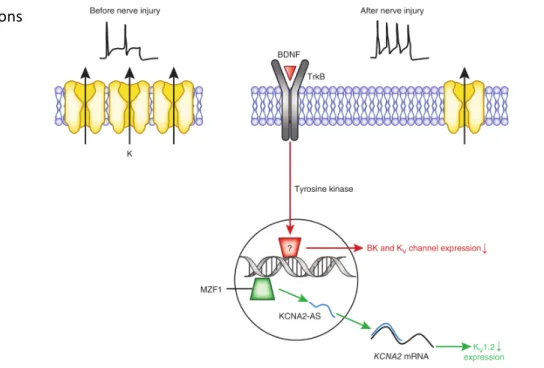
neurons of the pain pathway, having a prominent role on DRG hyperexcitability, thus starting to reveal its potential as a novel therapeutic target (Tsantoulas & McMahon 2014; Cao et al. 2011; Rasband et al. 2001; Wulff & Castle 2009). Multiple studies have demonstrated downregulation of expression of K+ channels in animal models of pain. For instance, a downregulation of Kv1.2 channels in DRG neurons in response to nerve injury has been reported (Fan et al. 2014) and is linked to myeloid zinc finger protein 1 (MZFP1) mediated expression of an endogenous antisense-like RNA (see Figure 5). As a result, excitability of afferent fibers is enhanced, leading to neuropathic-like pain behavior that can be attenuated by delivery of Kv1.2 sense fragments. Chronic bladder inflammation increases the excitability of C fiber bladder afferent neurons through
suppression of Kv1.4 channels (Yoshimura & Groat 1999), which suggests these A-type
Figure 5 Examples of mechanisms of nerve injury – induced inhibition of K+ channel expression. Under normal physiological circumstances, the activities of Kv channels limits the firing of afferent fibers (top left). In response to nerve injury BDNF activates TrkB receptors, which then regulate the expression of Kv channels at the transcriptional level, leading to reduced KV1.2 protein expression. The reduction of potassium channel expression leads to increased firing of injured neurons (top right). Reproduced from Waxman & Zamponi 2014.
17
channels are a major determinant of C fiber excitability. This is of particular importance when considering pharmacological treatment of pain. An alternative to reducing nociceptor excitability through blockade of action potential depolarization may lie in enhancing or prolonging activity of Kv1.4. This might be accomplished by interfering with the ball-and-chain N-type inactivation (explained below at section 1.3.8) that among Kv1 subunits is unique to Kv1.4 (Wells et al. 2007).
It has also been shown that Kv1.3 is present in DRG nociceptive neurons, which is a channel partly responsible for the slowly inactivating K+ current. This channels has been shown to be downregulated in whole lysates of DRG (Yang et al. 2004) in sciatic nerve transection chronic pain model. Also, Abdulla and Smith (2001) observed a significant decrease in steady-state mRNA level of Kv1.3 (about 50%) in all types of DRG neurons in axotomized rats that led to increased neuroexcitability. Although this channel appears to be downregulated in chronic pain models, the slowly inactivating current does not appear to be as affected as the A-type current due to their contributing channel proportion in small-diameter DRG neurons (Fukuoka et al. 2012; K Ishikawa et al. 1999)
Altogether, these various studies associate Kv channels as modulators of inflammatory and neuropathic pain signaling in afferent neurons. Specifically, they suggest a reduction of both slowly inactivating and A-type K+ currents through the downregulation
of Kv channels.
18
1.3.6. KV CHANNELS CONFORMATIONS
All Kv channels share a high level of similarity. Each Kv channel gene encodes one α-subunit and functionality requires four, hence Kv channels present a homotetrameric structure (with all α-subunit being identical) (Grizel et al. 2014). The transmembrane domain of the Kv channel α-subunit consists of six helices: S1 – S6. These helices form two structurally and functionally different parts of the tetrameric channel (Long et al. 2005):
1) K+-conducting domain (pore domain) – helices S5–S6 located in the center;
2) Voltage-sensing domain – helices S1–S4 located on the channel periphery.
The pore domain includes a channel gate and a selective filter that only allows K+ to enter through the channel. It is constituted by carbonyl groups which are spaced to interact with unsolvated K+. This way the passage of a Na+ through the channel is energetically unfavorable because the Na+ is too small for optimal interaction with the carbonyl groups.
Figure 6 Secondary, tertiary and quaternary structure of Kv channels. A. Scheme of a single α-subunit of a Kv
channel: Transmembrane segments S1–S6 and pore-forming P-region are marked. Charged Arg of the membrane voltage sensor S4 are marked with “+” signs. B. Crystal structure of a single α-subunit: S1–S6 segments, cytoplasmic domain T1, linker connecting the transmembrane portion with the T1 domain (T1–S1), as well as N- and C-termini are marked. Charged Arg residues of the membrane voltage sensor S4 are indicated by blue circles. C. Crystal structure of the Kv1.2 channel in a complex with the β-subunit (grey). D. Upper and Lower gates. Only two opposite subunits of Kvα are shown for clarity reasons. Figure replicated from (Grizel et al. 2014)
19
The channel gate is formed by crossing C-termini of the S6 helices that block passage of ions when the channel is closed (Doyle et al. 1998; Choe 2002).
It is known that the voltage sensing domain and the pore domain are covalently bound by the S4–S5 linker, which is a helix connected to the C-terminus of S6 helix and the next subunit (Choe 2002). This is a highly conserved region, which plays an important role in opening/closing the channel gates due to its flexibility. (Grizel et al. 2014).
Kv channels have two gates (Figure 6.D):
1) Lower gate formed by crossing the S6 helices on the intracellular side: this is the main activation gate controlled by external stimuli, such as the membrane potential; 2) Upper (inactivation) gate formed by the P-region of the selectivity filter on the extracellular side. The cytoplasmic part does not contain highly conserved regions and is different for Kv channels from different families (Pischalnikova & Sokolova 2009). β subunits (or auxiliary proteins), with distinct sequences, also often interact with the principal subunits complex, usually altering their electrophysiological or biophysical properties, expression levels or expression patterns (Coetzee et al. 1999).
1.3.7. ACTIVATION OF KV CHANNELS
Kv channels (and voltage-gated channels in general) exist typically in three different conformations that are inter-commutable: the activated state, associated with an open conformation – both gates open; inactivated state, associated with a non-conducting conformation – only one gate open and the other closed; and a quiescent state, associated with a closed conformation – both gates closed (Armstrong 2003)
The channel does not conduct the ions in the quiescent state. Depolarization of the membrane results in positive charge of its intracellular part, causing conformational rearrangements of Kv channels and making an open conformation energetically favorable, i.e., activation (Sigworth 1994; Coetzee et al. 1999; Grizel et al. 2014). In case the membrane remains depolarized, the majority of Kv channels switch to the inactivated non-conducting state.
20
which is the voltage sensor. Its cationic properties are due to its high arginine and lysine amino acids residue constitution. At rest, this positively charged domain is attracted to the inner residues of S2 and S3. When a depolarization occurs, the intracellular side of the membrane becomes less negative. As the S4 is positively charged, during depolarization this domain is electrostatically repelled upward, into the membrane, away from the cytoplasm. This allows ion conductance through the channel (Southan & Robertson 2000; Miller 2000). The exact mechanistic behind the activation of Kv channels is yet to be understood (Horn 2005; Grizel et al. 2014).
1.3.8. INACTIVATION OF KV CHANNELS
There were described two inactivation mechanisms for Kv channels during prolonged depolarization: 1. Fast N-type inactivation: The inactive state is mainly achieved through fast inactivation, by which a channel transitions rapidly from an open to an inactivated state. The model proposes that the inactive state, which is stable and non-conducting, is caused by the physical blockage of the pore. The blockage is mediated by an inactivation peptide positively charged folded into a globule and attached by a linker to the N-terminus. As the S4 is repelled into an upward position, the globule is dragged into the open pore of the channel and blocks ion traffic (Nicholls, J. G., Martin, A. R., & Wallace 1992). Importantly, Kv1.4 is the only Kv1 isoform that has this kind of inactivation. 2. Slow C-type inactivation: the selectivity filter acts as the second gate, as the C-terminus often carries motifs for channel modulation other than voltage, such as phosphorylation and Ca2+ binding (Robbins 2001). In cases of prolonged depolarization, the pore closes, preventing the entry of ions (Leung 2012). The channels completely return to the closed conformation after the inactivation when the potential drops to the resting potential level. The Kv1.3 channel shows a slow C-type inactivation and recovery (Southan & Robertson 2000).
1.3.9. KV CURRENTS IN DRG
Early electrophysiological characterization of DRG neurons revealed two types of voltage-gated K+ currents: transient (inactivating) A-type current (I
21
inactivating (Islow) (Mathie et al. 1998; Kostyuk et al. 1981; Yang et al. 2004). Within both
types of currents further biophysically and/or pharmacologically distinct components can be identified and these differ between the sensory neuron types. Criteria for their identification include rapid activation and inactivation, dependence on the holding potential, and sensitivity to both 4-AP and DTx (Wu & Barish 1992). For instance, Ifast currents
can be dissected from the sustained current using two different prepulse voltages with identical stimulation pulse protocols (Everill et al. 1998).
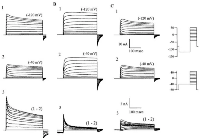
Figure 7 shows current families found in all DRG neurons from L4-L5 ganglia recorded at voltages between −40 and +50 mV after a 500 ms conditioning prepulse to −120 mV or to −40 mV.
•
Figure 7A1 demonstrates the main composition of currents occurring in ~58% of cells examined (Everill et al. 1998). Subtraction of responses (Figure 7A3) revealed fast and slow activating and inactivating components that were sensitive to the prepulse variation.•
Figure 7B shows that in some of the neurons the K+ current components(~24%, Everill et al. 1998), Ifast and Islow, were clearly defined after subtraction.
•
Figure 7C3 displays the slower inactivating component. Cells lacking Ifast, butshowing the slower inactivating component were seen in ~12% of the cells examined (Everill et al. 1998).
22 Figure 7 Voltage-dependent potassium currents of DRG neurons. Recordings from 3 categories can be seen in A1, B1, and C1 recorded after the −120-mV prepulse protocol; A2, B2, and C2 were initiated via the −40-mV prepulse protocol. A: typical type of traces (of this shape and amplitude) that were recorded in the majority of experiments. This cell type manifested ≥3 distinct components of current. B1–B2: this cell type has a large amount of fast inactivating component in its complement of currents. C1–C2: cell type that lacks the fast inactivating component but has a slower inactivating voltage-activated component sensitive to the conditioning potential. Lower scale bars relate to subtractions only (subtractions are enlarged for clarity of comparison). These are typical traces taken from the mean of each group. Reproduced from Everill et al. 1998.
Functional investigations pointed out that Ifast is typically involved in setting the
interspike interval (Hille 2001), while Islow is essential for fast repolarization of action
potentials and consequently contributes to repetitive firing pattern (Armstrong 2003).
23
1.4.
Animal Pain Models
Rodent behavioural models are important tools for expanding the understanding of the physiology underlying nociception and pain (Mogil et al. 2010). Rat models result in nociceptive sensitization similar to the key components of those experienced in humans (Barrett 2015). The use of rodents to study chronic pain is also adequate because drugs that shown some efficacy in chronic pain patients have also demonstrated activity in animal models of pain (Li et al. 2015).
The rat behaviour in response to noxious stimuli can be consistently and objectively scored through different tests. However, the most reliable and commonly scored behaviours are simple reflexes or innate responses (such as paw withdraw, linking the sensitized zone, tail flick) (Mogil et al. 2010).
1.4.1. NEUROPATHIC PAIN: CHRONIC CONSTRICTION INJURY
(CCI)
Neuropathic pain models rely on cutting, stretching or compressing the nerve and therefore, rely on a surgical approach (Mogil 2009). Following traumatic nerve injury spontaneous activity develops initially in myelinated and subsequently in unmyelinated sensory axons (Kajander et al. 1996; Austin et al. 2012). The onset of this spontaneous activity is associated with the emergence of pain-related sensory changes in animal models (Nashmi & Fehlings 2001).
One of the most usual neuropathic pain model used in rodent is the chronic constriction injury (CCI) in which the sciatic nerve is constricted with four loose ligations. The resulting constriction does not stop the blood flow and produces an inflammation, leading to the physiological changes inhered to chronic neuropathic pain in the DRGs located in the 4th, 5th and 6th lumbar vertebrae (L4-L6) (Austin et al. 2012).
24 One limitation of this model is the degree of variation amongst the rats subjected to CCI, due to variability in the tightness of the constrictions produced by tying knots with the sutures. This can be partially overcome by having an experienced researcher performing the surgery in a consistent way. Furthermore, the type of suture material used for ligating the nerve can also contribute to variability (Attal et al. 1990).
1.4.2. INFLAMMATORY PAIN: COMPLETE FREUND’S ADJUVANT
(CFA)
Complete Freund’s Adjuvant (CFA) is a solution of antigen emulsified in mineral oil and consists of inactivated and dried mycobacteria. The CFA injection is a simple procedure which reproducibly induces inflammation, as shown in previous studies (Spears et al. 2005; Guan et al. 2005). However, single-dose CFA injections in previous work were primarily associated with transient inflammatory changes in the temporomandibular joint (Ren & Dubner 1999). CFA is effective in stimulating cell-mediated immunity and leads to potentiation of T helper cells, which culminates in the production of certain immunoglobulins and effector T cells (Koo et al. 2013) therefore leading to inflammation and pain. The pain information is processed by the L3, L4 and L5 DRGs. Figure 8 Photograph of the 4 loose ligations on mid-thigh sciatic nerve of a 65 days old female Wistar rat.
25
1.4.3. NEUROPATHIC PAIN: PAINFUL DIABETIC NEUROPATHY
(PDN)
In order to induce Painful Diabetic Neuropathy (PDN), streptozotocin (STZ) has been largely used as an experimental paradigm for its great stability and relative lack of extrapancreatic toxicity (Sik Nam et al. 2014). STZ indirectly activates an apoptotic program that destroys pancreatic β-cells and all the cells expressing the GLUT2 transporter (Wattiez et al. 2012), leading to diabetes mellitus type I. Diabetes can lead to both mechanical hyperalgesia and hypoalgesia (Heng et al. 2015).
1.4.4. HOW TO EVALUATE PAIN IN ANIMALS
The area tested is the mid-plantar surface of the hind paw (Figure 9), which falls within the sciatic nerve distribution. Mechanical withdrawal threshold is assessed by mechanically stimulating both injured and uninjured hind paws using an electronic dynamic plantar von Frey aesthesiometer or manual von Frey filaments (Austin et al. 2012).
The mechanical withdrawal threshold is the maximum pressure applied (in grams) that elicits paw withdrawal. This quantifiable measurement allows direct comparisons of paw sensitivity to mechanical stimuli across different treatment options, and between different strains of rats.
It should be noted that other behavioural assays, such as the pin prick test, cold allodynia, vertical activity and open field exploration are also used for testing pain hypersensitivity.
26
2.
GOALS
The neurons of the dorsal root ganglia (DRG), are the main experimental model in studies of the physiology of pain. DRG neurons, and in particular the small-diameter DRG neurons, have great interest to the pharmaceutical industry dedicated to the research and development of new analgesics since its activity is involved in the processing of pain perception, which is exacerbated in chronic pain situations. A most promising therapeutic approach for chronic pain is to reduce neuronal hyperexcitability that occurs in this condition, which can be done by altering the current of the main ion channels that modulate the electrical activity of DRG neurons. The knowledge about the voltage-gated K+ (Kv) channels is essential since these channels control excitability and some of them are already seen as molecular targets for the development of new analgesics. This thesis aims to fill in the gap on Kv channels in small DRG neurons and deepen the understanding of their role in chronic pain, identifying the key subtypes, thus validating them as potential therapeutic targets.
This project focuses on the electrophysiological characteristics of K+ currents in DRG neurons that have direct relevance to the study of chronic pain. In particular, I aimed to study the biophysics and pharmacology components of the currents that are directly involved in chronic pain process. Therefore, the overall goal of the present dissertation was to study alterations in Kv channel mediated currents in small DRG neurons from rat models of neuropathic (CCI of the sciatic nerve and STZ-induced PDN) and inflammatory (CFA-induced monoarthritis) chronic pain.
More specifically, this study aimed to:
1. Optimize and validate chronic neuropathic and inflammatory pain models through behavior tests, specifically with the quantification of mechanical hyperalgesia;
2. Identify the biophysical and pharmacologic patterns that are altered following induced CP, by the study of K+ currents in small DRG neurons (derived from CCI, CFA and control rats) using voltage-clamp under whole-cell configuration;
3. Study the Kv channels expression, namely the expression of Kv1.3 and Kv1.4 isoforms (previously identified by electrophysiological studies), in DRG neurons derived from CCI neuropathic and CFA inflammatory rat models through Western assays.
27
Overall, results showed a development of hyperalgesia and an abnormal function and expression of Kv channels in small DRG neurons following neuropathic and inflammatory pain in rat models, contributing to their validation as potential targets for novel analgesics.
28
3.
METHODS
3.1.
Animal Pain Models
In the present work were used male and female Wistar rats with ages comprehended between 60 and 180 days. Three conditions have been studied – naïve control animals, procedure control (sham) animals and pain model animals, who were maintained for 4 weeks or 2 weeks after the procedure, depending on being neuropathic or inflammatory pain model, respectively. Animals were purchased from NMS|FCM and caged in a 12h light/dark cycle, at room temperature (21 ± 3°C) with water and food ad libitum. Animal care and experimental studies were in accordance with Directive 2013/63/EU, and the procedures were approved by the Ethical Committee of the NMS|FCM.
The animals were divided in four different groups: 1) chronic constriction injury (CCI), 2) complete Freund’s adjuvant (CFA) and 3) painful diabetic neuropathy (PDN) and respective 4) naive controls. After establishment of the animal pain models, the onset of mechanical hyperalgesia was studied. Consequently animals were sacrificed for following harvesting of DRG ganglia. The biophysical properties of K+ currents were studied by whole-cell voltage-clamp in up to 24h DRG neurons and protein expression of Kv channels in DRG ganglia was assessed by Western blot technique.