Escola de Ciências
Joana Isabel da Silva Tulha Moreira
Study of the influence of lipid rafts in
acetic acid-induced apoptosis
Tese de Mestrado
Mestrado em Genética Molecular
Trabalho efectuado sob a orientação de:
Professora Doutora Célia Ferreira
Professora Doutora Cândida Lucas
Outubro, 2011
ii Nome: Joana Isabel da Silva Tulha Moreira Endereço electrónico: [email protected] Número do Bilhete de Identidade: 13347163
Título da tese: “Study of the influence of lipid rafts in acetic acid-induced apoptosis” Orientador(es):
Professora Doutora Célia Ferreira Professora Doutora Cândida Lucas Ano de conclusão: 2011
Designação do Mestrado: Mestrado em Genética Molecular
É AUTORIZADA A REPRODUÇÃO PARCIAL DESTA TESE/TRABALHO APENAS PARA EFEITOS DE INVESTIGAÇÃO, MEDIANTE DECLARAÇÃO ESCRITA DO INTERESSADO, QUE A TAL SE
COMPROMETE.
Universidade do Minho, ___/___/______
iii
Embora esta tese seja, pela sua finalidade, um trabalho de carácter individual, a realização da mesma não teria sido possível sem a ajuda e apoio de algumas pessoas. A todas elas quero dizer um: Muito Obrigada!
Às minhas orientadoras, a Doutora Célia Ferreira e a Doutora Cândida Lucas, por me terem proporcionado a possibilidade de realização deste trabalho.
À Célia por todo o apoio e tempo pacientemente cedido assim como por todas as críticas e sugestões durante este ano de trabalho que tanto contribuíram para a minha evolução.
À Professora Cândida pela disponibilidade, orientação, experiência e conhecimentos transmitidos em momentos chave durante a realização deste trabalho.
À professora Manuela Côrte-Real pelas sugestões que ajudaram a melhorar este trabalho.
A todos os meus colegas de laboratório, Joana, Rui, Fábio e Alain, pela alegria, companhia e por tornarem o ambiente laboratorial ainda mais agradável. Basicamente por me aturarem todos os dias, e só se queixarem um bocadinho. Assim não custa vir trabalhar!
Um agradecimento especial ao Fábio, pela boa disposição, pela partilha de conhecimentos, pelas intermináveis discussões científicas, pelas horas no microscópio, mas principalmente pela paciência
Aos restantes colegas do departamento de Biologia pela disponibilidade para ajudar sempre que foi preciso.
Aos meus pais, madrinha e avó, pelos sacrifícios que fizeram e fazem todos os dias para me possibilitarem esta oportunidade. E pelo conforto de estar em casa.
Por último, mas o mais importante, ao Dário por… TUDO!
Este trabalho foi parcialmente financiado pelo projecto da FCT (FCOMP-01-0124-FEDER-007047).
v
Summary
Lipid rafts are sphingolipid-sterol rich micro-domains of the plasma membrane that have been associated with different cellular processes, including apoptosis. During the past years, yeast has been successfully established as a model to study mechanisms of programmed cell death in mammalian cells. Saccharomyces cerevisiae commits to cell death showing typical hallmarks of metazoan apoptosis in response to different stimuli. Gup1p, a pleiotropic O-acyltransferase first associated with glycerol metabolism and transport, is involved in lipid metabolism, rafts integrity and assembly, as well as in GPI anchor remodeling, among a wide range of cellular processes. While Gpd1p/Gpd2p are the two isoforms of glycerol-3-phosphate dehydrogenase that have a prime role in the production of glycerol, Rvs161p is a lipid raft protein mainly involved in the regulation of actin cytoskeleton polarization and secretory vesicle trafficking. Actin cytoskeleton together with vesicle trafficking have been suggested to play a crucial role in yeast apoptosis. Therefore, it is conceivable that lipid rafts may also play a role in yeast cell death and understanding their function in such process in yeast may provide further insights on their role in mammalian apoptosis. For this purpose we used two known apoptosis inducing conditions, chronological aging and acetic acid, and analyzed several apoptotic markers in ∆gup1, ∆gpd1∆gpd2 and ∆rvs161 mutant strains. We found that ∆gup1 presents a significantly reduced chronological life span as compared to Wt and is also highly sensitive to acetic acid treatment. According to the apoptotic markers analyzed, cells lacking GUP1 seem to be incapable of undergoing apoptosis. Instead this mutant appears to be experiencing a necrotic cell death process. The ∆gpd1∆gpd2 (but not single mutants) and ∆rvs161 mutant strains are also sensitive to acetic acid. However, the apoptotic markers examined suggest that these mutants die by apoptosis, differently from that we observed for gup1 mutant. The ergosterol distribution in Wt and in the acetic acid-sensitive mutant strains gup1, ∆gpd1∆gpd2 and ∆rvs161 was subsequently observed by filipin staining. An altered ergosterol distribution was visualized in all mutants studied, particularly in gup1 mutant. We also observed that acetic acid induces a rearrangement in the distribution of ergosterol.
Altogether, our results indicate that lipid rafts seems to be a key component in apoptotic induction/signaling, probably even essential in some circumstances for the correct development of apoptosis in yeast.
vii
Sumário
Os rafts, domínios da membrana plasmática ricos em esfingolípidos e ergosterol, têm sido associados com diversos processos celulares, incluindo a apoptose. Nos últimos anos, a levedura tem sido aceite como um bom modelo para o estudo dos mecanismos de regulação da morte celular programada. Vários estudos demostraram que, em resposta a diferentes estímulos, a levedura Saccharomyces cerevisiae apresenta uma morte celular com os fenótipos típicos da apoptose de mamíferos. A proteína Gup1p, uma O-aciltransferase anteriormente associada com o metabolismo e transporte de glicerol, está envolvida, de entre vários processos, no metabolismo lipídico, na arquitectura/integridade dos rafts, assim como na remodelação das âncoras GPI. As proteínas Gpd1p/Gpd2p são duas isoformas da glicerol-3-fosfatodesidrogenase, enzima responsável pela produção do glicerol. O Rvs161p é uma proteína dos rafts envolvida na regulação da polarização da actina e no tráfego de vesículas. Os filamentos de actina assim como o trafego de vesículas têm sido apontados como importantes intervenientes na apoptose em leveduras. Assim, é possível que os rafts exerçam um papel importante na morte celular em leveduras. O conhecimento da sua função neste processo poderá ajudar a compreender a importância dos rafts neste tipo de morte celular em mamíferos. Para estudar esta hipótese, foram analisados diferentes marcadores apoptóticos nos mutantes ∆gup1, ∆gpd1∆gpd2 e ∆rvs161, em duas condições indutoras de apoptose, o envelhecimento cronológico e o tratamento com ácido acético. Verificamos que, quando comparado com a Wt, este mutante apresenta uma redução significativa na longevidade sendo também extremamente sensível ao ácido acético. Os marcadores apoptóticos estudados indicam que as células deletadas no gene GUP1 são incapazes de morrer por apoptose. Nas condições mencionadas, a estirpe ∆gup1 parece morrer por necrose. Os mutantes ∆gpd1∆gpd2 (mas não os mutantes simples) e ∆rvs161 são também sensíveis ao ácido acético. Contrariamente ao que se observou para o mutante gup1, estes mutantes morrem por apoptose. Por último, examinou-se a distribuição do ergosterol nos mutantes sensíveis ao ácido acético. Observou-se alterações na sua distribuição em todos os mutantes, particularmente o mutante gup1. Verificou-se também que o tratamento com ácido acético induz um reorganização da distribuição do ergosterol. De um modo geral, os resultados obtidos sugerem que os rafts exercem uma função determinante na indução/sinalização da apoptose, sendo portanto, a sua integridade essencial no desencadear do processo apoptótico.
ix Table of Contents Agradecimentos ……….. Summary……….. Súmario ………. Table of contents ……….……... I. Introduction ……….…... I - 1 Cell death ……….……….…… I - 2 Apoptosis ……….…. I - 2.1 The morphology of apoptosis ……… I - 2.2 The biochemistry of apoptosis ……….……. I - 2.2.1 Caspases ………..……….. I - 2.2.2 Bcl-2 family ……….………… I - 2.3 Mechanisms of apoptosis ……….……….
I - 2.3.1 Activation phase ……….……….. I - 2.3.1.1 Extrinsic apoptotic pathway ………..………… I - 2.3.1.2 Intrinsic apoptotic pathway ………..………. I - 2.3.2 Execution phase . ……….………... I - 3 Apoptosis in yeast Saccharomyces cerevisiae ……….………..
I - 3.1 Yeast apoptotic triggers ……….……. I - 3.2 Benefits to a unicellular organisms ………... I - 4 Chronological aging in yeast ………...
I - 4.1 Apoptosis in chronological aging ………... I - 5 Acetic acid in Saccharomyces
cerevisiae………...
I - 5.1 Acetic acid-induced apoptosis ………. I - 6 Lipid rafts ………... I - 7 Scope of the thesis ………... II. Material and Methods ………...
II - 1 Strains and growth conditions ………. II - 2 Sensitivity to acetic acid ………... II - 3 Acetic acid treatment ………..…………. II - 4 Chronological aging ……….. II - 5 Apoptotic markers ………... II - 5.1 PI staining ……… II - 5.2 FITC-coupled Annexin V staining ………... II - 5.3 DiOC6 staining ……… II - 5.4 DAPI staining ………. II - 6 Assessment of ROS ………. iii v vii iv 1 3 4 5 6 6 7 8 8 8 10 11 11 12 13 14 17 18 19 22 25 27 29 29 30 30 30 30 31 31 32 32 32
x
II - 7 Filipin staining ………. III. Results and Discussion ……….
III - 1 Programmed cell death in Saccharomyces cerevisiae is hampered by the deletion of
GUP1 gene . ………..
III - 1.1 Abstract ……….. III - 1.2 Results ……… III - 1.2.1 ∆gup1 mutant cells exhibit a reduction in chronological life span ……... III - 1.2.2 Chronological aged ∆gup1 mutant seems to be incapable of dying by
apoptosis ……… III - 1.2.3 ∆gup1 mutant cells are extremely sensitive to acetic acid ………. III - 1.2.4 Acetic acid induces cell death similar to that triggered by chronological
aging………... III - 1.2.5 ∆gup1 mutant cells accumulate large amounts of ROS during chronological
aging and acetic acid treatment ………. III - 1.3 Discussion………... III - 2 Involvement of rafts in acetic acid-induced apoptosis …..……….
III - 2.1 Abstract ………. III - 2.2 Results ………... III - 2.2.1 ∆gup1, ∆gpg1∆gpd2 and ∆rvs161 mutants are very sensitive to acetic acid… III - 2.2.2 Acetic acid-induced death displays characteristic markers of apoptosis in
∆gpd1∆gpd2 and ∆rvs161 strains, but not in ∆gup1 mutant ………
III - 2.2.3 ∆gup1, ∆gpg1∆gpd2 and ∆rvs161 mutants present different ROS accumulation during acetic acid treatment ……… III - 2.2.4 ∆gup1, ∆gpg1∆gpd2 and ∆rvs161 mutant are affected in sterol-lipid
distribution ……… III - 2.3 Discussion ………. IV. Final considerations ……… V. References ……… VI. Supplementary data ……… VI - 1 Treatment with different acetic acid concentrations ……….. VI - 2 Ceramide C2 treatment ……….. VI - 3 Sensitivity to myriocin ………... VI - 4 DRMs isolation ……….. VI - 5 Supplementary references ……….. 35 37 37 37 38 39 41 43 45 46 49 49 50 50 52 54 56 58 61 67 79 81 82 83 84 85
Chapter I
3
I - 1 Cell death
Soon after being recognized that organisms are made of cells, it was discovered that cell death is an important part of development. Cell death was first observed during amphibian metamorphosis (Vogt, 1842). Several years later, inhibitors of RNA and protein synthesis were found to inhibit cell death, indicating that an active metabolism is necessary to carry out this process. The term programmed cell death (PCD) was first used by Lockshin and Williams in 1964 to describe the cell death that occurs in predictable places and at predictable times during mammalian development, emphasizing that death is somehow programmed into the development plan of the organism (Lockshin and Williams, 1964). It is clear, however, that cell death can be prevented by substances released by other tissues, indicating that the death is not inevitable and apparently can be suppressed by signals from other cells (Saunders, 1966).
Accordingly, the term PCD is used nowadays to define all modes of death whose execution is carried out in a regulated manner and is under molecular control. This requires that the process follows a specific, orchestrated choreography, in which one event is triggered by another. Moreover, such process should implicate an advantage for the particular organism or cell population. Thus, PCD is a normal physiological form of cell death that plays a key role in both the maintenance of adult tissues and embryonic development. In adult tissues, for instance, PCD is involved in the maintenance of homeostasis, in the control of cell number, and also provides a defense mechanism by which damaged and potentially dangerous cells can be eliminated. During development, PCD plays a key role by eliminating unwanted cells from a variety of tissues, like the elimination of the tissue between the digits during the formation of fingers, and in the selection of neurons that present correct connections with their target cells (Galluzzi et al., 2007).
For several years, apoptosis was wrongly understood as synonymous for PCD. However, it must be kept in mind that although apoptosis is the most common form, dying cells may follow other types of PCD, including programmed necrosis, autophagic cell death and mitotic catastrophe (Table I-1). Yet, unlike apoptosis the other types of
4
PCD are still difficult to distinguish unambiguously and remain poorly characterized (Galluzzi et al., 2007).
Besides its role in the development and homeostasis of cell populations, PCD has also been associated with the occurrence of several human and mammalian diseases. Since then, the study of different types of PCD has increased exponentially among the scientific community, aiming to use the emerging knowledge in the treatment of several pathologies.
I - 2 Apoptosis
The term apoptosis was first proposed in 1972 by Kerr, Wyllie, and Currie to describe a morphologically distinct mechanism of “controlled cell deletion”, which plays a complementary but opposite role to mitosis in the regulation of animal cell populations (Kerr et al., 1972). Since then, apoptosis has been recognized and accepted as an Cell death type Characteristic morphological aspects
Apoptosis - Rounding up the cell
- Reduction of cellular and nuclear volume - Retraction of pseudopods
- Nuclear fragmentation
- Little modification of cytoplasmic organelles - Plasma membrane blebbing
Necrosis - Cytoplasmic swelling
- Rupture of plasma membrane - Swelling of cytoplasmic organelles - Moderate chromatin condensation
Autophagy - Lack of chromatin condensation
- Massive vacuolization of the cytoplasm (double-membrane autophagic vacuoles)
Mitotic catastrophe - Micronucleation - Multinucleation
5
important physiological process of PCD. Nowadays, apoptosis is considered a universal event in the normal development and aging of multicellular organisms, acting as a homeostatic mechanism that removes mutated, infected or dispensable cells and contributes to tissue growth, maintenance and renewal. Albeit its importance to the normal functioning of the organisms, a defective regulation of apoptosis is, usually, associated to the occurrence and progression of several pathologies (Rudin and Thompson, 1997). The inappropriate activation of apoptosis may cause or contribute to a variety of diseases including acquired immunodeficiency syndrome (AIDS) (Ameisen et al., 1995), ischemic strokes (Raff et al., 1993), and neurodegenerative disorders, such as Alzheimer’s (Smale et al., 1995) or Huntington’s diseases (Hickey and Chesselet, 2003). Conversely, a defective activation of apoptosis is responsible for some autoimmune diseases (Tan, 1994) and is commonly recognized as a determinant step in the development of cancers (Lowe and Lin, 2000; Evan and Vousden, 2001; Reed, 2003). Apoptosis has been placed under the spotlight of scientific investigation in the last years, mainly due to the growing understanding of its important role on the emergence and prevention of diseases, increasing the interest in apoptosis as a therapeutic target.
I - 2.1 The morphology of apoptosis
Apoptosis has been suggested as an active programmed phenomenon that can be triggered by a variety of stimuli, which include genotoxic agents (responsible for DNA damage), loss of extracellular survival factors or the specific activation of death receptors (Nagata, 2000). Despite the process responsible for triggering apoptosis, the ultimate morphological and biochemical characteristics of the process are similar. Thus, apoptotic cells are distinguished by membrane blebbing, cell shrinkage, chromatin condensation and intranucleosomal cleavage of DNA, followed by cell fragmentation into membrane-bound apoptotic bodies, and the loss of lipid asymmetry with phosphatidylserine exposure on the surface of the plasma membrane. Phosphatidylserine externalization is known to be responsible for ‘calling’ neighboring cells to phagocyte apoptotic cells, preventing the uncontrolled release of intracellular
6
components to the extracellular milieu (secondary necrosis) and inflammation (Kerr et al., 1972; Fleury et al., 2002).
I - 2.2 The biochemistry of apoptosis
Many of the genes that control apoptosis have been identified, and the molecular mechanisms underlying these processes were proved to be evolutionarily conserved (Metzstein et al., 1998). Central to the execution of the suicide program are the cysteine-dependent aspartate-specific proteases, so called caspases (Shi, 2002). The Bcl-2 family, a second set of apoptotic regulators, is also important to the regulation of apoptosis (Adams and Cory, 1998).
I - 2.2.1 Caspases
Most of the morphological changes that were observed during apoptosis are caused by a set of cysteine proteases that are activated specifically in apoptotic cells. These death proteases are homologous to each other, and are part of a large protein family known as the caspases (Cysteine dependent Aspartate-specific Proteases) (Shi, 2002). Caspases are highly conserved through evolution. Over a dozen caspases have been identified in humans; about two-thirds of these have been suggested to function in apoptosis (Earnshaw et al., 1999).
All known caspases have cysteine residues at their active sites crucial for its proteolytic activity, that selectively cleave target proteins in specific positions (after an aspartate residue). As for most proteases, caspases are synthesized as enzymatically inert zymogens called pro-caspases. These inactive proteins are then activated by proteolytic cleavage of its pre-form, at the aspartate residues. The smaller fragments dimerise to form the active enzyme (caspases). Besides cleaving several proteins, these enzymes can also act upon other caspases activating them and leading to a phenomenon known as the cascade of caspase activation (Chowdhury et al., 2008). Regulation of caspases is, therefore, central to determine the cell’s fate. Once caspases are initially activated, there seems to be an irreversible commitment towards cell death.
7
Caspases have been divided into subfamilies based on their substrate preference, extent of sequence identity and structural similarities. Apoptotic caspases are usually divided into two different groups: the initiator caspases (that include caspases-2, 8, 9 and 10) and the effector caspases (caspasese-3, 6 and 7) (Rai et al., 2005). In general, initiator caspases, which act at the apex of the signaling cascade, are characterized by its long pro-domains (Death Effector Domain (DED) in caspases-8 and 10 and Caspase Recruitment Domain (CARD) in caspases-2 and 9), with more than 90 amino acid residues, while the effector caspases possess shorter pro-domains with only 20-30 residues. (Budihardjo et al., 1999; Chowdhury et al., 2008).
I - 2.2.2 Bcl-2 family
The Bcl-2 family proteins are also key regulators of apoptosis. All proteins of the Bcl-2 family share homologue regions known as BH domains. Thus, at the first time, this family was divided into three groups based on structural similarities: Anti-apoptotic members (share sequence similarity in all four conserved BH domains (BH1–4)); the pro-apoptotic members (have similarity in three domains (BH1–3)); and the ‘‘BH3-only’’ subfamily (display sequence similarity only in the BH3 domain) (Adams and Cory, 2001; Borner, 2003). Presently, with new results obtained for the sub-group “BH3-only” proteins, they are divided into four categories accordingly to their function: the anti-apoptotic Bcl-2 proteins (A1, Bcl-2, Bcl-w, Bcl-xL and Mcl-1); Bcl-2 effector proteins, (Bak and Bax); direct activators BH3 only proteins (Bid, Bim and Puma); and sensitizers/de-repressors proteins (Bad, Bik, Bmf, Hrk and Noxa) (Chipuk et al., 2010).
Bcl-2 family members are critical for the regulation of the mitochondrial pathway of apoptosis (Roset et al., 2007). However, the molecular actions of Bcl-2 family members are still controversial. The ability of these proteins to form homo- or heterodimers suggests that there is a neutralizing competition between both members, therefore the ratio between the two subsets will determine the cell’s fate (Gross et al., 1999). These proteins are also subjected to post-translational modifications, for instance phosphorylation, that affect their function. Such modifications can be mediated by the interaction with other cytoplasmic and mitochondrial proteins, such as the protein kinase C (PKC) isoforms (Gutcher et al., 2003).
8
I - 2.3 Mechanisms of apoptosis
The cascade of events leading to this PCD process can be sub-divided in activation and execution phase. During the activation phase, multiple signaling pathways lead to the central control of the cell death machinery and activate it. This is followed by the execution stage, in which the activated machinery acts on multiple cellular targets resulting on the destruction of the cell (Elmore, 2007).
I - 2.3.1 Activation phase
Currently, two well-understood mechanisms of apoptosis activation are documented: the extrinsic apoptotic pathway and the intrinsic apoptotic pathway (Fig I-1). Moreover, there is evidence that the two pathways are linked and that the molecules of one pathway can influence the other (Igney and Krammer, 2002).
I - 2.3.1.1 Extrinsic apoptotic pathway
The extrinsic pathway, also called death receptor pathway, depends of transmembrane receptor-mediated interactions. This pathway involves death receptors that are members of the tumor necrosis factor (TNF) receptor gene superfamily (Locksley et al., 2001). All members of the TNF receptor family share similar cysteine-rich extracellular domains and a cytoplasmic domain of about 80 amino acids called the death domain (DD) (Ashkenazi and Dixit, 1998). This DD plays a crucial role in spreading the death signal from the cell surface to the intracellular signaling pathways.
The death receptors are activated by specific extracellular ligands that promote clustering of the receptors. After ligand binding, cytoplasmic adapter proteins such as FADD (Fas-Associated protein with Death Domain) or TRADD (Tumor necrosis factor Receptor type 1-Associated Death Domain) are recruited, which in turn also have a DD and an additional interaction domain, the death effector domain (DED). These adapter proteins are responsible for connecting the death receptor, through the interaction between the DDs, to pro-caspase-8, via dimerization of the DEDs. Association between
9
the extracellular ligand, the death receptor, the adaptor protein and the pro-caspase 8 forms a complex called the Death Inducer Signaling Complex – DISC. The concentration of pro-caspase-8 in the DISC results in its cleavage and subsequent activation(Kischkel et al., 1995). Once active, caspase-8 initiates the cascade of caspase activation by processing caspases-3, -6 and -7, which will act upon different substrates (Thorburn, 2004; Chowdhury et al., 2008). The death receptors can be inhibited by a protein called c-FLIP, which binds to FADD and caspase-8, rendering them ineffective (Kataoka et al., 1998; Scaffidi et al., 1999).
Fig. I-1. Apoptotic pathways. The extrinsic pathway is initiated through the stimulation of the death receptors. Association between the death receptor, the adaptor protein and the pro-caspase 8 forms the DISC, responsible for the caspases 8 activation. The intrinsic pathway is activated by internal stimuli that depend on the MMP. The MMP facilitates the release of pro-apoptotic proteins to the cytoplasm, such as cytochrome c that combines with Apaf-1 and pro-caspase 9 to form the apoptosome, resulting in caspases 9 activation.
10
I - 2.3.1.2 Intrinsic apoptotic pathway
The intrinsic pathway (also called mitochondrial pathway) initiates within the cell and is triggered by stress stimuli such as DNA damage, caused by radiation, toxins, hypoxia, hyperthermia, viral infections and free radicals, or by absence of certain growth factors, such as hormones and cytokines. The activation of this pathway depends on the mitochondrial membrane permeabilization (MMP). Several mechanisms underlying MMP have been proposed including the formation of pores in the mitochondrial membrane mediated by pro-apoptotic members of the Bcl-2 family, such as Bax and Bak (Dejean et al., 2005). The MMP, and consequent loss of the mitochondrial membrane potential, facilitates the release of several pro-apoptotic proteins to the cytoplasm. The first pro-apoptotic proteins that are released from mitochondria include cytochrome c, Smac/DIABLO (Second Mitochondria derived Activator of Caspases/Direct Inhibitor of Apoptosis Protein (IAP)-Binding protein with Low PI), and the serine protease HtrA2/Omi (Cai et al., 1998; Du et al., 2000; van Loo et al., 2002; Garrido et al., 2006). Latter, the pro-apoptotic proteins AIF (Apoptosis-Inducing Factor), endonuclease G and CAD (Caspase-Activated Deoxyribonuclease) are also released.
The first group of proteins activates the caspase dependent mitochondrial pathway. After being released from the mitochondria, cytochrome c combines with Apaf-1 and dATP to form a protein complex known as the apoptosome. This complex, in turn, activates caspase-9 thus initiating the cascade of caspase activation which leads to cellular death (Acehan et al., 2002). Smac/DIABLO and HtrA2/Omi are reported to promote apoptosis by inhibiting IAP (Inhibitors of Apoptosis Proteins) activity (van Loo et al., 2002; Schimmer, 2004).
The second group of pro-apoptotic proteins is released only after the cell has committed to die. AIF and Endonuclease G are both translocated to the nucleus causing nuclear fragmentation and condensation (Joza et al., 2001; Li et al., 2001) in the so-called “stage I” (Susin et al., 2000).Both proteins function in a caspase-independent manner. CAD is subsequently released from the mitochondria and also translocates to the nucleus where, after cleavage by caspase-3, it leads to DNA fragmentation and a more pronounced and advanced chromatin condensation (Enari et al., 1998), referred to as “stage II” condensation (Susin et al., 2000).
11
Besides allowing the release of pro-apoptotic factors, loss of mitochondrial membrane potential and mitochondria permeabilization also induces the loss of cellular homeostasis: ATP synthesis stops, redox molecules such as NADH, NADPH and glutathione are oxidized, and the production of reactive oxygen species (ROS) increases (Fleury et al., 2002).
I - 2.3.2 Execution phase
Both the extrinsic and intrinsic pathways converge on the same terminal, or execution phase. This final phase of apoptosis begins when the execution caspases are activated. Execution caspases (3, 6 and 7) activate cytoplasmic endonuclease, which degrades nuclear material, and active as well proteases that degrade the nuclear and cytoskeletal proteins, being responsible for the typical morphological and biochemical changes seen in apoptotic cells (Slee et al., 2001).
Caspase-3 is considered to be the most important of the execution caspases. As mentioned above, it is responsible for specific activation of endonuclease CAD. Caspase-3 also induces the cytoskeletal reorganization and the disintegration of the cell into apoptotic bodies. This caspase cleaves gelsolin, an actin binding protein, and the fragments of gelsolin, in turn, cleave actin filaments resulting in disruption of the cytoskeleton, intracellular transport, cell division, and signal transduction (Kothakota et al., 1997). The phagocytosis of apoptotic cells is the last stage of apoptosis. Externalization of phosphatidylserine on the surface of apoptotic cells is the hallmark of this phase.
I - 3 Apoptosis in yeast Saccharomyces cerevisiae
The first evidence suggesting the presence of an endogenous PCD in yeast was obtained by Madeo and his co-workers, in 1997, who described a cell cycle cdc48S565G mutant of Saccharomyces cerevisiae exhibiting a cell death process that presented typical characteristics of apoptosis (Madeo et al., 1997). The finding that yeast can undergo apoptosis made possible to investigate this process of PCD in a genetically tractable
12
eukaryotic organism, constituting an ideal model to study the cell death regulatory network of higher organisms, including the switches between apoptotic, autophagic, and necrotic pathways. Since this discovery, several orthologous of crucial mammalian apoptotic proteins have been identified in yeast. For example, the metacaspase Yca1p (Madeo et al., 2002), an HtrA2/Omi homologue, Nma111p (Fahrenkrog et al., 2004), an IAP (Walter et al., 2006), Bir1p and two AIF/AMID homologues, Aif1p and Ndi1p (Wissing et al., 2004; Li et al., 2006) have been described. Although each of these proteins has been shown to function in yeast apoptosis under certain conditions, the pathways in which they are involved remain to be elucidated.
I - 3.1 Yeast apoptotic triggers
Several stimuli have been described as initiators of yeast apoptosis. Such stimuli can be divided into: a) exogenous triggers in the form of chemical or physical stress, natural triggers like mating pheromone exposure and via heterologous expression of human pro-apoptotic proteins; and b) endogenous triggers as part of lethal signal transduction pathways (Carmona-Gutierrez et al., 2010).
H2O2 and acetic acid are the major exogenous triggers commonly used to induce apoptosis in yeast (Madeo et al., 1999; Ludovico et al., 2001). However, a variety of additional agents have also been reported to induce an apoptotic phenotype, namely ethanol, hypochlorous acid, high salt concentrations, heavy metals, UV radiation and heat stress. Several pharmacological agents, such as aspirin, paclitaxel, edelfosine, arsenic, and bleomycin can also induce yeast apoptosis. In addition, several compounds, which normally constitute nutrients or oligo-elements, can also trigger apoptosis when they are applied at toxic concentrations. This includes glucose, sorbitol (hyper-osmotic stress), copper, manganese, and iron. Apoptotic death may also be triggered by toxins from either non-clonal enemy strains in competition for nutrients (killer strain attack), or by higher eukaryotes in their defense against pathogenic fungi (plant or animal attack) (Carmona-Gutierrez et al., 2010). Heterologous expression of human pro-apoptotic proteins, essentially proteins from the Bcl-2 family, leads to an pro-apoptotic cell death as well. The expression of the pro-apoptotic protein Bax in yeast resulted in cell death with an apoptotic nature, which could be prevented by the co-expression of the
13
anti-apoptotic Bcl-2 or Bcl-xL proteins, suggesting that the apoptotic pathways might be conserved from yeast to mammalians (Madeo et al., 2004). In contrast to the previous studies, other authors showed the absence of typical apoptotic markers in Bax induced yeast cell death. Instead they revealed that autophagy is activated (Kissova et al., 2006).
Endogenous triggers have also been associated with apoptotic death. Defects in several cellular processes such as N-glycosylation, chromatid cohesion, mRNA stability, and ubiquitination can trigger cell death in yeast (Carmona-Gutierrez et al., 2010). Moreover, DNA damage (resulting from oxygen metabolism and ROS generation) and replication failure can stimulate the activation of yeast cell death programs. Apoptosis may also play a role in both replicatively (Laun et al., 2001) and chronologically (Fabrizio et al., 2004; Herker et al., 2004) aged cells. In addition, apoptosis has also been demonstrated to occur during development of colonies on solid media (Vachova and Palkova, 2005).
I - 3.2 Benefits to a unicellular organism
Although being the most extensively studied, S. cerevisiae is not the only unicellular organism that can exhibit typical markers of apoptosis. In fact, apoptosis has been described for several other model organisms such as, Schizosaccharomyces pombe, Dictyostelium discoideum, Trypanosoma cruzi, and Tetrahymena thermophile (Frohlich and Madeo, 2000). Thus, the question arises: why a unicellular organism should have developed and conserved a suicide program during evolution in a context in which the demise of a single cell can be viewed as the death of the organism.
Yeast populations should not be interpreted just as a group of partitioned unicellular organisms that do not communicate among each other, but rather as a multicellular community of interacting individuals. Several microorganisms cluster together to survive nutrient depletion, forming multicellular communities called biofilms (Vachova and Palkova, 2005). In such community, the benefit of a cellular suicide program seems evident: the self-destruction of virus infected, damaged, and old cells, which consume dwindling nutrients or spread an infection, contributes to the viability and reproductive
14
success of fitted members of the community harboring similar genomes (Buttner et al., 2006).
It has been hypothesized that apoptosis in unicellular organisms, even in a liquid culture, serves an altruistic purpose, whereby old and sick cells self-sacrifice for the good of the population. Under certain circumstances, the death of a single cell might be beneficial for the whole population, thus promoting the survival of other cells (Frohlich and Madeo, 2000; Gourlay et al., 2006). Several physiological scenarios in which altruistic death of single cells promotes survival of the population as a whole support this idea (Buttner et al., 2006). For instance, a) the pheromone signaling leads to the apoptotic death of cells that fail mating, therefore eliminating infertile or damaged cells (Severin and Hyman, 2002); b) death of old cells within the colony center feeds the young cells at the colony margin (Vachova and Palkova, 2005); c) the death of chronologically old cells preserves resources, releases nutrients, and allows adaptive “re-growth”, whereas replicatively old cells die for the good of young cells, which inherit the undamaged cellular material upon cellular division. Such findings reinforce the idea of an altruistic behavior in yeast, as a reasonable explanation for the self-sacrifice of unicellular organisms.
I - 4 Chronological aging in yeast
In the past 50 years, the yeast S. cerevisiae has been widely used as a model to study cellular aging (Mortimer and Johnston, 1959; Bitterman et al., 2003; Kaeberlein, 2006; Piper, 2006). Since then, two forms of aging have been described: replicative aging and chronological aging. Replicative aging was defined as the number of daughter cells produced by each mother cell before senescence, whereas chronological aging is the length of time that a yeast cell can survive in a non-dividing state (Fabrizio and Longo, 2003). Studies in both yeast aging models demonstrated a relation between environmental nutrients and longevity. Reduction of the number of calories consumed is known to increase both replicative life span (RLS) and chronological life span (CLS) (Lin et al., 2002; Fabrizio and Longo, 2003). Under the CLS model, calorie restriction
15
is induced either by incubation on water, or by growing the cells in synthetic medium containing low doses of glucose (0.05% to 0.5%) (Smith et al., 2007). In fact, calorie restriction promotes longevity in a variety of organisms other than yeast, including worms, flies and rodents (Masoro, 2005), yet the anti-aging molecular mechanisms activated by this practice are still not completely understood.
Studies in S. cerevisiae CLS led to the discovery of three life span-regulatory pathways, namely the Sch9, TOR, and Ras/PKA pathways (Fabrizio et al., 2001; Powers et al., 2006; Fabrizio and Longo, 2008), which are partially conserved in higher eukaryotes (Longo and Fabrizio, 2002) (Fig I-2).
Fig. I-2. Pathways that regulate life span in S. cerevisiae. Nutrients activate the Sch9, TOR, and Ras/PKA pathways, which in turn promote the down-regulation of Msn2p/Msn4p and Gis1p-dependent stress resistance systems, through repressing Rim15p. In calorie restriction conditions occurs the down-regulation of the Sch9, TOR, and Ras/PKA pathways and consequent activation of Rimp15p-controlled Msn2p/Msn4p and Gis1p protection system.
16
These pathways are highly regulated by the availability of nutrients, such as glucose. Therefore, when glucose or other nutrients are present, these pathways are activated and generate signaling cascades that promote cell growth and division. Otherwise, if the nutrients are scarce, the reduction of the Sch9, TOR, and Ras/PKA signaling causes cell division arrest and the activation of mechanisms responsible for cellular protection. Deletion of the gene SCH9 gene, coding a serine/threonine kinase, extends the yeast CLS and promotes stress resistance (Fabrizio et al., 2001). Likewise to Sch9p, the serine–threonine activity of Tor1p is promoted by the presence of nutrients and its inactivation, as well as the inactivation of other proteins from TOR pathway, leads to yeast CLS extension and also promotes protection against stress (Powers et al., 2006). Moreover, protein Tor1p, an element of the TORC1 complex, has been described as the protein that directly phosphorylates and promotes the activation of Sch9p (Urban et al., 2007). This information indicates that the pro-aging role of Tor1p is, at least in part, due to stimulation of Sch9p (Fabrizio and Longo, 2008). Similarly, lack of Ras2p causes CLS extension and stress resistance. Together, these results confirm the association between longevity extension and ability to respond to stress. This association has been observed in a variety of organisms, from yeast to flies and mice, suggesting that increasing cellular protection against damage and possibly increasing repair may be conserved molecular strategies to delay aging (Longo, 1999; Longo and Fabrizio, 2002).
The Sch9, TOR, and Ras/PKA pathways have been intensively studied and some of the main mediators of these life span-regulatory pathways have been discovered. A recent study by Wei and collaborators revealed that the chronological life span extension caused by RAS2, TOR1 and SCH9 gene deletions or by calorie restriction is totally dependent on the activity of Rim15p (Wei et al., 2008).Lack of Rim15p causes the total reversion of the longevity extension phenotype observed the on three mentioned long-lived mutants indicating that the pro-aging pathways controlled by Sch9p, Tor1p, and Ras2p converge on this protein (Wei et al., 2008). Rim15p, a serine/threonine kinase, is responsible for activation of the stress resistance transcription factors Msn2p, Msn4p and Gis1p. These transcription factors stimulate the expression of stress resistance proteins. Up-regulation of these transcription factors therefore leads to an increase of superoxide dismutase (SOD) and catalase levels, thereby minimizing oxidative stress and cellular damage (Gorner et al., 1998).
17
I - 4.1 Apoptosis in chronological aging
Chronologically aged yeast cells die exhibiting markers of apoptosis, such as nuclear condensation/fragmentation, phosphatidylserine exposure, caspase activation, cytochrome c release and accumulation of ROS (Fabrizio and Longo, 2003; Herker et al., 2004). However, the process that determines which cells have to die in a chronologically aged population is not well understood. Allen and collaborators showed that only older cells, harboring two or more bud scars, kill themselves in times of starvation, demonstrating that during chronological aging, apoptosis selectively removes older individuals from the population (Allen et al., 2006). Furthermore, dying aged yeast cells stimulate directly the survival of the fittest cells by releasing substances into the medium (Fabrizio et al., 2004; Herker et al., 2004). In fact, several lines of evidence have demonstrated that cellular suicide represents a survival strategy for the group (Fabrizio et al., 2004; Longo et al., 2005). Some studies, reported a phenomenon referred to as “adaptive re-growth”, in which a “re-growth” is observed after the majority of population is dead (Fabrizio et al., 2004). Its major features are the dependency on DNA mutations that accumulate during aging and the requirement of the nutrients released by dead cells. Both features are dependent of superoxide, which accelerates cell death and consequent nutrients accumulation, and causes DNA damage that facilitates the appearance of genetic mutants with the ability to reentry the cell cycle in conditions that normally do not promote growth (Fabrizio et al., 2004).
Further analysis of yeast apoptosis during chronological aging has revealed other aspects that influence cell survival. During chronological aging (in glucose), cells initially ferment glucose to ethanol, which accumulates in the extracellular environment. After glucose depletion, cells begin to metabolize ethanol by mitochondrial respiration (Kaeberlein, 2010). This results in an increased of ROS that contributes to the chronological aging process. As cells continue to use ethanol as a secondary carbon source, acetic acid is secreted into the extracellular milieu, leading to acidification of the growth medium (Kaeberlein, 2010). A recent study from Burtner and co-workers pointed to the importance of acetic acid as a mediator of yeast apoptosis during aging. The authors showed that buffering the aging culture to a higher pH or removing acetic acid from the expired medium is sufficient to extend CLS. Moreover, transferring post-mitotic yeast to water containing acetic acid, suppresses this life span extension,
18
indicating that acetic acid is both necessary and sufficient to promote chronological aging (Burtner et al., 2009).
I - 5 Acetic acid in Saccharomyces cerevisiae
Acetic acid is a normal end product of the alcoholic fermentation carried out by S. cerevisiae. This yeast is capable to use acetate, resulting from dissociation of acetic acid, as the only carbon source. Acetate metabolism, as for most of yeast carbon sources, is subjected to glucose repression. Acetate can be metabolized to acetyl Co-A which is oxidized in the tricarboxylic acid cycle. It is also used to produce succinate replenishing the cell with biosynthetic precursors by entering in the glyoxylate cycle. Moreover, acetyl Co-A is used for the synthesis of macromolecules, which requires active gluconeogenesis (Kruckberg and Dickinson, 2004).
Several studies have contributed to the characterization of the mechanisms involved in the transport of acetic acid through the plasma membrane (Casal et al., 1996; Casal et al., 1999; Paiva et al., 1999; Mollapour and Piper, 2007) (Fig. I-3). Acetic acid entry into the cells depends on the extracellular pH and growth conditions according to glucose regulation of the transporters.
Fig I-3. Proteins involved in acetic acid transport across the plasma membrane. The undissociated form enters through Fps1p channel, which is regulated by Hog1p. The dissociated form of acetic acid is transported by Jen1p or by a more general monocarboxylate permease.
19
The anionic form is transported by an active transport process that involves an acetate-proton symporter, encoded by JEN1 (Casal et al., 1999), or by a more general monocarboxylate permease (Paiva et al., 1999). The protein Ady2p was found to be essential for acetate transport activity as well (Paiva et al., 2004). At low pH, when acetic acid is mainly in its undissociated form (pKa is 4.75), it enters essentially by simple diffusion (Casal et al., 1996) through the aquaglyceroporin Fps1p (Mollapour and Piper, 2007). Fps1p is a channel that has an important role in the control of acetic acid movement, and is regulated by Hog1p, a mitogen-activated protein kinase (MAPK) from the HOG pathway. When yeast cells are subject to acetic acid, Hog1p is activated and phosphorylates Fps1p, targeting this channel for endocytosis and consequent degradation in the vacuole. This adaptive response decreases the entry of acetic acid into the cell, protecting yeast from its toxic effects (Mollapour and Piper, 2007).
Once inside the cell, acetic acid dissociates leading to an intracellular acidification, an anion accumulation (Casal et al., 1996), and the inhibition of metabolic activity (Pampulha and Loureiro-Dias, 1989). Generated acetate induces osmotic stress that activates both the Hog1p and Slt2p stress-activated MAP kinases. Nevertheless, only active Hog1p, not Slt2p, is needed for the acquisition of acetate resistance (Mollapour and Piper, 2006). Moreover, the activity of several glycolytic enzymes such as hexokinase, phosphofructokinase and, mainly, enolase is also affected by acetic acid (Pampulha and Loureiro-Dias, 1990). It is well established now that when present in high concentrations, acetic acid can also compromise the viability of yeast cells, resulting in cell death (Pinto et al., 1989; Ludovico et al., 2001).
I - 5.1 Acetic acid-induced apoptosis
In S. cerevisiae, acetic acid-induced apoptosis was primarily described by Ludovico and co-workers in 2001. The authors showed that low doses of acetic acid trigger PCD in exponentially grown cells exhibiting typical markers of mammalian apoptosis, namely, chromatin condensation along the nuclear envelope, exposure of phosphatidylserine on the outer leaflet of the cytoplasmic membrane, and occurrence of DNA strand breaks. On the other hand, high doses of acetic acid lead to a necrotic death (Ludovico et al., 2001). Subsequent studies demonstrated that, like in mammalian cells, the yeast
20
apoptosis triggered by acetic acid can be mediated by mitochondria. Yeast cells exposed to this compound present accumulation of ROS, transient hyperpolarization followed by loss of mitochondrial membrane potential, decrease in cytochrome oxidase activity affecting mitochondrial respiration, and release of lethal factors like cytochrome c (Ludovico et al., 2001).
The mitochondrial cytochrome c release seems to be dependent on the presence of the yeast orthologues of adenine nucleotide translocator ADP/ATP carrier (AAC), a protein involved in the mitochondrial outer membrane permeabilization through the so-called permeability transition pore (PTP) (Pereira et al., 2007). In mammalian it has been proposed that major components of the PTP are the adenine nucleotide transporter (ANT), the voltage dependent anion channel (VDAC), and cyclophilin D (Crompton, 1999; Martinou et al., 2000; Bras et al., 2005). Yeast genetic approaches revealed that, while deletion of POR1 (VDAC yeast homologue) enhances apoptosis triggered by acetic acid, absence of ADP/ATP carrier (ANT yeast homologue) protects cells exposed to acetic acid (Pereira et al., 2007).
On the other hand, a recent study has proven that the majority of cytochrome c release in acetic acid-induced apoptosis occurs from intact coupled mitochondria in the absence of mitochondrial PTP (Giannattasio et al., 2008). So far, the exact process by which cytochrome c is released from mitochondria and its function in yeast remains unclear. Disruption of cytochrome c partially prevents acetic acid-induced cell death, which is linked to increased mitochondrial membrane potential and loss of cytochrome c oxidase activity (Ludovico et al., 2002). Consistently, cells that lack mitochondrial DNA, which are respiration-deficient, display resistance against acetic acid (Ludovico et al., 2002). Besides, it has been described that yeast acetic acid-induced PCD can occur without cytochrome c release (Pereira et al., 2007; Guaragnella et al., 2010). Other mitochondrial proteins have been associated with apoptosis induced by acetic acid, such as, Fis1p, Dnm1p and Mdv1p involved in fission/fusion (Fannjiang et al., 2004), Nuc1p (yeast endonuclease G) (Buttner et al., 2007) and Ysp2p (Sokolov et al., 2006).
Mitochondrial fragmentation and degradation has also been observed in response to acetic acid but, unlike mammalian cells, this is not dependent on the autophagy/mitophagy process. Instead, the vacuolar protease Pep4p (cathepsin D yeast homologue) is translocated to the cytosol and, together with the AAC proteins plays an
21
important role in mitochondrial degradation (Pereira et al., 2010). Another work also provides evidence of the vacuole involvement in the PCD process. Deletion of class C vacuolar protein sorting genes results in a drastically enhanced sensitivity to treatment with acetic acid and leads to a necrotic death (Schauer et al., 2009). These results unveil a complex interplay between mitochondria and the vacuole in yeast PCD.
The only yeast orthologue of mammalian caspases identified so far, the metacaspase Yca1p, is activated in cells undergoing acetic acid-induced apoptosis. This activation seems to be strongly dependent of the growth phase (Pereira et al., 2007). Still, while studying the response to acetic acid in yeast cells lacking Yca1p, Guaragnella and co-workers verified that these cells undergo apoptosis, although at lower rate than wild-type cells. Considering this, the authors proposed that a caspase-independent pathway may exist in yeast (Guaragnella et al., 2006).
In the past few years, numerous studies have also demonstrated that many different proteins are involved in apoptosis caused by acetic acid. The protease Kex1p, already known to participate in PCD caused by defective N-glycosylation, was also associated to the active cell death program induced by acetic acid (Hauptmann et al., 2006). Furthermore, the glycolytic enzyme phosphoglycerate kinase, Pgk1p, was found to act as a multicopy suppressor of apoptotic phenotypes, including in the death induced by acetic acid (Mazzoni et al., 2009). Another work, in which the yeast protein expression profile during acetic acid-induced apoptosis was analyzed, described alterations in the levels of proteins directly or indirectly linked with the target of rapamycin (TOR) pathway (Almeida et al., 2009). Transient proteasome activation is also necessary for protein degradation during acetic acid-induced apoptosis (Valenti et al., 2008), contrarily to what was observed in mammalian and plant cells where PCD is induced by proteasome inhibition (Shinohara et al., 1996; Kim et al., 2003).
Although acetic acid has been shown to induce PCD in S. cerevisiae, the players involved and the exact mechanisms by which this process is executed remain unknown. A major challenge in the future will be to understand how yeast cells signal and execute death programs and how the different players interact to decide the cell’s fate.
22
I - 6 Lipid rafts
In 1972, Singer and Nicolson described the plasma membrane as a fluid mosaic, with a homogeneous phospholipid bi-layer in which proteins diffuses freely (Singer and Nicolson, 1972). However, the plasma membrane of eukaryotic cells has a large variety of different lipid species and the quantities of these lipids exceed the levels required to form a simple bilayer, transcending the classic fluid mosaic model and raising the idea that lipids are organized in micro-domains (Bretscher, 1973). Since then, several studies that were designed to elucidate the temporal and spatial architecture of the plasma membrane have provided a new picture (Simons and van Meer, 1988; Simons and Ikonen, 1997). Many of these studies indicate that the plasma membrane is a mosaic of compartments that is maintained by an active cytoskeleton mesh. The membrane raft hypothesis has proposed a dynamic and heterogeneous compartmentalization in which specific lipids may associate with each other to form platforms commonly referred as lipid micro-domains or rafts (Simons and Ikonen, 1997).
Lipid rafts are regions of membranes with a distinct and, characteristic structural composition that appear to act as platforms to co-localize proteins involved in intracellular signaling pathways. Rafts are particularly rich in sphingolipids and cholesterol (yeast have ergosterol instead of cholesterol) (Fig I-4). The fatty-acid side-chains of the phospholipids present in lipid rafts tend to be more highly saturated than those in the surrounding membrane. This allows close packing with the saturated acyl chains of sphingolipids, and probably leads to phase separation (Brown and London, 2000). The empty spaces between sphingolipids are filled by cholesterol molecules (or ergosterol in yeast) via hydrogen bonds and van der Waals interactions between the 3-OH groups of cholesterol and the amide groups of sphingolipids (Filippov et al., 2006). Due to the presence of cholesterol, a lipid-ordered domain is formed exhibiting less fluidity than the surrounding plasma membrane (McMullen et al., 2004). This tight packing of lipids and phase separation is probably responsible for the principal property of lipid rafts: their insolubility in nonionic detergents at 4 °C (Brown and London, 1998).
23
Fig. I-4. Schematic representation of lipid rafts structure. Lipid rafts are specialized membrane micro-domains containing high concentrations of sphingolipids and cholesterol. This composition results in lateral phase separation and the generation of a liquid-ordered domain. Bulk plasma membrane contains less cholesterol and more phospholipids with unsaturated acyl chains, being more fluid than lipid rafts. A variety of proteins position themselves into lipid rafts: glycosylphosphatidylinositol (GPI)-anchored proteins; transmembrane proteins (TM) and acylated proteins.
Another important characteristic of lipid rafts is their ability to include or exclude proteins to variable extents. A large number of studies suggest that several protein modifications such as palmitoylation, myristoylation, dual N-terminal acyl-modification or glycosylphosphatidylinositol (GPI)-anchor can target proteins to lipid rafts (Zacharias et al., 2002; Smotrys and Linder, 2004; Pike, 2009). In contrast, proteins with transmembrane segments have been shown to be targeted to rafts by amino acid sequences in their extracellular (Yamabhai and Anderson, 2002), transmembrane (Scheiffele et al., 1997), or intracellular (Crossthwaite et al., 2005) domains. It has also been hypothesized that “lipid shells” surrounding transmembrane segments of proteins give them an enhanced affinity for cholesterol-enriched lipid rafts (Anderson and Jacobson, 2002). Thus, it was not surprising the finding that lipid rafts are enriched, for instance, in (GPI)-anchor proteins (Brown and London, 1998),doubly acylated proteins,
24
such as tyrosine kinases of the src family or the α-subunits of heterotrimeric G proteins (Resh, 1999), cholesterol-linked and palmitoylated proteins such as Hedgehog (Rietveld et al., 1999), and palmitoylated transmembrane proteins (Brown and London, 1998). Moreover, cytoskeletal and adhesion proteins were also identified in lipid rafts. Included in this group of proteins are actin, myosin, vinculin, cofilin, cadherin, filamin, and ezrin (Pike, 2009).
Various studies about lipid rafts have demonstrated their involvement in a wide range of important biological processes, including numerous signal transduction pathways (Pike, 2003), cell adhesion and migration (Huang et al., 2006), protein sorting during exocytosis (Salaun et al., 2004) and endocytosis (Nabi and Le, 2003), as well as in apoptosis (Legembre et al., 2006). In fact, recent publications have showed that several proteins involved in apoptosis regulation and signaling are also localized in lipid rafts, including Fas/CD95 receptor (Gajate and Mollinedo, 2001) and the pro-apoptotic protein of the Bcl-2 family, Bad (Ayllon et al., 2002). Protein Bad, known to be associated with mitochondria in apoptotic cells, presents itself linked to lipid rafts in proliferative cells. This suggests a dynamic involvement of rafts in Bad-dependent regulation of apoptosis (Ayllon et al., 2002). Gajate and Mollinedo also propose a role for lipid rafts in regulation of apoptosis. They showed that the Fas/CD95 receptor translocates to lipid rafts in order to amplify the Fas signaling (Gajate and Mollinedo, 2001; Hueber et al., 2002; Scheel-Toellner et al., 2002; Legembre et al., 2006).
Lipid rafts have been observed in several organisms, spanning mammalian cells and yeast (Kubler et al., 1996; Brown and London, 1998). In fact, lipid rafts have been identified in several fungal species, including the budding yeast S. cerevisiae, the fission yeast Schizosaccharomyces pombe as well as the pathogens Cryptococcus neoformans and Candida albicans. In yeast, lipid rafts, constituted by sphingolipids and ergosterol, are mainly involved in biosynthetic delivery of proteins to the yeast plasma membrane (Bagnat et al., 2000), cell polarity (Bagnat and Simons, 2002), signaling (Bagnat and Simons, 2002), as well as protein anchoring and for connection of the membrane to cytoskeleton, endoplasmic reticulum and Golgi apparatus.
25
I - 7 Scope of the thesis
In mammalian cells, lipid rafts have been intensively studied and recent pieces of evidence showed that, besides its importance in a wide range of vital biological process, these micro-domains plays a key role in apoptosis as well (Ayllon et al., 2002). However, to the date, its importance in yeast apoptosis is not known. Thus, the major aim of this thesis was to elucidate the influence of lipid rafts in yeast apoptosis. In order to accomplish this goal, we assessed apoptosis in several mutant strains, and rafts integrity/functionality was further analysed on the mutant strains in which apoptosis revealed affected. The mutants analyzed were the ∆gup1, ∆gpd1∆gpd2 and ∆rvs161.
Gup1p is a membrane-bound O-acyltransferase belonging to the MBOAT family (Hofmann, 2000; Neves et al., 2004) with multiple localizations at the plasma membrane and ER (Holst et al., 2000; Bleve et al., 2005), and that was associated to glycerol metabolism and transport (Holst et al., 2000). Gup1p was further implicated in a vast number of distinct cell structure and functional processes, like cell wall structure and biogenesis (Ferreira et al., 2006), lipid metabolism and sphingolipid-sterol rich domain’s integrity and assembly (Oelkers et al., 2000; Ferreira and Lucas, 2008), GPI anchor remodeling (Bosson et al., 2006; Jaquenoud et al., 2008), anaerobic sterol uptake (Reiner et al., 2006), secretory/endocytic pathway (Bonangelino et al., 2002), cytoskeleton polarization and bud site selection (Casamayor and Snyder, 2002), and telomere length (Askree et al., 2004). Recently, Abe and co-workers demonstrated that the mammalian Gup1p acts as a negative regulator for N-terminal palmitoylation of sonic hedgehog protein (Abe et al., 2008).
Gpd1p and Gpd2p are the two isoforms of glycerol 3-phosphate dehydrogenase, glycerol formation rate-controlling enzyme. These isoforms have different physiological roles: the Gpd1p is mainly involved in glycerol production during osmotic stress, whereas the Gpd2p is primarily involved in adjusting the NADH–NAD redox balance under anaerobic conditions (Albertyn et al., 1994; Ansell et al., 1997). Furthermore, they have distinct intracellular localization that explains their different roles. Gpd1p is partly cytosolic and partly peroxisomal, while Gpd2p is located in mitochondria (Valadi et al., 2004).
26
Rvs161p is a lipid rafts protein belonging to N-BAR (for Bin, amphiphysin, Rvs) family (Ren et al., 2006). Rvs161p is involved in the regulation of cell polarity (Durrens et al., 1995), actin cytoskeleton polarization (Sivadon et al., 1995), endocytosis (Munn et al., 1995), and secretory vesicle trafficking (Gammie et al., 1998; Breton et al., 2001).
Chapter II
29
II - 1
Strains and growth conditions
The Saccharomyces cerevisiae strains used in this study are listed in Table II - 1.
Yeast batch cultures were grown aerobically in minimal medium (0.67% (wt/v) YNB (Difco)) with 2% (wt/v) glucose and adequate quantities of auxotrophic requirements (Sherman, 2002). Incubation was performed at 30 °C, 200 rpm, orbital shaking and air/liquid ratio 3/1. Yeast strains maintenance was done on rich medium (YPD (Difco) with 2% agar), grown at 30 ºC for 48 h and kept at 4 ºC up to 5 days.
II - 2 Sensitivity to acetic acid
Drop tests were performed from mid-exponential YNB cultures containing approximately 1×10⁶ cells/ml. Ten-fold serial dilutions were made, and 5 μl of each suspension was applied on YNB medium supplemented with different acetic acid concentrations (50, 80 and 100 mM). Results were scored after 2 days of incubation at 30 °C.
Strain Genotype Origin
W303-1A MATa leu2-3 leu2-112 ura3-1 trp1-1 his3-11 his3-15 ade2-1
can1-100
Thomas and Rothstein, 1989
BHY54 Isogenic to W303-1A but gup1::HIS5+ Holst et al., 2000
YFA1 Isogenic to W303-1A but gpd1::KanMX4 Azevedo F., unpublished results
YFA2 Isogenic to W303-1A but gpd2::KanMX4 Azevedo F., unpublished results YSH642 Isogenic to W303-1A but gpd1::TRP1 gpd2::URA3 Holst et al., 2000
YFA3 Isogenic to W303-1A but rvs161::KanMX4 Azevedo F., unpublished results Table II - 1. Strains used in the present work.
30
II - 3 Acetic acid treatment
Yeast strains were grown until exponential phase (OD600 = 0.5–0.6) on YNB medium. Then the cultures were collected and resuspended to a final concentration of 107 cells ml-1 in fresh YNB adjusted to pH 3.0 with HCl and containing 160 mM acetic acid. Incubation took place for 180 min at 30 ºC as previously described (Ludovico et al., 2001; Guaragnella et al., 2006). At determined time points, 40 µl from a 10-4 cell suspension were inoculated onto YPD agar plates and colony forming units (c.f.u.) were counted after 48 h incubation at 30 °C. The percentage of viable cells was estimated considering 100% survival the number of c.f.u. obtained in time 0.
II - 4 Chronological aging
For aging experiments, pre-inoculum cultures grown overnight on YNB were used to start batch cultures at 0.05 (OD600nm). At the stipulated time points, culture aliquots were taken to assess growth through OD600 and c.f.u., and for apoptotic assays. c.f.u. were determined plating 1,000 cells, counted on a Neubauer chamber, on YPD agar, at previously described (Herker et al., 2004). Colonies were counted after 48 h incubation at 30 °C. No further colonies appeared after that incubation period.
II - 5 Apoptotic markers
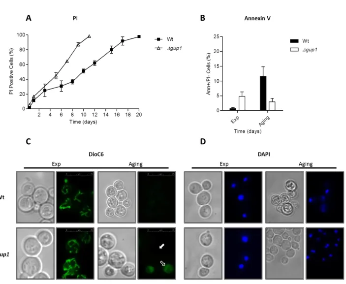
PI, Annexin V, DAPI and DiOC6 staining were performed both in cells treated with acetic acid and in aging cells as previously described, with some modifications (Madeo et al., 1997; Madeo et al., 1999; Ludovico et al., 2001; Gourlay and Ayscough, 2005).
II - 5.1 PI staining
Membrane integrity was assessed by PI (Propidium Iodide) staining. Cells were harvested, washed and resuspended in PBS (137 mM NaCl; 2.7 mM KCl; 100 mM Na2HPO4; 2 mM KH2PO4; pH 7.4) containing PI (4 µg/ml) (Sigma). The samples were
31
incubated for 10 min at room temperature in the dark and analyzed in an Epics® XL™ (Beckman Coulter) flow cytometer. At least 20,000 cells from each sample were analyzed.
II - 5.2 FITC-coupled Annexin V staining
Phosphatidylserine exposure was detected by an FITC-coupled Annexin V reaction with the ApoAlertAnnexin V Apoptosis Kit (CLONTECH Laboratories). For that, cells were primarily harvested and washed in digesting buffer (1.2 M sorbitol; 0.5 mM MgCl2; 35 mM K2HPO4; pH 6.8). To promote the drug course through cell wall, an incubation step with Zymolyase (20T) at 30 °C was performed. Phase-contrast microscopy was used to monitor that step, preventing this way damage to the unfixed spheroplasts. Cells were subsequently centrifuged (10 min at 1500 rpm) and resuspended in 200 µl of binding buffer (1.2 M sorbitol; 10 mM HEPES/NaOH, pH 7.4; 140 mM NaCl; 2.5 mM Cacl2). To 40 µl of this cell suspension, 2 µl Annexin V (1 µg/ml) and 1 µl PI (4 μg/ml) were added, and the mixture incubated for 20 min at room temperature in the dark. Finally, extra 400 µl of binding buffer were added to the mixture just prior to analysis in an Epics® XL™ (Beckman Coulter) flow cytometer. At least 20,000 cells from each sample were analyzed.
II - 5.3 DiOC6 staining
For evaluation of mitochondrial potential the probe DiOC6 (3,3′dihexyloxacarbocyanine iodide) (Invitrogen) was used. Cells were harvested, washed, and resuspended in DiOC6 buffer (10 mM MES; 0.1mM MgCl2; 2% (wt/v) glucose, adjusted to pH 6 set with Ca(OH)2) containing DiOC6 (20 ng/ml). Cells were visualized by light microscopy (LM) after 30 min at room temperature in the dark. Stained cells were visualized in a Leica Microsystems DM-5000B epifluorescence microscope with appropriate filter settings using a 100x oil-immersion objective. Images were acquired with a Leica DCF350FX digital camera and processed with LAS AF Leica Microsystems software. At least 300 cells were counted per sample.