Stéphanie Pereira Oliveira
Outubro de 2012
Study of the role of ceramide in
acetate-induced cell death in colorectal
carcinoma cell lines
UMinho|20 12 St éphanie P er eir a Oliv eir a Study of t
he role of ceramide in ace
tate-induced cell deat
h in colorect
Stéphanie Pereira Oliveira
Outubro de 2012
Dissertação de Mestrado
Mestrado em Genética Molecular
Study of the role of ceramide in
acetate-induced cell death in colorectal
carcinoma cell lines
Escola de Ciências
Trabalho realizado sob a orientação da
Professora Ana Arminda Lopes Preto Almeida
e da
Professora Maria Manuela Sansonetty
Gonçalves Côrte-Real
ii
Declaração
NOME:
Stéphanie Pereira Oliveira
ENDEREÇO ELECTRÓNICO: [email protected] TELEFONE: 918510161 / 252916278
NÚMERO DO BILHETE DE IDENTIDADE: 13642017
TÍTULO DA TESE:
Study of the role of ceramide in acetate-induced cell death in colorectal carcinoma cell lines
ORIENTADORES:
Professora Ana Arminda Lopes Preto Almeida
Professora Maria Manuela Sansonetty Gonçalves Côrte-Real
DESIGNAÇÃO DO MESTRADO:
Mestrado em Genética Molecular
ANO DE CONCLUSÃO: 2012
É AUTORIZADA A REPRODUÇÃO INTEGRAL DESTA TESE APENAS PARA EFEITOS DE INVESTIGAÇÃO, MEDIANTE DECLARAÇÃO ESCRITA DO INTERESSADO, QUE A TAL SE COMPROMETE;
Universidade do Minho, ___/___/____
iii Acknowledgments/Agradecimentos
“Vous savez ce que c’est que la recherche : on part sur une question et on trouve en cours de route des faits qui vous en posent une autre.” – Philippe Meyer
A conclusão deste projeto representa, não só mais um ciclo de estudos, mas também o início de uma nova fase. Mais do que uma simples experiência no laboratório, este projeto foi uma grande ajuda em termos de crescimento pessoal. Obrigado a todos os que, de alguma forma, me ajudaram a concretizá-lo!
À Professora Ana Preto, pela oportunidade de realização deste projeto, integrando a sua equipa de investigação, pela orientação, confiança, incentivo, boa disposição e por todos os ensinamentos e momentos disponibilizados para me apoiar. Muito obrigada!
À Professora Manuela Côrte-Real, por ter aceitado ser minha co-orientadora, pela disponibilidade, pela atenção, pela simpatia e pelas sugestões sempre proveitosas que me deu ao longo do projeto. Obrigada!
À Suellen, Lisandra e Paula, por terem sido ótimas colegas de trabalho, cuja simpatia, entreajuda, disponibilidade e apoio sempre lembrarei. Obrigada, também, pelas gargalhadas, brincadeiras, boa disposição e pelos lanches! Obrigada, de forma geral, a todos os colegas do laboratório de Biologia Animal (Sara Alves e João Silva, inclusive), pelo auxílio, dicas e paciência! A todos o meu sincero agradecimento!
À todas as pessoas que fazem parte e trabalham no Departamento de Biologia, pelo bom ambiente, pela ajuda preciosa sem a qual a realização deste projeto não seria a mesma!
À minha família (especialmente ao “Papi” que sei que está a olhar por mim, onde quer que esteja), pela compreensão e suporte incondicionais, pelas palavras de reconforto quando nada parecia funcionar. Obrigado por todo o amor e amizade! Sem vocês não sei o que seria!
Às minhas “parceiras”, pelos grandes momentos partilhados, pelos desabafos, pelos almoços, saídas, aventuras e por estarem sempre lá quando eu preciso! "Family does not necessarily mean you share DNA. It means you share memories, dreams and love!" – Jared Leto.
iv
Este trabalho foi financiado em parte pela Fundação para a Ciência e Tecnologia através do projeto PTDC/BIA-BCM/69448/2006, pela FEDER através da POFC – COMPETE e pelos fundos nacionais da FCT através do projeto PEst-C/BIA/UI4050/2011.
v Abstract
Colorectal carcinoma (CRC) has one of the highest incidences and mortality on the modern society, but a growing impact on less developed and economically transitioning nations. Prevention, surgery and radio/chemotherapy are the principal approaches to overcome the disease. Finding new efficient/specific treatments in CRC, better tolerated by patients is mandatory.
News strategies to be used against colorectal cancer emerged when it was found that certain strains of Propionibacterium could protect against CRC. Those bacteria, living in the human intestine, produce short-chain fatty acids (SCFAs), namely acetate, butyrate and propionate that are able to induce apoptosis specifically on CRC cells. The mechanisms behind these occurrences are not fully understood. Ceramide is an important signalling molecule, vital in cellular processes and may play a role in regulation of apoptosis in CRC cells. Previous research executed by our laboratory showed that acetate-induced apoptosis in CRC cell lines and lysosome membrane permeabilization (LMP) with cathepsin D release. The fact that ceramide promotes LMP and activates cathepsin D, led us to hypothesize that ceramide might be generated in response to acetate and thus mediate apoptosis induced by this lethal stimulus, in CRC cells.
The main objective of the project was to assess the involvement of ceramide in acetate-induced apoptosis on CRC derived cell lines (HCT-15 and RKO). In order to reach that goal, cells were treated with acetate and/or inhibitors of the ceramide biosynthesis pathways (GW4869, fumonisin B1 and myriocin) and cellular viability was analysed by MTT assay.
The inhibition of the three major pathways of ceramide generation (de novo, salvage and sphingomyelinase pathways) did not change significantly the cellular viability, on both cell lines. Indeed the expected increase in cellular viability did not occur when cells were treated with acetate and one or more inhibitors of ceramide synthesis pathways. Further studies are necessary to confirm the results obtained, namely determining ceramide cellular levels, and the activity of enzymes involved in ceramide synthesis treated with acetate and/or ceramide inhibitors.
Summing up, the results obtained so far suggest that acetate-induced cell death in HCT-15 and RKO CRC cells might not be mediated by ceramide.
vii Resumo
O carcinoma colo-rectal (CCR) tem uma das maiores incidências e mortalidade na sociedade moderna, mas um impacto crescente em países menos desenvolvidos e em vias de desenvolvimento. A prevenção, cirurgia e a rádio/quimioterapia são as principais abordagens usadas no combate à doença. É importante encontrar novos tratamentos mais eficazes, específicos e melhor suportados pelos pacientes.
Novas estratégias para serem usadas contra o CCR surgiram aquando da descoberta de que algumas estirpes do género Propionibacterium poderiam fornecer proteção contra o CCR. Essas bactérias, que vivem no intestino do Homem, produzem ácidos gordos de cadeia curta, como o acetato, o butirato e o propionato, que são capazes de induzir a apoptose específica das células do CCR. Os mecanismos que levam a essas ocorrências não estão bem compreendidos. A ceramida é uma molécula de sinalização importante e vital em processos celulares, e poderá desempenhar um papel na regulação da apoptose em células de CCR. Investigação prévia do nosso laboratório, mostrou que o acetato induz apoptose em linhas de CCR, bem como a permeabilização da membrana lisossomal (PML) com libertação de catepsina D. O facto de se saber que a ceramida promove a PML e activa a catepsina D, levou-nos a colocar a hipótese de que a ceramida poderia ser gerada em resposta ao acetato e poderia mediar a apoptose induzida por esse estímulo letal, em células do CCR.
O principal objectivo do projeto foi avaliar o envolvimento da ceramida na apoptose induzida pelo acetato, em células do CCR (HCT-15 e RKO). Para atingir essa meta, as células foram tratadas com acetato e um ou mais inibidores das vias de síntese da ceramida (GW4869, fumonisin B1 e myriocin) e a viabilidade celular foi analisada recorrendo a ensaios de MTT.
A inibição das três principais vias de produção de ceramida (de novo, selvagem e das esfingomielinases) não alterou significativamente a viabilidade celular em ambas as linhas celulares. De facto, o esperado aumento na viabilidade celular não ocorreu quando as células foram tratadas com acetato e com um ou mais inibidores das vias de síntese da ceramida. Mais estudos são necessários para confirmar os resultados obtidos, nomeadamente a determinação dos níveis celulares da ceramida e a actividade de enzimas envolvidas na síntese da mesma em células tratadas com acetato e um ou mais inibidores das vias de síntese da ceramida.
Resumindo, os resultados obtidos até agora sugerem que a morte celular induzida pelo acetato em células HCT-15 e RKO de CCR parece não ser mediada pela ceramida.
ix Contents: Acknowledgments/Agradecimentos ... iii Abstract ... v Resumo ... vii Contents... ix List of Abbreviations... xi List of Figures ... xv
List of Tables ... xvii
Chapter 1 - General Introduction 1.1 Cancer: incidence, genetics and hallmarks ... 3
1.2 Colorectal carcinoma ... 4
1.2.1 Colorectal carcinogenesis ... 5
1.2.2 Colorectal carcinoma: risk factors, prevention and therapy ... 7
1.3. Relation between intestinal flora and colorectal carcinoma ... 10
1.3.1 Bacterial production of acetate in human gut ... 10
1.3.2 The role of acetate and other SCFAs in colorectal carcinoma cell death ... 11
1.4 Apoptosis: the programmed cell death ... 14
1.4.1 Apoptosis in yeast and its relation to mammalian and colorectal carcinoma cells ... 16
1.5 Ceramide: a lipid second messenger molecule ... 17
1.5.1 Ceramide synthesis: major biosynthetic pathways ... 18
1.5.1.1 The de novo synthesis pathway ... 19
1.5.1.2 The sphingomyelinase pathway ... 19
1.5.1.3 The salvage or sphingosine pathway ... 20
1.5.2 Inhibitors of ceramide biosynthesis ... 21
1.5.3 Role of ceramide in vital cellular processes and apoptosis ... 22
1.5.4 Role of ceramide in colorectal carcinoma ... 25
Chapter 2 - Objectives 2.1 Rationale of the project ... 29
2.2 Objectives ... 30
Chapter 3 - Material and Methods 3.1 Cell culture: cell lines and culture conditions ... 33
x
3.2 MTT reduction assay ... 34 3.3 Cell treatment with acetate and ceramide pathway inhibitors: GW4869, fumonisin B1 and
myriocin ... 36 3.4 Statistical analysis ... 39
Chapter 4 - Results
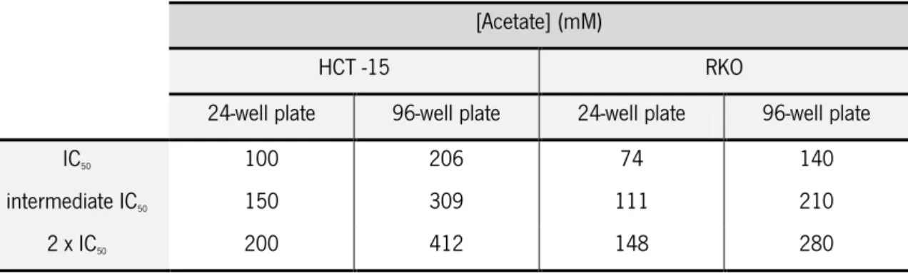
4.1 Optimization of different concentrations of ceramide pathway inhibitors in CRC cell lines ... 43 4.2 Determination of acetate half maximum inhibitory concentration (IC50) for HCT-15 and RKO
cell lines ... 47 4.3 Effect of ceramide pathway inhibition on acetate-induced cell death in CRC cells ... 49
4.3.1 Effect of GW4869 and fumonisin B1 in the acetate response on CRC cell lines ... 49
4.3.2 Effect of GW4869, fumonisin B1 and myriocin in acetate-induced cell death in CRC cells
... 53
Chapter 5 - Discussion
5.1 Effect of ceramide pathways inhibitors on acetate-induced cell death ... 61
5.1.1 Inhibition of the sphingomyelinase pathway: effect of GW4869 on cellular viability of CRC cell lines ... 63
5.1.2 Inhibition of de salvage and de novo synthesis pathways: effect of fumonisin B1 on
cellular viability of CRC cell lines ... 65
5.1.3 Inhibition of the de novo synthesis pathway: effect of myriocin on cellular viability of CRC cell lines ... 67
5.1.4 Effect of inhibiting different ceramide pathways on the cellular viability of HCT-15 and RKO cell lines ... 68
Chapter 6 - Conclusion and Future Perspectives
6.1 Final conclusion ... 73 6.2 Future perspectives ... 73
Chapter 7 - References
xi List of Abbreviations:
µL: Microlitre µM: Micromolar
Akt: Protein kinase B (synonym of PKB) ANT: Adenine nucleotide translocator AP1: Activator protein 1
APC: Adenomatous polyposis coli APO-1: Apoptosis antigen 1 aSMase: Acid sphingomyelinase ATCC: American type culture collection BID: BH3 interacting-domain death agonist BMI: Body mass index
BRAF: v-Raf murine sarcoma viral oncogene homolog B1 BrdU: Bromodeoxyuridine (5-bromo-2'-deoxyuridine) C1PP: Ceramide-1-phosphatase
CAPK: Ceramide activated protein kinases CAPP: Ceramide activated phosphatase
CD95: Cluster of differentiation 95 (synonym of APO-1) CDase: Ceramidase
CerS: Ceramide synthase CIN: Chromosomal instability CK: Ceramide kinase
CO2: Carbon dioxide
CRC: Colorectal carcinoma CRS: Cerebrosidase DAG: Diacylglycerol
xii
DES: Dihydroceramide desaturase DISC: Death inducing signalling complex DMH: 1,2-dimethylhydrazine
DMSO: Dimethyl sulfoxide
ED50: Half maximal effective concentration
ER: Endoplasmic reticulum
ERK: Extracellular signal-regulated kinase FACS: Fluorescence-activated cell sorting FAP: Familial adenomatous polyposis
FB1: Fumonisin B1
GCS: Glucosylceramide synthase GW: GW4869
h: Hours
H2O2: Hydrogen peroxide
HMA: Human-microbiota associated
HNPCC: Hereditary nonpolyposis colorectal cancer
IC50: Half maximal inhibitory concentration
IGF2R: Insuline-like growth factor 2 receptor
IPATIMUP: Institute of Molecular Pathology and Immunology of the University of Porto JNK1: Mitogen-activated protein kinase 8 (MAPK8 synonym)
KRAS: v-Ki-ras2 Kirsten rat sarcoma viral oncogene homolog LDL: Low-density lipoprotein
LMP: Lysosomal membrane permeabilization M: Molar
MAMs: Mitochondria-associated membranes microRNA: Micro ribonucleic acid
xiii
min: Minutes mL: Millilitre mM: Milimolar
MMP: Mitochondrial membrane permeabilization MMR: Mismatch repair
MOMP: Mitochondria outer membrane permeabilization MSI: Microsatellite instability
mTOR: Mammalian target of rapamycin
MTT: Methyl-thiazolyl-tetrazolium (3-(4,5-Dimethylthiazol-2-yl)-2,5-diphenyltetrazolium bromide) Myr: myriocin
NF-Kβ: Nuclear factor Kβ nM: Nanomolar
nSMase: Neutral sphingomyelinase
O2: Oxygen
p53: protein 53 (or tumour protein 53) PBS: Phosphate buffered saline (solution) PC: Phosphatidylcholine
PI: Propidium iodide PKB: Protein kinase B PKC: Protein kinase C PP1: Protein phosphatase 1 PP2A: Protein phosphatase 2A
PTPC: Permeability transition pore complex Rb: Retinoblastoma (protein)
ROS: Reactive oxygen species
xiv
S1P: Sphingosine-1-phosphate S1PP: S1P phosphatase SCFA: Short-chain fatty acid SD: Standard deviation
SEM: Standard error of the mean SK or SphK: Sphingosine kinase SMase: Sphingomyelinase SMS: Sphingomyelin synthase Sph: Sphingosine SPT: Serine palmitoyltransferase SRB: Sulforhodamine B
TGFβ: Transforming growth factor-β TGFβR2: TGFβ receptor 2
TLC: Thin layer chromatography TNF: Tumour necrosis factor TNF-α: Tumour necrosis factor-α
TRAIL: TNF-α-related apoptosis-inducing ligand t-test: Student’s t test
TUNEL: Terminal deoxynucleotidyl transferase dUTP nick end labelling UV: Ultraviolet
VDAC: Voltage-dependent anion channel vMIA: Viral mitochondrial inhibitor of apoptosis
xv List of Figures:
Figure 1.1 – The hallmarks of cancer, emerging hallmarks and enabling characteristics. ... 4
Figure 1.2 – Adenoma-carcinoma sequential model for chromosomal instability, in colorectal cancer. ... 5
Figure 1.3 – Effects of short-chain fatty acids (SCFAs), on colonic epithelial cells, at different phases of the adenoma-carcinoma sequence. ... 12
Figure 1.4 – Representation of the apoptotic intrinsic and extrinsic pathways. ... 15
Figure 1.5 – Ceramide structure representation. ... 17
Figure 1.6 – Pathways of sphingolipid metabolism. ... 18
Figure 1.7 – Ceramide effector pathways relevant to apoptosis. ... 24
Figure 3.1 – (A) Representation of the chemical structure of MTT and its reduced formazan product; (B) absorption spectra of MTT and MTT formazan ... 34
Figure 3.2 – Representation of the molecular structure of GW4869 (A), fumonisin B1 (B) and myriocin (C). . ... 37
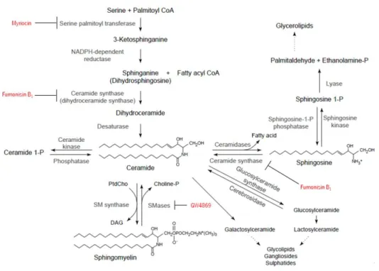
Figure 3.3 – Ceramide biosynthesis pathways and inhibitors: GW4869, fumonisin B1 and myriocin. ... 37
Figure 4.1 – Effect on cellular viability of different concentrations of GW4869 on HCT-15 (A) and RKO (B) cell lines, by MTT assay. . ... 44
Figure 4.2 – Effect on cellular viability of different concentrations of fumonisin B1 (FB1) on HCT-15 (A) and RKO (B) cell lines, by MTT assay. . ... 46
Figure 4.3 – Effect of different concentrations of acetate on cellular viability of HCT-15 (A) and RKO (B) cell lines, determined by MTT assay. . ... 48
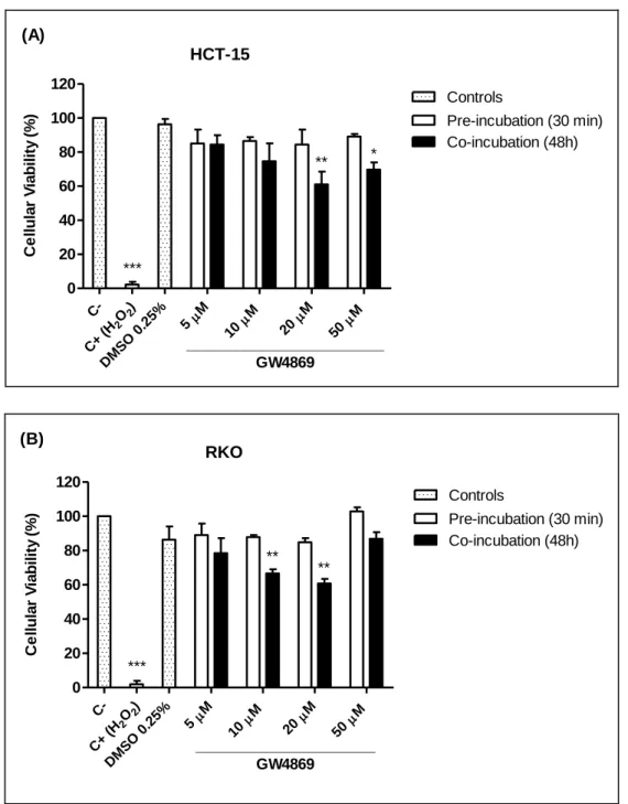
Figure 4.4 – Incubation of HCT-15 (A) and RKO (B) cell lines with GW4869 and acetate and determination of cellular viability, using MTT assay. . ... 50
Figure 4.5 – Incubation of HCT-15 (A) and RKO (B) cell lines with fumonisin B1 and acetate and determination of cellular viability, using MTT assay. ... 52
Figure 4.6 – Incubation of HCT-15 (A) and RKO (B) cell lines with GW4869, fumonisin B1, myriocin and acetate and determination of cellular viability using MTT assay. ... 54
Figure 4.7 – HCT-15 cells, incubated (48h) with acetate and inhibitors of ceramide metabolism (GW4869, fumonisin B1 and myriocin), observed under the microscope, on phase contrast (100x). . ... 56
xvi
Figure 4.8 – RKO cells, incubated (48h) with acetate and inhibitors of ceramide metabolism
(GW4869, fumonisin B1 and myriocin), observed under the microscope, on phase contrast
(100x). . ... 57
Figure 5.1 – Signalling roles of neutral sphingomyelinases in response to potential activators..
... 64
Figure 5.2 – Schematic representation of the sphingolipid rheostat. ... 66
xvii List of Tables:
Table I – Concentrations of acetate used in the MTT assays. ... 38 Table II – Concentrations of acetate used in 96-well cell culture plates for MTT assay. ... 49
1
3
1.1 Cancer: incidence, genetics and hallmarks
Nowadays, in the modern and developed countries and increasingly in developing nations, cancer is a disease with great incidence and affecting a growing number of people.
In 2008, 12.7 million of new cases of cancer and 7.6 million cancer deaths were estimated, with 56% and 63% of new cases of cancer and cancer deaths, respectively, occurring in the less developed regions of the world (Ferlay, Shin et al., 2010). A new tendency has been noticed, while mortality rates are decreasing in many western countries, like the Unites States of America, this rate is growing in less developed and economically transitioning nations, due mostly to the adoption of unhealthy habits more known to western lifestyles (Jemal, Center et al., 2010). More particularly, in Europe, 3.2 million of new cases of cancer and 1.7 millions of resulting deaths were estimated, being colorectal cancers the most common form of the disease found (13.6% of the total) and also the second most common cause of death (12.3% of the total) (Ferlay, Parkin et al., 2010).
Cancer is a complex disease with multiple causes. Nevertheless it could be defined as an atypical growth of cells, caused by several changes in gene expression leading to a disequilibrium between cell proliferation and cell death and, in some advanced cases, evolving into a population of cells capable of tissue invasion and metastization, far from the original location, resulting in an elevated morbidity and, if not treated, death of the host (Ruddon, 2007).
Several events are required to transform a normal cell into a malignant one and such process is called carcinogenesis. As a multi-step disease, cancer is caused by alterations that generally occur in somatic cells (although mutations in germ-line cells can also prompt a person to inherited cancer) affecting oncogenes, tumour suppressor and microRNA genes (Croce, 2008).
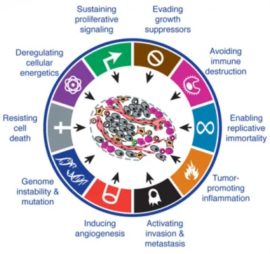
Some traits seemed to be shared by most forms of tumours. In fact, these acquired capabilities are the main consecutive alterations that enable carcinogenesis. There are six principal hallmarks that dictate cancer growth: sustaining proliferative signalling, evading growth suppressors, resisting cell death, enabling replicative immortality, inducing angiogenesis, and activating invasion and metastasis - Figure 1.1 (Hanahan and Weinberg, 2000). More recently, two other characteristics, involved in the pathogenesis of some and maybe all cancers, were added to the previous list, as emerging hallmarks: reprogramming of energy metabolism and evading immune destruction. Additionally, tumour-promoting inflammation and genomic instability and mutation are consequential properties of tumours that facilitate the acquisition of core and emerging hallmarks - Figure 1.1 (Hanahan and Weinberg, 2011).
4
Figure 1.1 – The hallmarks of cancer, emerging hallmarks and enabling characteristics. Adapted from (Hanahan
and Weinberg, 2011).
1.2 Colorectal carcinoma
Among all the different forms of cancer, colorectal carcinoma (CRC) has a great incidence and mortality in world population. According to the GLOBOCAN studies, in 2008, (Ferlay, Shin et al., 2010), CRC was the third most common type of cancer and the fourth cause of death, in both sexes, worldwide, with a major prevalence (60%) in developed countries.
There are two principal forms of colorectal cancer: the sporadic form and the inherited forms, which include the familial adenomatous polyposis (FAP) and the hereditary nonpolyposis colorectal cancer (HNPCC) (Potter, 1999).
The sporadic form is the most frequent form of colorectal cancer (70% of the cases), beginning from somatic mutations and evolving into a tumour. Inherited susceptibility to colorectal cancer may underlie 30% of all diagnosed types: 5% with an evident hereditary base predisposing to the disease and the remaining percentage only showing an elevated propensity of family members to be affected by the disease, without in fact presenting recognizable hereditary syndromes (Rustgi, 2007; Tops, Wijnen et al., 2009). FAP is an autosomal dominant inherited disease, characterized by the emergence of hundreds to thousands of adenomas or adenomatous polyps, during the second and third decade of patients’ lives. Even though these tumours are usually benign and individually not life threatening, some will eventually progress
5
and origin malignant forms of cancer (Kinzler and Vogelstein, 1996). HNPCC (or also called Lynch syndrome), is another inherited autosomal dominant syndrome, that exhibits a much less well characteristic phenotype than FAP and is often confused with sporadic polyposis (Potter, 1999). HNPCC involves a predisposition not only to colorectal cancer, but also to other seven types of cancer, among them: the small bowel, endometrium, ovaries, stomach, brain, hepatobiliary epithelium and uroepithelial epithelium (Lynch, Guirgis et al., 1977). The FAP and HNPCC conditions account to 5% or less of total CRC cancers: less than 1% for FAP and, approximately, 5% for HNPCC (de la Chapelle, 2004).
1.2.1 Colorectal carcinogenesis
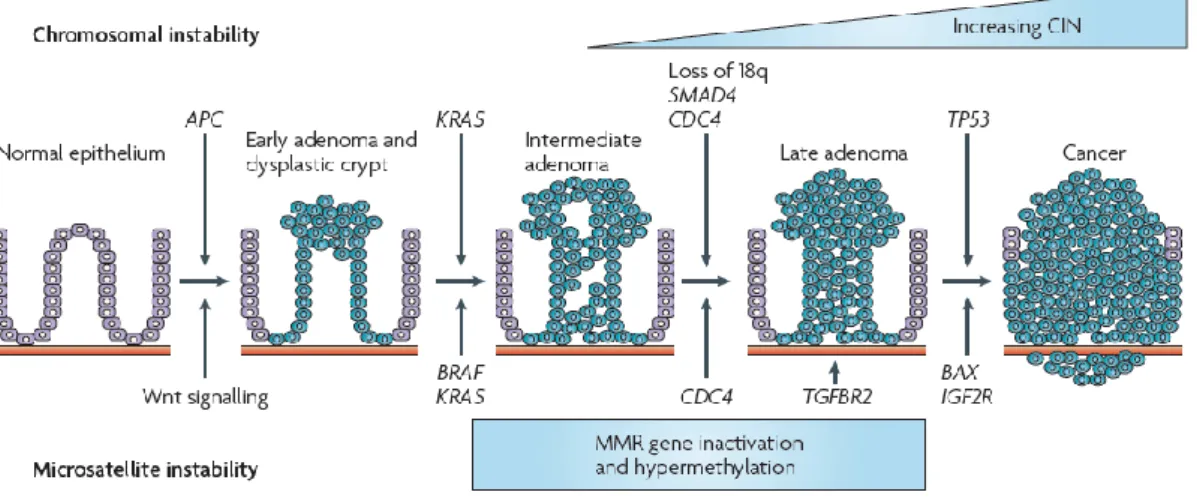
Many genetic and structural alterations are necessary to the development of colorectal carcinoma. In 1990, Fearon and Vogelstein proposed a genetic model for colorectal tumourigenesis. That model evokes a sequential multistep changes, affecting several types of genes, represented in Figure 1.2, and that would consequently lead to progressive morphologic shifts of the colon and rectal apparatus (Fearon and Vogelstein, 1990).
Figure 1.2 – Adenoma-carcinoma sequential model for chromosomal instability, in colorectal cancer. Adapted from
(Walther, Johnstone et al., 2009). APC: adenomatous polyposis coli; TGFβ: transforming growth factor – β; MSI: microsatellite instability; CIN: chromosomal instability; MMR: mismatch repair; TGFβR2: transforming growth factor – β receptor 2; IGF2R: insulin-like growth factor 2 receptor.
To guarantee the CRC evolution, at least, four sequential genetic alterations or “hits” in specific oncogenes and tumour-suppressor genes need to occur. KRAS, an oncogene, and APC,
6
SMAD4 and TP53, three tumour-suppressor genes are the main targets of these changes (Fodde, Smits et al., 2001).
The APC gene has a key role in CRC. Mutations of the germ-line in the APC gene are responsible for the FAP and are also found in non-hereditary colorectal cancers (Tanaka, 2009). The APC gene encodes a protein that has a preponderant role in Wnt signalling pathway, given that APC regulates cell proliferation. The mechanism by which this regulation is made depends on APC binding and degrading of β-catenin to promote cell proliferation (Ilyas, 2005). If APC is mutated this cannot occur and β-catenin is translocated to the nucleus, where it binds to a transcription factor (T-cell factor/lymphocyte enhancer factor). This will activate c-myc, cyclin D1 and c-junc genes and induce cell proliferation (Tanaka, 2009).
Other important functions have been attributed to APC such as the control of intracellular adhesion, that might be achieved by regulating the stability and subcellular localization of β-catenin (a constituent of adherens junctions), being the link between α-β-catenin and E-cadherin, that binds actin and its associated proteins (Ben-Ze'ev and Geiger, 1998). Furthermore, the direct association between APC with the microtubule cytoskeleton was also demonstrated (Munemitsu, Souza et al., 1994; Smith, Levy et al., 1994).
KRAS gene is also an important piece in the carcinogenesis process of CRC. It encodes a protein involved in G protein-mediated transduction. KRAS protein has a GTPase activity, which transmit signal across the cellular plasma membrane through the activation of the RAS-RAF-MEK-ERK-MAPK signalling cascade (Roberts and Der, 2007). The most widespread types of KRAS mutation, in colorectal cancers, are the substitution of glycine to aspartate on codon 12 (p.G12D), glycine to valine on codon 12 (pG12V) and glycine to aspartate on codon 13 (p.G13D) (Neumann, Zeindl-Eberhart et al., 2009). Mutation of KRAS is responsible for unregulated and increased cell proliferation and malignant transformation (Bos, 1989).
BRAF mutations have been identified in several types of human cancers, including colorectal tumours. The frequency of this mutation associated to CRC varies between 1% and 20%, and BRAF mutation is specially connected to tumours with a dysfunction in mismatch repair (MMR) activity (Wellbrock, Karasarides et al., 2004). A transversion (T into A) is the most common BRAF mutation (90% of BRAF mutant tumours), originating a valine to a glutamic acid alteration - V600E (Davies, Bignell et al., 2002). In consequence, there is a major increase in in vitro kinase activity and the stimulation of cell proliferation and transformation (Wan, Garnett et al., 2004). Recently, it has been demonstrated that the inhibition of BRAF in CRC cell lines
7
interfered with the proliferation and apoptosis mechanisms. In BRAFV600E microsatellite unstable
(MSI) cell lines, the suppression of BRAF, using the RNA interference technique, significantly induced apoptosis and inhibited proliferation. In contrast, no significant differences were observed in proliferation and apoptosis in cell lines harbouring a KRASG13D mutation, when
submitted to BRAF inhibition (Preto, Figueiredo et al., 2008). Besides, the study from Preto and collaborators also found that, in BRAFV600E cell lines under BRAF inhibition, the levels of
proliferation associated molecule p27Kip1 are increased and the levels of the anti-apoptotic protein
Bcl-2 are decreased.
The mutation of the TP53 gene is preponderant not only in CRC cases, but also in the majority of known cancers. It is thought to be a relatively late event in the carcinogenesis process of CRC and determinant in the progression from adenoma to a malignant tumour. This gene is mutated in up to 70% of colorectal cancers (Baker, Preisinger et al., 1990). In 1979, TP53 tumour suppressor gene was the first one to be identified. It encodes a transcription factor - p53 protein - implicated in cell cycle control. The p53 protein specifically binds to a recognition sequence in a variety of genes, including p21, Bax, and Bcl-2, when the DNA is damaged or the cell is under stress, if not the p53 network is usually not active (Vogelstein, Lane et al., 2000). p53 has a protective role toward cells, as well as a destructive one. Different stimuli, such as basal low-levels of p53, stress, DNA damage and oncogenic signals can all elicit the p53 system, resulting in different cell responses such as transient cell cycle arrest, autophagy, repair, apoptosis, senescence and necrosis. It is clear, that a shift in p53 output, from a death signalling to a repair one, can promote survival of mutated and damaged cells and increase the probability of tumour cell survival (Junttila and Evan, 2009).
The APC, KRAS, BRAF and TP53 are thought to be the major mutations found in CRC, but the carcinogenesis process is a complex set of events and all the interveners are not known. Additionally, we must be aware of the cross-talk between pathways that augment the tumourigenesis complexity. The more profound is our knowledge about CRC mutations, the bigger is the probability to find new strategies and new approaches to cancer therapy.
1.2.2 Colorectal carcinoma: risk factors, prevention and therapy
The survival rates of CRC have significantly improved in the past years. The acquired knowledge about the disease, the prevention, the early detection and the development of treatments are fundamental reasons that sustain this increase.
8
A crucial part in the diminution of the CRC incidence is the identification of potential dangerous factors that contribute to the malady apparition. Risk factors behind CRC development can be divided in two categories: non-modifiable and modifiable risk factors. Non-modifiable risks factor are age and predispositions factors (incidental polyps, inherited disorders, inflammatory bowel disease, some of these already discussed in the last section). The major part of CRC cases occur in older individuals (age above 50 years), principally because the carcinogenesis process requires several mutations that gather over time. It is known that it would take up to ten years for cells to undergo significant mutations and become malignant (Scholefield, Ritchie et al., 2005). Interestingly ethnicity is also considered a non-modifiable risk factor for CRC. For example, the Ashkemazi Jews are a highly predisposed group in terms of the disease development, due to the I1307K APC mutation that runs among (6-7%) of the population (Locker, Kaul et al., 2006).
Worldwide patterns and variations of CRC incidence are also consequence of modifiable risks. Obesity is an undeniable risk factor for CRC. Some studies showed that the body mass index (BMI) is connected to the incidence of CRC, and that risk suffers an elevation of 25% and 50% in overweight and obese men, respectively (Moghaddam, Woodward et al., 2007). The link between the BMI and colorectal cancers seems to be weaker in women (Murphy, Calle et al., 2000). If obesity is a risk factor, certainly the lack of physical activity is also a factor to be considered, as they are tightly linked. The presence of central adiposity and the absence of regular physical activity is associated with poorer survival rates, after CRC diagnosis (Haydon, MacInnis et al., 2006).
Diet has proven to be a decisive feature to either prevent or be, at least, in part responsible for the development of colorectal cancers. Currently, it is believed that a diet constituted of red meat with low portions of vegetables and fruits are likely to be increasing the risk of cancer (van Duijnhoven, Bueno-De-Mesquita et al., 2009).
The two last modifiable factor risks important to be referred are alcohol consumption and smoking. The uncontrolled alcohol consumption on a regular basis, as well as the habit of smoking, are thought to be associated with the increase in CRC incidence. An increased risk of 16% and 41% is observed in individuals that drink 30-45 g/day and ≥45 g/day, respectively (Cho, Smith-Warner et al., 2004). Concerning the smoking, it very well known that several carcinogenic compounds are found in tobacco, being an important factor in CRC, despite being far more preponderant in other cancer types (Liang, Chen et al., 2009).
9
Having in mind all the risks factors, prevention seems the best way to avoid and decrease the incidence of colorectal cancers. Several research works revealed that diet, as discussed above, could be important to prevent CRC and other form of tumours. Phytochemicals, included in the chemopreventive agent’s category, are found in vegetables and fruits and demonstrated to have a preventive effect on proliferation, to induce apoptosis and to inhibit growth factor signalling pathways. In addition, phytochemicals play a role in reversing chemoresistance and radioresistance, among other positive effects (Dorai and Aggarwal, 2004). A spice, curcumin, common in India has proven to be relevant in CRC prevention by promoting apoptosis, cell cycle arrest and participate in other essential pathways too (Johnson and Mukhtar, 2007). Resveratrol (grapes), capsaicin (chilli peper) and gingerol (ginger) are all natural compounds able to stop tumour cell proliferation and malignant transformation by targeting and suppressing the overexpression of NF-κB (nuclear factor κB) and AP1 (activator protein 1) transcription factors, respectively (Surh, 2003). In addition, the dietary phytochemicals quercetin, luteolin and ursolic acid demonstrated to have anti-proliferative and pro-apoptotic effects on CRC derived cell lines (HCT-15 and CO115) and seemed to act on KRAS and PI3K. Quercetin and luteolin decreased ERK (extracellular-signal-regulated kinase) phosphorylation, in HCT-15 cells, while the three compounds led to decreased Akt phosphorylation on CO115 cells (Xavier, Lima et al., 2009).
A set of products called probiotics and prebiotics are thought to be relevant in the prevention of colorectal carcinoma, for example, the consumption of milk and its derivatives, products containing lactobacilli or bifidobacteria, seems to diminish the CRC incidence (Wollowski, Rechkemmer et al., 2001). Among the effects of pre and probiotics, that appear to reduce the risk of CRC, we can count the changes in the pH conditions in the colon, the lower production of probable carcinogenic product by gut microflora, the modulation of the immune response, the production of anticancer compounds, inter alia (Lim, Ferguson et al., 2005; Fotiadis, Stoidis et al., 2008).
Chemopreventive agents are also able to improve the efficiency of common cancer therapy i.e. the combined use of preventive agents and chemotherapy or radiotherapy (Sarkar and Li, 2006). Further investigation is needed to understand the mechanisms of synergy between chemopreventive agents and usual therapy, to take benefit of it.
10
1.3. Relation between intestinal flora and colorectal carcinoma
Human intestines are colonized by a large and diverse population of microorganisms. Bacteria are the predominant type and are indispensable for our survival and the healthy functioning of our organism. Gut microflora is responsible for multiple tasks as, for instance, saving nutrients and energy acquired from food, controlling the epithelial cell proliferation and differentiation, balancing the immune system homeostasis and protecting, in certain cases, against possible pathogens (Guarner and Malagelada, 2003).
The intestinal flora influence on colorectal carcinogenesis has become increasingly evident. Bacteria have been associated to CRC by production of toxic and genotoxic metabolites that affect intracellular signal transduction (Fasano, 1999). N-Nitroso compounds and bile acids are examples of such metabolites (Rowland, 2009). Other studies show that the metabolic activity of gut microbiota produces toxic products, such as reactive oxygen species (ROS), which lead to DNA damage and chronic inflammation contributing to tumourigenesis. Increasing findings demonstrated that the same agents are implicated in changes in host glycosylation, altering the cellular and sub-cellular distribution of glycans, and those alterations are linked to the neoplastic process (Hope, Hold et al., 2005).
On the other hand, short chain fatty acids (SCFAs) – acetate, propionate and butyrate – seem to have a protective effect against colorectal carcinoma (Scheppach, 1994), as it will be addressed in the next sections.
1.3.1 Bacterial production of acetate in human gut
A substantial part of carbohydrates consumed in human daily routine are fermented by several microorganisms in the colon. This is a result of the ingestion of cellulose (plant cell wall polysaccharides), hemicelluloses and pectins, vulgarly known as dietary fibre (Cummings, 1981).
Bacteria present in human gut are capable of fermentation, not only of carbohydrates (polysaccharide and oligosaccharide) but also of proteins, peptides and glycoproteins precursors in the colon, resulting into pyruvate formation and its conversion into a series of intermediary and end products such as butyrate (butyric acid), acetate (acetic acid), propionate (propionic acid), carbon dioxide, methane, hydrogen and water (Miller and Wolin, 1979; Cummings, 1981).
Between SCFAs products, acetate is the most produced (Cummings and Macfarlane, 1991). There are a vast set of bacteria species involved in acetate formation; among them are
11
the following ones: Anaerotruncus colihominis, Bacteroides goldsteinii, Bifidobacterium, Cetobacterium somerae, Clostridium asparagiforme, Clostridium hathewayi, Dorea longicatena, Eubacterium, Lactbacillus, Streptococcus, Ruminococcus luti, and Victivallis vadensis (Miller and Wolin, 1979; Duncan, Louis et al., 2007).
Short chain fatty acids are absorbed in various zones of the colon and later metabolized at three specific sites in the organism: the ceco-colonic epithelium, the liver and muscles (Hijova and Chmelarova, 2007). Butyrate is used as a substrate to maintain the energy of producing pathways by cells belonging to the ceco-colonic epithelium. In the liver, SCFAs (residual butyrate and propionate) are metabolized and used in gluconeogenesis, also a great part of acetate (50% to 70%) is taken up by this organ. Additionally, a residual amount of acetate is utilized in muscle cells to generate energy from its oxidation. In general, SCFAs are important nutrients for the colonic epithelium cells. They have a role not only in the regulation of gene expression, cell proliferation and differentiation but also as modulators of colon ion transport, cell volume and intracellular pH (Cook and Sellin, 1998). As these compounds are rapidly absorbed in the colon, they are connected to bicarbonate excretion (Binder, Rajendran et al., 2005) and improve sodium absorption (Trinidad, Wolever et al., 1996).
Focusing in acetate metabolism, it is known that it is absorbed in the colon and transported to the liver. Once it enters the systemic circulation, acetate can be a part of lipogenesis that occurs in mammary glands (Hijova and Chmelarova, 2007). Acetate is the most abundant SCFA in the blood and it is the primary substrate for the synthesis of cholesterol, as well as it might be absorbed and used in peripheral tissues (Pomare, Branch et al., 1985).
1.3.2 The role of acetate and other SCFAs in colorectal carcinoma cell death
As active compounds in the organism, SCFAs could have a beneficial effect on health especially in the fight against CRC. In fact, research on this area is evolving and every day a little more is known on the subject.
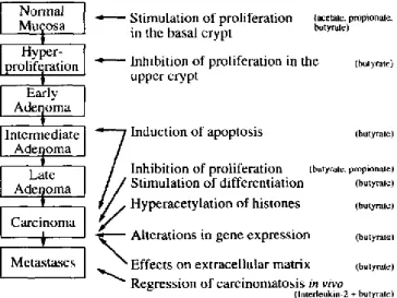
Sources of SCFAs that come from alimentation are nearly entirely dairy products. Once in the organism, they have numerous effects on colonic epithelial cells at different moments: growth, development, transformation and cell death (Cook and Sellin, 1998). Such interactions could explain the preventive effect of SCFAs on colorectal carcinoma (Figure 1.3).
Studies support the notion that production of endogenous SCFAs might diminish the risk of cancer in colonocytes through processes such as apoptosis (Heerdt, Houston et al., 1994) and
12
regulation of histone acetylation (Hinnebusch, Meng et al., 2002). Some experiments realized with HT-29 cells, confirmed that butyrate and propionate are able to enhance histone acetylation and might contribute to biological effects connected to cancer prevention or progression (Kiefer, Beyer-Sehlmeyer et al., 2006).
Figure 1.3 – Effects of short-chain fatty acids (SCFAs), on colonic epithelial cells, at different phases of the
adenoma-carcinoma sequence. Adapted from (Scheppach, Bartram et al., 1995).
On the other hand, programmed cell death is a fundamental process to keep the balance between cell generation and cell death. Interruption or defects on this process would alter this balance and favour tumour progression. All three major SCFAs, acetate, butyrate and propionate, have all proven to induce apoptosis in several adenoma and carcinoma cell lines, but butyrate seemed to be the most efficient and the most studied (McBain, Eastman et al., 1997; Cook and Sellin, 1998). Some interesting studies have been made showing, for instance, that propionibacteria are able to induce apoptosis, in vitro, in two colorectal carcinoma cell lines (HT-29 and Caco 2) by means of acetate and propionate produced by fermentation (Jan, Belzacq et al., 2002). Two species of bacteria were object of the study P. acidipropionici and P. freudenreichii. The effects of propionate and acetate were tested alone or in combination, with a half maximal effective concentration (ED50) of 11 mM and 20 mM respectively, in HT-29 cells.
The SCFAs-induced apoptosis was confirmed by the observation of ROS generation, caspase-3 processing and nuclear chromatin condensation. In addition, the oncoprotein Bcl-2 (known to prevent apoptosis via mitochondria) and the viral mitochondrial inhibitor of apoptosis (vMIA), which interacts with the adenine nucleotide translocator (ANT), both inhibited cell death induced
13
by SCFAs, suggesting that mitochondria is decisively involved in this cell death process (Jan, Belzacq et al., 2002). Others studies also denote the induction of apoptosis of CRC cells by SCFAs (acetate and propionate) involving the mitochondria (Heerdt, Houston et al., 1997; Jan, Belzacq et al., 2002). It is evident that this process may be decisive in the attempt of finding a manner to prevent or treat colorectal cancers.
Another investigation reinforces this finding demonstrating a main impact of a shift in extracellular pH, on the mode of propionibacteria SCFA-induced cell death of HT-29 cells, leading to SCFA-induced cell cycle arrest and apoptosis (Lan, Lagadic-Gossmann et al., 2007) and even leading to apoptosis in vivo (Lan, Bruneau et al., 2008). In 2007, Lan and collaborators demonstrated that P. freudenreichii SCFAs led to apoptosis and induced cell cycle arrest in G2/M phase prior to programmed cell death at pH 7.5. This pH also caused all typical morphological changes known in apoptosis (membrane blebbing, chromatin condensation and fragmentation, phosphatidylserine exposure and formation of apoptotic bodies). Otherwise at a pH of 5.5, the necrotic process was observed, in HT-29 cells, manifested by rapid swelling and disruption of internal organelles and plasma membrane lyses without chromatin fragmentation (Lan, Lagadic-Gossmann et al., 2007).
In 2008, Lan and co-workers also noticed that Propionibacterium freudenreichii could be able of not only killing cells in vitro, but also in vivo, with a remarkable selectivity. Apoptosis and proliferation of colonic epithelial cells were analysed in human microbiota-associated (HMA) rats, fed with P. freudenreichii TL133 strain on a daily routine, 48h hours after the induction of carcinogenesis with 1,2-dimethylhydrazine (DMH). The strain survived in the gastrointestinal tract of rats and demonstrated to induce apoptosis only in DMH-damaged cells, revealing the specificity of these bacteria protective role (Lan, Bruneau et al., 2008).
Recent results, obtained by our laboratory, showed that SCFA and more particularly acetate is able to in induce apoptosis and to inhibit proliferation in HCT-15 and RKO CRC derived cell lines. This SCFA is also responsible for triggering lysosomal membrane permeabilization (LMP) and cathepsin D release to the cytosol (Marques, Oliveira et al., 2012 ; submitted).
All the combined results showed above contribute to the idea that probiotics, such as propionibacteria and their respective metabolic products, could be used as strong agents for colorectal carcinoma prevention and perhaps therapy.
14 1.4 Apoptosis: the programmed cell death
Apoptosis is a cellular process of programmed cell death and is deeply important in development, immune response and tissue homeostasis. More and more studies link this process to the successful triggering of cancer cells death, namely CRC cells, when those seem “immortal”.
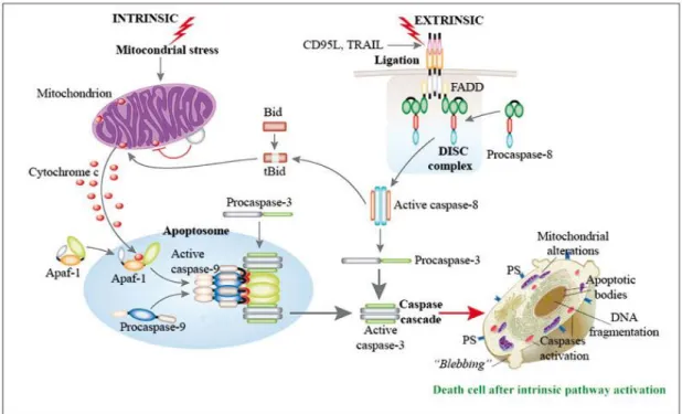
This type of death is characterized by a set of morphological changes beginning with cell shrinkage and chromatin condensation. Cell blebbing occurs and the “budding” process takes place, in other words, cell fragments are separated into apoptotic bodies that consists of cytoplasm (enclosed within an intact plasma membrane), containing tightly packed organelles with or without nuclear fragments (Elmore, 2007). These changes are caused by several cysteine proteases, known as caspases, that participate in two (intrinsic and extrinsic) pathways described for apoptosis (Figure 1.4).
The intrinsic and extrinsic pathways of apoptosis differ in some points. The first pathway is activated by many different types of cellular stress, including some chemotherapeutic compounds, UV and γ-irradiation, reactive oxygen species (ROS), radicals generated as by-product of normal cell metabolism, oncogene activation and DNA damage. Basically, the death stimuli origin comes from within the cell (Yang, Sales et al., 2009). Mitochondria has a central role in this pathway, as the apoptotic stimuli converge in the mitochondrial membrane permeabilization (MMP), releasing some pro-apoptotic proteins into the cytoplasm (such as cytochrome c), thereby triggering the activation of a cascade of caspases (Kroemer and Reed, 2000; Riedl and Shi, 2004).
On the other hand, the extrinsic pathway is activated by the so-called “death ligands”, for example, tumour necrosis factor-α (TNF-α) and TNF-α-related apoptosis-inducing ligand (TRAIL) (Yang, Sales et al., 2009). When they bind to their “death receptor”, a protein complex, called the death inducing signalling complex (DISC), is formed and the activation of the caspase cascade occurs (Riedl and Shi, 2004). In some cases, activation of caspase-8 directly leads to caspase cascade launch while, in other situations, caspase-8 modulates apoptosis through the cleavage of BH3 interacting-domain death agonist (BID), which in turn allows the MMP and
consequent programmed cell death (Li, Zhu et al., 1998).
The programmed cell death is object of a rigorous control. This regulation include proteins of the Bcl-2 family, that contains both agonists (Bax, Bak, Bad, Bcl-XS) and antagonists
15
(Bcl-2, Bcl-XL) of apoptosis (Ligr, Madeo et al., 1998). Those proteins also exercise control over
MMP enhancing or inhibiting it (Kroemer, 2003).
Besides mitochondria, other organelles and cellular compartments have been associated to the regulation of apoptosis: the endoplasmic reticulum (ER) and the lysosome.
The stress upon the ER and its dysfunction (due to the accumulation and aggregation of unfolded proteins, for example) can result in cellular death, through apoptosis, if the stress is prolonged and the response fails restoring the normal ER function (Szegezdi, Logue et al., 2006).
Figure 1.4 – Representation of the apoptotic intrinsic and extrinsic pathways. Adapted from (Calvino-Fernández
and Parra-Cid, 2010).
The lysosome is also involved in programmed cell death. In response of certain stimuli, including ROS, TNF receptor ligation, p53 activation and lipid second messenger sphingosine, lysosome partial membrane permeabilization has been shown to initiate an apoptosis response (Jäättelä, Candé et al., 2004; Boya and Kroemer, 2008). After stimulation, lysosomal membrane permeabilization (LMP) occurs through caspase-dependent mechanisms and releases cathepsins and other hydrolases in the cytosol (Kroemer, Galluzzi et al., 2007). It is thought that cathepsins are able to cleave Bid, leading to Bax activation and consequent release of apoptotic factors from mitochondria, initiating the programmed death of the cell (Cirman, Oresic et al., 2004).
16
1.4.1 Apoptosis in yeast and its relation to mammalian and colorectal carcinoma cells
Over the years, the apoptosis process has been studied using a not so complex model such as the mammalian one: Saccharomyces cerevisiae has been largely used as a eukaryotic model for the understanding of programmed cell death.
In S.cerevisiae, the apoptotic phenotype was found to be triggered by acetic acid (an end product of the alcoholic fermentation realized by this type of yeast), involving mitochondria and the release of cytochrome c in the process (Ludovico, Sousa et al., 2001; Ludovico, Rodrigues et al., 2002). It was also demonstrated that acetic acid had a stimulating effect on the mitochondrial outer membrane permeabilization (MOMP), requiring ADP/ATP carrier (AAC) proteins, which are orthologues of the adenine nucleotide translocator (ANT) of mammalian cells (Pereira, Camougrand et al., 2007). Moreover, cell death triggered by acetic acid in yeast was found to be responsible for the Pep4p (orthologue of mammalian cathepsin D) translocation from the vacuole to the cytosol (Pereira, Chaves et al., 2010).
The mechanisms described above, also happen in mammalian cells. It is believed that MOMP event is due, at least in part, to the formation of the permeability transition pore complex (PTPC), composed by several proteins, in which ANT and the voltage-dependent anion channel (VDAC) are included. The PTPC formation occurs between the two mitochondrial membranes, ANT and VDAC being located in the inner and the outer membrane, respectively (Halestrap, McStay et al., 2002). And with the opening of PTPC, proteins (such as cytochrome c) are released and apoptosis is engaged (Henry-Mowatt, Dive et al., 2004).
The fact that similar events occurs both in yeast and mammalian cells is interesting, but is acetic acid/acetate capable of triggering such events in mammalian cells to? As mentioned in a previous section (1.3.2), evidences point to a certain kind of bacteria, more precisely propionibacteria, that are able to survive in the human intestines and kill colorectal carcinoma cells. These microorganisms, through their metabolism, produce metabolites, SCFAs (acetate, butyrate and propionate) that seem to be responsible for the death of CRC cells, by triggering apoptosis.
In results obtained by our group, it was found that acetate lead to CRC apoptosis and that LMP and release of cathepsin D (to the cytosol) occurred in CRC cells undergoing apoptosis (Marques, 2010 ; Master thesis). The inhibition of cathepsin D with pepstatin A proved to lead to the increase in acetate-induced apoptosis in the HCT-15 and RKO CRC derived cell lines
17
(Marques, Oliveira et al., 2012 ; submitted). In a yeast model, in accordance with the mammalian studies, deletion of the Pep4p confers higher susceptibility to acetic acid, while the overexpression of this molecule confers higher resistance to yeast cells (Pereira, Chaves et al., 2010). Thus, the role of cathepsin D may be protective over the process of acetate-induced apoptosis. Moreover, the overexpression of cathepsins have been linked to cancer, poor prognosis and higher risk of recurrence (Palermo and Joyce, 2008), although studies have been showing the emerging role of these proteases as tumour suppressor (López-Otín and Matrisian, 2007).
Every day, more and more is known about the mechanisms and routes of signalizations used by SCFAs to induce programmed cell death, but more studies are needed in order to unveil such processes and maybe provide a new strategy of CRC therapy/prevention.
1.5 Ceramide: a lipid second messenger molecule
Ceramide belongs to the sphingolipid family and is considered as a biologically active molecule due to its role as lipid second messenger. Ceramide and other active sphingolipids are involved in the regulation of diverse cellular responses to exogenous stimuli (Spiegel, Foster et al., 1996). A regulating role has been attributed to ceramide in many signalling pathways involving the cell cycle, differentiation, senescence and apoptosis. This is the main reason why ceramide action and the regulation of its production have recently attracted the scientific community attention. In major part, effects of ceramide are antagonistic to growth and survival (Ruvolo, 2001).
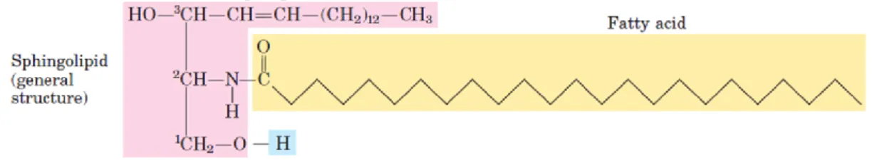
Figure 1.5 – Ceramide structure representation. Adapted from (Nelson and Cox, 2004).
Sphingolipids molecules are constituents of all eukaryotes as membrane cells components and they can be found in milk fat globule membranes, skin lamellar permeability barrier and lipoproteins (Schmelz, 2004). Their basic constitution is one molecule of the long-chain amino alcohol – sphingosine (or one of its derivatives) and a long-long-chain fatty acid. A polar head group is connected to the structure by a glycosidic or a phosphodiester linkage, according
18
to the situation. The ceramide formation occurs when a fatty acid is attached in an amide linkage to the –NH2, on the C-2 (Figure 1.5), and this represents the structural origin of all sphingolipids (Nelson and Cox, 2004).
1.5.1 Ceramide synthesis: major biosynthetic pathways
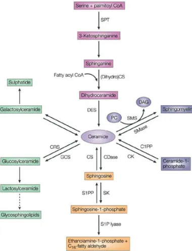
Ceramide is the central point of sphingolipid metabolism. It serves as a crucial local of sphingolipid accumulation and it is a precursor for all major sphingolipids in eukaryotic cells. Synthesis of ceramide can occur through three major pathways: the de novo synthesis pathway, the sphingomyelinase (SMase) pathway and the salvage or sphingosine pathway (Reynolds, Maurer et al., 2004), represented in Figure 1.6.
Figure 1.6 – Pathways of
sphingolipid metabolism. Adapted from (Ogretmen and Hannun, 2004). The de novo pathway (pink); sphingomyelinase (SMase) pathway (blue); salvage pathway (orange); cerebrosides (green). Ceramidases, CDases; sphingosine kinases, SKs; sphingosine-1-phosphate, S1P; C1PP, ceramide-1-phosphate phosphatase; CRS, cerebrosidase; CK, ceramide kinase; CS, ceramide synthase; DAG, diacylglycerol; DES, dihydroceramide desaturase; GCS, glucosylceramide synthase; PC, phosphatidylcholine; S1PP, S1P phosphatase; SMS, sphingomyelin synthase; SPT, serine palmitoyltransferase.
19
1.5.1.1 The
de novo
synthesis pathwayThe de novo pathway begins with the condensation reaction between the amino acid serine and palmitoyl-CoA resulting in 3-ketosphinganine formation. After that, the compound is reduced by the 3-ketosphingamine reductase enzyme, forming sphinganine and, by the action of ceramide synthase, dihydroceramide is formed. Finally, dihydroceramide is converted into ceramide, adding to it a trans-4,5 double bond, by the action of a desaturase (Merrill, 2002). In an alternate manner, this pathway may re-utilize sphingosine released after the degradation of more complex sphingolipids (Reynolds, Maurer et al., 2004).
The de novo synthesis is believed to occur in the endoplasmic reticulum (ER) and probably in ER-associated membranes including the perinuclear membrane and mitochondria-associated membranes – MAMs (Hannun and Obeid, 2008).
The stimulation of the pathway can be achieved by drugs and ionizing radiation that normally results in a prolonged ceramide elevation (Liao, Haimovitz-Friedman et al., 1999). Some chemotherapeutic agents can also enhance the de novo pathway stimulation; etoposide and daunorubicin are examples of them (Bose, Verheij et al., 1995; Perry, Carton et al., 2000) . Others inducers of apoptosis like the B-cell receptor, seem to have the same activating effect (Kroesen, Jacobs et al., 2003). Obviously, the metabolic loading of either serine or palmitate can lead to ceramide production, as well as oxidized low-density lipoprotein - LDL (Kitatani, Nemoto et al., 2002), cannabinoids (Gómez del Pulgar, Velasco et al., 2002) and heat stress (Jenkins, Cowart et al., 2002). Studies realized have largely implicated this pathway in mediating some effects of the inducers, described above, on stress responses and apoptosis (Kitatani, Idkowiak-Baldys et al., 2008).
1.5.1.2 The sphingomyelinase pathway
Ceramide can be generated through the activation of sphingomyelinases (SMases), a special class of phospholipid phosphodiesterases, found in cell membranes. These enzymes are responsible for hydrolyzing sphingomyelin to originate ceramide and phosphocholine (Jones, Hannun et al., 2005).
Different types of SMases exist, differentiated mainly by their optima (acid, neutral or alkaline) pH. The acid SMase (aSMase) has an optimum pH between 4.5 and 5.0 and was initially considered to be strictly located in the lysosome. Nevertheless, an isoform has been
20
described to be present in vesicles near the plasma membrane and, under certain conditions, the aSMase could also relocate to the outer leaflet of the plasma membrane (Grassmé, Schwarz et al., 2001; Hannun and Obeid, 2008). Furthermore, aSMase was shown to be secreted extracellularly (Schissel, Keesler et al., 1998). In sum, there may be three types of aSMase: the acidic lysosomal aSMase (responsible for the sphingomyelin metabolism), the secretory aSMase (associated with inflammation and stress responses) and the receptor-activated aSMase (that translocates to the outer cell membrane, after activation by various cell surface receptors and hydrolyzes membrane sphingomyelin into ceramide).
Two neutral SMases (nSMases) have been identified: nSMase1 and nSMase2. Although both proteins are capable of converting sphingomyelin into ceramide, evidences are against a role for nSMase1 in regulating this reaction in cells (Tepper, Ruurs et al., 2001). The nSMase2 has an optimum pH of 7.4 and appears to be localized to the inner leaflet of the plasma membrane (Hannun and Obeid, 2008).
The alkaline SMase (alk-SMase) has an optimal alkaline pH at 9 and its activity is dependent on bile salts (Hertervig, Nilsson et al., 1997). This enzyme is expressed in the intestinal mucosa in many species and human bile too. In the intestinal tract, alk-SMase activity is superior in the jejunum and lower in the duodenum and colon, being responsible for the hydrolysis of dietary sphingomyelin (Duan, 2006).
Sphingomyelinase pathway is stimulated in response to cell treatment with TNF-α, Fas ligand or oxidative stress (Hannun, Luberto et al., 2001; Marchesini and Hannun, 2004).
1.5.1.3 The salvage or sphingosine pathway
Another important mechanism for ceramide generation is the salvage pathway, in which sphingosine (produced from the metabolism of complex sphingolipids, mostly in the lysosome) is re-cycled to ceramide, localized then in the ER or associated membranes (Hannun and Obeid, 2008). Various enzymes are part of this pathway including ceramidases, (dihydro)ceramide synthase, SMases and probably glucocerebrosidase (acid-β-glucosidase).
This alternative via of ceramide generation is thought to be crucial for sphingolipid breakdown, in sphingolipid synthesis/turnover as well as in cellular signal transduction. These assumptions are based on the fact that the salvage pathway, leading to the re-regeneration of sphingolipids, has been estimated to contribute in 50% to 90% in sphingolipid biosynthesis (Tettamanti, Bassi et al., 2003).
21
Salvage pathway is stimulated in response to cell treatment with TNF-α (Kim, Linardic et al., 1991), Fas ligand (Brenner, Ferlinz et al., 1998) or oxidative stress (Goldkorn, Balaban et al., 1998).
1.5.2 Inhibitors of ceramide biosynthesis
Certain compounds and molecules have an inhibitory action upon ceramide synthesis and are able to stop those pathways at different levels. The same effect can be obtained using other techniques such as the knockdown of a particular enzyme of the pathway, or using microRNA. Some noteworthy inhibitors will be briefly described below.
Ceramide de novo synthesis can be inhibited at the serine palmitoyltransferase (SPT) level by an antifungal molecule called sphingofungin B (Zweerink, Edison et al., 1992). Myriocin, an antifungal antibiotic, also stops the action of SPT. It was described as possessing immunosuppressant properties (Miyake, Kozutsumi et al., 1995) and, after some developments, an analogue of this molecule was created – FTY720 (Adachi, Kohara et al., 1995).
Fumonisins (Desai, Sullards et al., 2002), originated by Fusarium verticilioides and Fusarium moniliforme; the fumonisins related AAL-toxins, produced by the fungus Alternaria alternata (Winter, Gilchrist et al., 1996); australifungins (Mandala, Thornton et al., 1995), micotoxins coming from Sporormiella australis are among the most studied inhibitors of ceramide synthase (CerS). Among all, fumonisin B1 (FB1) is the most representative member of
this class of compounds. This molecule is a powerful inhibitor of CerS, acting in both de novo and salvage pathways of ceramide production (Kitatani, Idkowiak-Baldys et al., 2008).
Inhibition can also happen in the SMase pathway, by cessing the activity of the different SMases. For example, desipramine is a tricyclic antidepressant that acts upon aSMase inducing its proteolysis (Elojeimy, Holman et al., 2006). Several compounds have been described as inhibitors of nSMase. Manumycin A irreversibly inactive the nSMase (Arenz, Thutewohl et al., 2001) while Scyphostatin has a reversible effect, but also reversibly inhibits the aSMase (Nara, Tanaka et al., 1999; Arenz and Giannis, 2000). Sphingolactones, a new family of molecules, are also reported as irreversible inhibitors of the neutral SMase (Wascholowski and Giannis, 2006). During a high throughput screening on Mg2+ dependent nSMase, a compound called GW4869
was discovered and exhibited inhibitory properties against this enzyme (Delgado, Casas et al., 2006).
22
1.5.3 Role of ceramide in vital cellular processes and apoptosis
Sphingolipid metabolism and, more particularly, ceramide have been implicated in many cellular processes. Ceramide has been seen as a coordinator of cellular stress responses, taking part of mechanisms as the cell cycle, differentiation, cell senescence and apoptosis.
During a growth phase, ceramide and phosphatidylcholine are converted into sphingomyelin (Hannun, 1994) and diacylglycerol (DAG), a product of this reaction, is a strong protein kinase C (PKC) activator (Nishizuka, 1995). This activation of PKC pathways by DAG sustain cell survival (Ruvolo, 2001). On the opposite, upon stress conditions, ceramide is produced by the breakdown of sphingomyelin (by SMases) (Hannun and Luberto, 2000). Sphingomyelin synthase appears to regulate DAG and ceramide possibly balancing the fate of cells between growth and apoptosis (Luberto and Hannun, 1998).
Some studies reported that differentiation of leukaemia cells happened when they were treated with ceramide and its analogues, imitating the effects of TNF-α, 1α, 25-dihydroxyvitamin D3 and γ-interferon, on the cell line studied (Okazaki, Bielawska et al., 1990).
Ceramide also demonstrated to be important when cells enter in the senescence phase, since its quantity increases (4-fold) in a human fibroblast cell line (Venable, Lee et al., 1995), but much remains to be elucidated about the mechanisms behind those observations.
Autophagy is a vital procedure in a living cell and it has also been connected to ceramide. Some models have shown that ceramide-induced autophagy is a response to starvation, resulting from nutrient transporter downregulation (Peralta and Edinger, 2009). Moreover, ceramide-mediated macroautophagy resulted in up-regulation of beclin 1 and inhibition of protein kinase B - PKB (Scarlatti, Bauvy et al., 2004). Besides, after proving that ceramide signalling occurs through the activation of Akt/PKB, upstream of mTOR, it was shown that ceramide stimulates JNK1 to phosphorylate Bcl-2, after dissociation of beclin 1 (Pattingre, Tassa et al., 2005; Pattingre, Bauvy et al., 2008; Pattingre, Bauvy et al., 2009).
Over the years, ceramide role in apoptosis has attracted many attentions due to the finding of its pro-apoptotic role.
Environmental stresses and several cytokines (that trigger apoptosis) such as TNF, CD95/Fas/APO-1, ultraviolet-C, ionizing radiation, oxidative stresses, heat shock, chemotherapeutic agents, among others, appear to quickly induce ceramide production (Pettus, Chalfant et al., 2002). In fact, it is important to refer that the doses of these agents necessary to produce ceramide are really close to those required to induce apoptosis (Kolesnick and Kronke,