Accidental cystectomy during laparoscopic excision of
prostatic utricle cyst - a rare complication
_______________________________________________
Vikash Kumar
1, Chirag Punatar
1, Kunal Jadhav
1, Sharad Sagade
11 Department of Urology, P. D. Hinduja National Hospital and Medical Research Centre, Mumbai, Maharashtra, India
ABSTRACT
ARTICLE
INFO
______________________________________________________________ ______________________
Prostatic utricle cyst is a rare congenital anomaly. Symptomatic cysts require treat-ment. Surgical excision is the treatment of choice, but is challenging due to close prox-imity to vas deferens, ejaculatory ducts, bladder, prostate, rectum and pelvic nerves. Complications include rectal injury, ureteral injury, impotence, infertility and faecal incontinence. We here report a rare complication in which bladder was accidentally re-moved during laparoscopic excision of prostatic utricle cyst. To best of our knowledge such a complication has never been reported previously. We also describe the possible cause of this accident and suggest ways to prevent this disastrous complication.
INTRODUCTION
Prostatic utricle cyst is a rare congenital anomaly. Cysts vary in size and presentations di-ffer. Symptomatic cysts require treatment (1). Sur-gical excision is treatment of choice. Surgery can have its own complications. We report a rare case of accidental urinary bladder cystectomy during laparoscopic excision of prostatic utricle cyst.
CASE REPORT
A 24 year old male presented to us with urinary diversion by bilateral percutaneous ne-phrostomies (PCN), performed six months ago. He
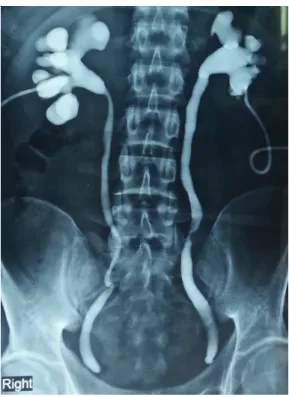
had undergone laparoscopic surgery for removal of prostatic utricle cyst elsewhere. Postoperatively he developed anuria. A sonogram revealed bilate-ral hydro-ureteronephrosis. Bladder was not com-mented upon. This acute crisis was treated by bi-lateral PCN. Nephrostomogram revealed complete cut-off of both lower ureters (Figure-1).
He had history of lower abdominal pain with burning micturition on and off since two ye-ars. Investigations had revealed a prostatic utricle cyst with infection. Following conservative mana-gement, he was asymptomatic for about 18 mon-ths. Recurrence of symptoms was associated with increase in cyst size (Figure-2). Surgical treatment was advised at this time. Laparoscopic cyst
exci-Keywords:
Cystectomy; Intraoperative Complications; Prostate
Int Braz J Urol. 2018; 44: 826-30
_____________________
Submitted for publication: May 19, 2017
_____________________
Accepted after revision: July 08, 2017
_____________________
Figure 1 - Nephrostomogram showing complete cut off at the level of lower ureter on both sides (done after being operated elsewhere).
Figure 2 - Preoperative CT scan showing the bladder anteriorly and prostatic utricle cyst posteriorly.
sion was undertaken which resulted in anuria leading to emergency bilateral PCN. Patient presented to us six months later.
Systemic examination was normal. Abdomi-nal examination revealed port site scars, bilateral ne-phrostomies and coronal hypospadias. Investigations revealed normal hemogram and creatinine. Bilateral
were dissected, prostatic utricle cyst was mar-supialized, it’s opening into urethra closed and Studer’s orthotopic ileal neobladder was cons-tructed.
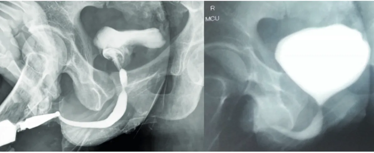
Postoperative MCU showed good capaci-ty neobladder and no extravasation (Figure-5). Nephrostomies were clamped and removed. lower ureteric injury was the suspected diagnosis ini-tially. Ascending and micturating cysto-urethrogram (MCU) showed smooth walled bladder with mildly reduced capacity and normal urethra (Figure-3).
With evidence of bilateral ureteric cut-off and normal lower urinary tract, bilateral ureteric re--implantation was planned. Urethrocystoscopy sho-wed normal anterior urethra. There was an opening on verumontanum, which accommodated 17 French cystoscope sheath easily. This lead to a smooth walled cavity containing about 200 mL of turbid fluid. The epithelium was not like normal urothelium. Ureteric orifices were not seen. Then we realized that this ca-vity was indeed the cyst which was falsely mistaken as bladder on MCU. The proximal urethra was com-pletely cut off below the level of bladder neck, ending blindly. A situation of accidental urinary bladder cys-tectomy and not prostatic utricular cyscys-tectomy was realized. Further surgery was abandoned.
At follow-up, he was voiding well with minimal residue and no incontinence. His outco-mes in terms of ejaculation are yet not known.
DISCUSSION
Prostatic utricles are remnants of Mulle-rian ducts. Normally MulleMulle-rian ducts regress un-der influence of mullerian regression factor and is represented by appendix testis (cephalad part) and utricle (caudal part). Utricular anomalies result from incomplete regression of Mullerian ducts or incom-plete androgen mediated closure of the urogenital sinus in form of prostatic utricular cyst (2).
Figure 3 - Ascending cysto-urethrogram and MCU showing normal anterior urethra and a smooth walled small capacity bladder (which in retrospect was actually the prostatic utricle cyst).
Figure 4 - MRI showing the ureters anteriorly and the prostatic utricle cyst posteriorly. The bladder is absent.
Mullerian duct remnants are uncommon. Incidence of enlarged prostatic utricle is 11-14% in association with hypospadias or intersex anomalies, more so with perineal hypospadias (>50%). About 10-25% show an association with renal agenesis / dysgenesis and 25% cases with hypospadias (1).
The relation of cyst size and symptoms is deba-ted (1, 2).
Symptomatic cysts require intervention. Modalities include endoscopic deroofing, trans-rectal ultrasound guided or transperineal cyst aspiration and sclerotherapy, endoscopic fulgu-ration of cyst lining (1, 2), marsupialisation of cyst into bladder (3), open / laparoscopic (4) excision of cyst, and recently robot assisted surgical excision (5).
Surgical excision is the treatment of choice, but is challenging due to close proximi-ty to vas deferens, ejaculatory ducts, bladder, prostate, rectum and pelvic nerves. Approa-ches described include abdominal extravesical, transvesical (transtrigonal), perineal and ante-rior or posteante-rior transrectal sagittal approaches. Complications include rectal injury, ureteral injury, impotence, infertility and faecal incon-tinence (2).
Figure 5 - Postoperative MCU showing a good capacity neobladder with no extravasation. The urethra appears normal. There is reflux of contrast on both sides.
In our patient there was accidental re-moval of urinary bladder. To best of our kno-wledge, such complication has never been re-ported. Possibly Foley catheter was lodged into utricular cyst, misleading the surgeon to excise urinary bladder.
Preoperative cystourethroscopy could have shown that opening of utricle cyst was directly in line with urethra, bladder neck was high at an angle, and would have more clearly shown anatomical relationship of cyst with bla-dder.
Preoperative cannulation of cyst has been described to facilitate laparoscopic identi-fication and mobilization of cyst (4).
Intraoperative flexible cystoscopy helps in identifying the bladder from cyst, by seeing light of cystoscope during laparoscopy. By chance, if bladder gets injured, it could be iden-tified immediately.
Preoperative cannulation of ureters with ureteral catheters helps in identifying them intraoperatively. By seeing the structure into which ureters enter, bladder could have been differentiated from cyst. Ureteral injury could be identified intraoperatively.
Adopting above mentioned measures could potentially avoid such disastrous complication.
ACKNOWLEDGEMENTS
The National Health and Education Society (work done at Hinduja Hospital, Mumbai)
CONFLICT OF INTEREST
None declared.
REFERENCES
1. Coppens L, Bonnet P, Andrianne R, de Leval J. Adult müllerian duct or utricle cyst: clinical significance and therapeutic management of 65 cases. J Urol. 2002;167:1740-4.
3. Moore v, Howe GE. Müllerian duct remnants in the male. J Urol. 1953;70:781-8.
4. Yeung CK, Sihoe JD, Tam YH, Lee KH. Laparoscopic excision of prostatic utricles in children. BJU Int. 2001;87:505-8. 5. Goruppi I, Avolio L, Romano P, Raffaele A, Pelizzo G.
Robotic-assisted surgery for excision of an enlarged prostatic utricle. Int J Surg Case Rep. 2015;10:94-6.