ISSN 0103-8478
Nicolás Oliveira MegaI Luís Fernando ReversII
Developing a rapid, efficient and low cost method for rapid DNA extraction from
arthropods
Desenvolvimento de um método rápido, eficiente e de baixo custo para extração de DNA de artrópodos
IPrograma de Pós-graduação em Genética e Biologia Molecular, Universidade Federal do Rio Grande do Sul (UFRGS); Departamento de Zoologia, 91501-970, Porto Alegre, RS, Brasil. E-mail: [email protected]. Autor para correspondência.
IICentro Nacional de Pesquisa de Uva e Vinho, Empresa Brasileira de Pesquisa Agropecuária, Bento Gonçalves, RS, Brasil.
ABSTRACT
Here, it is presented a rapid and efficient method to obtain good quality DNA from small samples of arthropod tissues generating low quantities of hazardous wastes. This new method was compared with another homemade protocol using phenol and other two commercial kits. The quality of DNA obtained was checked by spectrophotometer and evaluated by an AFLP assay. Low shearing DNA was obtained from all samples and the best readings were observed to DNA recollected with the new method. The AFLP assay indicated that DNA obtained with all methods were suitable for use in molecular biology techniques sensitive to contaminants. However, homemade protocols were more efficient in recollect DNA than commercial kits, without lose any quality of samples. Also, they were less time and fund consuming, with costs ten times cheaper than commercial kits. The quicker, less pollutant and cheaper protocol was the one described here (USD 0.52 per sample).
Key words:arthropod DNA, high quality DNA, molecular biology, AFLP.
RESUMO
Aqui, é apresentado um método rápido e eficiente para obtenção de DNA de boa qualidade a partir de pequenas amostras de tecidos de artrópodos, gerando pequenas quantidades de resíduos perigosos. Comparamos a eficiência do método com outro protocolo caseiro utilizando fenol e com dois kits comerciais. A qualidade do DNA obtido foi verificada em espectrofotômetro e avaliada por um ensaio de AFLP. Foi obtido DNA pouco fragmentado a partir de todas as amostras, mas as melhores leituras foram obtidas para o DNA extraído com o novo método. O ensaio de AFLP indicou que os DNAs
obtidos estavam adequados para uso em técnicas de biologia molecular sensíveis a contaminantes. Porém, os protocolos caseiros foram mais eficientes em extrair DNA do que kits comerciais, sem perder nenhuma qualidade na pureza das amostras. Além disso, eles foram mais rápidos e baratos, chegando a custar dez vezes menos que os kits comerciais. O protocolo mais rápido, menos poluente e mais barato foi o descrito aqui (USD 0,52 por amostra).
Palavras-chave:DNA de artrópodos, DNA de alta qualidade,
biologia molecular, AFLP.
INTRODUCTION
concern on these methods have been the quantity and quality of DNA. The quality of samples depends on biological material as well as on protocol followed (REINEKE et al., 1998; CHEN et al., 2008).
Homemade DNA extraction protocols are cheap and very efficient to recollect DNA from samples, but are usually time-consuming; commercial kits are generally very fast but too expensive and generate large quantities of contaminant wastes, such as beads, filters, columns and microcentrifuge tubes. Also, homemade DNA extraction protocols are less efficient in the removal of proteins/carbohydrates and other organic contaminants from samples or extraction solutions used, thus causing enzymatic inhibition in further molecular analysis. In these cases the use DNA clean-up steps are required, which can be time and fund consuming. An ideal protocol should optimize DNA yield, minimize degradation, and be efficient in terms of cost, time, labor, and supplies (CHEN et al., 2010).
REINEKE et al. (1998) evaluated several DNA extraction protocols using insect tissue samples and determined the quality of DNA recollected by Amplified Fragment Length Polymorphism (AFLP). The study also investigated the effect of additional clean-up steps on the improvement of DNA quality, concluding that the best protocols to obtain good quality DNA for AFLP are phenol or phenol-chloroform based methods, but for these protocols additional purification steps are recommend. Cationic detergent cetyltrimethyl ammonium bromide (CTAB)-based methods, as described in MOELLER et al. (1992), demonstrated to provide good quality DNA samples, using less hazardous chemicals, by combining its use with some proteinase and ribonuclease (RNAse), which can cause more fund consuming.
Here, it is described a method to obtain good quality DNA from small samples of freshly and frozen insect tissues. The method is CTAB-based and does n ot use ph enol, a hazardous chemical an d a contaminant in DNA samples. Sodium Chloride (NaCl) has been used to remove polysaccharides (FANG et al., 1992), and polyvinyl pyrrolidone (PVP) to purge polyphenols from arthropods diet (MALIYAKAL, 1992). To improve DNA quality it was used cross-linked polyvinyl pyrrolidone (PVPP) to remove alkaloids, terpen oids, glycosides, ph enols, ph en azin es polyketides and peptides, as well as protein and carbohydrate (HAAF et al., 1985). The main objective of this research was to develop a quick, cheap and less hazardous alternative to protocols available on literature and market.
MATERIAL AND METHODS
Species and tissue samples
Two flies, two butterflies, one moth, two spiders and three isopods species were used as biological material for test the protocol’s efficiency to extract DNA from tissue samples. The species used were, respectively, Anastrepha fraterculus, Drosophila immigrans, Dryas iulia alcionea, Heliconius erato phyllis, Grapholita molesta, Paratrechalea azul, Paratrechalea ornata, Benthana cairensis, Balloniscus glaber, Balloniscus sellowi. All specimens were collected from nature and weighted. The tissue sources used for DNA extraction were whole body (flies, isopods and moth), head and thorax (butterflies) and cephalothorax (spiders). Each sample was frozen in liquid nitrogen and immediately crushed into a fine powder in 1.5ml sterile microcentrifuge tubes. The DNA extraction protocol
Crushed tissues were mixed with PVPP (Sigma, P6755) in a proportion of 100mg of PVPP per 1g of grinded tissue, 600L of extraction buffer (50mM Tris pH 8.0, 20mM EDTA pH 8.0, 1.1M NaCl, 0.4M LiCl, 1% CTAB, 2% PVP40 (Sigma), 0.5% Tween 20, 0.2% -mercaptoethanol) in 1.5ml microcentrifuge tubes and mixed thoroughly. The tubes were incubated at 60°C for 25 minutes and inverted each 10 minutes for good solution of the crushed tissues with the buffer. The tubes were cooled at room temperature and 600L of chloroform: isoamyl alcohol mixture (24:1) was added. The tubes were then mixed gently by inversion to form an emulsion during 4 minutes. After that the tubes were centrifuged (Eppendorf Centrifuge 5415R) at 10.600g for 5 minutes at room temperature. The aqueous phase present in each tube (~500L) was transferred to a new 1.5ml microcentrifuge tube gently, to avoid DNA shearing. To each tube was added 250L of 5M NaCl and 750L of cold (-20°C) isopropanol. The solution was kept in the freezer at -20ºC for 20 minutes to improve precipitation nucleic acids. Samples were then centrifuged at 10.600g (Eppendorf Centrifuge 5415R) for 10 minutes at room temperature and the aqueous poured off. DNA pellets were washed with cold (4°C) 76% ethanol (1000L). After the washing step, the samples were spun quickly and the excess of ethanol removed with a micropipette. Washed DNA pellets were dried by leaving the tubes uncovered in a 37°C stove for 20 minutes. DNA samples were dissolved in 50L TE (10mM Tris HCl pH 8.0, 0.1mM EDTA pH 8.0) and treated with 1L of RNAase A (10mg mL-1) at 50°C for
Quality and quantification tests
Integrity of the DNA samples was analyzed in 0.8% ethidium bromide-stained agarose gels, using 1Kb Ladder (Fermentas) as control for DNA shearing. Visual patterns of DNA samples were photographed under ultraviolet light and scanned with Stratagene EagleEye II still video system. The DNA samples were quantified using a spectrophotometer (GeneQuant PRO, Amersham Biosciences) in the absorbance spectrum for nucleic acids (260 m). To access DNA purity, 230 m and 280 m readings were recorded and compared with 260 m readings. The 230 m reading can indicate the presence of organic impurities and buffer contaminants, once phenols, carbohydrates, peptides, urea, thiocyanates, -mercaptoethanol, humic acids and other buffer components absorb light at this wavelength (YEATES et al., 1998; SAMBROOK & RUSSELL, 2001; HANSEN et al., 2007). The 280 m reading indicate the presence of proteins originated from the cells where the nucleic acids were extracted (YEATES et al., 1998; HANSEN et al., 2007), once tyrosine, tryptophan and phenylalanine amino acids are absorbed at this wavelength (SAMBROOK & RUSSELL, 2001). It was considered as having good DNA quality, the samples which exhibited 260/230 and 260/280 m rates higher than 1.7. Background correction was done using the 320 m reading to compensate the turbidity.
AFLP assay and evaluation of method’s efficiency against other DNA extraction protocols
The quality of some DNA samples was also validated by an AFLP assay (VOS et al., 1995), a sensitive multi-step molecular technique based on the detection of genomic restriction fragmen ts by polymerase chain reaction (PCR), which requires high quality DNA samples, free of organic compounds, proteins and RNA. During the AFLP assay, it was compared the AFLP profiles of the DNA samples obtained with protocol presented here with DNA samples obtained with another three protocols. The first one was a homemade sodium dodecyl sulfate (SDS)/phenol-chloroform based protocol, modified from RIED (2002). The other two protocols were guanidine hydrochloride/mini-column based protocols from commercial kits available on market for animal genomic DNA isolation: a silica membrane purification method from Macherey-Nagel GmbH & Co. KG (NucleoSpin® Tissue, Ref. no 740952.10), and a resin
membrane purification method from Norgen Biotek Corp. (Genomic DNA Isolation Kit, Ref no 24700)
Additional information on DNA shearing and spectrophotometer absorbance were also provided for
the DNA samples. All DNA samples used in this comparative test were obtained from Heliconius erato
(Lepidoptera) specimens (numbered from H01 to H08) collected from a natural population near Porto Alegre, southern Brazil (lat -30.06681, log -51.122582), and were prepared as described for the other species. The selective amplification step of the AFLP assays were performed using four set of primers EcoRI+3 and MseI+3 nucleotides (set 1, EcoRI+ACC / MseI+CGC; set 2, EcoRI+AGG / MseI+CCG; set 3, EcoRI+ATA / MseI+CCT; and set 4, EcoRI+ACT / MseI+CCC) and visualized in silver stained 6% polyacrylamide denaturing gels according to CRESTE et al. (2001). It was considered as having good quality DNA the samples which produced clear AFLP patterns.
RESULTS AND DISCUSSION
The DNA extraction protocol
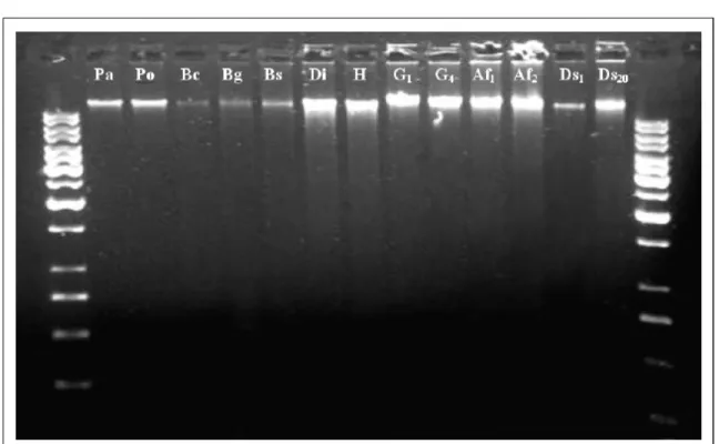
After the protocol application, DNA was obtained from all arthropod species and samples exhibited a transparent to very light color. In the 0.8% ethidium bromide-stained agarose gels the DNA samples revealed good quality (Figure 1). The DNA-shearing effect was not prominent for any sample and only low smearing fragments were observed. This low shearing effect is expected, since the tissue samples were mech anically gr in ded an d subjected to homogenization procedures. The RNAse treatment was efficient to eliminate RNA from the samples, once no RNA was observed in any sample.
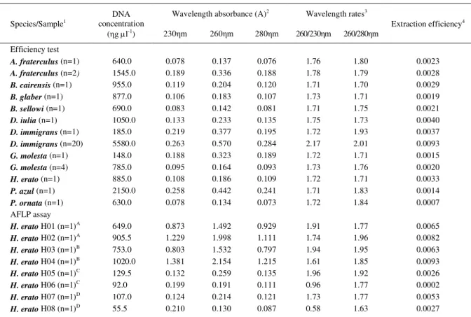
The table 1 shows the results for the protocol’s efficiency test with the different species. The yield of DNA recollected from the tissue samples shown a significant correlation between the weight of tissue sample used and the quantity of DNA recollected was observed (r=0.842; P<0.001). All samples tested shown high quality DNA after extraction procedure (Table 1). No significant amounts of proteins were detected in the samples, showing that protein removal was very efficient using the protocol presented here, without the necessity of using phenol or any other proteolytic method. Organic compounds presented in DNA samples also were inexpressive, dispensing the need of purification step after the extraction.
AFLP assay and evaluation of method’s efficiency against other DNA extraction protocols
As obser ved for the other arth ropod species, all DNA samples used for the AFLP assay revealed good quality. DNA samples obtained with the four methods compared shown similar results, but the yield of DNA recollected with the commercial kits was lower than homemade protocols. Wavelength readings
and wavelength rates also were very similar, excepting the samples H06 and H08, which were obtained with the commercial kits; those samples revealed some excess of organic impurities or contaminants (Table 1). No expressive amounts of proteins were observed in any sample.
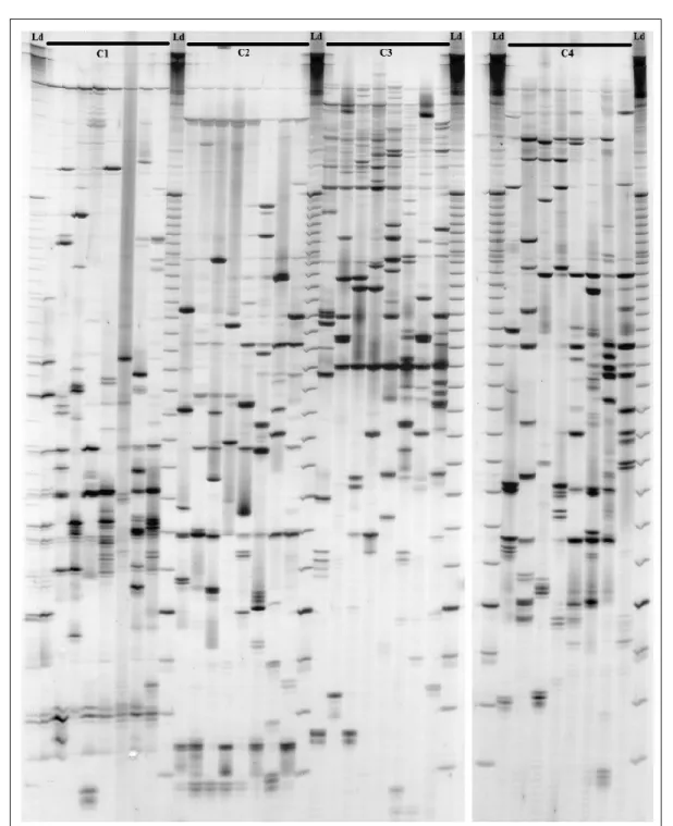
The figure 2 shows the AFLPs patterns obtained for the DNA samples used in the AFLP assay. All four set of primers tested exhibited a great number of loci with good visualization. Overall, it was observed 251 distinguished loci, being 240 polymorphic. Regarding the DNA quality for molecular application, all four methods provided good quality, proving to be suitable for enzyme digestion, linkage and PCR, once AFLP has those three steps and is very sensitive to poor quality DNA samples (REINEKE et al., 1998; CHEN et al., 2008).
The results obtained with the AFLP assay indicate that the use of different methods may produce good quality DNA. Homemade protocols provide an alternative to low budget projects, but must be optimized to produce good results. Commercial kits are very convenient, but also too expensive. These statements become clear when time/fund consumption
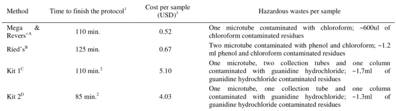
and toxic wastes produced are concerned (Table 2). The homemade protocol based on SDS/phenol-chloroform extraction showed similar runtime and results to the CTAB/PVPP/chloroform method presented here, and also was very inexpensive as well. Moreover, the use of phenol demands an additional step of organic solvent cleaning which generates higher amounts of contaminated wastes when compared with the method developed here. On the other hand, the commercial kits showed lower efficiency in recollect DNA from tissue samples when compared with the homemade protocols, without gain any advantages on samples purity. Despite having less time consuming, they were ten times more expensive than the homemade protocols. They did not generate hazardous wastes containing phenol and chloroform, dispensing the need of a fume hood to be applied, but the use of guanidine hydrochloride solutions also produces noxious wastes and some care on the disposal of residues should be taken as well. Scoring the benefits and costs of all protocols compared, the results indicate that the method presented here is the best choice to obtain good quality DNA.
Figure 1 - The 0.8% ethidium bromide-stained agarose gel showing DNA samples after DNA extraction from freshly and frozen tissues. Pa, Paratrechalea azul; Po, Paratrechalea ornata; Bc, Benthana cairensis; Bg, Baloniscus glaber; Bs, Baloniscus sellowii; Di, Dryas iulia; H, Heliconius erato; G1, Grapholita molesta (n=1), G4, Grapholita
molesta (n=4); Af1, Anastrepha fraterculus (n=1); Af2, Anastrepha fraterculus (n=2); Ds1, Drosophila immigrans
High yields of clean DNA were obtained from arthropod tissues by using the DNA extraction procedure presented here. The strategy of using NaCl to r emove polysacch ar ides an d PVP to pur ge polyphenols from arthropods’ diet proved to be very efficient. The PVPP proved to be and proficient tool to isolate high quality DNA from tissue samples, especially by removing protein s and organic contaminants, without the need of further purification steps. It was also essential to keep apart cellular debris from the aqueous phase containing nucleic acid solution, by forming a compact and consistent interface.
Although the DNA extraction protocol developed here had shown promising results, some care must be taken to maximize the yield and quality of
the DNA samples. First, the tissue sample must be completely dried before the grind procedure starts. The formation of ice crystals can prejudice the grinding procedure. Second, excess or lack of grinded tissue can generate samples with excess of proteins and organic compounds or residual buffer components, respectively. Although, as shown in the AFLP assay, the residual buffer components presented in the DNA samples did not interfere in the AFLP technique. Excess of tissue can cause pigmentation of DNA samples after extraction, but also increase the amount of DNA and other substances. The pigmentation does not affect the quality of DNA suitable to sensitive molecular techniques, but to obtain best results it is recommend increasing the dilution factor of DNA samples (75-200µl of TE). According to the observations best results are achieved when 0.10-0.06g of grinded tissue are used.
Table 1 - Analysis of concentration, wavelength absorbance readings and rates, and extraction efficiency for the biological samples used in the efficiency test and in the AFLP assay.
Wavelength absorbance (A)2 Wavelength rates3 Species/Sample1
DNA concentration
(ηg l-1) 230ηm 260ηm 280ηm 260/230ηm 260/280ηm
Extraction efficiency4
Efficiency test
A. fraterculus(n=1) 640.0 0.078 0.137 0.076 1.76 1.80 0.0023
A. fraterculus(n=2) 1545.0 0.189 0.336 0.188 1.78 1.79 0.0028
B. cairensis (n=1) 955.0 0.119 0.204 0.120 1.71 1.70 0.0029
B. glaber (n=1) 877.0 0.106 0.183 0.107 1.73 1.71 0.0019
B. sellowi (n=1) 690.0 0.083 0.142 0.081 1.71 1.75 0.0021
D. iulia (n=1) 1050.0 0.133 0.233 0.135 1.75 1.73 0.0040
D. immigrans(n=1) 185.0 0.219 0.377 0.195 1.72 1.93 0.0037
D. immigrans (n=20) 5580.0 0.263 0.570 0.284 2.17 2.01 0.0093
G. molesta(n=1) 148.0 0.188 0.323 0.189 1.72 1.71 0.0015
G. molesta(n=4) 785.0 0.095 0.164 0.093 1.73 1.76 0.0020
H. erato (n=1) 885.0 0.108 0.186 0.109 1.72 1.71 0.0033
P. azul (n=1) 2150.0 0.258 0.442 0.241 1.71 1.83 0.0014
P. ornata (n=1) 630.0 0.078 0.134 0.073 1.72 1.84 0.0007
AFLP assay
H. erato H01 (n=1)A 649.0 0.873 1.492 0.929 1.91 1.77 0.0065
H. erato H02 (n=1)A 905.5 1.229 1.998 1.111 1.74 1.96 0.0082
H. erato H03 (n=1)B 753.0 0.803 1.532 0.797 1.94 1.95 0.0063
H. erato H04 (n=1)B 1020.0 1.381 2.154 1.215 1.61 1.85 0.0093
H. erato H05 (n=1)C 129.5 0.132 0.259 0.135 1.96 1.92 0.0026
H. erato H06 (n=1)C 92.0 0.199 0.191 0.111 0.96 1.77 0.0002
H. erato H07 (n=1)D 107.0 0.124 0.214 0.121 1.73 1.77 0.0053
H. erato H08 (n=1)D 55.5 0.210 0.130 0.087 0.58 1.63 0.0027
1
Numbers in brackets indicate the quantity of specimens used.
2 Absorbance were measured by the optical density of the sample in relation to ultrapure distilled water.
3 Wavelength rate higher than 1.7 indicates pure samples and lower than 1.7 indicate samples with significant levels of buffer/organic components (260/230ηm) and proteins (260/230ηm).
4 In grams of DNA / grams of tissue.
A Mega e Revers’ method (CTAB/PVPP/chloroform - described here). B
Ried’s method (SDS/phenol-chloroform).
CONCLUSION
It was con cluded that th e pr otocol presented here is a good method to obtain high quality
DNA without the necessity of further steps of proteolysis and purification. The protocol has an approximate cost of USD 0.52 per sample, including all chemicals and plastics, which is very inexpensive if
compared with the commercial kits. Also, hazardous wastes at the end of extraction procedure totaled one microcentrifuge tube with cellular debris and 600l of chloroform and isoamyl alcohol mixture per sample. Thus, researchers from Agrarian and Biological Sciences may study arthropod DNA at low costs, generating lower quantities of toxic waste when compared with other methods and commercial kits available on market.
ACKNOWLEDGMENTS
The researchers are grateful to Alessandra Russi, Felippe Gomes, Gisele Passaia, Ivan Ceriotti, Rafaela Nalin and Vanessa Buffon for providing laboratory support, and Aline Quadros, Lisandra Robe, Luis E. Schmidt and Wilson Morandi Jr. for providing specimens for DNA extraction. This study was funded by CNPq, EMBRAPA Uva e Vinho and FAPERGS. REFERENCES
BACKER, L.E. et al. A silica-based mitochondrial DNA extraction method applied to forensic hair shafts and teeth.
Journal of Forensic Sciences, v.46, n.1, p.1-5, 2001. CHEN, M. et al. Methodological comparison of DNA extraction from Holcocerrus hippophaecolus (Lepidoptera: Cossidae) for AFLP analysis. Forestry Studies in China,
v.10, n.3, p.189-192, 2008. Available from: <http:// w w w. s p r i n g e r l i n k . c o m / c o n t e n t / h 8 1 7 2 0 p 7 4 p 8 2 t 4 6 7 > . Accessed: Jun. 21 2011. doi: 10.1007/s11632-008-0035-5. CHEN, H. et al. Evaluation of Five Methods for Total DNA Extraction from Western Corn Rootworm Beetles. PLoS One, v.5, n.8, p.e11963, 2010. Available from: <http://www.plosone.org/
article/info%3Adoi%2F10.1371%2Fjournal.pone.0011963>. Accessed: Jun. 21 2011 doi:10.1371/journal.pone.0011963. CRESTE, S. et al. Detection of single sequence repeat polymorphisms in denaturing polyacrilamide sequencing gels by silver staining. Plant Molecular Biology Reporter, v.19, p.299-306, 2001.
FANG, G. et al. A quick and inexpensive method for removing polysaccharides from plant genomic DNA. BioTechniques, v.13, n.1, p.52-56, 1992.
GRACHEV M.A. et al. A method for the isolation of pure DNA for PCR. Molecular Biology, v.40, n.1, p.159-161, 2006. Available from: <http://www.springerlink.com/content/ 075u65x3w8687374/>. Accessed: Jun. 21 2011. doi: 10.1134/ S0026893306010213.
HAAF, F. et al. Polymers of N-Vinylpyrrolidone: Synthesis. Characterization and Uses. Polymer Journal, v.17, n.1, p.143-152, 1985. Available from: <http://www.jstage.jst.go.jp/ article/polymj/17/1/143/_pdf>. Accessed: Jun. 21 2011. doi:10.1295/polymj.17.143.
HANSEN, T.O. et al. Collection of blood, saliva. and buccal cell samples in a pilot study on the Danish nurse cohort: comparison of the response rate and quality of genomic DNA.
Cancer Epidemiology, Biomarkers & Prevention, v.16, n.10, p.2072-2076, 2007. Available from: <http:// cebp.aacrjournals.org/content/16/10/2072.abstract>. Accessed: Jun. 21 2011. doi:10.1158/1055-9965.EPI-07-0611. LEE, P.L.M.; PRYS-JONES, R.P. Extracting DNA from museum bird eggs, and whole genome amplification of archive DNA.
Molecular Ecology Resources, v.8, n.3, p.551-560, 2008. Available from: <http://www3.interscience.wiley.com/cgi-bin/ fulltext/120090725/HTMLSTART>. Accessed: Jun. 21 2011. doi: 10.1111/j.1471-8286.2007.02044.x.
Table 2 - A comparative oftime, costs and hazardous wastes generated per sample with the four methods used to recollect DNA from fresh arthropod tissues.
Method Time to finish the protocol1 Cost per sample
(USD)3 Hazardous wastes per sample
Mega &
Revers’A 110 min. 0.52 One microtube contaminated with chloroform; ~600ul ofchloroform contaminated residues
Ried’sB 125 min. 0.67 Two microtube contaminated with phenol and chloroform; ~1.2
ml phenol and chloroform contaminated residues
Kit 1C 110 min.2 5.10
One microtube, two collection tubes and one column contaminated with guanidine hydrochloride; ~1,7ml of guanidine hydrochloride contaminated residues
Kit 2D 85 min.2 4.03
One microtube, one collection tube and one column contaminated with guanidine hydrochloride; ~1.3ml of guanidine hydrochloride contaminated residues
1 Grinding tissue time and solution preparation time not considered. 2 Sample incubation time of one hour.
3 Calculated on February 2011, from lowest Brazilian market prices. A CTAB/PVPP/chloroform - described here.
B
SDS/phenol-chloroform.
LEFORT, F.; DOUGLAS, G.C. An efficient micro-method of DNA isolation from mature leaves of four hardwood tree species Acer, Fraxinus, Prunus and Quercus. Annals of Forest Science, v.56, n.3, p.259-263, 1999. Available from: <http:/ /www.afs-journal.org/articles/forest/pdf/1999/03/AFS_0003-4312_1999_56_3_ART0008.pdf>. Accessed: Jun. 21 2011. doi: 10.1051/forest:19990308.
LODHI, M.A. et al. A simple and efficient method for DNA extraction from Grapevine cultivars and Vitis species. Plant Molecular Biology Reporter, v.12, n.1, p.6-13, 1994. MALIYAKAL, E.J. An efficient method for isolation of RNA and DNA from plants containing polyphenolics. Nucleic Acids Research, v.20, n.9, p.2381, 1992. Available from: <http:// w w w. n c b i . n l m . n i h . g o v / p m c/ a r t i c l e s / P M C 3 1 2 3 6 4 / p d f / nar00083-0172.pdf>. Accessed: Jun. 21 2011. doi:10.1093/ nar/20.9.2381.
MOELLER E.M. et al. A simple and efficient protocol for isolation of high molecular weight DNA from filamentous fungi. fruit bodies. and infected plant tissues. Nucleic Acids Research, v.22, n.22, p.6115-6116, 1992.
OLIVEIRA, L.F.V. et al. Isolation of high quality DNA: a pr otocol combining “r ennet” a nd glass milk. Electronic Journal of Biotechnology, v.12, n.2, p.1-6, 2009. Available from: < http://www.ejbiotechnology.info/index.php/ ejbiotechnology/article/view/v12n2-4/709>. Accessed: Jun. 21 2011. doi: 10.2225/vol12-issue2-fulltext-4.
REINEKE A. et al. Preparation and purification of DNA from insects for AFLP analysis. Insect Molecular Biology, v.7,
n.1, p.95-99, 1998. Available from: <http:// www3.interscience.wiley.com/journal/119130668/abstract>. Accessed: Jun. 21 2011. doi: 10.1046/j.1365-2583.1998.71048.x.
RIED, T. DNA preparation from fresh/frozen tissue.
Bethesda: Section of Cancer Genomics, Genetics Branch, NCI, National Institutes of Health, MD, 2002. Protocols. Available on Internet from:http://www.riedlab.nci.nih.gov/publications/ DNA%20Prep_TC%20%20High%20Salt.doc.pdf. Online. Accessed: Jun. 21 2011.
ROHLAND, N. et al. Nondestructive DNA extraction method for mitochondrial DNA analyses of museum specimens.
Biotechniques, v.36, n.5, p.814-821, 2004. Available from: <http://www.biotechniques.com/BiotechniquesJournal/2004/ M a y / N o n d e s t r u c t i v e D N A e x t r a c t i o n m e t h o d f o r -m i t o c h o n d r i a l - D N A - a n a l y s e s - o f - -m u s e u -m - s p e c i -m e n s / biotechniques-116773.html>. Accessed: Jun. 21 2011. doi 10.2144/3605A0814.
SAMBROOK, J.; RUSSELL, D.W. Molecular cloning: a laboratory manual. Woodbury: Cold Spring Harbor Laboratory, 2001. 3v, 999p.
VOS, P. et al. AFLP: a new technique for DNA fingerprinting.