(Annals of the Brazilian Academy of Sciences) ISSN 0001-3765
www.scielo.br/aabc
Biotransformation of sucrose into 5-hydroxy-2-hydroxymethyl-
γ
-pirone
by
Aspergillus flavus
NELSON R. FERREIRA1,3, MARIA INEZ M. SARQUIS2, CLÁUDIO N. ALVES3 and ALBERDAN S. SANTOS1,3
1Pós-graduação em Ciência e Tecnologia de Alimentos, Universidade Federal do Pará Rua Augusto Corrêa, 01, Guamá, 66075-110 Belém, PA, Brasil
2Laboratório de Coleção de Cultura de Fungos, Instituto Oswaldo Cruz/Fiocruz-RJ Av. Brasil, 4365, Pv. Rocha Lima, sala 525, Manguinhos, 21045-900 Rio de Janeiro, RJ, Brasil
3Laboratório de Investigação Sistemática em Biotecnologia e Química Fina, Programa de Pós-graduação em Química Universidade Federal do Pará, Rua Augusto Corrêa, 01, Guamá, 66075-110 Belém, PA, Brasil
Manuscript received on June 5, 2008; accepted for publication on April 30, 2010
ABSTRACT
The sucrose hydrolysis and the preference of consumption of glucose instead of fructose were investigated for the production of 5-hydroxy-2-hydroxymethyl-γ-pyrone (HHMP) in the presence ofAspergillus flavusIOC 3974 cultivated in liquid Czapeck medium. Standardized 0.5g of pellets were transferred as inoculum into twelve conical flasks of 250 ml containing 100 ml of medium with different sucrose concentration, which was kept at 120 rpm and 28◦C for 16 days without pH adjustment. Aliquots of 500µl of the broth culture were withdrawn at 24 h intervals and analyzed.
The major yield of HHMP was 26g l−1in 120g l−1of sucrose. At these conditions,A. flavusproduced an invertase capable of hydrolyzing 65% of total sucrose concentration in 24h, and an isomerase capable of converting fructose into glucose. In this work, it focused the preference for glucose and, then, of fructose byA. flavusand the strategy used to produce HHMP.
Key words:Aspergillus flavus, biotransformation, kojic acid, secondary metabolite, fructose-isomerase.
INTRODUCTION
Fungi are lower eukariotes microorganisms that have been important in both ancient and modern biotechno-logical processes. They are known as excellent metabo-lites producer agents of antibiotics, alcohols, enzymes, organic acids, pharmaceuticals and several organic com-pounds (Wang et al. 2005). Fungi present a different way of nutrition. They secrete in the environment a wide range of secondary metabolites and powerful enzymes, such as peroxidase and hydrolases, among others, which oxidize and hydrolase lignins, cellulose and polysac-charides into micro molecules, such as phenyl alcane and glucose, and absorb them as foodstuff (Papagianni
Correspondence to: Alberdan Silva Santos
E-mail: [email protected]; [email protected]
2003). Based on this mode of action, fungi are classified in the fifth kingdom (Bennett 1998), and can be found as yeasts, molds and mushrooms that produce metabo-lites and enzymes with biological activities. In this as-pect, the generaAspergillus,Penicillium,Peacylomyces
andFusariumpresent high potential in producing active
metabolites and enzymes that must be screened in a great range of strain to select a great producer (Mercier et al. 1998). This kind of work developed with fungi is known as Mycotechnology, which is one part of Biotechnology operating at the scientific frontier approaching medicine, food industry, agriculture and cosmetic by the forefront of molecular biotechnology (Bennett 1998).
is a secondary metabolite produced from carbohydrate sources, mainly from those with pyranosidic structures, by aerobic fermentation (Ariff et al. 1997). However, other fungi that belong toAspergillus generawere
de-scribed to produce the same metabolites: A. oryzae (Wa-kisaka et al. 1998),A. tamarii (Rosfarizan et al. 1998) and some strains ofA. parasiticus(Varga et al. 2003).
This substance is industrially interesting (Park et al. 2003) for presenting an inhibitory activity against tyro-sinase (Kim et al. 2004) and other several correlated en-zymes, such as the polyphenol oxidases (PPO) (˙
Iyido-˘
gan and Bayindirli 2004). Previous studies showed that this substance presented antibiotic activity. It also pres-ents an potential application as a precursor of flavor en-hancer and as an antioxidant agent by inhibiting oxida-tions of polyphenol (Ariff et al. 1996). Several works have been developed to discover new microorganisms and substrates that can be used in the production of HHMP (5-hydroxy-2-hydroxymethyl-γ-pyrone)
(Bur-dock et al. 2001). However little is known about the mechanisms of HHMP formation, and only biosynthesis discussions have been published without characterizing the enzymes, the biotransformation of different sources of carbohydrate to HHMP, and the kinetic parameters for glucose and sucrose (Mohamad and Ariff 2007).
During sucrose fermentation by A. flavus, the
HHMP is synthesized by the direct conversion of glucose through multistep enzyme reactions. Although the en-zyme system involved in HHMP biosynthesis was found to be very stable under a chemically defined resuspended cell system, the action of fructose-isomerase was never observed before.
The present study was undertaken to investigate the different sucrose concentrations on the biotransforma-tion of this disaccharide to HHMP and monosaccharides preference of consumption during filamentous fungus cultivation after the hydrolysis of the glucose by an in-vertase produced fungus.
MATERIALS AND METHODS
MICROORGANISM ANDMEDIUM
Aspergillus flavusIOC 3974 used in this work was
ob-tained from the laboratory of collection of fungi of the Oswald Cruz Institute in Rio de Janeiro (Brazil). The
conidia were suspended in a sterile solution of NaCl (1% w/v) and used as the initial inoculum (S1). In this work, three types of culture media were used to investi-gate the adaptation and better mycelial development of
A. flavus: Czapek Dox agar (CDA), potato dextrose agar (PDA), and Sabouraud agar (SBA) (Keller et al. 2003). All of the media were sterilized at 121◦C (1kgf cm−2)
for 15 minutes. The sucrose used as a carbon source was added into the media in different concentrations, and the pH was adjusted to 5.5 with NaOH (1 mol l−1) before
sterilization.
CULTIVATION OFAspergillus flavusFOROBTAINING CONIDIA(SPORES)
Amounts of 500µl of the initial spores suspension (S1)
of A. flavus (±108 conidia ml−1) were transferred to
Petri dishes containing 20 ml of CDA with a concentra-tion of sucrose of 30 g l−1and incubated at 28◦C for 10
days. A volume of 20 ml of a sterilized solution of NaCl (1% w/v) was used on the plates for obtaining a second suspension of spores (S2).
EVALUATION OFAspergillus flavusGROWTH ONSOLIDMEDIA
Amounts of 50µl of the suspension (S2) were
transfer-red to a 5 mm disk of cellulose, centralized on the plates containing CDA medium with different sucrose concen-trations: 30, 60, 120, 240 and 360g l−1 and incubated
at 28◦C for 10 days. This procedure was made in
trip-licate and repeated for the PDA and SBA media. The mycelial growths were evaluated by the biometric or-thogonal axes method. The culture media containing the different sucrose concentrations were evaluated at every 24 h intervals by the measurement of mycelial growth diameters in the two directions of the orthogonal axes. Statistical analysis was applied, as well as the formation of the conidia was evaluated qualitatively. The culture media that presented better mycelial growth and good spores formation was selected and used as a medium of
A. flavuscultivation (E1).
STANDARDIZATIONMETHOD FOROBTAININGA. flavus INOCULUM IN THELIQUIDMEDIACULTIVATION
An amount of 1 ml (±108conidia ml−1) of the spores
better growth (E1) was transferred to five conical flasks of 250 ml containing 100 ml of Czapek liquid medium (pH 5,5) with concentrations of 30, 60, 120, 240 and 360g l−1of sucrose properly sterilized at 121◦C for 15
min. Then they were incubated in a shaker at 120 rpm with the controlled temperature at 28◦C for 72h. Each
conical flask was submitted to a vacuum filtration with a Büchner funnel with a quantitative filter paper. After the filtration, mycelium as pellets was used as inoculum. Standardization was carried out by transferring 0.5 g of pellets amounts from a flask containing 6% of sucrose to 250 ml conical flasks containing 100 ml of Czapek culture medium with the addition of 30, 60, 120, 240 and 360g l−1of sucrose. This experiment was prepared
in triplicate and further incubated at 120 rpm and 28◦C
for 16 days.
EVALUATIONMETHOD OFDIFFERENTCONCENTRATIONS OFSUCROSE ON THEMYCELIALDEVELOPMENT
OFA. flavusANDHHMP PRODUCTION
Standardized 0.5 g amounts of pellet as inoculum were transferred from the flask containing 6% of sucrose to twelve conical flasks of 250 ml, each one containing 100 ml of Czapek medium with different sucrose con-centrations: 60, 120, 240 and 360g l−1. The cultivation
was kept at 120 rpm and 28◦C for 16 days without a
pH adjustment. Aliquots of 500µl of the broth culture,
without mycelium, were withdrawn at 24 h intervals, and transferred into glass vials of 10 ml. This experiment was prepared in triplicate and samples were analyzed for the quantification of HHMP and residual saccharides.
METABOLITEIDENTIFICATION
The identification of the metabolite was performed by comparing the sample with the standard. We used carbon-13 nuclear magnetic resonance spectroscopy (VARIAN/MERCURY 300 MHZ), and the dimethyl
sul-foxide (DMSO) as solvent, and Infrared Spectroscopy (Spectrometer SHIMADZU – IR 740).
QUANTIFICATION OFKOJICACID
The quantification curve was built by quantifying the absorbance for different HHMP concentrations. Thus, solutions were prepared at concentrations of 100, 200, 400, 600, 800, 1000 and 1200µg ml−1standard. The
ab-sorbances were determined in triplicate by UV-Vis spec-trophotometry (GBC 911 system) at 269 nm.
Amounts of 50µl of the samples were transferred
to a volumetric bottle of 50 ml, and the volume was com-pleted with deionized water. A quantitative analysis was performed as described in (Gomara et al. 2004). Each sample was analyzed in triplicate.
QUANTIFICATION OF THETOTALREDUCINGSUGARS
Amounts of 250µl of the samples and 200µl of HCl 2N
were transferred to volumetric bottles of 50 ml and the residual non-reducing sugar (sucrose) was hydrolyzed at 70◦C for 10 minutes in a water bath. After cooling,
the solution samples were neutralized with 200µl of a
solution of NaOH 1N. The bottles were completed with deionized water. Amounts of 1.5 ml of this solution were transferred to glass tubes and aliquots of 0.5 ml of an al-kaline solution of 3,5-dinitrosalicilic acid (DNS) were added. The analysis was performed in a Quimis model Q798 spectrophotometer. Each sample was analyzed in-dividually in triplicate. The pH was measured at the first and the 16thdays of incubation. Fructose was quantified
by Saliwanoff, and glucose was quantified by difference.
QUANTIFICATION OFFRUCTOSE BYSELIWANOFFREAGENT
The standard curve was built as follows: fructose was measured by the adapted Seliwanoff’s method described by Souza et al. (2007). From a standard solution of fructose (150 mg/100 ml), aliquots were withdrawn and transferred to individual glass tubes and diluted with dis-tilled water to reach 1, 10, 20, 30, 40-100mg/100 ml of fructose and ready to reach 200µl of individual solution.
4ml of Seliwanoff reagent were added, and the solutions were boiled for 3 minutes and analyzed by spectropho-tometer at 486nm after reaching the room temperature. Samples were measured in the same way, replacing fruc-tose solution by broth medium aliquots.
RESULTS AND DISCUSSION
MEASUREMENT OFMYCELIALGROWTH OFAspergillus flavusINSOLIDMEDIA
A. flavuswas cultivated in Petri dishes forming
TABLE I
Comparison of mycelial diameter growth ofA. flavusin different solid media.
Diameters of mycelial growth in (cm) Sucrose 9 days of cultivation concentrations (g l−1) Different culture media
CDA PDA SBA
30 6.063±0.294 5.150±0.203 6.567±0.341 60 5.550±0.165 5.327±0.228 6.900±0.274 120 7.817±0.305 7.500±0.195 7.550±0.445 240 5.433±0.306 7.067±0.318 6.800±0.681 360 5.783±0.289 7.163±0.222 6.700±0.376
which is called “biometric method of orthogonal axes”, made possible to evaluate the mycelial growth speed in the culture media CDA, PDA and SBA in different su-crose concentrations: 30, 60, 120, 240 and 360g l−1.
The results showed that CDA presented the best mycelial growth (Table I). The sucrose concentrations above 120g l−1 of sucrose presented smaller mycelial
growth. Several factors might have influenced the de-crease of the metabolism, like the osmotic pressure and the high fructose concentration after sucrose hydrolysis. The analysis of the different concentrations of the sub-stratum showed that sucrose 30g l−1presented a smaller
mycelial growth when compared with the concentrations of 60 and 120g l−1.
The results were important to evaluate the mycelial growth profile of A. flavusin different media with
dif-ferent sucrose concentrations in a period of 9 days to select the one which presents the best conditions forA. flavus adaptation. In this case, CDA with 120 g l−1
of sucrose was the chosen medium for the cultivation of this microorganism, even in a solid to produce the conidia.
STANDARDIZATION OF THEINOCULUM WITHPELLETS
The culture medium CDA with 60g l−1of sucrose
pre-sented more spherical shape and an uniform pellet size with a diameter interval of 1.5 mm = D = 3 mm. The standardization of the inoculum was accomplished with fixed amounts of 0.5g (0.0169g of dry weight withdrawn from medium containing 6g l−1cultivated at 120 rpm for
72 h), and of fresh mass of pellets, properly drained and transferred to liquid medium. This procedure allowed
a better standardization and quantification of the inocu-lum in the biotechnological process for the production of HHMP. However, the use of conidia had a huge margin of error due to its fast sedimentation in the suspension. For this problem, a standardization of the inoculum was developed with pellets.
IDENTIFICATION OFHHMP
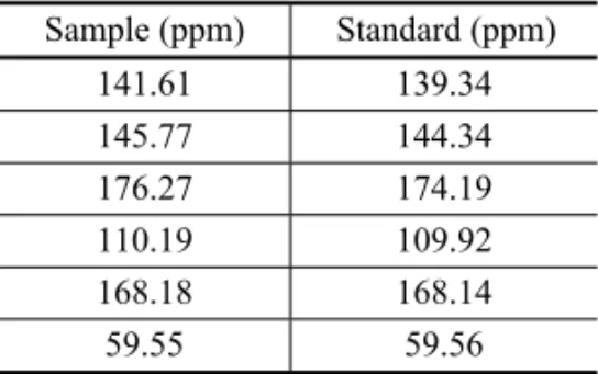
The identification of HHMP was performed using car-bon-13 nuclear magnetic and Infrared Spectroscopy. The spectra of samples were compared with the spectra of standards HHMP. These spectra showed similar chemi-cal shifts (Table II).
TABLE II
Chemical shifts of the sample and standard in C-13 NMR. The chemical shifts are reported
in relation to tetramethylsilane (TMS).
Sample (ppm) Standard (ppm) 141.61 139.34 145.77 144.34 176.27 174.19 110.19 109.92 168.18 168.14 59.55 59.56
car-bonyl. Absorption in the aromatic region was not ob-served.
These results confirm the authenticity of the iden-tification of the metabolite produced byA. flavusas the
5-hydroxy-2-hydroxymethyl-γ-pirone.
QUANTIFICATION OFHHMP PRODUCTION
The cultivation ofA. flavusin solid medium showed that the level of sucrose concentrations is equal to 240g l−1,
and 360g l−1 do not permit an identical metabolism as
that one for 120g l−1. These results were the basis for
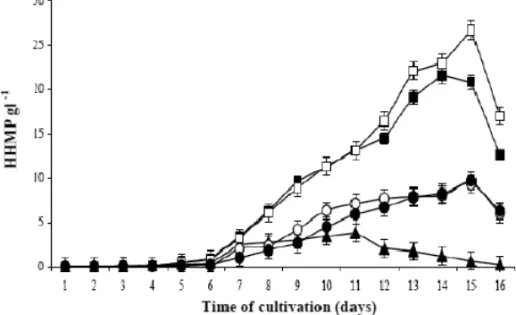
the investigation of the development of mycelial growth in liquid medium. In this case high concentrations of sucrose increased the viscosity and the osmotic pres-sure. This aspect also influenced a smaller diffusion of the molecular oxygen, affecting the biosynthetic route of HHMP production. The maximum metabolite produc-tion was reached at the 15thday of cultivation, influenced
by different concentrations of sucrose (Fig. 1).
This production has started from at the 6thday for
all sucrose concentrations. However, 60g l−1and 120g
l−1 of sucrose presented a better production, reaching
out 21g l−1and 26g l−1of kojic acid, respectively
(Ta-ble III). The best yield coefficient was 0.367g of HHMP
pergram of added sucrose. These results are indica-tive of an important information, so that they established better conditions of cultivations for high HHMP produc-tion in a biotechnological process.
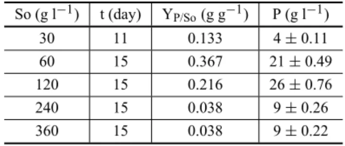
TABLE III
Kojic acid data production related to different sucrose concentrations. Y: yield coefficient of product formation,
So: initial concentration of substrate, t: time (day) and P: maximum production of metabolite.
So (g l−1) t (day) YP/So(g g−1) P (g l−1) 30 11 0.133 4±0.11 60 15 0.367 21±0.49 120 15 0.216 26±0.76 240 15 0.038 9±0.26 360 15 0.038 9±0.22
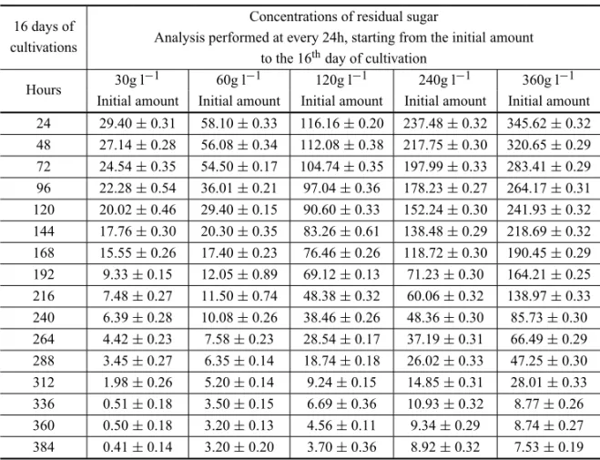
The quantification of residual sugar concentration was developed to estimate the material balance between sugar converted to HHMP and sugar converted to my-celial biomass. At the 15th day, the yield of HHMP
reached 26g l−1, and the residual sugar measured was
3.8% (w/w) from the initial amount that was added into the medium (Table IV).
The smallest adaptation of the filamentous fungus in PDA could have happened due to the absence of min-eral salts, mainly phosphate, which is a very important nutrient for glycolytic route and the Krebs Cycle. On the other hand, Sabouraud and Czapek media possess enough amounts of the nutrients that are necessary for the development of the mycelium. The difference was that, in this specific investigation, the standard HHMP presented pale yellow coloration in solution, so that it can be confused with the coloration of the own culture medium. Because of the hydroxyl and keto groups in the position C-4 and C-5, this structure presents poten-tial to chelate transition metals, mainly iron, producing red coloration. A number of works proved the capacity ofγ-pyrones to form complexes with metals such as iron
(Marwaha and Sohi 1994). In this process, sucrose was used as a source of carbon and was hydrolyzed by inver-tase to form glucose and fructose in the culture medium by the microorganism. The glucose was consumed im-mediately and acted as a precursor for HHMP. Fructose was then isomerized to glucose and follows the same biotransformation. The production of fructose was mea-sured by the method of Seliwanoff, which was adapted and described as Souza et al. (2007). HHMP presents a six-member ring and all the evidences indicate that it was produced by the biotransformation of glucose in few main steps, without a break of the monosaccharide chemical structure.
This study was developed to optimize the process production of this metabolite, starting from sucrose. It presented innovative results from the biochemical point of view. In this study, it was observed that the sucrose had been 65% w/w of its total concentration in the hy-drolyzed culture medium, being obtained fructose and glucose by an invertase produced fungus. During the sucrose fermentation in a submerged cultivation, the
Aspergillus flavusinitially consumed the glucose, which
hap-Fig. 1 – Profiles of HHMP production in liquid medium CDA with different sucrose concentrations. Sucrose 30g l−1(N), sucrose 60g l−1(), sucrose 120g l−1(), sucrose 240g l−1(◦), sucrose 360g l−1(•).
Fig. 2 – Profiles of sucrose hydrolyze, fructose isomerase into glucose and its consumption: Sucrose(•), HMPP(◦), glucose(), fructose(♦).
pened because an isomerase converted the fructose into glucose, which could be evidenced by the increase of the glucose concentration starting from 168h, and remain-ing constant up to 264h in the culture medium. In fact, the sucrose was hydrolyzed totally in 168h. It is clearly evident that it could not produce glucose a different way and, then, convert the fructose. The increase of the
con-centration of this monosaccharide felt in function of the isomerase activity. This enzyme was not characterized in this study yet.
TABLE IV
Residual sugar concentration in a Czapek Dox liquid medium.
16 days of Concentrations of residual sugar
cultivations Analysis performed at every 24h, starting from the initial amount to the 16thday of cultivation
Hours 30g l−1 60g l−1 120g l−1 240g l−1 360g l−1 Initial amount Initial amount Initial amount Initial amount Initial amount 24 29.40±0.31 58.10±0.33 116.16±0.20 237.48±0.32 345.62±0.32 48 27.14±0.28 56.08±0.34 112.08±0.38 217.75±0.30 320.65±0.29 72 24.54±0.35 54.50±0.17 104.74±0.35 197.99±0.33 283.41±0.29 96 22.28±0.54 36.01±0.21 97.04±0.36 178.23±0.27 264.17±0.31 120 20.02±0.46 29.40±0.15 90.60±0.33 152.24±0.30 241.93±0.32 144 17.76±0.30 20.30±0.35 83.26±0.61 138.48±0.29 218.69±0.32 168 15.55±0.26 17.40±0.23 76.46±0.26 118.72±0.30 190.45±0.29 192 9.33±0.15 12.05±0.89 69.12±0.13 71.23±0.30 164.21±0.25 216 7.48±0.27 11.50±0.74 48.38±0.32 60.06±0.32 138.97±0.33 240 6.39±0.28 10.08±0.26 38.46±0.26 48.36±0.30 85.73±0.30 264 4.42±0.23 7.58±0.23 28.54±0.17 37.19±0.31 66.49±0.29 288 3.45±0.27 6.35±0.14 18.74±0.18 26.02±0.33 47.25±0.30 312 1.98±0.26 5.20±0.14 9.24±0.15 14.85±0.31 28.01±0.33 336 0.51±0.18 3.50±0.15 6.69±0.36 10.93±0.32 8.77±0.26 360 0.50±0.18 3.20±0.13 4.56±0.11 9.34±0.29 8.74±0.27 384 0.41±0.14 3.20±0.20 3.70±0.36 8.92±0.32 7.53±0.19
the yield of HHMP reached 26g l−1, and the residual
sugar measured was 3.8% (w/w) from the initial amount (Table III). In the same way, the percentage of HHMP measured in relation to initial sugar concentration was 21.67% (w/w). It was demonstrated that 78.4% (w/w) of sugar were converted to mycelial biomass, carbon di-oxide and other macromolecules not quantified.
CONCLUSION
The following conclusions can be drawn from the above results:
A. flavuscould hydrolyze sucrose, isomerize fruc-tose into glucose, and biotransform this monosaccha-rides to HHMP. In this case, it was not possible to quan-tify the yield of isomerization due to microorganism just consuming the produced glucose. However, the maxi-mum concentration of fructose was 15g l−1 in the
cul-ture medium. This phenomenon showed that this micro-organism produced an invertase and an isomerase capa-ble to hydrolyzed glucose and convert fructose to glucose in a dynamic process.
ACKNOWLEDGMENTS
This work was supported by Conselho Nacional de Desenvolvimento Científico e Tecnológico (CNPq) and Secretaria Estadual de Desenvolvimento Científico e Tecnológico (SEDECT).
RESUMO
preferencialmente no consumo da glicose frente à frutose por A. flavuse na estratégia de produção do HHMP.
Palavras-chave: Aspergillus flavus, biotransformação, ácido kójico, metabólito secundário, frutose-isomerase.
REFERENCES
ARIFFAB, SALLEHNMS, GHANIB, HASSANMA, RUS-SULGANDKARIM MI. 1996. Aeration and yeast ex-tract requirements for kojic acid production byAspergillus flavuslink. Enzyme Microb Technol 19: 545–550.
ARIFFAB, ROSFARIZANM, HERNGLS, MADIHAHSAND KARIMMI. 1997. Kinetics and modeling of kojic acid production byAspergillus flavuslink in batch fermenta-tion and resuspended mycelial system. World J Microbiol Biotechnol 13: 195–201.
BENNETTJW. 1998. Mycotechnology: the role of fungi in biotechnology. J Biotech 66: 101–107.
BURDOCKGA, SONIMGANDCABINIG. 2001. Evaluation of health aspects of kojic acid in food. Regul Toxicol Pharmacol 33: 80–101.
GOMARAFL, CORRER CJ, SATOMANDPONTAROLOR. 2004. Development and valdation of a spectrophotometric method for the quantification of kojic acid. Ars Phar 45: 145–153.
˙
IYIDOGAN˘ NFANDBAYINDIRLIA. 2004. Effect of L-cys-teine, kojic acid and hexylresorcinol combination on in-hibition of enzimatic browning in Amasya apple juice. J Food Eng 62: 299–304.
KELLERFA, HAMILTONJEANDNGUYENQA. 2003. Mi-crobial Pretreatment of Biomass. Appl Biochem Biotech 105: 27–41.
KIM H, CHOI J, CHO JK, KIM SYAND LEEYS. 2004. Solid-phase synthesis of kojic acid-tripeptides and their tyrosinase inhibitory activity, storage stability and toxic-ity. Bioorg Med Chem Lett 14: 2843–2846.
MARWAHASANDSOHIG. 1994. Organomercury (II) com-plexes of kojic acid and maltol: synthesis, characteriza-tion, and biological studies. J Inorg Biochem 54: 67–74.
MERCIER RR, MOUGIN C, SIGOILLOT LC, SOHIRE L, CHAPLAINVANDASTHERM. 1998. Wet sand cultures to screen filamentous fungi for the biotransformation of polycyclic aromatic hydrocarbons. Biotechnol Tech 12: 725–728.
MOHAMADRAND ARIFF A. 2007. Biotransformation of various carbon sources to kojic acid by cell-bound enzyme system ofA. flavusLink 44-1. Biochem Eng J 35: 203– 209.
PAPAGIANNIM. 2003. Fungal morphology and metabolite production in submerged mycelial processes. Biotechnol Adv 22: 189–260.
PARK YD, LEE JR, PARK KH, HAHN HS AND HAHN MJ. 2003. A new continuous espectrophotometric as-say method for DOPA oxidase activity of tyrosinase. J Protein Chem 22: 473–480.
ROSFARIZANM, MADIHAHSANDARIFFAB. 1998. Iso-lation of a kojic acid producing fungus capable of using starch as a carbon source. Lett Appl Microbiol 26: 27–30. SOUZARF, PEREIRAEOLANDSANTOSAS. 2007. Adap-tação e utilização do reagente de Seliwanoff na análise quantitativa de frutose presente em méis de abelha. In: 59a Reunião Anual da SBPC, 2007, Belém. Livro de Resumos 59: 101–102.
VARGAJ, RIGÓK, TÓTHB, TÉRENJANDKOZAKIEWICZ Z. 2003. Evolution relationships amongAspergillus spe-cies producing economically important mycotoxins. Food Tech Biotechnol 4: 29–36.
WAKISAKAY, SEGAWAT, IMAMURAK, SAKIYAMATAND NAKANISHIK. 1998. Development of a cylindrical appa-ratus for membrane-surface liquid culture and production of kojic acid usingAspergillus oryzaeNRRL484. J Fer-ment Bioeng 85: 488–494.