Samples Collection and Transport
Texto
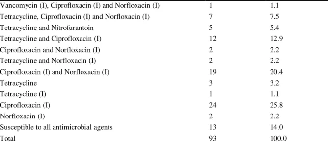
Imagem

Documentos relacionados
Neste trabalho o objetivo central foi a ampliação e adequação do procedimento e programa computacional baseado no programa comercial MSC.PATRAN, para a geração automática de modelos
the distribution of CRISPR1 -cas, CRISPR2 and CRISPR3 -cas in non-clinical strains of Enterococcus faecalis and Enterococcus faecium isolates from food and fecal
O problema desta pesquisa é a aplicação prática dos roteiros apresentados nas monografias de Santi (2015) sobre o desenvolvimento de orçamento de custos via BIM 5D
O diagnóstico diferencial da prenhez tubária deve ser fei- to com: prenhez uterina com aborto iminente ou incompleto, prenhez intra-uterina normal, rotura de cisto
Trabalho de Conclusão de Curso (Graduação em Engenharia Mecânica) - Universidade Federal de Uberlândia, Uberlândia, Brasil. O objetivo do trabalho foi projetar novos selos
Os modelos desenvolvidos por Kable & Jeffcry (19RO), Skilakakis (1981) c Milgroom & Fry (19RR), ('onfirmam o resultado obtido, visto que, quanto maior a cfiráda do
As doenças mais frequentes com localização na cabeça dos coelhos são a doença dentária adquirida, os abcessos dentários mandibulares ou maxilares, a otite interna e o empiema da
Este artigo discute o filme Voar é com os pássaros (1971) do diretor norte-americano Robert Altman fazendo uma reflexão sobre as confluências entre as inovações da geração de
