UNIVERSIDADE DA BEIRA INTERIOR
Ciências
Disposable immunosensor for diagnosis of Human
Cytomegalovirus infection
Antibodies immobilization
Hugo Flávio Cunha da Silva
Dissertação para obtenção do Grau de Mestre em
Biotecnologia
(2º ciclo de estudos)
Orientador: Prof. Doutora Ana Cristina Dias-Cabral
Co- Orientador: Prof. Doutora María Julia Arcos Martínez
Acknowledgements
The accomplishment of one’s thesis requires great dedication, hard work and personal commitment. I think that for its realization, a great deal of inspiration, patience, curiosity and reflection must be present throughout the way. However, this is a product of teamwork, and, without it, this goal would not be achievable. For all these reasons, I want to thank and dedicate this work to all who helped me in this project.
Primarily, I’d like to thank my supervisor Dr. Ana Cristina Dias-Cabral, for the opportunity she gave me in developing this study, for the unconditional support and guidance during my academic journey.
I’d also like to acknowledge my co-supervisor, Dr. María Julia Arcos Martínez, for all her availability, transmission of knowledge, constant encouragement in discovering new things and her extraordinary motivation. I am also extremely grateful for all the advice and suggestions given towards solving problems.
Moreover, I would like to thank all my group colleagues, especially Filipa Pires, for all the advices, enlightenment and support they gave me.
I’d also like to thank all my friends for the support they gave me in achieving my goals, especially to Chegão, Cláudia, Micha and Dri for the long lost hours, and also my roommates for putting up with me all these years.
Finally, my most sincere gratitude is extended to my family, who have given me their utmost support and conditions for all that I have tried to achieve. I can’t ever repay them enough. Thank you.
Abstract
Human Cytomegalovirus (HCMV) is a herpes virus that establishes a lifelong latent infection which, in most of the immunocompetent individuals is normally subclinical. Severe infections occur more frequently in immunocompromised ones, or those with immature immune system, for which can be fatal. Glycoprotein B (gB) is the dominant antigen of HCMV envelope being regarded as a promising component in the establishment of new diagnostic tests.
Nowadays there are several diagnosis methods, however, these are expensive, or/and require long time to perform, or/and need skilled operators, or even leads to the possibility of false results. So, in previously work, a disposable immunosensor was developed based on electrochemical silver oxidation as response for gB concentration increasing, using screen-printing carbon electrodes. This method allows a faster and inexpensive way to detect that viral protein. Despite that, a lack on device reproducibility and sensitivity, due to the randomly antibody adsorption was found. It is known that the oriented immobilization has become critical for optimized antigen detection on solid surfaces; therefore, those results lead us to employ other immobilization techniques to improve the immunosensor performance.
In this work the immobilization of antibodies was carried out through glutaraldehyde cross-linking, by covalent immobilization using diazonium salts and finally by using the boronic acid affinity towards the carbohydrate present on the antibody molecules. The results were conclusive only for the cross-linking, which has disadvantages when compared with adsorption.
Additionally, the importance of the gB on diagnosis tests for HCMV, lead us to study its isolated electrochemical behavior. Questionable results were found demonstrating the impossibility of its use in the determination of gB in biological samples.
Keywords
Immunosensor, antibody immobilization, Human cytomegalovirus, glutaraldehyde, diazonium salts, boronic acid.
Resumo alargado
O Citomegalovírus humano (HCMV) é um herpes vírus que pode originar infeções primárias, a partir das quais a reativação poderá ser frequente. Quando os hospedeiros deste agente viral são indivíduos imunocompetentes, é geralmente associada uma latência do HCMV que eventualmente poderá resultar em sintomas subclínicos. No entanto, em indivíduos imunocomprometidos, tais como os portadores do vírus da imunodeficiência humana (HIV) e os sujeitos a terapêuticas imunossupressoras ou ainda em indivíduos cujo sistema imune é imaturo, como é o caso de fetos e recém-nascidos, a infeção adquire proporções graves, podendo resultar na morte dos mesmos.
Este vírus apresenta um invólucro rico em proteínas virais das quais a glicoproteína B (gB) se destaca, pois está presente em aproximadamente 100% dos indivíduos infetados, sendo também reconhecida por desencadear a produção de anticorpos neutralizantes que levam à eliminação das células infetadas. Assim, esta proteína pode ser vista como um componente essencial no diagnóstico da infeção por HCMV.
A definição de um diagnóstico ideal para o HCMV tem sido difícil de implementar devido às desvantagens apresentadas pelos métodos existentes, que se baseiam em informações clinicas e imunológicas. Definido como método convencional, o isolamento do vírus em culturas de fibroblastos, obtido a partir de biópsias ou de fluidos dos hospedeiros, tem associadas as desvantagens de exigir assepsia total e longos períodos de tempo para a sua execução. Para contrapor as dificuldades desta técnica, um método idêntico, o “Shell-vial”, reduz o tempo do ensaio através de um passo de centrifugação que aumenta a penetração do HCMV nos fibroblastos, que por sua vez pode ser avaliada por imunofluorescência. No entanto, o sucesso deste método continua a depender das condições assépticas usadas. Por outro lado, o PCR (“Polimerase Chain Reaction”), analisa amostras clínicas com uma rápida performance e elevada sensibilidade, conseguida na amplificação de ADN viral. Contudo, o elevado custo e a dificuldade de realização contrapõem-se ao seu uso como técnica de diagnóstico corrente. Outra técnica, ELISA (“Enzyme-Linked Immunosorbent Assay”), deteta a presença de anticorpos específicos no sangue, o que pode levar a falsos positivos devido a reações cruzadas com o fator reumatoide, anticorpos antinucleares e outros membros da família
herpesviridae. Usado para medir a afinidade e avidez do anticorpo para o antigénio viral, o
“Western Blotting” apresenta uma baixa disponibilidade comercial conjugada também com a possibilidade de falsos positivos. Por último, testes citológicos/histológicos permitem observar inclusões virais em biópsias de tecidos do hospedeiro, contudo apresentam igualmente uma baixa sensibilidade tendo apenas 50% de sucesso na identificação de falsos negativos.
Todos os métodos têm associadas desvantagens quanto a falsos resultados, ou equipamentos e/ou procedimentos caros, que podem estar ou não relacionados a uma difícil manipulação e elevado tempo de realização. Desta forma, para contrariar estes inconvenientes, num estudo anteriormente realizado neste grupo de investigação, foi desenvolvido um imunossensor eletroquímico descartável para a deteção do HCMV, tendo por base uma imunorreação do tipo sandwich na qual o anticorpo secundário estava marcado com nanopartículas de ouro (AbNPs). Esta marcação permitiu a posterior deposição catalítica de nanopartículas de prata (AgNPs), que geraram um sinal eletroquímico na sua redissolução anódica por voltametria de pulso diferencial. O uso de elétrodos serigrafados (SPEs) como base para o immunosensor, acrescenta vantagens tais como a miniaturização, baixo custo, versatilidade e principalmente a possibilidade de uso como “point of care”. Adicionalmente, a facilidade de produção destes dispositivos por impressão sequencial de camadas de tintas, oferece vantagens na manipulação de padrões e geometrias conforme o pretendido.
O maior desafio na construção deste tipo de biossensores, passa pela imobilização dos anticorpos na superfície dos elétrodos. Está descrito na literatura que a imobilização de anticorpos de forma orientada resulta numa melhor exposição dos locais de ligação aos antigénios, exibindo melhores capacidades de ligação e posterior deteção dos mesmos. De facto, foram detetadas algumas limitações relacionadas com este passo de construção do imunossensor, uma vez que os anticorpos anti-HCMV se encontravam adsorvidos de uma forma aleatória na superfície do elétrodo de trabalho. Desta forma, as moléculas de anticorpo apresentam uma orientação nem sempre ideal afetando a reprodutibilidade e a sensibilidade do dispositivo na resposta a concentrações de gB do HCMV.
Assim, com o objetivo de melhorar as características do immunosensor descartável, neste trabalho foram aplicadas várias técnicas para a imobilização dos anticorpos anti-HCMV, tais como a reticulação, imobilização covalente através de sais de diazónio e por último usando a afinidade do ácido borónico para os açúcares presentes nas moléculas de anticorpo. O glutaraldeído, usado como agente reticulante, permitiu a imobilização das moléculas de anticorpo anti-HCMV na superfície dos SPEs. O sistema mostrou responder às concentrações incubadas de gB, levando a respostas dependentes das mesmas. Comparativamente aos resultados obtidos para a adsorção, a reticulação demonstrou ter associadas algumas desvantagens em termos de reprodutibilidade e de sensibilidade, devido à imposição da ligação dos anticorpos pelos domínios de ligação ao antigénio. Por outro lado, em condições favoráveis, a adsorção pode resultar numa orientação favorável dos anticorpos, uma vez que as moléculas têm a liberdade para se adaptarem à superfície pela conjugação de diversos fatores, nomeadamente a sua reorientação favorável na zona de saturação. Quanto à imobilização covalente foram encontradas interferências na ativação da superfície dos elétrodos. As vias condutoras de prata presentes nos elétrodos serigrafados geram uma espécie desconhecida de prata que impossibilita o estudo da eficiência de imobilização,
uma primeira abordagem foi realizada para a imobilização dos anti-HCMV através da afinidade do ácido borónico para os resíduos de açúcar presentes na estrutura dos anticorpos. Os resultados preliminares demonstraram ser promissores para uma futura aplicação neste imunossensor.
Finalmente, a reconhecida importância da gB no diagnóstico do HCMV remeteu-nos para o estudo do seu comportamento eletroquímico. Este estudo permitiu ainda inferir sobre a possibilidade de esta proteína viral interferir no sinal obtido durante a redissolução anódica das AgNPs no immunosensor. A análise foi primeiramente conduzida em SPE, os quais não possibilitaram a procura de um sinal associado à gB, permitindo no entanto concluir que não há interferência desta molécula no imunoensaio. Quando aplicado num sistema eletroquímico convencional, a gB gerou um sinal em meio tamponado, o que não se confirmou quando aplicado a amostras reais (urina).
Palavras-chave
Imunossensor, imobilização de anticorpos, Citomegalovírus humano, glutaraldeído, sais de diazónio, ácido borónico.
List of Contents
I. Introduction ... 1
1. Human cytomegalovirus ... 1
1.1. General characteristics ... 1
1.2. Viral infection and transmission ... 2
1.2.1. Congenital infection ... 2
1.2.2. Non-congenital infection ... 3
1.2.3. Prevention of virus transmission ... 3
1.3. HCMV immune response ... 4
1.3.1. HCMV specific antibodies ... 4
1.3.2. Glycoprotein B as dominant antigen ... 5
1.4. Diagnosis of HCMV infection ... 6
1.4.1. Virus isolation culture ... 7
1.4.2. Shell-vial ... 7
1.4.3. Polymerase chain reaction (PCR) ... 7
1.4.4. Enzyme-Linked Immunosorbent Assay (ELISA) ... 8
1.4.5. Western Blotting ... 8
1.4.6. Cytology/Histology ... 8
2. Biosensors ... 9
2.1. Biosensors principles ... 9
2.2. Biosensors classification ... 10
2.2.1. Transduction process and measurement techniques ... 11
2.2.1.1. Non-electrochemical transducers ... 11
2.2.1.2. Electrochemical transducers ... 11
2.2.1.3. Measurement techniques of electrochemical signals ... 12
2.2.2. Biological specificity conferring mechanism... 13
2.2.2.1. Biocatalytic recognition element ... 13
2.2.2.2. Bioaffinity recognition element ... 15
2.3. Screen-Printed Electrodes (SPE) ... 16
2.3.1. SPEs fabrication ... 17
2.3.2. Materials for SPE construction ... 18
2.3.2.1. Non-biological materials ... 19
2.3.2.2. Biological material ... 20
3. Immunosensors ... 21
3.1. Immunosensors principles and components ... 21
3.2. Electrochemical immunosensors ... 22 3.2.1. Potentiometric immunosensors ... 22 3.2.2. Impedimetric immunosensors ... 22 3.2.3. Amperometric immunosensors ... 23 3.2.4. Voltammetric immunosensors ... 23 3.3. Antibodies immobilization ... 23
3.3.1. Random immobilization techniques ... 26
3.3.1.1. Adsorption ... 27
3.3.1.2. Cross-linking ... 27
3.3.2. Oriented immobilization techniques ... 28
3.3.2.1. Covalent immobilization ... 28
3.3.2.2. Bioaffinity immobilization ... 29
3.4. Immunoassay signaling ... 29
3.4.1. Antibodies labelling ... 29
3.4.2. Nanoparticles in signal amplification ... 30
II. Groundwork and Aim ... 32
III. Materials, Apparatus and Methods ... 35
1. Reagents ... 35
2. Apparatus and software ... 36
3. Methods ... 37
3.1. Immunosensor construction ... 37
3.1.1. Antibodies labelling ... 37
3.1.1.1. Preparation of gold nanoparticles (AuNPs) ... 37
3.1.2. Antibodies immobilization procedures ... 38
3.1.2.1. Glutaraldehyde cross-Linking ... 38
3.1.2.2. Covalent immobilization using diazonium salts ... 38
3.1.2.3. Immobilization using the affinity of boronic acid (APBA) ... 39
3.1.3. Sandwich immunoreaction ... 41
IV. Results and Discussion ... 43
1. Electrochemical behavior of HCMV glycoprotein B ... 43
1.1. Electrochemical behavior in SPCEs ... 43
1.1.1. Electrochemical interference of gB on immunoassay ... 45
1.2. Electrochemical behavior in conventional electrodes ... 46
1.2.1. Buffer electrochemical screening ... 46
1.2.2. Glycoprotein B deposition Potential ... 47
1.2.3. Deposition time ... 48
1.2.4. HCMV glycoprotein B concentration determination ... 48
1.2.4.1. Calibration ... 49
1.2.4.2. Reproducibility ... 50
1.2.4.3. Limit of detection (LOD) ... 51
1.3. Urine samples testing ... 51
2. Anti-HCMV antibodies immobilization on SPCE ... 54
2.2. Anti-HCMV cross-linking through glutaraldehyde ... 54
2.2.1. HCMV glycoprotein B concentration determination ... 54
2.2.1.1. Calibration ... 56
2.2.1.2. Reproducibility ... 57
2.2.1.3. Method sensitivity ... 58
2.2.1.4. Limit of detection (LOD) ... 58
2.2.2. Immunoreaction time optimization ... 59
2.3. Covalent immobilization of Anti-HCMV through diazonium salts ... 60
2.3.1. Functionalization of SPCEs ... 61
2.3.1. HCMV glycoprotein B concentration determination ... 62
2.3.1.1. Interference studies ... 63
V. Conclusions ... 69 VI. Future Perspectives ... 72 VII. References ... 74
I. Introduction
1. Human cytomegalovirus
Human cytomegalovirus (HCMV) is a highly host-specific herpes virus that provides a primary infection followed by the establishment of lifelong latent infection, from which periodic reactivation is common (Cannon and Davis, 2005; Khattab et al., 2009). During reactivation it can be a potential killer. For this reason is regarded a virus of paradoxes (Revello and Gerna, 2002).
1.1.
General characteristics
Cytomegalovirus (CMV) (from the Greek cyto-, ‘cell’, and –megalo-, ‘large’) is a viral genus of the Herpes virus group, which belongs to the Betaherpesviridae subfamily of herpesviridae. In humans, the designated Human cytomegalovirus (HCMV) or human herpes virus 5 (HHV-5) is a common virus that infects most people at some point during their lives (Khattab et al., 2009). Known as the largest human virus, it is morphologically indistinguishable from other human herpes virus (Revello and Gerna, 2002). In this specific type, the virions consist in three major structural regions (Figure 1.1.). The icosahedral viral capsid, containing the double-stranded DNA; the tegument composed of many proteins, which help in viral replication and that are highly immunogenic. And at last, enclosing the capsid and the tegument, a lipid bilayer envelope consisting of two glycoprotein complexes, which plays a vital role in infection (Sanchez et al., 2000; Vadlapudi et al., 2012).
It is a ubiquitous double-stranded DNA virus and its genome, which has approximately 230 kb in size, encodes 200 proteins including glycoprotein B (gB) and glycoprotein H (gH), found only in Betaherpesvirus (Boeckh and Geballe, 2011; Malm and Engman, 2007).
Like others human herpesviruses, the assembly of this infectious HCMV particle is a complex and poorly understood process. It is generally accepted that virus establish interactions, to enter in host cells, by binding and fusing with the plasma membrane receptors, including integrins and possibly growth factor receptors, in a process mediated by virion glycoproteins (gB and gH) (Boeckh and Geballe, 2011; Sanchez et al., 2000).
1.2.
Viral infection and transmission
HCMV’s infection, first detected in newborn’s urine during the early 20th century, is endemic
and occurs throughout the year, having no seasonal pattern and occurring early in childhood, being about 40-80% of individuals seropositive at puberty (Almeida et al., 2001; Boeckh and Geballe, 2011). This opportunist virus, as previously described, can provide an initial infection, known as primary infection, followed by establishment of a lifelong latent infection, from which periodic reactivation is common (Cannon and Davis, 2005). The primary infection is defined as the detection of HCMV in an individual previously found to be HCMV seronegative, whereas reactivation or recurrent infection is defined as new detection of virus in patient, due to a reactivation of a latent virus or reinfection, by respectively endogenous or exogenous causes (Ljungman et al., 2002). After a primary infection, the virus can remain in the body fluids for months to years (Cheeran et al., 2009; Nassikas and Tsaples, 2013). It is believed that humans are the only receptor for HCMV, and transmission occurs by direct or indirect contact through multiples routes, like contaminated urine, saliva, semen, cervical secretion, breast milk, tears, sexual activity and organ transplantation. The transmission between individuals can also be achieved during blood transfusions by infected lymphocytes and mononuclear cells, establishing a latent infection in mononuclear leucocytes and organs like kidneys and heart (Domingues et al., 2005). Seroprevalence is due to hygienic circumstances, geographic location and socio-economic factors (Almeida et al., 2001; Malm and Engman, 2007).
1.2.1. Congenital infection
HCMV infection is one of the most common viral causes of congenital infections (Malm and Engman, 2007), in which a pregnant mother transmits the virus via placenta to the fetus, being a major public health concern (Nassikas and Tsaples, 2013).
Different incidences are found for a primary or a recurrent infection. The seroconversion, defined as the detection of anti-HCMV antibodies that were previously absent, during the pregnancy is reported in 1-4% of seronegative women and the risk for viral transmission to the fetus is 30-40% (Malm and Engman, 2007; Manicklal et al., 2013). On the other hand, a recurrent maternal infection is moderately protective against sequeal, but these infants are
risk of transmitting the virus to the fetus is about 1-3% (Malm and Engman, 2007). Furthermore, the infection time relative to the pregnancy is a crucial factor in establishing the risk to the fetus in utero transmission (Cheeran et al., 2009). That is because, during early pregnancy, HCMV has a teratogenic potential to the fetus (Malm and Engman, 2007). Therefore, the infection during the first semester is more likely to cause HCMV disease, once organogenesis takes place in this period (Nassikas and Tsaples, 2013), being associated with more severe symptoms, sequelaes and abnormalities (IJpellar, 2010).
A variety of disabilities, alone or in combination, may be cause by congenital infection in children, resulting in clinical manifestations such as mental retardation, autism, learning disabilities, cerebral palsy, epilepsy, deafness or hearing impairment, visual deficit or blindness, (Malm and Engman, 2007) microcephaly, small body size and hepatomegaly (Domingues et al., 2005).
1.2.2. Non-congenital infection
Despite concerns about the congenital infection, HCMV can be a problematic issue to other individuals. Throughout the world; 40-100% of adults in different populations become infected by the fourth decade of their life (Eddleston et al., 1997), principally due to sexual relations and occasionally via blood transfusions or transplanted organs, or even via saliva and urine from children (Boeckh and Geballe, 2011).
In immunocompetent individuals, a primary infection is normally subclinical, producing a mononucleosis-like syndrome, which is generally mild and self-limiting and rarely develops a fulminant infection (Eddleston et al., 1997). Severe infections occurs more frequently in immunocompromised individuals like transplantation receptors and those infected with HIV (Human immunodeficiency Virus), or others with immature immune system like newborns (Domingues et al., 2005), being a common cause of death among them (Eddleston et al., 1997). For these individuals the diagnosis is pneumonia, retinitis, colitis and encephalopathies (Domingues et al., 2005).
With this worrying pathology and multiples routes of infection and transmission, HCMV is capable of affecting many types of population in endemic scale, being a global concern for prevention and treatment.
1.2.3. Prevention of virus transmission
The greatest importance has been given to the management of those infected individuals, mainly to confine the disease spreading. Particularly in congenital infection, this management is further difficult, first because mothers are unaware of HCMV disease and second because the associated symptoms are non-specific, turning HCMV infections undiagnosed.
Seroconversion can occurs at mucosal surfaces, especially via infected urine and saliva (Nassikas and Tsaples, 2013). Thus, to minimize the transmission risk of the virus to the seronegative mothers, behavioral changes on hygienic practices should be adopted, like hand-washing and cleansing of environmental surfaces when interacting with young children. Predominantly the women who work in daycare or preschool centers should wear gloves in certain activities of child caring (Kenneson and Cannon, 2007). Concomitantly, educational interventions among population, should be adopted in preventing HCMV transmission (Kenneson and Cannon, 2007).
The major promising and priority in prevention of HCMV disease, is the possibility of developing a vaccine that can avoid infection. The Institute of Medicine declared that the development of a HCMV vaccine should be a primary goal for controlling the virus (Boeckh and Geballe, 2011). Although an available licensed vaccine is not imminent (Kenneson and Cannon, 2007), some researches have promising results to this problematic.
The most promising approach is a subunit vaccine, using the HCMV envelope gB, who has been recognizing as candidate target, with the goal of inducing a potent virus-neutralizing antibody response (Nyholm and Schleiss, 2010). Pass and co-workers developed a vaccine formulation that consists on envelope gB with MF59 adjuvant. Results of a phase II, placebo-controlled, randomized, double-blind trial showed 50% efficacy in preventing CMV acquisition of primary CMV infection in young mothers (Pass et al., 2009).
1.3. HCMV immune response
As aforementioned, HCMV infection mostly occurs in immunocompromised patients possibly due to immunosuppressive drugs or to the presence of underlying disease; or in individuals with immature immune system, evidencing that humoral immunity may be important in the role of infection (Rasmussen et al., 1982).
1.3.1. HCMV specific antibodies
As a response to HCMV infection, there is an increase of antibody production that usually sheds the virus asymptomatically, rather than developing clinically significant diseases (Rasmussen et al., 1982). High levels of virus neutralizing antibody (NAb) have been associated with a favorable clinical course. Indeed, titers of NAb have been reported to be lower in mothers who transmit HCMV to their fetus, than those who did not transmit. The protective immunity to HCMV due to humoral antibody response, plays an important role, particularly in maternal-fetal transmission and transplant recipients (Rasmussen and Cowan, 2003).
Two antibodies are produced in presence of HCMV, the immunoglobulin M (IgM) and the immunoglobulin G (IgG). This immunoglobulin production is associated with different phases of infection. The IgM antibody production may be related to the severity of the infection once is carried out during active infection, both primary and reactivated, observable with high titers of IgM. This immunoglobulin was also detected early in the course of HCMV infection, being associated with the initial excretion of the virus and providing a marker for its replication in patients. On the other hand, IgG antibody titers persist during and after symptomatic infection being similar to those asymptomatic positive individuals (Rasmussen et
al., 1982).
1.3.2. Glycoprotein B as dominant antigen
In acute HCMV infection, an antibody production is triggered against to numerous structural and non-structural viral proteins. Nevertheless, between more than 100 proteins encoded by HCMV, just B and H glycoproteins have the ability to induce neutralizing antibodies that will result in virus neutralization and elimination of the infected cell (Domingues et al., 2005). Glycoprotein B (gB) is the major antigen of HCMV envelope and, as was said above, has an important role in infection, being implicated in host entry, cell-to-cell virus transmission and fusion of the infected cells, as well as, being an important target for humoral and cellular immune responses. This protein has also been shown to be a major target for the production of neutralizing antibodies that comprise 40 to 70% of the total neutralizing activity against HCMV (Fan et al., 2011). In fact, this envelope protein has been recognized as the immunodominant target for humoral immune response, because nearly 100% of the infected people who are seropositive have antibodies against gB (Domingues et al., 2005).
Structurally gB has 3 recognition sites (figure 1.2.), that allows antibodies linkage: the antigenic domain 1 (AD-1), which lies between a.a. 552 and 635; the AD-2 between a.a. 50 and 77; and the AD-3 between a.a. 783 and 906. The AD-2 includes two sites, local I (residues 68-77) and local II (residues 50-54). A number of additional non-linear epitopes is also found in this glycoprotein. This 906 amino acid polypeptide can be proteolytically cleaved into two subunits gp116 and gp58, covalently linked by disulphide bonds (Domingues et al., 2005; Speckner et al., 2000).
Figure 1.2. Schematic representation of gB and the antigenic domains.
The immune response is due to the antigenic domains, mainly AD-1 and site II of AD-2, that induce neutralizing antibodies production in a natural infection. AD-1 has been recognized as the immunodominant site of gB, once all seropositive individuals have antibodies against this domain, whereas only 47% of serological samples from individuals with past infection, are against local II of AD-2. The occurrence of antibody binding requires the entire AD-1 sequence and the formation of intramolecular disulfide bond between cysteine 573 and cysteine 630. This antigenic domain is highly conserved because, unlike other domains, it does not show considerable variations. This is also evidenced when the recognition ability is not affected by the glycosylation of gB, because the antibodies also recognize non-glycosylated protein form. Thus, the gB, more specifically the AD-1 fraction, plays an important role in diagnostic tests to verify the presence of neutralizing antibodies (Domingues et al., 2005).
1.4. Diagnosis of HCMV infection
Diagnosis of HCMV disease has a vital importance in the management of patients, with great improved advances obtained during the past two decades. While viruses still account for substantial morbidity, mortality and associated costs, it is still difficult to establish an ideal clinical diagnosis (Boeckh and Geballe, 2011). However, there are several diagnostic methods available, which are based on clinical and immunological data (Jahan, 2010; Junqueira et al., 2008). These methods must provide the ability to detect the virus or viral components on several types of samples (Ljungman et al., 2002). Thus, generally the diagnosis of HCMV infection can be substantiated by virus isolation culture, shell-vial, polymerase chain reaction (PCR), Enzyme-linked immunosorbet assay (ELISA), western blotting and by cytological/histological methods. Some disadvantages of these methodologies, described below, are in conflict with the requirements for an efficient diagnosis. So, avenues for improving outcomes in HCMV diagnosis must provide rapid, efficient, simple, sensitive, sensible, specific and low cost methodology, which allows the clinician to predict which patients are infected with HCMV.
In order to avoid the over treatment with immunosuppressive drugs and to guide antiviral therapy, diagnosis of active HCMV infection is of great importance (Jahan, 2010). In fact, early diagnosis is essential in order to start preventive treatments and reduce consequent sequels (Albanna et al., 2013).
1.4.1. Virus isolation culture
Considered as the gold standard method, it is based on the virus isolation predominantly from urine or biopsy tissues. Urine was found to be the ideal specimen for detection of HCMV for containing moderate to large amounts of infectious virus particles (Albanna et al., 2013). HCMV grow in human diploids fibroblast cell cultures at 36°C and HCMV inclusions in cells are identified. This technique leads to a limitation on timing, once it is required 1-2 weeks, or 6 weeks when little amount of virus is present for a visible cytopathology. Moreover, this method is associated with difficulties on technique procedures, once required aseptic techniques (Jahan, 2010; Junqueira et al., 2008).
1.4.2. Shell-vial
To deal with the excessive time consumption of cells culture, this similar technique was developed to reduce the time for detection of cytopathology. Using indirect immunofluorescence monoclonal antibodies, which enable the detection of a nuclear HCMV antigen in fibroblast, the revelation time is decreased to 24, 48 or 72 hours. Unlike conventional culture, these cells suffer an additional centrifugation step, which enhance the absorption of the virus, increasing four fold the infectivity of the vial inoculums. This method has been reported to exceed the sensitivity of the conventional culture (Jahan, 2010; Junqueira et al., 2008).
1.4.3. Polymerase chain reaction (PCR)
Known as, highly sensitive and rapid technique (~6 hours), the PCR is based on selective amplification of specific nucleic acid sequences. This method measures the HCMV DNAemia, which is correlated with risk and severity of HCMV disease in patients. It is versatile and can be used either qualitatively in diagnostic PCR, or quantitatively, to measure the viral load, which is proportional to the level of HCMV DNA. With sensitivity and sensibility similar to the virus isolation cultures, it brings some advantages in obtaining rapid results and enabling the possible use of freeze samples. PCR disadvantages are the highly cost, difficulty to perform and its great sensitivity, once it can detect low levels of HCMV that are not always predictive of disease. Despite its disadvantages the PCR is widely used (Jahan, 2010; Junqueira et al., 2008).
1.4.4. Enzyme-Linked Immunosorbent Assay (ELISA)
ELISA technique, detects antibodies in blood with 100% of sensibility and 86% of specificity. This method is available from several manufacturers, is accurate in determining serologic status and it is easier to perform, with results in a few hours. However, there is the possibility of false positive results, caused by cross-reactions with some virus of herpesviridae family, rheumatoid factors and antinuclear antibodies. Also, because IgG and IgM antibodies pass through placenta, that technique is not able to be used in congenital HCMV infection (Jahan, 2010; Junqueira et al., 2008).
1.4.5. Western Blotting
Western blotting allows to measure affinity and avidity of the antibody for the viral antigen. In that way it is used in detection of a primary infection. The major disadvantaged of this method is the lower commercial availability and the false positive results (Junqueira et al., 2008).
1.4.6. Cytology/Histology
Cytological techniques may be applied in specific diagnose of HCMV involvement in an organ, attempting to find characteristically intranuclear inclusions. Inclusion-bearing cells may be found in saliva, milk, cervical and tracheal secretions and in preparations from biopsy tissues. The microscopic hallmark of HCMV infection is the large, central, basophilic intranuclear inclusion, which is referred as “owl’s eye”; concomitantly, clusters of small intracytoplasmic inclusions are also observed. This technique has lower sensitivity relative to virus isolation, and has false negative results with only 50% successful ones. The histological examination of a small piece of tissue obtained by biopsy is prone to sampling errors (Jahan, 2010).
2. Biosensors
The modern concept of biosensors represents a rapidly expanding and active field of instruments to determine the concentration of substances and other parameters of biological interest (Turner et al., 1987). The major markets for biosensors generally are the clinical and health care fields, with increasing interest in analytical devices with greater sensitivity, specificity, reproducibility, and reliability (Frew and Hill, 1987). This emerging technology results from the apparently alien marriage of two contrasting disciplines, which combines the specificity and the sensitivity of biological systems with the computing power of microprocessor (Turner et al., 1987).
2.1. Biosensors principles
Defined as an analytical device that integrates a biological recognition element with a physical transducer to generate a measurable signal, proportional to the concentration of analytes (Su et al., 2011), biosensors, in recent years, takes a very important role on medicine, biology and biotechnology (Koyun et al., 2012).
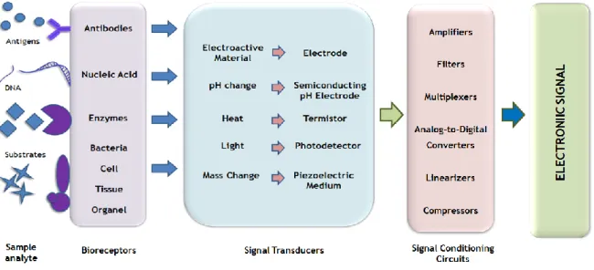
Since the first approach for glucose detection by immobilized glucose oxidase, developed by Clark and Lyon in 1962 (Clark Jr. and Lyons, 1962), biosensors have been intensively studied and extensively used in various applications. The applications range include environmental monitoring and control, chemical measurements in the agriculture, food analysis, drug industries, bioprocess and clinical diagnosis (Mehrvar and Abdi, 2004; Zhang et al., 2000). Biosensors have recognized advantages such as, the improved performance, the elimination of slow preparation and expensive reagents, providing a low cost, portable, simple-to-operate and real-time analysis, over the conventional analytical instruments. On the other hand, some limitations are known such as, electrochemically active interferences in the sample, weak long-term stability and troublesome electron-transfer pathways (Mehrvar and Abdi, 2004). These analytical devices contains two basic components connected in series, the biological recognition system which lead to an biochemical mechanism; and a physicochemical transducer, which is capable to provide selective quantitative or semi-quantitative analytical information (Figure 2.1.) (Thévenot et al., 2001). Both components play an important role in the construction of a sensitive and specific device for the analyte analysis.
The biological recognition system, also known as bioreceptor, consists an immobilized sensitive biological element (e.g. enzyme, DNA, probe, antibody) recognizing the analyte (e.g. enzyme substrate, complementary DNA, Antigen). Its immobilization in proximity of the transducer allows the translation of information from the biochemical domain, usually an analyte concentration, into a chemical or physical output signal with a defined sensitivity.
Biosensors can also be self-contained, that is, all parts are packaged together in the same unit, with the biorecognition element contacted directly with the transducing element. The main purpose of the recognition system is to provide a sensor with a high degree of selectivity for the analyte to be measured. Enzymes are the major biological elements employed in biosensors, nevertheless other biomolecules are widely used (Koyun et al., 2012; Thévenot et
al., 2001).
The transducer part of the sensor serves to transfer the output signal resulting from the interaction of the analyte with the bioreceptor, to the electrical domain by converting the biochemical signal into an electronic one. The intensity of generated signal is directly or inversely proportional to the analyte concentration. With transducer as preferred term, it component is also called as detector, electrode or sensor; in any case, should provide bi-directional signal transfer (non-electrical to electrical and vice versa) (Thévenot et al., 2001).
Figure 2.1. Schematic representation of biosensor device.
A third component based on computer-assisted mathematical modeling, describe the very complex biochemical processes that occur in the boundary region close to the sensor interface (Zhang et al., 2000). Finally, the resulting processed signal has to be presented through an interface to the human operator (Grieshaber et al., 2008).
2.2. Biosensors classification
The existing high range of biorecognition molecules and transducers, leads to high possibility of combinations to construct biosensors. These devices may be classified according to the biological specificity conferring mechanism, based on a catalyzed chemical reaction, or on an equilibrium reaction with, macromolecules that have been isolated, engineered or present in their original biological environment. Biosensors may be further classified according to the
mode of physicochemical signal transduction, and to the analytes or reactions they monitor (Mehrvar and Abdi, 2004; Thévenot et al., 2001).
2.2.1. Transduction process and measurement techniques
As aforementioned, transducer domain will respond to an analyte concentration, converting the biochemical signal into an electronic one. Nowadays, biosensors are miniaturized and self-contained, with all parts packaged together in the same unit, with the biological recognition element in direct spatial contact with the transducing element. Although electrochemical transducers are widely used in biosensors development, the devices can also be based on piezoelectric, calorimetric or optical detection (Koyun et al., 2012; Thévenot et
al., 2001).
2.2.1.1. Non-electrochemical transducers
Optical biosensors are particularly attractive for the application in direct detection systems. The light resulting of a biological and/or chemical reaction can be measured with high sensitivity. However, this sensitivity may result in a lack on turbid media analysis. Also a disadvantage is the relatively long assay time, the longest of all the biosensors, and the lack of sensitivity.
Calorimetric biosensors are based on the heat evolved from biochemical reactions of the analyte with a suitable biological active substance. In terms of performance, those biosensors have also some lack once the majority of heat evolved is lost to the surrounding media without being detected, leading to lowers sensitivities.
Finally, the piezoelectric transducers operate through the coating of the surface of the biosensor that selectively binds biologically active substances. This surface is placed in a solution containing analytes that bind to the binding substance, increasing the mass of the crystal while resonance frequency of oscillation decreases proportionally. This methodology has also some disadvantages like, lack on specificity, selectivity and sensitivity (Mehrvar and Abdi, 2004).
2.2.1.2. Electrochemical transducers
Electrochemical biosensors emerge as the most commonly used biosensors in monitoring and diagnosis tests in different areas, including clinical analysis (Mehrvar and Abdi, 2004). For extracting information from biological systems to measure electrical proprieties (Grieshaber
et al., 2008), those transducers are able to converts biochemical information to a processed
and quantifiable electric signal. They provide selective quantitative or semi-quantitative analytical information using a biological recognition element (Thévenot et al., 1999), offering
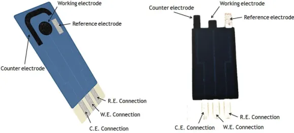
advantages such as, low cost, simple design, small dimensions (Koyun et al., 2012), compatibility with instrumental sensitivity and the ability to operate in turbid media. Those transducers are also more amenable to miniaturization having the biological environment directly connected to the electronic device with a rapid response (Mehrvar and Abdi, 2004). An electrochemical sensing device usually requires in its composition, a reference electrode, a counter or auxiliary electrode and a working electrode, shown later in figure 2.2. for a screen-printed electrode as example. The reference electrode is a kind of standard electrode and is kept at distance from the reaction site in order to maintain a known and stable potential, being commonly made from Silver/Silver chloride (Ag/AgCl) electrically stable. This functions as a redox electrode for the reaction between Ag metal and its salt, AgCl.
The working electrode where the reaction occurs in the electrochemical system, is also known as sensing or redox electrode, and serves as the transduction element in biochemical reaction. Based on Platinum (Pt), Gold (Au) or Carbon (C) (Thévenot et al., 2001), they can be referred as either cathodic or anodic, depending if the reaction is a reduction or an oxidation, respectively. The working electrode is where the immobilization procedures for bioreceptor molecules are conducted.
The third component, a counter electrode, establishes a connection to the electrolytic solution, so that a current can be applied to the working electrode. Thus, the counter electrode functions are cathodic, whenever the working electrode is operates as anode, or vice versa. Both, working and counter electrodes should be conductive and chemically stable, therefore, platinum, gold, carbon and silicon compounds are commonly used, depending on the analyte (Grieshaber et al., 2008; Koyun et al., 2012).
2.2.1.3. Measurement techniques of electrochemical signals
Depending on electrochemical property measured by the detector system, (Bio)-electrochemical sensors can be divided into amperometric, conductometric or potentiometric, according to the detection method of biochemical changes in solution. When the reactions under investigation generate a measurable current, the amperometry is the technique in operation, which is correlated to the reduction or oxidation of an electroactive product on the surface of a working electrode. It is usually performed by maintaining a constant potential between the working electrode and the reference electrode, generating a signal correlated with the proportional concentration of target compounds. Conductometric or capacitive techniques are able to measure conductive properties of a medium, measuring chemical changes in the conductance between electrodes, at constant voltage. These devices need to perform an additional measurement, in an identical device
without the bioreceptor, due to parallel conductance of the sample solution, which hinder the biochemical signal analysis.
Potentiometry investigate a potential or a charge accumulation between the working and a reference electrode, measuring changes in pH and ion concentration, in function of the analyte interaction with immobilized bioreceptor. Thus, the response is a logarithmic function of analyte in sample.
Amperometric biosensors have advantages of being more highly sensitive, rapid, inexpensive, and disposable in comparison to the conductometric and potentiometric biosensors (Grieshaber et al., 2008; Mehrvar and Abdi, 2004; Thévenot et al., 2001).
2.2.2. Biological specificity conferring mechanism
The bioreceptor, usually biomolecules such as, enzymes, DNA probes and antibodies, or even living cells, can interact with the analyte through two types of mechanisms, a biocatalytic one or a bioaffinity mechanism.
2.2.2.1. Biocatalytic recognition element
This category is represented for enzymes, whole cells and tissue as immobilized biocatalyst, which are incorporated into the sensor, allowing the continuous monitoring of substrate(s) consumption. Those macromolecules are present in their original biological environment or have been isolated or manufactured previously.
Enzymatic biosensors, which can be mono- or multi-enzyme, are the most common and well
developed recognition system, which are not consumed and can be reused. Their use as biological recognition elements is very popular owing to their commercial availability and the facility of isolation and purification from diverse sources. Other advantages, such as rapid response, selectivity, sensitivity, precision, the possibility of regeneration and the simplicity involved in constructing devices, are also recognized. The operating principle, based on the recognized specificity of enzymes to the limited range of substrates (Frew and Hill, 1987), enables the monitoring of the catalyzed reaction. The union of the substrate to the active center of the enzyme, allow the formation of the products, which when released, allows the initiation of a new reaction cycle. Thus, those are used to detect the presence of any of the substrates that participate in the reaction by detecting the disappearance of it, or in the appearance of a known product (Cock and Aponte, 2009).
Enzymes as thin as 10 – 200 µm, are incorporated in sensors for it combination in close proximity to the active surface of the transducer, leading to a great selectivity for target species. However, some difficulties in direct electron transfer between enzymes and electrodes are found as limitation. This encourages the use of small-molecule and
electroactive mediators, because they shuttle efficiently electrons between these two elements, enhancing the rate at which the electrons are transferred (Frew and Hill, 1987). In the common applications, the detection limits are satisfactory or excessive, but the stability of enzymes and their capacity to maintain enzymatic activity over a long period of time continues to be problematic. Generally, it is resolved by enzyme immobilization allowing the increase of affinity to the substrate, reducing inhibition, increasing the optimal pH range and reducing possible microbial contamination. However, bacteria, fungus, protozoa and higher organisms become preferred as recognition molecules, when the purification of such enzymes is too difficult and costly, or when the stability is not sufficient to achieve the desirable results (Cock & Aponte, 2009).
Whole cells (microorganisms, cell organelles or particles) can be immobilized in membranes
or trapped in matrix in a simpler and more economical way than enzymatic immobilization. With value in clinical analysis of biological fluids and for monitoring fermentation processes, these devices can provide a simple and rapid means of measuring analytes that previously were undetectable by electrochemical methods (Frew and Hill, 1987). These systems have the advantage of being directly used as biorecognition element, instead to be purified, having a reducing cost, improved stability and even offering the possibility of regeneration with the increase of electrode lifetime. As well, they are capable to metabolize different organic compounds, due to multi-cellular enzymatic systems that possess, generating distinct products. Furthermore, cells offer the facility to be modified genetically to improve their activity or to produce specific enzymes that do not normally appear. Nevertheless, limitations like the reduced diffusion of substrates and products result in a slower response comparing to the purified enzyme biosensor, leading to lengthy recovery times between measurements, referring their use as disposable devices. In addition, is also observed a lost on specificity owing to reactions catalyzed by others enzymes present in the cell (Cock and Aponte, 2009; Frew and Hill, 1987).
Tissue biosensors (plant or animal tissue slice), are based on determined tissues that
according to their physiological function in the organism, produce specific enzymes or enzymatic systems. Some examples of used tissues are roots, leaves, fruits or seeds from plants, or animal tissues as beef liver or cortex of porcine kidney, in sliced or homogenized form (Cock and Aponte, 2009; Wijesuriya and Rechnitz, 1993). These structures have recognizable high concentrations of biocatalytic activity, due to their functions in the live being.
Low cost, simple construction, high catalytic activity, good shelf and electrode lifetime are offered by these types of sensors. Like whole cells biosensors, slow responses due to diffusional limitations; and a poor selectivity are also found. The presence of multiple enzymes, lead to the need of changing external conditions like, the use of different
substrates, enzyme inhibitors or activators and stabilizing agents (Cock and Aponte, 2009; Rechnitz and Ho, 1990).
2.2.2.2. Bioaffinity recognition element
Antibodies and nucleic acids are the biorecognition elements integrated in this category. Based on the interaction of the analytes with macromolecules or organized molecular assemblies, the biosensor reach the equilibrium and there is no further net consumption of the analyte by the immobilized biocomplexing agent, which have been isolated from their biological environment or engineered. In some cases this biocomplexing reaction is itself monitored using a complementary biocatalytic reaction. The equilibrium reaction signals are then monitored by the integrated detector, being often unable to monitor continuously the analyte concentration (Thévenot et al., 2001, 1999).
Antibodies and Antigens have been used for the specific detection of their complementary
partners through immunochemical reactions, where an antigen (Ag) is bound to a specific antibody (Ab). The biosensors concerned with monitoring solely Ab-Ag interaction, are also termed immunosensors, and the detection needs to be performed under conditions where non-specific interactions are minimized. Similarly to conventional immunoassays, these devices are based on the principles of solid-phase immunoassay, with either antibody or antigen immobilized at the sensor surface. These, can be divided into heterogeneous assays, in which antibody-bound antigen is separated from free antigen during the procedure, and homogeneous assays, in which there is no such separation step (Frew and Hill, 1987). Each determination requires the production of a particular Ab, its isolations and, usually, its purification. However, the Ab-Ag complexes formed have a particularly high binding or affinity constant, which make regeneration of the surface difficult, turn such systems irreversible (single-use biosensors). Notwithstanding, the Ab surface can be regenerated by dissociation of complexes, placing within environments with chaotropic agents, such as glycine-HCl buffer at pH 2.5.
The basic feature of immunoassay that provides the desired specificity under favorable conditions, is the complementary reaction (both on chemical binding and spatial orientation of reactive groups) between antigen and antibody. Thus, the sensitivity and specificity of an immunosensor are determined by the affinity and specificity of the binding agent. In some cases, the use of labels has been incorporated into immunosensor design to enhance sensitivity. Thus, the choice of label is another important feature, once such labelling requires an additional chemical synthesis step; and can be performed using radioisotopes, fluorescent, luminescent, light-scattering, enzyme or redox labels. In the case of labelled Ab, these devices will operate at equilibrium to quantify indirectly the amount of complexes produced (Morgan et al., 1996; Thévenot et al., 2001). Thus, an voltammetric based detection of immunoreactions depends on label type, assay format and electrochemical
technique, which can achieve excellent detection limits on small sample volume (Frew and Hill, 1987).
Based on the hybridization concept, nucleic acids (e.g. DNA and RNA) also have been used, once a known DNA sequence (probe) and an unknown counterpart (analyte) have a complementary degree between both chains. This biomolecular recognizing event occurs directly on the surface of the physical-transducer due to an immobilized single-chain of DNA specific to the target DNA analyte chain. The hybridization is stronger and more specific with the increase of complementarity degree between the two DNA chains, reaching a maximum in the case of full 100% recognition. That hybridization results in measurable changes in electrical parameters, allowing the electrochemical analysis. Like immunosensors, in this DNA-sensors design can be used various types of labels to improve the sensitivity (He et al., 2011; Wang et al., 1997).
2.2.3. Analytes or reactions monitored
Biosensors classification according to the target analytes or reactions that they monitor should be differentiated between, the direct monitoring of analytes and the indirect monitoring of inhibitors or activators.
Direct monitoring of the analyte: Known as the main application of biosensors, are based on
the direct measurement, continuously or sequentially, of a physical phenomenon that occurs during a biochemical reaction on the transducer surface. Those devices measure the production or consumption of a given compound, being a useful tool for enzyme or living cell activities monitoring.
Indirect monitoring of inhibitor or activator of the biochemical receptor: These devices are
irreversible, and their use is limited to applications which not require exact measurement of the analyte concentration. They are referred to as single-use biosensors and were developed for detecting substances, such as inhibitor or activator of the bioreceptor, which interferes with biocatalytic properties. One example of those devices applications are the monitoring of organic pesticides and inorganic substances (Mehrvar and Abdi, 2004; Thévenot et al., 1999).
2.3. Screen-Printed Electrodes (SPE)
During the past decades, due to their advantages, the importance of electrochemical techniques has increased, being the main class of used of biosensors (Mehrvar and Abdi, 2004). However, in the analytical chemistry field these techniques have several restrictions and practical difficulties such as the cleaning and the surface regeneration processes needed in solid electrodes. On the other hand, conventional based stripping measurements had relied
Additionally, there is a growing need of rapid performance and in situ analyses. This challenges resulted in an emerging electroanalytical field of small sized, disposable, sensitive, selective and low-cost devices, the Screen-Printed Electrodes (SPE) produced by thick-film technology (figure 2.2.) (Metters et al., 2011; Nyholm and Schleiss, 2010; Renedo
et al., 2007; Tudorache and Bala, 2007).
These microelectronics adapted devices, offer important benefits facing the traditional methods mainly the possibility of sample direct analyses in its environment without alteration of the natural conditions (Tudorache and Bala, 2007), reinforcing their use to a more effective real life applications on industrial, clinical and environmental fields. SPE allows short assays with lowest energy consumption, making possible a small fluid manipulation, with less reagent at lower cost, increasing the sample throughput, which is also increased by the additional possibility of employing many parallel on-chip systems (Nyholm and Schleiss, 2010; Sin et al., 2014).
Figure 2.2. Electrochemical biosensor as a screen printed device and its constitution. Representation of working, counter and reference electrodes. At left, is represented an SPCE device produced at Burgos University.
2.3.1. SPEs fabrication
The automated and easy fabrication of these devices results in a large range of possibilities for design. The thick-film technology consists on the sequential deposition of layers of special inks or pastes onto an insulating support or substrate, being widely used on large-scale fabrication (Renedo et al., 2007; Tudorache and Bala, 2007).
The deposition of different inks is carried out on a substrate using the appropriate screen, which is made from a finely mesh of different materials including stainless steel, polyester or nylon mounted under tension on a metal frame. The screen has open-mesh areas through which the desired pattern can be printed. The printing of SPE, shown in figure 2.3., is
conducted by a squeegee which slowly moves from the back to the front part of the screen, forcing the ink or paste through the open areas, depositing the required pattern onto the substrate surface. To obtain the desired electrode, between the printing patterns, is carried out a step of ink drying at an adequate temperature to allow the evaporation of the ink solvents. The requirement of this step is justified by the screen-printing inks which usually contain various organic solvents added with the aim of producing the accurate viscosity for screen-printing process. Thus, the substrate retains a rigid pattern, in the range of 20 to 100 µm of thickness (Metters et al., 2011) that is relatively resistant to smudging. The combination of different screens and inks give rise to the definition of the different electrodes (working, reference and auxiliary) in the same configuration unit. The available range of different screens, originates different types of SPEs configurations, used according to the analytical problem characteristics by choosing the adequate fabrication materials.
Figure 2.3. Screen printed device fabrication process scheme. A - Components for SPE fabrication, Squeegee, porous screen and substrate; B - Screen-printing step by squeegee traversing, C - Ink deposited on the substrate surface.
To be converted into a biosensor device, the SPE are further modified with the biosensing material. This modification, held at the working electrode, implies several steps in order to assure the robustness and durability of the developed biosensor (Metters et al., 2011; Renedo
et al., 2007).
2.3.2. Materials for SPE construction
The performance of sensors fabricates by thick-film technology depends critically on the materials used and the manufacturing process. In addition, an electrode substrate of suitable condition, size, geometry, and immobilization technique has usually been taken into consideration to design appropriate electrochemical biosensors (Mehrvar and Abdi, 2004). Thus, the configuration of the SPE comprises material defining the basic structure of the electrode, the non-biological materials; and the biorecognition element with their related compounds and additives and/or cross-linkers necessary for developing the electrochemical signal (Tudorache and Bala, 2007).
2.3.2.1. Non-biological materials
Depending on the measurement technique, different materials can be used for the preparation of surfaces for biosensing applications. Currently, a wide diversity of materials is used, however, they need to fulfill special requirements, such as electrical conductivity for the electrochemical techniques (Grieshaber et al., 2008). This category encompasses the materials for substrates, inks or pastes and the immobilization matrixes, additives or cross-linkers used for the bioreceptor element (Tudorache and Bala, 2007).
The first consideration is the composition of the substrate, the base on which the functional sections of the sensor are printed. The most common and well known materials used for substrate composition are alumina, ceramics, Poly(vinyl chloride) (PVC), cardboard coated with acrylic paint, and polycarbonate. The substrate must provide good electrical insulation, be chemically inert (Tudorache and Bala, 2007) and an inexpensive support (Renedo et al., 2007).
On the other hand, the inks or pastes which represent the conducting part of the electrode, has an important role on the SPE performance. Commercial carbon and metal ink formulations are commonly used for printing the working electrodes, whereas silver-based inks are used for obtaining the reference electrode (Renedo et al., 2007; Wang et al., 1998). In fact, a broad range of pastes with different properties are available commercially, however, they are commonly adjust to make them suitable for particular purposes. These pastes usually contain binding agents, solvents and additives for dispersion, printing and adhesion tasks, that provide functional characteristics. To increase the conductivity properties, powdered metals, such as gold, platinum, silver, or palladium, or even non-metallic conductors like graphite, are added (Tudorache and Bala, 2007). Thus, the composition of the various inks used determines the selectivity and the sensitivity required for each analyses, resulting in versatile SPEs (Metters et al., 2011; Renedo et al., 2007). The carbon inks, composed by graphite particles are widely used, owing to their electrochemical advantages and improvements in response, detection limit and cost. Moreover, they have an easily handling and easy preparation. Differences in ink composition (e.g., Type, size or loading of graphite particles), and in the printing curing conditions, may strongly affect the electron transfer reactivity and overall analytical performance of the resulting carbon sensors (Tudorache and Bala, 2007; Zhang et al., 2000).
Attributes such as good adhesion, similar coefficients of expansion, to prevent stress-relates damage during sudden heating and cooling, the ability to retain their characteristics through the fabrication process, the ready availability, and low cost must be found at all materials mentioned above (Tudorache and Bala, 2007).
Apart from the biomolecule, some other materials can be used to functionalize the working electrode surface, as example the gold, silver, nickel, bismuth and mercury coating-films,
which increase the function of SPEs. Among the materials used, the metallic nanoparticles (Nps) are highlighted due to their important properties and multiple applications. Au, Pt and Ag have an important role on nanoparticle field for biosensing application and can be obtained by chemical or electrochemical synthesis or by UV light (Renedo et al., 2007). Due to their high surface-to-volume ratio, conductivity, catalytic and magnetic properties the NPs are attractive means of signal amplification for the improvement of sensitivity detection rates and versatility of biosensing devices (Nyholm, 2005).
Furthermore, the possibility of modifying the SPE by means of depositing various substances on the surface of the electrodes, as example the immobilization matrixes, additives (e.g. cellulose acetate) and cross-linkers (e.g. glutaraldehyde) are also often involved in structures to improve the sensitivity, selectivity, stability, and reproducibility of the biosensor (Renedo
et al., 2007; Tudorache and Bala, 2007).
2.3.2.2. Biological material
The active part of the sensors, the layer of the biorecognition element, must be connected to different conductive pads for transducing and conversing signals. These elements, as mentioned may be enzymes, antibodies, DNA, RNA, cells, mediators and cofactors existing different forms for its deposition onto SPEs (Tudorache and Bala, 2007). Special attention will be given to antibodies in the next chapter.
3. Immunosensors
Bioanalytical assays such immunoassays (IAs), are transcendent to a variety of fields, including the biosensor field, in which offers applications for biological and medical research, diagnostic medicine, genetics, forensics, drug and pesticide testing (Frew and Hill, 1987; Renedo et al., 2007). Firstly introduced by Yalow and Berson (Yalow and Berson, 1959), the concept of monitoring the specific antigen (Ag) and antibody (Ab) complexation, was used on an IA for plasma insulin in human subjects (Holford et al., 2012).
3.1. Immunosensors principles and components
As already mentioned, biosensors can integrate different types of biomolecules as sensing elements. In the case of immunosensors, antibodies or antibody fragments with high specificity to detect the presence of its specific antigen, by forming a stable complex (Figure 3.1) are employed. The high affinity of the Ab is an important advantage of immunosensors, resulting in very low detection limits. However, this type of biosensor presents a general problem which is the difficulty to reversibly regenerate the sensing surface without rendering the device unusable by removing or denaturing the biological recognition element. Thus, due to the high affinities of Ab to Ag (Kd’s generally lie between 105–1011 M), efforts are to the
construction of disposable or single-use devices (Holford et al., 2012; Tudorache and Bala, 2007).
Figure 3.1. Schematic representation of complexation reaction between antibody and its specific antigen. Adapted from http://drcercone.iculearn.com/bio2/wpcontent/uploads/Lectures/immune
Immunosensors are small and portable instruments, designed for use by untrained personnel, for a rapid and sensitive assay. In accordance with the definition of a biosensor, the immunosensors also incorporate a transducer in which the biological component is immobilized allowing the occurrence of a biochemical or biophysical event onto the surface.