CRISTINA MARIA OLIVEIRA FERREIRA
“ANALYSIS AND CHARACTERIZATION OF DE NOVO CENTRIOLE BIOGENESIS IN ACENTRIOLAR DROSOPHILA SAS-4 -/- CELLS”
Dissertação de Candidatura ao grau de Mestre em Oncologia submetida ao Instituto de Ciências Abel Salazar da Universidade do Porto.
Orientador – Dr. Helder Maiato
Categoria – Professor Auxiliar Convidado
“Life begins at the end of your comfort zone” Anonymous Para os meus Pais, Emília e Carlos À Olga Afonso
Agradecimentos
Por vezes, pequenos elementos mudam o rumo das nossas vidas.
No meu caso, esse elemento foi um simples envelope com um conjunto de cartas deixado, em meados de Outubro do ano 2011, na secretaria de um instituto científico localizado na cidade do Porto.
O envelope dirigia-se ao investigador Hélder Maiato e não continha mais do que três cartas que forneciam informação sobre uma estudante de mestrado, licenciada em Biologia, e entusiasta em relação a ciência.
As probabilidades de ser contactada eram reduzidas, mas nunca foram impedimento para o atrevimento.
Dois anos passaram, e dentro das inúmeras aprendizagens, existe uma conclusão: a vida não avança, se a acompanhá-la não existir atrevimento.
É inacreditável a quantidade de informação que absorvi e aprendi nestes últimos dois anos. Acredito agora mais que nunca, que a faculdade é simplesmente o ponto de partida e a base para um conhecimento generalizado. No entanto, é no laboratório onde se “talham” os cientistas.
Esta tese de mestrado é o resultado de dois intensivos anos de trabalho laboratorial. Mas, acima de todo o trabalho cientifico elaborado, é o reflexo de muita persistência.
Por sua vez, a persistência advém de um conjunto de pessoas que proporcionaram o ambiente ideal para que eu fosse capaz de ultrapassar os momentos mais complicados.
Todos os elementos do laboratório do Hélder foram, sem excepções, elementos cruciais para o meu sucesso e bem-estar. Ana Pereira, António Pereira, Cristina Madureira, Danica Drpic, Elsa Logarinho, Filipe Sousa, Joana Macedo, Jorge Ferreira, Luísa Ferreira, Margarida Gomes, Marin Barisic, Martina Barisic, Nina Schweizer, Olga Afonso, Zaira Garcia e o próprio, Hélder Maiato.
Em especial, agradeço à Cristina Madureira e à Zaira Garcia por todos os ensinamentos relativos à componente de biologia molecular.
Obrigado à Ana Pereira por ter, sem dúvida, enriquecido o meu leque literário. Também agradeço aos elementos do laboratório do Cláudio Sunkel pela hospitalidade com que me receberam, especialmente à Tália Figueiredo, Carlos Conde e João Barbosa.
Agradeço à Zita Carvalho-Santos pelos valiosos comentários e discussões, principalmente em relação à análise por microscopia electrónica.
E agradeço imenso ao Hélder pela oportunidade que me deu de desenvolver este trabalho científico como parte da minha tese de mestrado, mesmo tendo consciência da minha imensa inexperiência e falta de conhecimento. Foi, sem dúvida, o elemento crucial e influenciador das decisões que acabei por tomar ao longo destes últimos dois anos.
No entanto, existe uma pessoa que, acima de todas, considero ser a base de toda a minha aprendizagem, orientação, e que estará sempre associada a todos os “passos” deste projecto. Essa pessoa é a Olga Afonso.
É de facto louvável a atitude da Olga que, debatendo-se com o seu doutoramento, assumiu tal responsabilidade. É, devido a ela, que considero ter sido possível executar este projecto e, consequentemente, escrever esta tese. Se actualmente sou portadora de “algum” conhecimento científico, é inteiramente devido a ela.
Muito obrigado, Olga, por toda a tua dedicação, carinho, paciência, orientação para comigo. Todos os méritos que, eventualmente, possam reconhecer a este trabalho, serão sempre partilhados contigo.
Em especial agradeço, também, a – André Levi, Diogo Pedrosa, Gilda Carvalho, Jorge Correia de Castro, Maria Pedro, Patrícia, Rita Rocha, Tiago Ferreira - que sempre me apoiaram e incentivaram ao longo destes anos de amizade.
Ao António Dias, Dennis Herrmann, Gabriela Fioreze, Jovin Jacobs, Lorenza Calcaterra e Tomás Cruz por tornarem os meus dias Lisboetas muito melhores.
Ao Luís Pedro, quero agradecer todo o carinho, paciência e atenção ao longo destes anos, especialmente nos momentos mais delicados.
À minha irmã, Ana Bárbara, que é um motivo de orgulho e admiração todos os dias.
Por fim,
Aos meus pais por toda a dedicação e afecto ao longo destes vinte e quatro anos.
São voçês, sem dúvida, o “motor” da minha vida. Os meus “incentivadores”. Aqueles que, independentemente das circunstâncias, sempre acreditarão em mim.
Os elementos essenciais para a minha construção enquanto pessoa.
Abstract
Centrioles, the key elements on centrosome biogenesis and function, are replicated during S phase of each cell cycle. Two main mechanisms of centriole assembly have been described: 1) a template or canonical, and 2) a de novo. While the template mechanism has been assigned as the major mode of centriole assembly in somatic cell divisions, the nature of the de novo mechanism of centriole assembly is less well understood, especially in proliferating somatic cells. Moreover, the function of the mother centriole as a template in the centriole duplication process has been recently brought into question (Rodrigues-Martins et al., 2007b). Here, we show that expression of DSas-4-GFP in somatic acentriolar DSas-4 mutant Drosophila cells led to de novo centriole formation. Mitotic DSas-4 rescued cells showed amplification and co-localization of de
novo DSas-4-GFP foci with the centriolar and PCM proteins DSas-6, Ana1, D-PLP, Asl
and Cnn, respectively. Live cell imaging analysis showed that the newly formed DSas-4-GFP foci nucleate discrete astral microtubule and mitotic spindle assembly seemed to occur through an outside-inside process. Moreover, we rescued the expression levels of the centriole-specific proteins Asl and DSas-6, suggestive of an activation of the centriole biogenesis pathway. Electron microscopy analysis revealed the presence of centriole-like structures as well as clouds of electron-dense material lacking centrioles.
This study reinforces the notion that de novo and template mechanisms of centriole assembly may be variations of a common pathway based on the same molecular machinery. Centriole biogenesis is a template-free process in which the mother centriole may function as a platform for regulatory proteins involved in the centriole duplication process whereby offering an ideal environment for centriole duplication. Therefore, control of centriole number, spatial restriction along with a kinetic advantage might be the key factors that the mother centriole offers to the centriole biogenesis process.
Key words: centrioles; centrosomes; de novo centriole biogenesis; DSas-4 protein; Pericentriolar material; Drosophila Sas-4 mutant cell line
Resumo
Os centríolos, elementos chave na formação e funcão do centrosoma, são replicados durante a fase S de cada ciclo celular. Existem dois principais mecanismos de duplicação: 1) através de um molde, e 2) o de novo. Enquanto que o mecanismo molde tem sido reconhecido como o principal modo de formação de centríolos nas células somáticas em divisão, a natureza do modo de novo é ainda pouco compreendida, principalmente durante a proliferação de células somáticas. Além disso, o papel do centríolo mãe como modelo no processo de duplicação dos centríolos foi recentemente questionado (Rodrigues-Martins et al., 2007b). O presente estudo mostra que a expressão da proteína DSas-4-GFP em células acentriolares de Drosophila mutantes para a proteína DSas-4 leva à formação de novo de centríolos. Células mitóticas a expressar DSas-4-GFP mostram amplificação e co-localização dos foci de DSas-4-GFP com as proteínas centriolares e centrosomais DSas-6, Ana1, D-PLP, Asl e Cnn, respectivamente. Análise através de microscopia em células vivas revelou que os foci de DSas-4-GFP são capazes de gerar feixes de microtúbulos astrais e a formação do fuso mitótico parece ocorrer através de um processo de fora para dentro. Adicionalmente, foi verificado um aumento nos níveis de expressão das proteínas especificamente associadas com os centríolos, Asl and DSas-6, o que poderá sugerir uma re-activação do processo de formação de centríolos. A análise por microscopia electrónica revelou a presença de estruturas similares a centríolos, bem como locais com material pericentriolar sem centríolos.
Este estudo reinforça a ideia de que os processos de novo e molde são potencialmente variações de um mesmo processo que tem por base a mesma maquinaria molecular. A biogénese dos centríolos é um processo que não necessita de um molde, e em que o centríolo mãe poderá funcionar como uma plataforma para proteínas reguladores involvidas na biogénese de centríolos, assegurando um ambiente ideal para a duplicação dos centríolos. Assim, o controlo do número de centríolos, restrição espacial e uma vantagem cinética poderão ser os factores principais que o centríolo mãe oferece ao processo de biogénese de centríolos.
Palavras-Chave: centríolos; centrosoma; biogénese de novo de centríolos; proteína DSas-4; material pericentriolar; linha celular mutante Drosophila Sas-4
Abbreviations
aMTOCs – acentriolar microtubule organizing centres Ana1-3 – anastral spindle 1-3
Asl – asterless
BSC-1 –African green monkey kidney epithelial CDK-2 – cyclin-dependent kinase 2
CDK5RAP2 – CDK5 regulatory subunit-associated protein 2 CENPJ – Centromere protein J
CEP135 – centrosomal protein CEP152 – centrosomal protein CEP192 – centrosomal protein CHO – Chinese hamster ovary Cnn – Centrosomin
CP110 – Centriolar coiled-coil protein
CPAP – centrosomal P4.1- associated protein D-PLP – Drosophila pericentrin-like protein
DSas-4 – Drosophila spindle assembly abnormal 4 DSas-6 – Drosophila spindle assembly abnormal 6 GDP – guanosine diphosphate
GTP – guanosine triphosphate
hSAS-6 – human spindle assembly abnormal 6 LECA – last eukaryotic common ancestor MTOC – microtubule organizing centre MTs – microtubules
NAB – nuclear associated body PCM – Pericentriolar material PLK1 – polo-like kinase 1 PLK4 – polo-like kinase 4 S2R+ - S2 receptor plus
SAS-4 – spindle assembly abnormal 4 SAS-5 – spindle assembly abnormal 5 SAS-6 – spindle assembly abnormal 6 SPB – spindle pole body
SPD-2 – spindle defective 2
TCP10 – T-complex protein 10 γ-TuRCs – γ-tubulin ring complexes
1. INTRODUCTION 23
2.DISSECTING CENTROSOMES AND CENTRIOLES 26
2.1.HISTORY AND INTER-RELATIONSHIP 26
2.2.ROLE IN SPINDLE ASSEMBLY DURING CELL DIVISION 27
2.3.ACENTRIOLAR ORGANISMS AND CELL LINES 29 3.THE CENTROSOME 31
3.1.FROM WHAT IS MADE A CENTROSOME ? 31 3.2.OVERVIEW OF CENTROSOME CYCLE 34 4.HOW TO BUILD A CENTRIOLE ? 37 4.1.CENTRIOLE ARCHITECTURE 37 4.2.CENTRIOLE ASSEMBLY PATHWAY: ACTION OF A CORE ANCESTRAL PROTEIN MODULE 40 5.SAS-4: A MULTIFACETED PROTEIN 45 6.CENTRIOLE BIOGENESIS:DIFFERENT ORIGINS, ONE GOAL 50 6.1. THE ROLE OF THE PARENTAL CENTRIOLE 52 OBJECTIVE 56
2. MATERIALS AND METHODS 60
3. RESULTS 67
PART 1 69
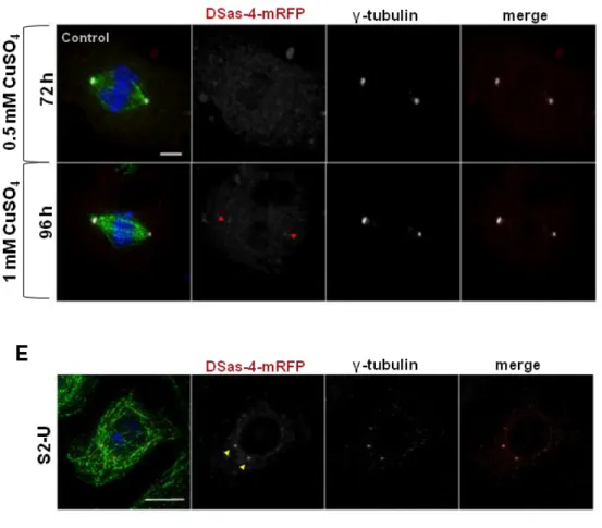
1.CHARACTERIZATION OF THE DROSOPHILA ACENTRIOLAR DSAS-4-/- CELL LINE #131 71
1.1.ACENTRIOLAR MITOTIC SPINDLE ASSEMBLY 71
1.2. RECRUITMENT OF PCM AND CENTRIOLAR PROTEINS IN DSAS-4-/- CELLS 72
PART 2 79
2.1. TRANSFECTION OF DSAS-4 PROTEIN IN THE DSAS-4-/- CELLS 81
2.1.1.DSAS-4-mRFP CONSTRUCT 81
PART 3 89 3. ANALYSIS OF DE NOVO CENTRIOLE FORMATION IN DSAS-4-/- RESCUED CELLS 91
3.1. RESCUE OF DSAS-4-/- CELLS THROUGH DSAS-4 PROTEIN REINTRODUCTION 91 3.2.DE NOVO FORMED DSAS-4-GFP FOCI COLOCALIZE WITH CENTRIOLAR PROTEINS 92 3.3.AMPLIFICATION OF DSAS-4-GFP FOCI IN DSAS-4-/- RESCUED CELLS 94
3.4.RECOVERY OF THE EXPRESSION LEVELS OF CENTRIOLAR PROTEINS IN DSAS-4-/- 96
RESCUED CELLS
3.5.THE DE NOVO FORMED DSAS-4-GFP FOCI ARE ABLE TO NUCLEATE MICROTUBULES 97 AND TO ACCUMULATE CENTROSOMIN DURING MITOSIS
3.6. DE NOVO CENTRIOLE-LIKE STRUCTURES ARE FORMED IN DSAS-4-/- RESCUED CELLS 100
4. DISCUSSION 104
DSAS-4-/- CELLS SHOW ABNORMAL SPINDLE MORPHOLOGY ACENTRIOLAR DSAS-4-/- CELLS RECRUIT PCM AND CENTRIOLAR COMPONENTS 107
TO THE POLES OF THE MITOTIC SPINDLE
ECTOPIC DSAS-4 PROTEIN RESCUES DSAS-4-/- ACENTRIOLAR PHENOTYPE 110
DE NOVO FORMED DSAS-4-GFP FOCI COLOCALIZE WITH CENTRIOLAR PROTEINS 112
AMPLIFICATION OF DSAS-4-GFP FOCI IN DSAS-4-/- RESCUED CELLS 114
RECOVERY OF THE EXPRESSION LEVELS OF CENTRIOLAR PROTEINS IN DSAS-4-/- 115
RESCUED CELLS
THE DE NOVO FORMED DSAS-4-GFP FOCI ARE ABLE TO NUCLEATE MICROTUBULES 117
AND TO ACCUMULATE CENTROSOMIN DURIING MITOSIS
DE NOVO CENTRIOLE-LIKE STRUCTURES ARE FORMED IN DSAS-4-/- RESCUED CELLS 120
5. CONCLUSIONS 124
REFERENCES 131
Figure Index Introduction Figure 1 32 Figure 2 38 Figure 3 44 Figure 4 45 Results Figure 5 71 Figure 6 73 Figure 7 74 Figure 8 76 Figure 9 82 Figure 10 86 Figure 11 91 Figure 12 93 Figure 13 95 Figure 14 96 Figure 15 98 Figure 16 100 Figure 17 101
1. Introduction
"Omnis cellula e cellula" was postulated two centuries ago by the German
physician Rudolf Virchow and it was the first approach defining that every cell arises from a preexisting parent cell. Back to our century, it is now known that Virchow's definition is undoubtedly correct and that cells need to proliferate to ensure progeny and the continuity of life (Rieder, 2006, pp.439).
Cell division is the result of a complex and strictly regulated process composed by distinct stages integrated in the cell cycle. In eukaryotes, the major goal of the cell cycle is to reproduce a daughter cell inheriting the same diploid number of chromosomes from the mother cell in a highly dynamic process called Mitosis. After DNA replication in interphase, sister chromatids of each chromosome have to be segregated to assure the euploidy of the daughter cells during mitosis. Therefore, an important biological machine was developed by the cells in order to organize chromosomes during metaphase and to accurate segregate them in anaphase - the Mitotic Spindle. The mitotic spindle is a symmetrical, dynamic and bipolar structure made up of microtubules (MTs) that interact with chromosomes via their kinetochores. Its assembly is orchestrated by centrosomes which are defined as the major microtubule-organizing centre (MTOC) in animal cells. Apart from its role in cell division, the centrosome is also a very important organelle in cell shape, polarity, motility, signaling, protein trafficking and in cilia/flagella formation (Bettencourt-Dias, 2013; Nigg, 2007; Nigg and Raff, 2009)
The advent of electron microscopy revealed the beautiful composition of the centrosome. This organelle is composed by a pair of barrel-shaped bodies called centrioles that are surrounded by an area of dense protein matrix termed the pericentriolar material (PCM) (Avidor-Reiss and Gopalakrishnan, 2013; Azimzadeh and Marshall, 2010; Debec et al., 2010; Nigg, 2007). As centrioles are at the basis of centrosome formation, it demands strict control of centriole number to ensure a correct number of centrosomes, and faithful chromosome segregation in mitosis. Indeed, it has been recognized the direct association between centriole number deregulation, centrosome overexpression and cancer (Nigg and Raff, 2009). Over the past decade, the mechanisms underlying centriole biology have been uncovered at a tremendous speed due to the development of more advanced imaging techniques and its combination with biochemical and cell biological approaches. A deeper knowledge of centriole structure, function and biogenesis is of major importance to a better understanding of centrosome biology and its role in disease.
2. Dissecting Centrosome and Centrioles 2.1. History and Inter-relationship
The centrosome was discovered by Boveri and Van Beneden in the late 1800s and it was coined as "the organ for cell division" with an important role in karyokinesis/mitosis and cytokinesis. Since that very first description, there was a long latency period in which research on centrosomes/centrioles languished. However, in the 80s and 90s, genetic and proteomic studies allowed the identification of specific centrosome and centriole proteins, as well as electron microscopy revealed the amazing architecture of centrioles within centrosomes.
Centrioles, which are the core components of the centrosome, are among the most conserved organelles through evolution in eukaryotic cells, having been lost in several lineages such as yeast, some amoebas and seed plants (Bettencourt-Dias, 2013; Carvalho-Santos et al., 2011). These organelles can also be converted into basal bodies when tethered to the cell membrane to template the axoneme required in cilia and flagella formation, indicating that centrioles are not only important structures in cell division, as they also perform a role in cell motility and signaling.
The evolutionary history of centrioles and centrosomes has just started to emerge. Recently, two phylogenetic studies (Carvalho-Santos et al., 2010; Hodges et al., 2010) unveiled the evolution of the centriole through the analysis of its protein components, from the centriole assembly machinery to the ciliary related proteins. Namely, Hodges and colleagues (2010) brought some important data supporting a previously held idea about centriole ancestral function, in which centrioles and centrosomes have not always coexisted during evolution. By analyzing the phylogenetic distribution of core centrosomal proteins across six major groups of eukaryotes, they found that the majority of the centrosomal proteins were restricted to Holozoa1, suggesting a scenario in which the animal centrosome functioning as a MTOC in cell division is an holozoan innovation, which implies that in the temporal scale of evolution, centrioles could have been present before centrosomes, performing an ancestral role only as basal bodies (in motility/sensory function and cell organization) (Bornens and Azimzadeh, 2007). Interestingly, the occurrence and requirement of centrioles seems to be strongly correlated with the presence of flagella/cilia rather than with the centrosome (Carvalho-Santos et al., 2011; Hodges et al., 2010). It is known that many non-flagellated species, like some amoeba and fungi are able to produce modified MTOCs with no centrioles, such as the nuclear associated body (NAB) (amoebas) and the spindle pole body (SPB) (fungi) (Bornens and Azimzadeh, 2007; Debec et al., 2010). These data suggest that centrioles are mostly
1
essential in axoneme nucleation rather than in centrosome formation. It is possible to speculate that, during evolution, the ancestral centriolar structure only took part in cell division as a parsimonious solution to efficiently control the equal chromosome segregation into the daughter cells, providing a selective advantage to primitive eukaryotic cells (Bornens and Azimzadeh, 2007). Furthermore, Carvalho-Santos et al. (2010) have shown that centriole biogenesis is controlled by an evolutionary conserved and ancestral protein module (UNIMOD) that might have emerged in LECA (last eukaryotic common ancestor), and its occurrence is correlated with presence or loss of centrioles.
2.2. Role in Spindle Assembly during Cell Division
A controversial question in centrosome biology has been to what extent the centrosome is an essential and mandatory organelle for cell division, specifically acting as an MTOC needed for mitotic spindle formation and correct chromosome segregation during mitosis in animal cells. In contrast to what was believed at the time of the first pioneering studies on the centrosome, it is now known that the centrosome is not an essential organelle for cell division and survival. As described above this organelle was naturally lost in some groups of the eukaryotic tree of life such as in some species of fungi (ex. Schizosaccharomyces pombe). However, even those species organize the microtubule cytoskeleton using different MTOCs which do not contain centrioles, as exemplified by the spindle pole body in S. pombe and Saccharomyces cerevisiae (Bettencourt-Dias, 2013; Bornens and Azimzadeh, 2007; Debec et al., 2010). This is indicative that an MTOC does not need to be necessarily a centrosome, whereas a centrosome is always an organelle responsible for MT nucleation and anchoring.
In animals, the most documented and thoroughly studied case of a natural absence of centrioles is in female meiosis. In this specific event, as in the case of humans, the oocyte looses centrioles during oogenesis (in some point before metaphase of meiosis I) retaining just the PCM, whereas the spermatocyte does not retain the PCM, but an incomplete centriole pair. A fully functional and intact centrosome will only born after fertilization by the combination of the sperm-derived centrioles and the PCM supplied by the egg (reviewed in Debec et al., 2010). In animal somatic cells, centriole loss or inactivation was reported in differentiated cells, such as myotubes (muscle cells), in certain types of epithelia cells such as Drosophila wing epidermal cells and, also in neuronal cells (Bartolini and Gundersen, 2006; Bettencourt-Dias, 2013; Cunha-Ferreira et al., 2009; Debec et al., 2010). Surprisingly, a striking case of natural centrosome inactivation occurs in Drosophila cells, in which the interphase MT cytoskeleton is not nucleated by the centrosome, which seems to be only active as MTOC during mitosis (Rogers et al., 2008). In these last cases, centriole inactivation or loss is coupled with the
existence of non-centrosomal MT arrays, and even in cells that normally have functional centrioles, those centrioles are not obliged to take part in cell division within the context of the centrosome. This characteristic has been proven over the last years by a series of experiments taking advantage of specific antibodies that disrupted centrioles or laser irradiation to ablate the centrosome, in which it was demonstrated the dispensability of centrioles in cell division (Bobinnec et al., 1998; Khodjakov et al., 2000; Mahoney et al., 2006). These studies imply that additional non-centrosomal mechanisms of MT nucleation must exist, even in cells that naturally contain functional centrosomes.
Two major centrosome-independent pathways used for spindle assembly were described. A chromatin pathway that generates and stabilizes microtubules in the vicinity of the chromosomes, facilitated by a RanGTP gradient centered around chromosomes that triggers the release of TPX2 that, together with γ-tubulin complexes, nucleate MTs (reviewed in Meunier and Vernos, 2012). The CPC complex (INCENP, Survivin, Borealin and Aurora B) might also be involved in MT assembly in the periphery of chromatin (Moutinho-Pereira et al., 2013; Sampath et al., 2004). Microtubules are also nucleated from preexisting microtubules through the action of the Augmin complex in Drosophila (HAUS in human cells) that associates and recruits γ-TuRCs to assemble MTs, promoting microtubule nucleation and amplification (reviewed in Meunier and Vernos, 2012). It is noteworthy that these alternative acentrosomal MT nucleation pathways are not backup mechanisms, but can coexist with the centrosomal pathway for the successful mitotic spindle assembly and chromosome segregation (Debec et al., 2010; Meunier and Vernos, 2012; Moutinho-Pereira et al., 2013). Moreover, the Golgi and the nuclear envelope may also participate in MT nucleation, although the mechanism for MT assembly from both pathways has not yet been clarified (Bettencourt-Dias, 2013; Efimov et al., 2007; Meunier and Vernos, 2012). Interestingly, the evidence of centrosome dispensability in cell division was in stark contrast with reports of its requirement for cell cycle progression from interphase to mitosis in mammalian cells. Hinchcliffe et al. (2001) proposed the potential existence of a "centriole-dependent checkpoint" to monitor the G1-S transition, since the
microsurgical removal of centrosomes from BSC-1 (African green monkey kidney cells) cells right before S phase led to a G1 arrest and blocked entry into S phase after the
completion of a first cell cycle (Hinchcliffe et al., 2001). Nevertheless, this hypothesis was later refuted because the previous arrest in interphase most likely reflected a stress response rather than a specific novel checkpoint (Uetake et al., 2007).
Although centrosomes can be viewed as dispensable "facilitators" that help in many aspects of cell´s life, there are some situations in which they seem to be strictly required. Early embryonic divisions (e.g. syncytial mitosis in Drosophila), asymmetric cell divisions and cilia/flagella formation are the three main cases in which the centrosome is
just not a "facilitator", but rather, an indispensable organelle for the successful and normal organism development (reviewed in Nigg and Raff, 2009 and Debec et al., 2010).
2.3. Acentriolar Organisms and Cell lines
Centriole removal in somatic cells by micromanipulation, laser ablation or antibody injection allowed to infer that centrosomes are not essential to drive bipolar spindle assembly during mitosis. Despite the valuable conclusions of the last studies, they did not allow to track the acentriolar-induced cells over many generations and infer about their fate. In fact, there was still an intriguing question that remained to be decipher. What
would be the effect of centrosome removal in the development of a whole organism?.
The model system Drosophila melanogaster has been intensively explored in the last years to address that question. Basto et al. (2006) successfully developed an acentriolar lineage of flies through the insertion of a P element that truncated a specific centriole assembly component (Drosophila SAS-4 or DSas-4). These mutant flies (DSas-4S2214), derived from heterozygous females, were able to progress through the first embryonic divisions owing to the maternal DSas-4 protein supply, and centrioles were lost as a result of the successive DSas-4 dilution over cell divisions ensuring the lack of a functional centrosome. Mutant flies were able to reach the adult stage in normal timing and morphology. Nevertheless, DSas-4S2214 mutants were highly uncoordinated and unfertile as a consequence of the lack of cilia in type 1 mechanosensory neurons and in sperm. Mitosis duration was slightly increased by 30-40 % in mutant cells as well as 30 % of dividing mutant neuroblasts produced two daughter cells of equal size or failed cytokinesis, although the development or the neuronal organization was not compromised. This study implies four major conclusions: (1) centrosomes/centrioles are not essential organelles in Drosophila development; (2) centrioles are essential for fly survival due to its role in cilia and flagella formation and not as an MTOC; (3) centrosomes have an important role in fly asymmetric cell divisions, and (4) centrosomes might promote the fidelity and favour the kinetics of cell division.
In Drosophila, the first cell line constitutively lacking centrioles (1182-4) was reported by Alain Debec and colleagues in 1982, although its origin still remains unclear (Debec, 1982). Recently, the same group has established and characterized new centriole-free Drosophila cell lines derived from homozygous Drosophila embryos for the mutation DSas-4 (Lecland et al., 2013). These cells exhibit a typical phenotype, in which the mitotic spindle usually presents broad poles with no recruitment of centrosomal and centriolar components such as D-PLP, Cnn, γ-tubulin and DSas-4. As in acentriolar
Drosophila flies, mitosis is delayed and lasts nearly three times compared to wild-type
Live imaging analysis and MT regrowth experiments revealed that the mitotic spindle is built from discrete foci close to chromatin by an "inside-out" process, and there is the absence of centrosomal asters (Lecland et al., 2013). Concomitantly, the establishment of vertebrate acentriolar cell lines was successfully performed in DT-40 (hyperrecombinogenic chicken B cell line) cells by disruption of the centriole assembly proteins CEP152 and STIL. Both cell lines lack intact centrioles and show acentriolar MTOCs (aMTOCs) composed of satellite clumps of PCM components (e.g. γ-tubulin) with residual MT nucleation activity, but unable to duplicate. Also, mitotic spindles are formed by the chromatin or augmin-dependent pathways and are characterized by displaying a disorganized and unfocused MT array. Furthermore, mitosis timing in acentriolar DT-40 cells is higher compared to wild-type cells, in which G2/M phases take more time to be
accomplished. Interestingly, 30% of these cells exhibit a tendency to missegregate chromosomes during anaphase, originating an aneuploid state or even chromosome instability (CIN) events (Sir et al., 2013). Strikingly, a slight increase in aneuploidy was also reported in DSas-4S2214 mutant flies (Basto et al., 2006). The features of these acentriolar invertebrate and vertebrate cell lines revealed that functional centrosomes are important eukaryotic organelles to ensure a normal mitotic timing and to promote fidelity in mitosis, which might be predominantly important features in organisms with high number of chromosomes. It would be particularly interesting to address the effects of centriole loss on vertebrate development, although the challenge of this work relies on the higher complexity of vertebrates relatively to invertebrates, and therefore the necessity of this organelle in important functions such as responding to extracellular signals and in organ development.
Lastly, recent reports addressed the question of centriole loss in model systems that usually contain centrioles, in which centriole function was successfully disrupted through genetic approaches. Since there was no reports of centriole absence during the entire development of an organism, the natural absence of centrioles in metazoans seemed to be confined to specialized cells such as female oocytes. The flatworm Planaria was the first metazoan revealing that centrioles can be absent from proliferating cells and they are not essential for the normal development of this organism at any stage. An elegant study conducted by Azimzadeh et al. (2012) on the planarian specie Schmidtea
mediterranea showed that centrioles are restricted to multiciliated cells and are absent
from the only two types of non-ciliated cells able to divide in planarians: embryonic cells and neoblasts (a population of totipotent stem cells). The silencing of critical proteins involved in centriole duplication (SAS-4 and PLK4) through RNAi (RNA interference) led to a defect in the gliding locomotion of these species, as a consequence of impaired ciliary function, whereas the same depletions did not affect tissue regeneration ability.
Interestingly, it was also shown that planarians did not retain a subset of specific centrosomal proteins (SPD-2/CEP192, Cnn/CDK5RAP2 and Nek2) present in humans and in Drosophila genome, suggesting that centrosome loss during evolution was in parallel with the loss of the previous centrosomal proteins subset and, probably, also with a change in the pattern of embryonic cleavages (Azimzadeh et al., 2012). This study represented an important step for a better understanding of centrosome and centriole function during evolution and in organism development, demonstrating that centrioles can be retained only to produce cilia and are dispensable as centrosomes in cell division. It is likely that the presence of centrosome is more related with the need of this organelle to coordinate specific developmental processes, rather than an essential cellular requirement.
3. The Centrosome
3.1. From what is made a Centrosome ?
The centrosome is a non-membrane bound organelle composed of a centriole pair surrounded by a proteinaceous scaffold containing a large number of proteins referred as pericentriolar material (PCM) (Figure 1). Since centrosomes are central players in MT nucleation and organization, many cellular activities including cell motility, polarity, shape, cell division, transport of vesicles and targeting of signaling molecules are performed by this organelle. Centrosome is a dynamic cell component whose size is tightly regulated along the cell cycle. In interphase, it is usually small and closely associated with the nucleus, whereas in preparation to mitosis increases in size and defines the two opposite poles of the mitotic spindle to ensure an accurate chromosome segregation (Bettencourt-Dias and Glover, 2007; Mahen and Venkitaraman, 2012; Schatten, 2008).
Despite over 100 years passed since its discovery, only recently, its composition and structure started to be elucidated. The complementarity of different techniques such as RNA-mediated interference (RNAi), mass-spectrometry-based proteomics and centrosome isolation revealed a vast inventory of proteins, including as far as 500 proteins (Andersen et al., 2003), the majority harboring coiled-coil domains that might be permanently or temporarily associated with the human centrosome. Many of these proteins may not be involved in centrosome-specific functions and might only use centrosomes as a "docking station" to regulate cell-cycle specific events (Schatten, 2008). Numerous centrosome-specific proteins and respective orthologues have been identified and described in human and Drosophila centrosomes, including PCM-associated and regulatory proteins (Andersen et al., 2003; Dobbelaere et al., 2008; Goshima et al., 2007). Principal among PCM components, which have been studied in detail, are the conserved centrosomal proteins γ-tubulin, pericentrin and centrosomin (Cnn). Both are key players in
centrosome integrity, function and in the maturation process at the onset of mitosis characterized by an enrichment of numerous PCM components into this organelle which influences its MT-nucleation ability.
γ-tubulin is a conserved eukaryotic protein known for its major role in MT nucleation and thereby vital for centrosome function. It is present in a ring-shape structure, the γ-tubulin ring complex (γ-TuRC) that promotes MT polymerization and organization (reviewed by Kollman et al., 2011). Drosophila centrosomin (or its mammalian orthologue CDK5RAP2) is an essential component for the recruitment of many PCM factors, such as γ-tubulin and pericentrin, and promotes the cohesion between centrioles and the PCM network (Buchman et al., 2010; Choi et al., 2010; Fong et al., 2008; Goshima et al., 2007; Lucas and Raff, 2007; Megraw et al., 1999; Vaizel-Ohayon and Schejter, 1999). Its depletion completely prevents centrosome maturation (Dobbelaere et al., 2008; Muller et al., 2010) and it was suggested that its Polo (PLK1 in
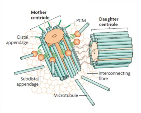
Figure 1. Centrosome structure. Schematic view of a typical animal centrosome, illustrating the mother and daughter centrioles that formed the centriole pair associated at each centrosome surrounded by a cloud of PCM proteins. Note that, specifically the mature “mother” centriole harbors distal and sub-distal appendages (adapted from Bettencourt-Dias and Glover, 2007).
mammalian cells) dependent-phosphorylation during mitosis initiates centrosome maturation in flies (Dobbelaere et al., 2008). Pericentrin and AKAP450 family make part of a group of proteins harboring a centrosomal targeting (PACT) domain and known for their role in docking and recruiting regulatory components involved in MT nucleation (e.g. γ-TuRC) and in PCM integrity (Bettencourt-Dias and Glover, 2007; Lawo et al., 2012; Mennella et al., 2012; Schatten, 2008). Drosophila pericentrin-like protein (D-PLP) is the only PACT domain protein identified in flies. d-plp Drosophila mutants, although viable, are severely uncoordinated and the sperm is nonmotile, suggesting an additional role in cilia and flagella formation (Martinez-Campos et al., 2004).
Recently, the combination of 3D structured illumination microscopy (3D-SIM) and STORM (stochastic optical reconstruction microscopy) allowed a deeper look on centrosome architecture revealing a conserved high-order structure within PCM, as opposed to the traditional "amorphous" description. PCM organization is characterized by two overlapped layers: a radial and a concentric, in which proteins either are framed at specific sites in a layered fashion or extend outward from centriole wall. The PCM surface is composed by proteins in a toroidal arrangement linked to the outer wall of mother centriole such as SAS-4, followed by proteins extending out to the PCM periphery like SPD-2 (CEP192 in mammalian cells), Cnn and γ-tubulin. The radial distribution is strongly defined by D-PLP that binds its C-terminal PACT domain to the centriole wall and the N-terminal domain extends outwards in order to form a matrix of D-PLP extended fibrils that function as a scaffold for the recruitment of other PCM components. Interestingly, D-PLP fibrils formed an open gap (150-200 nm) on mother centrioles, coinciding with the position of daughter centriole assembly, which was not noticeable in the early stages of the cell cycle that precede centriole duplication (Fu and Glover, 2012; Lawo et al., 2012; Mennella et al., 2012; Sonnen et al., 2012).
Centrioles are at the foundation of centrosomes. Thus, an interaction between the centriole pair and the surrounding PCM was expected to exist. A symbiotic relationship between PCM and centrioles has been reported by several studies. Only the mother centriole within the centriole pair is able to anchor MTs through the sub-distal appendages (Piel et al., 2000) and centrioles define centrosome size by the incorporation of Cnn into the PCM, which seems to be driven by the components Asterless (Asl) and Drosophila Spd-2 (DSpd-2) (Conduit et al., 2010). Furthermore, the levels of centriolar protein SAS-4 were reported to set centrosome size (Kirkham et al., 2003) and SAS-4 itself was found in complexes with some centrosomal proteins, such as Asl, Cnn and D-PLP, suggesting a scaffolding role to tether PCM components within the centrosome (Gopalakrishnan et al., 2011). Also, the interaction SAS-4/Tubulin was shown to control PCM recruitment depending on the tubulin guanine-bound state (discussed in chapter 5) (Gopalakrishnan
et al., 2012). In agreement with an influence of centrioles in PCM assembly, the transiently centriole disintegration in HeLa cells upon the microinjection of an antibody directly against glutamylated tubulin (GT335) led to PCM dispersion, suggesting that the presence of centrioles allow the correct segregation and organization of the pericentriolar material (Bobinnec et al., 1998). Alongside, the PCM also exhibits an influence over centrioles. It was demonstrated that overexpression of pericentrin in S-phase arrested CHO cells induced the formation of large PCM clouds containing variable number of centrioles (Loncarek et al., 2008). Moreover, depletion of PCM proteins in the model organism C. elegans resulted in failure of centriole assembly in ~50% of the time, and the centrioles who were formed failed to reach full size (Dammermann et al., 2004). Considering the combined data, there is a strong support to the idea of a symbiotic model in which the interaction between centriole and PCM is important to assemble structural and functional normal centrosomes during cell division.
3.2. Overview of Centrosome Cycle
In active proliferating cells, the two functional centrosomes ensure accurate chromosome segregation through the formation of a bipolar mitotic spindle during mitosis. In order to be in harmony with cell cycle progression, the centrosome cycle must be highly coordinated with the DNA cycle. The Centrosome cycle depends on centriole duplication and behavior along the different phases of the cell cycle. The centrosome-centriole cycle can be divided into the following discrete and critical stages: (1) centriole disengagement; (2) centriole duplication and elongation; (3) centrosome maturation and (4) centrosome separation (Nigg, 2007) (Figure 3a). In interphase, a typical G1 centrosome harbors two
centrioles that are structurally and functionally different - a mother/mature centriole and a daughter or immature centriole assembled during the previous cell cycle. Centriole duplication starts in late G1/early S transition with procentriole nucleation at right angle to
the proximal end of each parental centriole. From S to mitosis, after the establishment of the basic centriolar structure, each procentriole starts to elongate reaching ~80% of its full-length during mitosis and eventually matures after a second cell cycle. A mature centriole differs from the younger one due to the acquisition of distal/sub-distal appendages and PCM enrichment. This final complete version of centriole has the ability to dock to the plasma membrane to promote ciliogenesis and to nucleate astral MTs to build the mitotic spindle. Thus, the assembly of a mature centriole able to promote and work as an independent and functional centrosome is a step-by-step process that needs two consecutive cell cycles (Avidor-Reiss and Gopalakrishnan, 2013; Azimzadeh and Marshall, 2010; Brito et al., 2012). The molecular mechanisms regulating centrosome-centriole cycle are for the most part poorly understood, nevertheless some experimental
works have already elucidated some essential regulators acting during the four stages of centrosome cycle.
During metaphase, each centrosome defining one of the two spindle poles contains a pair of tightly associated parental-progeny centrioles. In spite of the unknown nature of that linker (S-M linker) (Nigg and Stearns, 2011), it is known that upon exit of M phase (or early G1 phase), the link between the two centrioles is lost in a process termed
"centriole disengagement". Interestingly, it was shown in Drosophila that DSas-6-Ana2 complex might be involved in S-M linker functionality since their co-overexpression in spermatocytes formed short tubules (SAStubules) linking the inner region of the daughter centriole to the outer surface of the parental centriole, which is lost in meiosis I, coincident with centriole disengagement (Stevens et al., 2010b).
It is now imperative to approach the licensing model based on two distinct rules that centrosome cycle must obey in order to promote a correct centriole/centrosome number over successive cell divisions: (1) centrosomes duplicate once and only once in each cell cycle (cell cycle control) and (2) only one progeny centriole must arise next to each parental centriole (copy number control) (Nigg, 2007). Centriole disengagement has been proposed to be a key event for centriole licensing, rendering both centrioles competent to duplicate in the following S phase. Like chromosomes, centriole separation also requires the activity of separase, a protease responsible for driving sister chromatids separation prior to anaphase, as well as the kinase PLK1 (Tsou and Stearns, 2006; Tsou et al., 2009). Centriole disengagement as a license model for centriole duplication is supported by a series of experimental works. Wong and Stearns (2003) in an elegant cell fusion assay have shown that when G2/S phase cells were fused, there was no centriole
duplication, but when the fusion was between G1/G2-phase or G1/S phase cells, the G1
centrosomes replicate. Furthermore, Loncarek et al. (2008), by laser ablating the daughter centriole in S phase arrested cells, thereby artificially mimicking centriole disengagement, showed that resident mother centrioles were able to generate new daughter centrioles. Thus, both studies imply three major conclusions: first, centriole disengagement is a centriole-inherent permissive state to duplication; secondly, there is a centrosome-intrinsic-block to re-duplication which probably manifests itself in the already-duplicated and engaged centrioles (since G2/S-phase fusions did not lead to centrosome duplication,
in spite of permissive cytoplasmic conditions in S phase) and third, the centrosome somehow "senses" the presence of an immature centriole, blocking re-duplication events. Coupled to DNA replication, the next stage in this sequence is centriole assembly in S phase in which each parental centriole "seeds" the growth of a new centriole (procentriole) which grows orthogonally in relation to the mother centriole. This process is controlled by a specific set of proteins discussed in detail in chapter 4. Nonetheless, the
cell cycle kinase CDK2 seems to be a critical factor to enhance centriole duplication (Hinchcliffe et al., 1999; Matsumoto et al., 1999; Meraldi et al., 1999). Formation of procentrioles coincides with an increase in CDK2 activity in S phase, and in complex either with cyclin A and E is essential to trigger centriole duplication through phosphorylation of nucleophosmin (NPM/B23) and Mps1 (Fisk and Winey, 2001; Okuda et al., 2000). However, the direct role of CDK2 in regulating centriole duplication is still controversial since it was shown that CDK2 activity is not essential for centriole duplication, but instead it seems to speed up procentriole formation (Duensing et al., 2006).
In the following cell cycle stages (from G2 phase to mitosis) the two recently
formed centrosomes must mature and separate in order to orchestrate the formation of a bipolar mitotic spindle. During late G2 phase, centrosome separation seems to require the
disintegration of the linker (G1-G2 tether) that mediates centrosome cohesion by linking
the proximal ends of the two parental or previously disengaged centrioles (but not the two centrioles within a pair) (Nigg and Stearns, 2011). This linker is assembled right after centriole disengagement and remains temporarily associated with centrioles throughout interphase to ensure centrosome cohesion and to avoid premature centrosome splitting prior to mitosis.
Ultrastructural analysis revealed that rootletin, a conserved component of the ciliary rootlet, and C-Nap1 are key structural elements whose interaction is important for the functionality of this linker. Whereas C-Nap1 localization is restricted to the proximal ends of centrioles, rootletin forms fibers emanating from the proximal ends of both centrioles (Bahe et al., 2005; Mayor et al., 2000; Yang et al., 2006), suggesting that C-Nap1 functions as a "docking site" from where rootletin-based fibers attach and emanate. Upon entry into mitosis, this fibrous linker is disassembled through phosphorylation of C-Nap1 by the mitotic kinase Nek2 allowing the separation of the two independent centrosomes (each containing a pair of centrioles) and consequently mitotic spindle formation (Fry et al., 1998; Helps et al., 2000). Concomitantly with centrosome separation, the younger of the two parental centrioles (originated from the previous cell cycle) acquires distal and sub-distal appendages and enlarges its PCM with the recruitment of γ-TuRCs and other matrix components, thereby reaching full maturity and promoting centrosome maturation.
4. How to Build a Centriole ? 4.1. Centriole Architecture
Centrioles are microtubule-based, cylindrical and evolutionary conserved eukaryotic cell structures that exhibit a distinct nine-fold symmetric radial array of stabilized microtubules. They are polarized along the proximo-distal axis with the base commonly referred as the proximal end and its tips as distal ends. The typical size for a human mature centriole is approximately 200 x 500 nm, although these measures vary between organisms, as it is the case of C. elegans and Drosophila centrioles that tend to be shorter (Gonczy, 2012; Pelletier et al., 2006). Nonetheless, the signature of centriole architecture is undoubtedly the conservation of the 9-fold symmetry across evolution. Cryo-electron tomography studies have brought new and instructive data concerning centriole/basal body structure (Guichard et al., 2010; Guichard et al., 2013; Li et al., 2012). How does the ninefold symmetry arise to build a highly ordered and complex
structure as the centriole?. In Drosophila, unicellular algae, many protozoa and
vertebrates, the basic structure of centrioles relies on microtubule blades displayed into a 9-fold radial array (Loncarek and Khodjakov, 2009). This symmetry is established by the cartwheel, which is a structure located in the very proximal end of the centriole and composed of a central hub with 20-25 nm in diameter and ~100 nm in height from which nine spokes radiate outwards (Gonczy, 2012; Guichard et al., 2010) (Figure 2). Each stroke ends in a structure so-called Pinhead which bridges the central hub with centriolar microtubules. The cartwheel is the central piece on centriole organization and the first intermediate of centriole structure to appear during the early events of centriole biogenesis. Interestingly, despite its presence in immature centrioles, the cartwheel disappears from mature centrioles in some organisms (e.g. humans). In most species, including human centrioles, nine sets of microtubule triplets decorate the outer surface of the centriole and are linked to the cartwheel through the stroke pinheads. From the inside out, each triplet comprises an A-microtubule, B-microtubule and a C-microtubule, in which only the A-microtubule is complete being formed by 13 protofilaments. The A-microtubule is oriented toward the center of the centriole and is connected to the pinhead of each stroke, B-microtubules attach to A-microtubules and consequently C-microtubules associate with neighboring B-microtubules. Moreover, A-microtubules can bind to C-microtubules from the previous triplet forming an A-C linker. This microtubule triplet pattern is only characteristic of the proximal-end of centrioles since the distal-end displays double microtubules (A- and B-microtubules) (Guichard et al., 2013; Li et al., 2012).
Figure 2. Centriole and Cartwheel architecture. (a,d) The ultrastructure of a resin-embedded centriole and the cartwheel purified from human cells and Chlamydomonas
reinhardtii. Part (a) shows the side view of a mature human centriole. The proximal and
distal ends of the centriole are indicated. The arrow points to distal and subdistal appendages present on the sides of the distal part of the centriole. Part (d) shows a cross-section of the proximal part of a C. reinhardtii centriole. Note the central tube from which nine spokes emanate that radiate towards the vicinity of triplet microtubules. The A-,B- and C-microtubule are indicated and the arrow points to the A-C linker. (e) Schematic representation of a centriole and procentriole pair in a human cell. (f) Schematic representation of the cartwheel viewed from the proximal end (adapted figure from Pierre Gӧnczy, 2012).
Interestingly, it was recently demonstrated that the pinhead structure plus the A-C linker are polarized structures that might be responsible for dictating the directionality of centriolar microtubule assembly, as well as the chirality of microtubule triplets (Guichard et al., 2013). An amazing exception to this cartwheel-based centriole structure is represented by C. elegans centrioles. Electron microscopy studies revealed that they do not comprise the cartwheel structure and, instead, follow a more simple architecture formed by a central tube on which nine sets of centriolar microtubules are directly attached (Pelletier et al., 2006). Additionally, the microtubule number can also vary between species. For example, C. elegans centrioles are made only by singlet
microtubules whereas Drosophila centrioles in embryos and most tissues are formed by doublet microtubules, and specifically in the spermatocytes of male germ line, centrioles do exhibit centriolar triplets of microtubules (Carvalho-Santos et al., 2010; Debec et al., 2010). In spite of these variations on centriole structure, the nine-fold symmetry is always maintained.
A typical interphase G1 cell harbors one centrosome built from a pair of centrioles
linked by a flexible connection. Through the following cell cycle stages, a series of events take place including centriole replication, elongation and maturation. A cryo-electron tomography analysis of centrosomes isolated from human lymphoblasts by Guichard and co-workers have revealed new insights about the initial structural events involved in centriole duplication. In the nascent procentriole, centriolar singlets A-microtubules were observed which seemed to be capped by a conical structure at their proximal or minus end resembling the γ-tubulin ring complex (γ-TuRC). This suggests, in association with other studies (Dammermann et al., 2008; Dammermann et al., 2004; Kleylein-Sohn et al., 2007), the involvement of γ-TuRC in nucleating each A-microtubule, which grows unidirectionally from the proximal to the distal (plus) end during centriole assembly. Accordingly, the distal end of A-microtubule is not closed and, instead, it showed outward curved extensions characteristic of growing microtubules. In contrast with the minus end of A-microtubule, B- and C- microtubules showed always open and outward curved extensions at both proximal and distal ends. Therefore, nucleation of these last microtubules (B and C) is not mediated by γ-TuRC, but it follows a template-dependent mechanism, in which A- and B-microtubules are the templates for a bidirectionally growth (from their plus and minus ends) of the B- and C-microtubules. Interestingly, the attachment of A-, B- and C-microtubules for the centriole wall formation occurs independently and without any specific order or position. As B- and C-microtubules reach the minus end of A-microtubules, their proximal ends become blunt and just the distal-ends continues to grow until completion of the microtubule triplet blades (Guichard et al., 2010). In a mature centriole, A-microtubules lose the closest conformation of the proximal end indicating that γ-TuRC is no longer necessary, being removed when the centriolar microtubule wall is fully developed. The composition of B- and C-microtubules is still unknown, but it seems that ε- and δ-tubulin are potential candidates to take in consideration, since their mutation in Chlamydomonas and Paramecium disrupts the MT triplet arrangement. Nevertheless these two tubulin isoforms are absent from D.
melanogaster proteome suggesting an alternative mechanism for B- and C-microtubules
nucleation (reviewed in Azimzadeh and Marshall (2010), Brito et al. (2012) and Gonczy et al. (2012)). The answer to this question awaits further experiments.
4.2. Centriole-assembly pathway: action of a core ancestral protein module Centrioles are structurally complex organelles, but surprisingly the mechanism orchestrating centriole assembly relies on a few and evolutionary conserved core of proteins. A long-standing question in centriole biology was focused on the players and the way they interacted to initiate and produce a centriole. The first cues concerning the molecular mechanism involved in centriole biogenesis came from studies in C. elegans embryos. Genetic analysis and RNAi-based screens revealed a hierarchical molecular
cascade in which only five proteins are specifically required for centriole duplication: the coiled-coil proteins SPD-2, SAS-4, SAS-5, SAS-6 and the kinase ZYG-1 (Dammermann et al., 2004; Delattre et al., 2004; Kemp et al., 2004; Kirkham et al., 2003; Leidel and Gonczy, 2003; O'Connell et al., 2001). During centriole assembly, these proteins are sequentially recruited to centrioles (Delattre et al., 2006; Pelletier et al., 2006), where procentriole nucleation is triggered by the PCM protein SPD-2 that recruits ZYG-1 to the procentriole. The protein kinase ZYG-1 is responsible for the recruitment of the structural proteins SAS-5 and SAS-6, who physically interact (Leidel et al., 2005) to build the central tube and to recruit SAS-4, which induces the assembly of nine singlet microtubules to the outer wall of the emerging centriole. However, given that C. elegans centriole architecture is atypical and divergent compared with Drosophila and mammalian centrioles (Pelletier et al., 2006), it became imperative to address if the same protein module governing centriole duplication in worms was transversal to other organisms.
An elegant study conducted by Kleylein-Sohn et al. (2007) unveiled how centriole biogenesis is governed in human cells. Taking advantage of centriole induction by PLK4 overexpression (Habedanck et al., 2005; Rodrigues-Martins et al., 2007b) in association with siRNA mediated depletion of individual centriolar proteins, a similar set of proteins associated with C. elegans centriole assembly were described to control this process in humans cells. Five proteins were identified to be essential for centriole duplication, specifically human SAS-6 (hSAS-6), CPAP (functional homologue of SAS-4/DSas-4 in C.
elegans and Drosophila), CEP135, CP110 and γ-tubulin. A putative sequential centriole
assembly pathway was described in which Polo-like kinase 4 (PLK4) is an upstream and key regulator essential to trigger procentriole assembly, and does not depend on the presence of any of the other proteins to localize to centrioles (Bettencourt-Dias et al., 2005; Habedanck et al., 2005; Kleylein-Sohn et al., 2007). Although PLK4 is not an homologue of C. elegans ZYG-1, it seems that both proteins work in an analogous way to initiate centriole duplication in different organisms. Thus, in human cells, PLK4 activation on the surface of the parental centriole triggers procentriole assembly and is crucial for the recruitment of hSAS-6 and STIL (functional homologue of C. elegans SAS-5 or Drosophila Ana2) (Vulprecht et al., 2012), which in turn are needed for CPAP loading in the
procentriole (reviewed by Gonczy, 2012). CPAP was shown to be a substrate for PLK2 and its phosphorylation (CPAP residues 589-595) is critical for procentriole assembly as well as for its stabilization in the nascent centriole (Chang et al., 2010).
Moreover, in agreement with Kleylein-Sohn et al. (2007), in which CEP135 was identified as a core component of centriole duplication, it was recently shown that CEP135 directly interacts with hSAS-6 and CPAP, indicating that these three proteins might regulate centriole biogenesis as a complex. It was also identified in its protein structure a MT-binding domain, elucidating a potential role for CEP135 in mediating CPAP-dependent centriolar microtubule assembly (Lin et al., 2013). Furthermore, it was shown that STIL N-terminal domain interacts and eventually recruits CPAP to the procentriole (Cottee et al., 2013; Vulprecht et al., 2012). Strikingly, as opposed to C. elegans and Drosophila, human STIL does not seem to form a stable complex with hSAS-6. Nevertheless, the siRNA-mediated depletion of STIL significantly decreased hSAS-6 levels at centrosome (Vulprecht et al., 2012), and STIL centriolar localization was also affected upon hSAS-6 depletion (Arquint et al., 2012), suggesting that STIL and SAS-6 might be partially interdependent for their localization at centrioles (Vulprecht et al., 2012).
SPD-2 ortholog in humans, called CEP192, seems also to be required for centriole duplication in human cells (Zhu et al., 2008). CEP192 was recently found to cooperate with CEP152 for the centriolar recruitment of PLK4 during centriole duplication, indicating an important and direct role of this PCM component in human centriole biogenesis (Kim et al., 2013; Sonnen et al., 2013).
As expected, the sequential model proposed in mammalian cells extends to D.
melanogaster. In a genome-wide screen to dissect centriole duplication and centrosome
maturation in Drosophila cells (S2R+), from among 119 centrosomal-related genes that were analyzed, only nine genes were identified to be directly involved in centriole assembly. From those nine genes, three coded for the well-known SAS-4, SAS-6 and SAK (Drosophila homologue of PLK4) proteins and another three genes coded for a set of proteins so-called Ana1, 2 and 3 (previously identified by Goshima et al., 2007). As emphasized previously, SAK (or PLK4 in human cells) is a master regulator in centriole assembly, whose depletion in Drosophila impairs centriole duplication and flagella formation (Bettencourt-Dias et al., 2005). Overexpression of Ana1 and Ana2 formed extra centrioles suggesting a potential role in centriole duplication in Drosophila cells (Dobbelaere et al., 2008). Latter, Ana2 was identified either as a conserved centriole duplication factor and as orthologue of STIL/SAS-5 based on weak sequence similarities (Stevens et al., 2010a), as well as Ana3 was described to be important for centriole structural integrity and cohesion, but not for centriole duplication. On the other hand, the precise function of Ana1 on centriole assembly is still not known. However, it has been
shown that its RNAi depletion in S2 Drosophila cells led to a significant decrease in SAS-6 levels at centrosome, indicative of an important role in centriole duplication (Goshima et al., 2007).
As opposed to mammalians and worms, SPD-2 is not required for centriole assembly in Drosophila (Dix and Raff, 2007), therefore flies and humans have one additional duplication factor, Asterless (Asl; human orthologue CEP152) (Blachon et al., 2008; Dobbelaere et al., 2008; Dzhindzhev et al., 2010). This coiled-coil protein was shown to be extremely important for centriole duplication in Drosophila and human cells since: (1) directly binds to SAK/PLK4's cryptic polo box domain (CPB, conserved motif in PLK4 orthologs) mediating its centrosomal localization in Drosophila and human cells; (2) interacts through N-terminal domain of CPAP or Drosophila Sas-4 (DSas-4), recruiting human CPAP to centrosomes and (3) its overexpression led to supernumerary centrosomes in Drosophila and human cells. Thus, it was proposed that Asl/CEP152 acts upstream of PLK4 during centriole biogenesis, acting as a scaffold protein for both PLK4 and SAS-4/CPAP and triggering procentriole nucleation in flies and humans (Cizmecioglu et al., 2010; Dzhindzhev et al., 2010).
As in the other model organisms, Drosophila SAS-6 (DSas-6) and Ana2 cooperation is crucial for centriole assembly (Stevens et al., 2010a). DSas-6 depletion in
Drosophila spermatocytes and S2 cells led to a reduction in centriole number, and the few
proportion of formed centrioles were smaller and lost the typical 9-fold symmetry. In opposition, DSas-6 overexpression in Drosophila embryos and unfertilized eggs resulted in de novo formation of MTOCs, in which were detected irregular tube-like structures that resembled incomplete centrioles with wrong orientation and symmetry (Rodrigues-Martins et al., 2007a). Moreover, only when DSas-6 was overexpressed in combination with its binding partner Ana2 in Drosophila spermatocytes, there was the assembly of ordered tubule-like structures (SAStubules) similar to the inner centriole cartwheel. Strikingly, in contrast to what was previously reported about DSas-6 overexpression in Drosophila eggs and embryos (Rodrigues-Martins et al., 2007a), SAStubules were not formed when DSas-6 and Ana2 were overexpressed individually or under combined DSas-4 / Asl overexpression in Drosophila spermatocytes, indicating that DSas-6 and Ana2 are indispensable players and do need to cooperate to drive the formation of the cartwheel in
Drosophila centrioles (Stevens et al., 2010b).
Besides the reported interaction between DSas-6/Ana2, DSas-4 is also a well-known interacting protein. Its depletion led to centriole loss in Drosophila flies and impaired centriole duplication in C. elegans embryos and human cells (Basto et al., 2006; Kirkham et al., 2003; Leidel and Gonczy, 2003; Tang et al., 2009). Recent structural analysis identified the TCP region as a conserved binding domain of CPAP/DSas-4/SAS-4