UNIVERSIDADE FEDERAL DO PARÁ INSTITUTO DE CIÊNCIAS BIOLÓGICAS
PROGRAMA DE PÓS-GRADUAÇÃO EM NEUROCIÊNCIAS E BIOLOGIA CELULAR
Estudos Citogenéticos em Roedores do Gênero
Oecomys
(Rodentia: Cricetidae)
Celina Coelho da Rosa
UNIVERSIDADE FEDERAL DO PARÁ INSTITUTO DE CIÊNCIAS BIOLÓGICAS
PROGRAMA DE PÓS-GRADUAÇÃO EM NEUROCIÊNCIAS E BIOLOGIA CELULAR
Estudos citogenéticos em Roedores do Gênero
Oecomys
(Rodentia: Cricetidae)
Dissertação de Mestrado submetido ao curso de Programa de Pós-graduação em Neurociências e Biologia Celular da Universidade Federal do Pará (UFPA), como requisito parcial para a obtenção do grau de Mestre em Neurociências e Biologia Celular.
Orientadora: Profª. Drª. Cleusa Yoshiko Nagamachi
i
ii
―Todo estado atual de uma substância
simples é naturalmente conseqüência de seu estado anterior, de tal modo que seu presente está impregnado no seu futuro.‖
iii
Aos meus melhores amigos, Clélia e Perilo
iv
AGRADECIMENTOS
Agradeço primeiramente a Deus por ter me dado a vida e aos meus pais, Clélia e
Perilo, pela compreensão e por terem fornecido condições para realizar meus sonhos;
À minha orientadora, Professora Drª. Cleusa, por ter confiado em mim e ter concedido
a oportunidade de realizar este trabalho;
Ao Prof. Dr. Julio pelo exemplo de capacidade e inteligência;
Às Professoras Drª Susana Milhomem da Paixão e Drª Renata Noronha por serem
sempre solicitas e competentes;
Ao Dr. Rogério Rossi, à Dra. Maria Iracilda Sampaio e à Tamara Flores, pela
identificação dos animais e análise molecular, pois sem eles esse trabalho não seria realizado;
Às minhas amadas irmãs, Carolina e Carla, pelo companheirismo, apoio e amor de
sempre;
Ao meu sobrinho mais que amado, Vinícius, por completar meu dia e me fazer mais
feliz a cada sorriso e beijo;
À minha avó querida, Celina, por todo amor e carinho que só a senhora sabe dar;
Às minhas amigas, Bruna, Bia, Maíra e Camila, pelo carinho, amizade e amor.
Ao Diego, por toda a paciência, tolerância, amor e respeito.
À amiga querida Natália, por todos os momentos bons e ruins que vivemos sempre
juntas, pelos conselhos, brigas e conversas.
Aos técnicos do laboratório: Conceição, pelo café de todos os dias e pela solidariedade
sempre a disposição; ao Jorge pelas culturas e pelos puxões de orelha muitas vezes merecidos
v
Aos colegas de Laboratório de Citogenética da UFPa, Anderson, Danillo, Pablo,
Patrícia, Ramon, Fernando, Adauto, Ingrid, Karina, Thayse, Mila, Jéssica e Anneiriane.
Aos colegas do grupo de roedores, Paulinho, Jamilly, Adenilson, Stella, Willan,
Marlyson, Geovanna e Vergiana por todo trabalho dividido e pelas inúmeras reuniões.
Aos familiares, amigos e professores que de alguma forma, direta ou indiretamente,
contribuíram para que a realização deste trabalho fosse possível.
À FAPESPA pelo apoio financeiro, através da bolsa de mestrado durante o
vi
SUMÁRIO
LISTA DE FIGURAS ... vii
RESUMO ... vii
ABSTRACT ... iv
1 INTRODUÇÃO ... 1
1.1 ORDEM RODENTIA ... 1
1.2 GÊNERO OECOMYS ... 5
1.2.1 História taxonômica do gênero Oecomys ... 7
1.2.2 Diversidade Cromossômica em Oecomys ... 7
2 REFERÊNCIAS BIBLIOGRÁFICAS ... 8
3 OBJETIVOS ... 11
3.1 OBJETIVO GERAL ... 11
3.2 OBJETIVOS ESPECÍFICOS ... 11
4 MANUSCRITO DO ARTIGO CIENTÍFICO ... 12
vii
LISTA DE FIGURAS
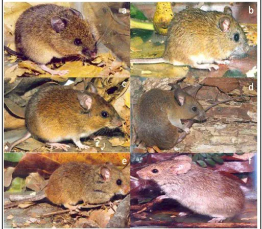
Figura 1: Roedores pertencentes à Subfamília Sigmodontinae; (a) Delomys collinus (b) Cerradomys subflavus (c) Euryoryzomys russatus (d) Hylaeamys sp. (e) Juliomys pictipes (f) Necromys lasiurus
2
4
5
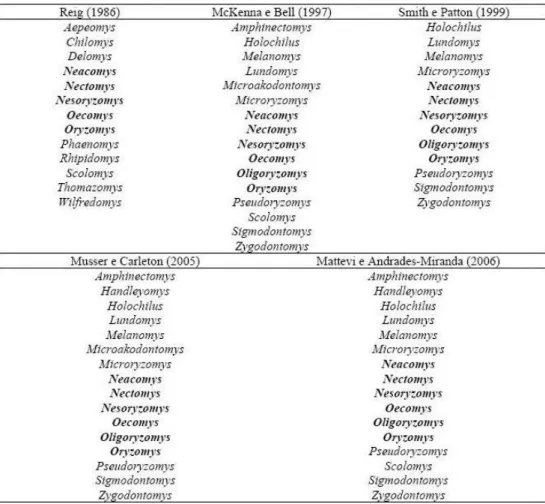
6 Figura 2: Classificação atual da Tribo Oryzomyini
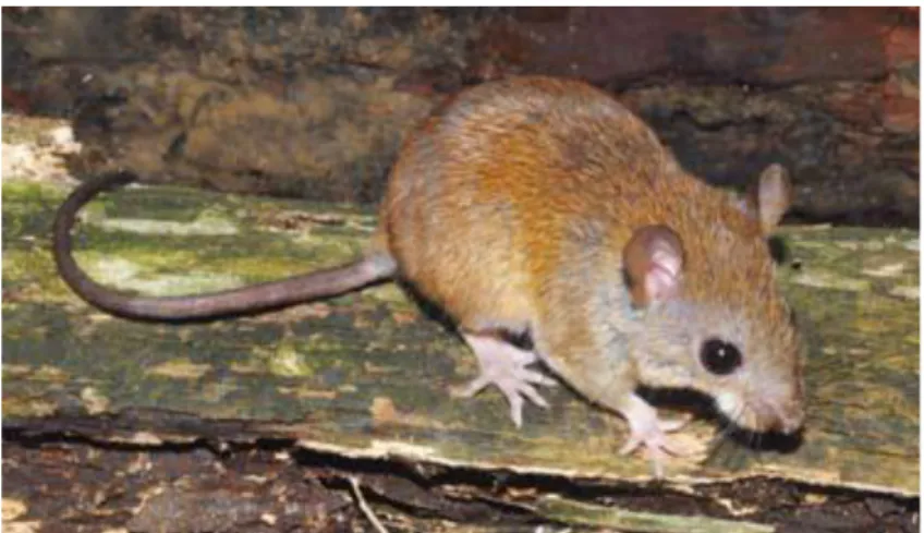
Figura 3: Exemplar do gênero Oecomys
viii
RESUMO
Os roedores representam o grupo de mamíferos viventes mais diversificados e com ampla diversidade de adaptações ecológicas. Os roedores, devido às características populacionais que apresentam, desenvolveram-se como o grupo mais especioso de mamíferos em florestas neotropicais e um dos mais interessantes para estudos da variabilidade genética e de evolução entre os vertebrados. Os roedores do gênero Oecomys compreendem aproximadamente 16
espécies que habitam floresta tropical e subtropical do Centro e do Sul da América. Destas, apenas seis têm ocorrência esperada para a Amazônia Oriental Brasileira. De acordo com a literatura, o gênero Oecomys apresenta uma grande diversidade cariotípica, com o número
diplóide variando entre 58 e 86. Neste estudo, espécimes de Oecomys paricola Thomas, 1904
de Belém e da Ilha do Marajó foram estudadas usando analises citogenética, molecular e morfológica. Três cariótipos foram encontrados, dois de Belém (2n=68, NF=72 e 2n=70, NF=76) e um da Ilha do Marajó (2n=70, NF=74). Não foi encontrada diferença molecular e morfológica entre indivíduos dos diferentes citótipos de Belém e da Ilha do Marajó. Espécies da cidade de Belém representam duas espécies crípticas, pois dois cariótipos diferentes estão presentes na ausência de diferenças significativas nas características morfológicas e moleculares. Populações da Ilha do Marajó e Belém representam espécies distintas que foram separadas há algum tempo, e estão em processo de diferenciação morfológica e molecular, como consequência do isolamento reprodutivo a nível geográfico e cromossômico.
iv
ABSTRACT
The rodents are one of the most diversified groups of living mammals and also have a large range of ecological adaptations. The rodents, because of yours population characteristics, developed as the most specious group of mammals in Neotropical forests and one of the most interesting for studies of genetic variation and evolution among vertebrates. The genus
Oecomys (Sigmodontinae) comprises approximately 16 species that inhabit tropical and
subtropical forests in Central and South America. Six of these species are expected to occur in eastern Brasilian Amazon. In literature, the genus Oecomys has a large karyotypic variation,
where the diploid number ranges from 58 to 86. In this study specimens of Oecomys paricola
1. INTRODUÇÃO
1.1. ORDEM RODENTIA
Os roedores são membros importantes de quase todas as faunas, sendo cosmopolitas e nativos na maioria das áreas terrestres, exceto em algumas ilhas árticas e oceânicas, Nova Zelândia e Antártica. Os representantes são, em sua maioria, de porte pequeno, compreendendo desde animais de poucos gramas até a grande capivara (Nowak, 1994). Esses animais apresentam corpo versátil e esguio, bem adaptado a diversos modos de vida e variados climas. A enorme variação na morfologia, na diversidade de habitats e climas e na alimentação, os tornou a mais numerosa e melhor sucedida evolutivamente entre as ordens de mamíferos (Emmons & Feer, 1997).
Os roedores são animais predominantemente herbívoros, porém apresentam uma grande variedade de hábitos alimentares, podendo ser insetívoros, piscívoros ou carnívoros. Esta versatilidade alimentar tem sido considerada um dos principais fatores no notável sucesso das radiações adaptativas obtidos por estes mamíferos (Landry, 1970).
Uma característica marcante dos roedores são os seus dentes. Esses animais possuem dois pares de dentes incisivos, um superior e um inferior, sendo o superior de crescimento ilimitado, que se sobrepõe ao par inferior. Não possuem outros incisivos ou caninos e apresentam poucos molares ou pré-molares, sendo os incisivos separados dos demais dentes por um espaço chamado de diastema (Nowak, 1994).
Inicialmente os roedores foram classificados em três grandes categorias subordinais: Sciuromorpha, Hystricomorpha e Myomorpha (Simpson, 1945; Anderson, 1967). Esta classificação levou em consideração as diferenças na estrutura craniana e nos padrões de especialização do músculo masseter e principalmente nas relações deste com o conduto infraorbitário. Entretanto, atualmente acredita–se que as especializações deste músculo possam ter surgido independentemente mais de uma vez, não sendo, desta forma, relevantes para a classificação dos roedores. A grande diversidade morfológica, o número considerável de ramos de descendência e a evolução paralela de caracteres muito similares em alguns grupos de roedores geraram controvérsias quanto à sua classificação taxonômica. A classificação mais atual baseia-se nas diferenças da musculatura craniana e formas da mandíbula e crânio, que agrupa os roedores em duas subordens: Hystricognathi e Sciurognathi (Woods, 1976; Hartenberger, 1981; Wood, 1955).
Nesomyidae, Platacanthomyidae e Spalacidae (Musser & Carleton, 2005). Os cricetídeos são considerados a família mais diversificada do Brasil.
Diversos estudos morfológicos mostraram uma série de diferenças entre os Cricetideos Norte e Sul– Americanos,como por exemplo, diferenças na morfologia peniana, na anatomia das glândulas acessórias do sistema reprodutor masculino, na morfologia do estômago, na microestrutura do pelo e parasitologia(Reig, 1984). Baseado nestas divergências Reig (1981) propôs elevar as tribos Sigmodontini e Neotomini (Hershkovitz, 1966, 1969, 1972) à condição de subfamílias da Família Cricetidae.
Cricetidae apresenta seis subfamílias, dentre as quais, Sigmodontinae é a segunda maior em número de espécies e representantes na biodiversidade de roedores (Swier et al., 2009). A subfamília Sigmodontinae (Figura 1) tem distribuição geográfica restrita às Américas, com grande maioria na América do Sul e abriga atualmente 377 espécies em 74 gêneros e nove tribos: Abrotrichini, Akodontini, Ichthyomyini, Oryzomyini, Phyllotini, Reithrodontini, Sigmodontini, Thomasomyini e Wiedomyini (Reig, 1980; Smith & Patton,
1999; Musser & Carleton, 2005; D’Elía et al., 2005).
Figura 1. Roedores pertencentes à Subfamília Sigmodontinae; (a) Delomys collinus (b) Cerradomys subflavus (c) Euryoryzomys russatus (d) Hylaeamys sp.
A tribo Oryzomyini foi cladisticamente definida por Voss e Carleton (1993) quando estabelecem cinco sinapomorfias para a tribo: ausência de vesícula biliar, presença de um par peitoral de mamas, ausência de cobertura timpânica, ausência da barra do alisfenóide e presença de um palato longo. Esta tribo compreende cerca de 35% das espécies de Sigmodontinae.
Atualmente existe uma grande controvérsia quanto ao número e quais os gêneros pertencentes à tribo Oryzomyini. Dependendo do autor, o número de gêneros varia entre 11 e 27 e de espécies descritas para a tribo Oryzomyini varia de 90 a 120 (Reig, 1986; McKenna e Bell, 1997; Smith e Patton, 1999; Musser e Carleton, 2005; Weksler et al., 2006). Esta
discrepância no número de gêneros é devida, principalmente, à inclusão ou retirada de gêneros de tribos mais ou menos relacionadas, especialmente Thomazomyini, Sigmodontini e Phyllotini. Porém, alguns gêneros sempre fizeram parte da tribo Orizomyini, independente do autor. São eles: Neacomys, Nectomys, Nesoryzomys, Oecomys, Oryzomys e Oligoryzomys,
este último não está nominado na classificação de Reig (1986) pelo fato de que na época ele era considerado subgênero de Oryzomys (Tabela 1).
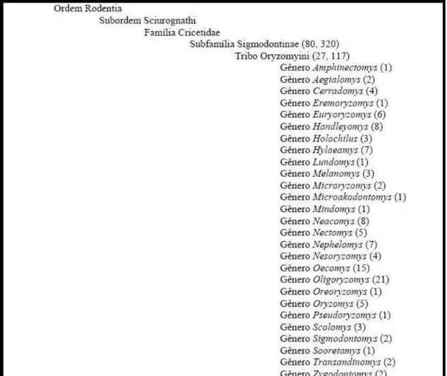
Atualmente a tribo Oryzomyini compreende 27 gêneros e cerca de 120 espécies que estão relacionadas na Figura 2, organizada segundo classificação básica de Voss e Carleton (1993) e Musser e Carleton (2005), incluindo propostas atuais que envolvem a descrição de novas espécies e, até mesmo, de novos gêneros. Weksler et al. (2006), através de estudos
morfológicos e com marcadores moleculares reclassificaram espécies do polifilético gênero
Oryzomys em dez novos gêneros: Aegialomys, Cerradomys, Eremoryzomys, Euryoryzomys, Hylaeamys, Mindomys, Nephelomys, Oreoryzomys, Sooretamys e Transandinomys. O gênero Oryzomys permaneceu com cinco espécies.
Figura 2. Classificação atual da Tribo Oryzomyini
al., 1971; Gardner e Patton, 1976). Seus hábitos alimentares vão de onívoros a insetívoros e
apresentam uma grande diversidade morfológica, variando no tamanho corporal, de pequenos com 10g até grandes com cerca de 300g. A maioria possui hábito escansorial, mas alguns podem desenvolver hábitos arbóreos ou até semi-aquáticos, constituindo assim um dos mais claramente definidos grupos multi-genéricos de muróides. A distribuição geográfica desta tribo é a mais ampla dentro dos Sigmodontinae, desde o extremo sul da América do Sul até o sudoeste dos Estados Unidos. Entre os 27 gêneros que a tribo Oryzomyini apresenta está o gênero Oecomys (Musser & Carleton, 2005).
1.2. GÊNERO OECOMYS
As espécies deste gênero (Figura 3) têm tamanho de pequeno a médio porte, sendo a cauda maior que o comprimento do corpo. O dorso varia de escuro a castanho-avermelhado. As laterais são mais claras que o dorso, com limite bem definido com o ventre. A pelagem do ventre pode ser completamente branca, creme com pêlos de base cinza, ou com este segundo padrão e manchas completamente brancas ou cremes na linha mediana do ventre. As patas são curtas, largas e claras e a cauda apresenta porção terminal pilosa, podendo ou não formar um pincel caudal (Reis, 2006).
Figura 3. Exemplar do gênero Oecomys (Bonvicino, 2008)
Este gênero apresenta roedores de hábitos noturnos e solitários. Comem frutas e sementes verdes e usa todos os níveis das florestas, inclusive o solo (Emmons e Feer, 1999).
De acordo com Musser & Carleton (2005) e Oliveira & Bonvicino (2006) o gênero
Oecomys conta com dezesseis espécies atualmente reconhecidas como válidas: Oecomys auyantepui Tate, 1939, O. bicolor Thomas, 1860, O. catherinae Thomas 1909, O. cleberi
1906, O. paricola Thomas, 1904, O. phaeotis Thomas, 1910, O. rex Thomas, 1910, O. roberti
Thomas, 1904, O. rutilus Anthony 1921, O. speciosus Allen & Chapman, 1893, O. superans
Thomas, 1911, O. sydandersoni Carleton, 2009 e O. trinitatis Allen & Chapmam, 1893,
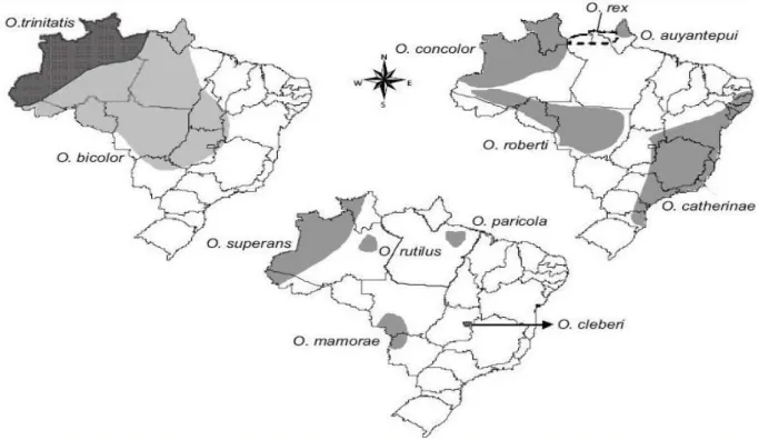
distribuídas em áreas de floresta tropical e sub-tropical das Américas Central e do Sul, incluindo Costa Rica, Trinidad, Panamá, Venezuela, Colômbia, Equador, Peru, Bolívia, Guiana, Guiana Francesa, Suriname e Brasil (Hershkovitz, 1960). A Figura 4 mostra a distribuição das doze espécies encontradas no Brasil (Bonvicino et al., 2008).
Figura 4. Distribuição das espécies de Oecomys no Brasil (Bonvincino et al., 2008)
Para a Amazônia brasileira são conhecidas nove espécies de Oecomys assim
distribuídas: Oecomys auyantepui, presente na Venezuela, Guianas e Brasil, no Estado do
Amapá; O. bicolor, presente do Panamá à Colômbia, Equador, Peru, Bolívia, Venezuela,
Guianas e no Brasil, nos Estados do Amapá, Roraima, Amazonas, Pará, Acre, Rondônia, Mato Grosso, Mato Grosso do Sul, Tocantins, Goiás, Bahia, Minas Gerais e no Distrito Federal; O. concolor, presente no Brasil, nos Estados do Amazonas e Roraima, e também na Venezuela,
Colômbia e Bolívia; O. paricola, registrado apenas para o Estado do Pará, nas proximidades de
Belém; O. rex, presente no norte dos estados do Amapá e Amazonas; O. roberti, presente no
superans, com ocorrência na Colômbia, Equador, Peru e Brasil, nos Estado do Acre, Amazonas
e Roraima e O. trinitatis, estendendo-se da Costa Rica até o Brasil, incluindo Guianas, Trinidad
e Tobago, Colômbia até o Perú, e no Brasil os Estados do Acre, Amazonas, Roraima e Pará (Bonvicino et al., 2008).
1.2.1. História taxonômica do gênero Oecomys
O gênero Oecomys foi primeiramente incluído no gênero Rhipidomys e posteriormente
considerado um subgênero de Oryzomys (Musser & Carleton, 1993). Porém, por ser fortemente
diferenciado sob o ponto de vista cariotípico, o subgênero Oecomys foi elevado ao nível de gênero
(Gardner e Patton, 1976). Entretanto, alegando similaridades entre as espécies de Oecomys e Oryzomys, Ellerman (1941) questionou a validade deste táxon, até mesmo como subgênero.
Hershkovitz (1960) realizou a única revisão taxonômica ampla para o gênero Oecomys,
o considerando subgênero de Oryzomys. Este autor, tomando por base as características como
tamanho do corpo, proporção do tamanho do pé e da cauda em relação ao do corpo e grau de desenvolvimento da crista temporal, alocou as 25 espécies reconhecidas na época para apenas duas: O. bicolor e O. concolor. Somente a partir do trabalho de Musser & Carleton (1993)
tornou-se consenso o status genérico de Oecomys.
1.2.2. Diversidade Cromossômica em Oecomys
Os roedores, devido às características populacionais que apresentam, desenvolveram-se como o grupo mais especioso de mamíferos em florestas neotropicais e um dos mais interessantes para estudos da variabilidade genética e de evolução entre os vertebrados. De acordo com a literatura, o gênero Oecomys apresenta uma grande diversidade cariotípica, com o
número diplóide variando entre 58 e 86, sugerindo um alto grau de rearranjo cromossômico. O primeiro estudo citogenético nesse gênero foi realizado por Gardner & Patton (1976), identificando três populações dentro de Oecomys, sendo uma relacionada a Oecomys bicolor
(2n=80, NF=134-136) e duas relacionadas a Oecomys concolor (2n=80, NF=108; 2n=60,
NF=62). Dentro do gênero Oecomys apenas oito espécies contam com o cariótipo descrito,
identificação taxonômica e na compreensão das relações filogenéticas entre os diferentes grupos.
3. OBJETIVOS
3.1. Geral:
O presente trabalho tem como objetivo geral estudar os cariótipos de espécies
Oecomys paricola de duas localidades, visando diagnosticar quais os possíveis rearranjos
cromossômicos envolvidos na evolução cariotípica das espécies deste gênero e auxiliar na elucidação de possíveis problemas taxonômicos.
3.2. Específicos:
Os objetivos específicos são:
a) Caracterizar os cariótipos (2n e NF) de roedores da espécie Oecomys paricola, organizando
os cromossomos de acordo com a classificação morfológica e em ordem decrescente de tamanho;
b) Identificar homologias cromossômicas por bandeamento G;
c) Analisar a distribuição da heterocromatina constitutiva por bandeamento C;
d) Identificar os sítios de rDNA ativos por meio de impregnação por Nitrato de Prata (marcação Ag-NO3);
e) Fornecer dados citogenéticos para auxiliar na identificação das espécies coletadas através do número diplóide (2n) e número fundamental (NF), bem como comparar os resultados obtidos com os já descritos na literatura;
2. REFERÊNCIAS BIBLIOGRÁFICAS
ANDERSON, S. Introduction to Rodents. In: Recent Mammals of the world. A Synopse of Families. Anderson, S. & Jones jr., J.K. The Ronald Press Company, New York, p. 206 – 209. 1967.
BIANCHI, N.O.; MOLINA, O.J.; DULOUT, F.N. Cytogenetics of the South American Akodont rodents (Cricetidae). I. A progress Report of Argentinian an Venezuelan Forms. Evolution. 25: 724 – 736. 1971.
BONVICINO, C. R.; DE OLIVEIRA, J. A.; D’ANDREA. Guia dos Roedores do Brasil, com chaves para gêneros baseadas em caracteres externos. Rio de Janeiro: Centro Pan-Americano de Febre Aftosa - OPAS/OMS, 2008.
D’ELÍA, G., LUNA, L., GONZÁLEZ, E. M., PATTERSON, B. On the Sigmodontinae
radiation (Rodentia, Cricetidae): An appraisal of the phylogenetic position of Rhagomy.
Molecular Phylogenetics and Evolution. 2005.
ELLERMAN, J. R. The families and genera of living rodents. Vol. II. Family Muridae. British Museum (Natural History), London. 1941.
EMMONS, L.H. & FEER, F. Neotropical rainforest mammals: a field guide. 2ª ed., University of Chicago, Chicago, 307p. 1997.
EMMONS, L.H. & FEER, F. Neotropical rainforest Mammals: a field guide. 2ª ed. University of Chicago Press, Chicago. IL. 1999.
GARDNER, A.L. & PATTON,J.L. Karyotipyc variation in chromosomal evolution in the Neotropical cricetine complex. Occasional Papers of the Museum of Zoology, Louisiana State University, 49:1-48. 1976.
HARTENBERGER, J.L. The Order Rodentia: Major Question on Their Evolutionary Origin, Relationships and Suprafamilial Sytematics. Montpellier, Institutt des Sciences de
l’Evolution, p.33. 1981.
HERSHKOVITZ, P. Mammals of northern Colombia, preliminary report no. 8: arboreal rice rats, a systematic revision of the subgenus Oecomys, genus Oryzomys. Proc USA. Natl Mus
110:513-568.1960.
HERSHKOVITZ, P. Mice, land bridges and Latin American faunal interchange. In: WENZEL, R.L. & TIPTON, V.J. eds. Ectoparasites of Panamá. Chicago, Field Mus. P. 725
– 751. 1966a.
HERSHKOVITZ, P. The evolution of mammals on Southern continents. VI. The recent mammals of the Neotropical Region: a zoogeographical and ecological reviw. Quart. Rev. Biol. 44: 1 – 70. 1969.
HOOPER, E. T. & MUSSER, G. G. The glans penis in Neotropical cricetines (family Muridae), with comments on classification of muroid rodents. Misc Publ Mus Zool. Univ Michigan 123:1-57.1964.
LANDRY jr. S.O. The Rodentia as Carnicores. Q. Ver. Biol.45: 351 – 372. 1970.
MCKENNA, M. C & BELL, S. K. Classification of Mammals above the species level. Columbia University Press, New York. 1997.
MUSSER, G. G. & CARLETON, M. D. Family Muridae. P.501-756. In : Mammals species of the world: a taxonomica and geographic reference, 2nd edition. Wilson D.E. and Reeder D.M. (Ed.). Washington, DC: Smithsonian Institution. 1207p. 1993.
MUSSER, G. G. & CARLETON, M. D. Super Family Muroidea. In: Mammal species of the world: a taxonomic and geographic reference. Wilson D.E. and Reeder D.M. (eds) Johns Hopkins University Press, Baltimore, pp 894-1531. 2005.
NOWAK, R. M. Walker´s Mammals of the World. 1 ed. Baltimore: The Johns Hopkins Univ. Press. p. 642. 1994.
OLIVEIRA, J.A. & BONVICINO, C.R. Ordem Rodentia. In: Mamíferos do Brasil. N.R. Reis, A.L. Peracchi, W.A. Pedro, & I.P. Lima (eds.). Imprensa da UEL, Londrina, p. 347-406. 2006.
REIG, O. A. A new fóssil genus of South American cricetid rodents allied to Wiedomys, with
an assessment of the Sigmodontinae. J Zool 192:257-281.1980.
REIG, O. A. Teoria del origin e desarollo de la fauna de mamíferos de América del Sur.
Naturae Mus. Munic. Cienc. Nat. Lorenzo Scaglia. 1:1 – 161. 1981.
REIG O. A. Distribuição geográfica e historia evolutiva dos roedores sulamericanos (cricetidae, sigmodontinae). Rev. Bras. Genet. 7:333 – 365. 1984.
REIG, O. A. Diversity patterns and differentiation of high Andean rodents. In: High Altitude Tropical Biogeography. Vuilleumier F and Monasterio M (eds). Oxford University Press. New York. NY. Pp 404-439. 1986.
REIS, N. R.; PERACCHI, A. L.; PEDRO, W. A.; LIMA, I. P. Mamíferos do Brasil. Londrina, Paraná.. 395-396p. 2006.
SIMPSON, G.G. The Principles of Classification and a classification of mammals. Bull. Amer. Mus. Nat. Hist., 85: 1 – 350. 1945.
SMITH, M.N.F. & PATTON, J.L. phylogenetic relationships and radiation of Sigmodontinae rodents in South America: evidence from cytochrome b. Journal of Mammalian Evolution.
6:89-128. 1999.
VOSS, R. S. & LINZEY, A. V. Comparative Gross morphology of male accessory glands among Neotropical Muridae (Mammalia: Rodentia) with comments on systematic implications. Misc Publ Mus Zool Univ Michigan 159:1-41. 1981.
VOSS, R.S. & CARLETON, M.D. A new genus for Hesperomys molitor wing and Holochilus magnus Hershckovitz (Mammalia, Muridae) with an analysis of its Phylogenetics
Relationships. Americam Museun of Novitates. 3085:1-39. 1993.
WEKSLER, M.; PERCEQUILLO, A.R.; VOSS, R.S. Ten new genera of Oryzomyine Rodents (Cricetidae, Sigmodontinae). American Museum of Novitates 3537, 29pp. 2006.
WOOD, A.E. A revised classification of the rodents. Evolution 19:115-30. 1955.
4. MANUSCRITO DO ARTIGO CIENTÍFICO
Genetic and morphological variability in
Oecomys paricola
(Sigmodontinae,
Rodentia): evidences for a complex of species.
Artigo submetido a revista Journal of Evolutionary Biology (JEB-2011-00273)
Rosa, C.C.1,2; Flores, T.3; Pieczarka, J.C.1,4; Rossi, R.V.5; Sampaio, M. I. C.4,6; Rissino, J.D.1; Amaral, P.J.S.1,7; Nagamachi, C.Y.1,4§
1 Laboratório de Citogenética, Instituto de Ciências Biológicas, Universidade Federal do Pará,
Belém, Brazil
2 FAPESPA Mastership Scholarship in Neurociences and Celular Biology, Belém, Brazil
3 CAPES Mastership Scholarship in Zoology;
4 CNPq Researcher, Belém, Brazil
5 Departamento de Biologia e Zoologia, Instituto de Biociências, Universidade Federal do
Mato Grosso, Brazil
6 Laboratório de Genética e Biologia Molecular, Universidade Federal do Pará, Campus
Universitário de Bragança, Pará, Brazil
7 FAPESPA Doctorship Scholarship in Genetic and Molecular Biology
ABSTRACT
The rodent genus Oecomys (Sigmodontinae) comprises approximately 16 species that inhabit
tropical and subtropical forests in Central and South America. In this study specimens of
Oecomys paricola Thomas, 1904 from Belém and Marajó Island, northern Brazil, were
investigated using cytogenetic, molecular and morphological analyses. Three karyotypes were found, two from Belém (2n=68, FN=72 and 2n=70, FN=74) and a third from Marajó Island (2n=70, FN=74). No molecular or morphological differences were found between the individuals with differing cytotypes from Belém, but differences were evident between the individuals from Belém and Marajó Island. O. paricola is a complex of species. Specimens
from the Belém City region may represent two cryptic species because two different karyotypes are present in the absence of significant differences in morphology and molecular characteristics. The Marajó Island and Belém populations represent distinct species that have been separated for some time, and are in the process of morphological and molecular differentiation as a consequence of reproductive isolation at the geographic and chromosomal levels.
INTRODUCTION
Rodents are important members of most faunal communities as they are cosmopolitan and native to most terrestrial areas. Their wide variation in morphology, diversity of habitats, climatic tolerance and food sources make them the most numerous and evolutionarily successful among the orders of mammals (Emmons and Feer, 1997).
The subfamily Sigmodontinae, which includes most of the species of South American rodents, occurs only in the Americas. It includes 386 species in 81 genera and nine tribes: Abrotrichini, Akodontini, Ichthyomyini, Oryzomyini, Phyllotini, Reithrodontini, Sigmodontini, Thomasomyini and Wiedomyini (Reig, 1980, 1984; Smith and Patton, 1999; Musser and Carleton, 2005; Weksler et al., 2006; D’Elía et al., 2007). The tribe Oryzomyini
comprises 28 genera (Weksler et al., 2006) and includes the genus Oecomys, which was first
described by Thomas as a subgenus of Oryzomys (1906), and later accepted as a full genus
(Thomas, 1909). In the only taxonomic review of Oecomys, Hershkovitz (1960) followed
Thomas (1906) in considering the group as a subgenus of Oryzomys, but others disagreed
with this taxonomic arrangement. Only after the study of Musser and Carleton (1993) the generic status of the genus Oecomys was largely accepted. Recent phylogenetic studies
including species of Oecomys have confirmed the monophyly of this genus (Patton and Da
Silva, 1995; Smith and Patton, 1999; Patton et al., 2000; Andrade and Bonvicino, 2003;
Weksler, 2003, 2006), which now includes 16 valid species (Musser and Carleton, 2005; Carleton et al., 2009).
The genus Oecomys occurs in tropical and subtropical rainforests in Central and South
America. In the Brazilian Amazon region there are nine species, of which only five have had their karyotype described (Andrade and Bonvicino, 2003; Langguth et al., 2005). Cytogenetic
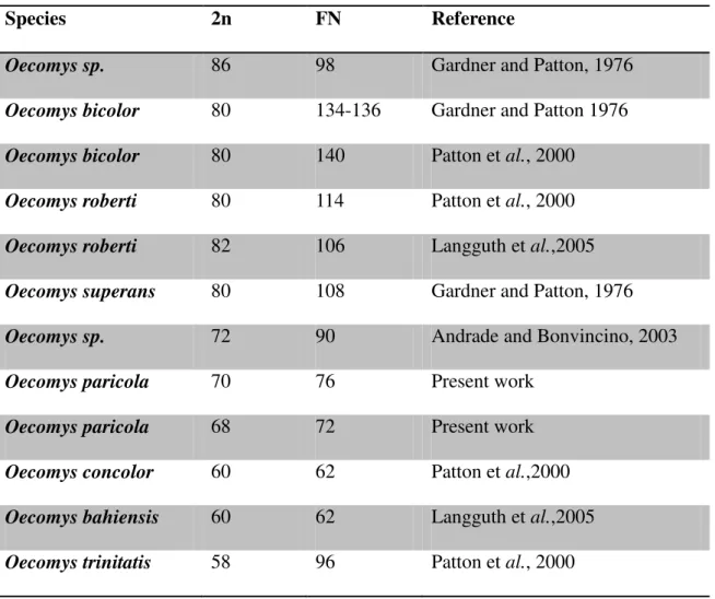
studies of Oecomys show that the diploid number ranges from 58 to 86 (Table 1), suggesting a
high level of chromosomal rearrangement among the karyotypes of these species (Gardner and Patton, 1976; Patton et al., 2000). This karyotypic variability demonstrates the
importance of chromosomal studies in the taxonomic identification of the species (Langguth
et al., 2005).
Oecomys paricola Thomas, 1904 is an Amazonic species with an incompletely defined
distribution south of the Amazon River and east of the Tocantins River (Voss et al., 2001;
Musser and Carleton, 2005). No karyotype information has been reported for the species. According to Voss et al. (2001), O. paricola has predominantly gray-based ventral fur, except
tuft of hairs 6–10 mm long. Cranially, O. paricola has a primitive pattern of carotid arterial
supply, separated accessory oval and buccinator−masticatory foramina; the subsquamosal fenestra are absent.
The identification of rodent species using morphological traits is very complex (Patton and Gardner, 1972; Gardner and Emmons, 1984), leading to uncertainties in the taxonomy of many genera of this group. Studies combining morphological, molecular and karyotype analyses have the potential to demonstrate the occurrence of a greater diversity of Oecomys
species, resulting in the identification of new species, and revalidation of previously described species (Patton and Da Silva, 1995; Smith and Patton, 1999; Andrade and Bonvicino, 2003; Weksler, 2006). Molecular studies using the cytochrome b gene as a marker in phylogenetic studies of Sigmodontinae rodents are particularly relevant (Smith and Patton, 1991, 1993, 1999; Patton et al., 2000; Andrade and Bonvincino, 2003; D’Elia, 2003; Miranda et al., 2007;
D’Elia et el., 2008; Catzeflis and Tilak, 2009). This gene has been reported to have
arrangements consistent with the species boundaries based on classic taxonomic studies (Johns and Avise, 1998; Avise and Walker, 1999), making it highly appropriate for biodiversity studies (Bradley and Baker, 2001).
In this study we undertook chromosomal, molecular and morphological analyses of O. paricola from the Belém City region and Marajó Island, in Pará State, Brazil. This is the first
description of the karyotype of the species, and provides new information on molecular and morphologic variation in the species.
MATERIAL AND METHODS Karyotype analysis
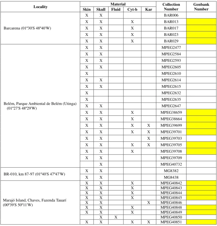
We karyotyped 6 specimens of Oecomys paricola (Table 2), of which 4 were collected
in the Environmental Park in Belém City (01°27′ S, 48°29′ W) and 2 (males) were collected at Marajó Island (01°00′S, 49°30′W), in the region of the Amazon River mouth (Figure 1). The specimens were collected using pitfall traps (buried 60 l buckets) connected by plastic tapes, and with conventional traps including the Sherman trap and cages on the ground containing baits of peanuts, flour and canned fish. Samples for metaphase chromosome analysis were taken in the field using the bone marrow technique (Ford and Hamerton, 1956). We also made laboratory culture of fibroblasts.
NOR staining, according to Seabright (1971), Sumner (1972), and Howell and Black (1980), respectively. The karyotypes were organized by size and morphology of the chromosome pairs.
Molecular analysis
We extracted 738 bp sequences of the cytochrome b gene from 17 ethanol-preserved samples of muscle tissue of O. paricola, including 4 of the 6 specimens we used for karyotype
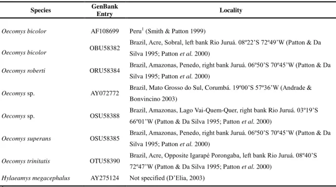
analysis (Table 2). We also used nine Oecomys sequences available in GenBank (several
species, Table 3), two sequences of O. auyantepui and O. rutilus (CN120 and CN123,
respectively) obtained in our laboratory, and one GenBank sequence of Hylaeamys megacephalus (AY275124), as the outgroup in the phylogenetic analysis.
DNA extraction involved the phenol–chloroform and proteinase K−RNase protocol (Sambrook et al., 1989). Cytochrome b fragments were isolated and amplified using the
polymerase chain reaction (PCR) with primers MVZ05 and MVZ16 (Smith and Patton, 1993). The amplification protocol consisted of initial denaturation at 94°C for 3 min, 35 denaturation cycles of 30 s at 94°C, 1 min of annealing at 45°C, 2 min of extension at 72°C, and a final extension at 72°C for 7 min.
Sequences were edited and aligned in BioEdit 7.0.5.2 (Hall, 1999) using ClustalW (Larkin et al., 2007), following the parameters proposed by Schneider (2006). We verified
sequence saturation using DAMBE 5.2.34 software (Xia and Xie, 2001), and undertook phylogenetic analyses. Using MrModeltest 2.3 (Nylander, 2004) in PAUP* 4.0 we found the best evolutionary GTR+I+G model fit to our sequences with a substitution rate of 6, a gamma distribution parameter of 0.5449, and an invariable sites proportion of 0.4145. Maximum likelihood (ML) was estimated from the above model using the online PhyML (Guindon and Gascuel, 2003) site to construct a BioNJ initial tree. Maximum parsimony (MP) was determined using PAUP* 4.0 with a heuristic search; the starting tree was obtained by stepwise addition and branch-swapping using a tree–bisection–reconnection (TBR) algorithm. Clade support was performed for both analyses using bootstrap with 1000 replicates. Estimates of evolutionary divergence of sequence pairs between and within Oecomys species
and populations were conducted using the GTR model in DAMBE 5.2.34 (Xia and Xie, 2001). All codon positions were included.
Morphological and morphometric analyses
We examined the morphological characteristics of 35 specimens of O. paricola from
Federal do Pará (UFPA), identified in this study by the abbreviation BAR (Barcarena Project), will be deposited in the MPEG. All six specimens for which we obtained karyotypes were included in the morphological and morphometric analyses (Table 2). For comparison, we also examined the following 15 MPEG specimens of O. auyantepui: 7169, 13132, 39793,
39794, 39798, 39804, 39816, 39817, 39825, 39831, 39836, 40076, 40078, 40084 and 40085. To enable more accurate morphological and morphometric comparisons the specimens were classified into three age classes based on the eruption pattern and the differential wear of the occlusal surface of the superior molars. These classes, modified from Voss (1991), and Brandt and Pessôa (1994), were: (i) Age class 1 (young)—M3 incompletely erupted or
unworn, (ii) Age class 2 (adult)—occlusal surface exhibiting slight to moderate wear, but still
tubercular; mesoflexus and paraflexus of M1 and M2 sometimes as enamel islands; all M3 flexus, except paraflexus, obliterated and sometimes as enamel islands, (iii) Age class 3 (old
adult)—occlusal surface flat or concave; only paraflexus, metaflexus, protoflexus and hypoflexus are present in M1 and M2; others, when present, are just enamel islands; paraflexus of M3 always as an enamel island, or totally absent.
We evaluated morphological characteristics of the skin and skull of two populations of
O. paricola (one from the Belém region and the other from Marajó Island) for use in
separating groups of individuals. Comparisons among specimens were made with respect to sex and age classes. To avoid misinterpreting ontogenetic variation as taxonomic variation we did not restrict comparisons to the specimens for which we have karyotypes, as they belonged to different age classes. The anatomical nomenclature used followed Pocock (1914), Hershkovitz (1962, 1977), Carleton and Musser (1984, 1989) and Voss (1988) for external morphology, and McDowell (1958), Hershkovitz (1962), Wahlert (1974), Carleton and Musser (1984, 1989), Voss (1988), Steppan (1995) and Weksler (2006) with respect to cranial morphology.
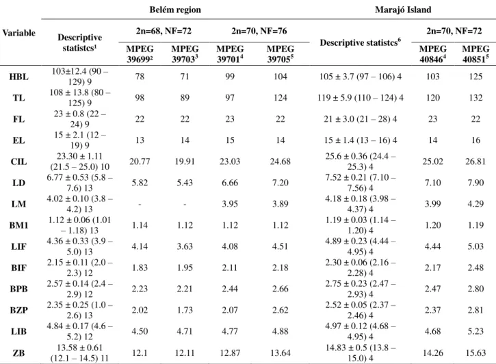
The following external measurements were obtained directly from the specimen labels, and were used for descriptive statistics only: head and body length (HBL), tail length (TL), foot length (FL), ear length (EL), weight (W). We also obtained 10 craniodental measurements (to the nearest 0.01 mm) from each of the 32 skull specimens using a digital caliper during skull examination with a stereomicroscope. These included: (i) Condyloincisive Length (CIL)—from the greater curvature of one upper incisor to the articular surface of the
condyle on the same side, (ii) Length of Diastema (LD) )—from the crown of the first cheek
tooth to the lesser curvature of the incisor on the same side, (iii) Length of Molars (LM) )—
the first upper molar (M1), (v) Length of Incisive Foramen (LIF) —the greatest anterior
posterior dimension of one incisive foramen, (vi) Breadth of Incisive Foramen (BIF)—the
greatest transverse dimension across both incisive foramina, (vii) Breadth of Palatal Bridge (BPB)—measured between the protocones of the right and left M1, (viii) Breadth of Zygomatic Plate (BZP)—the least distance between the anterior and posterior edges of the
zygomatic plate, (ix) Least Interorbital Breadth (LIB)—the least distance across the frontal
bones between the orbital fossa, and (x) Zygomatic Breadth (ZB)—the greatest transverse
dimension across the squamosal zygomatic processes.
We evaluated the presence of sexual dimorphism in our largest sample, from the Belém region (6 females and 7 males), using Hotelling's T-square test. We also used this test to evaluate morphometric differences between specimens from the Belém region and Marajó Island. The morphometric analyses were conducted on specimens of age class 2 only using PAST 2.02 (Hammer et al., 2001), as this class contained the greatest number of samples, and
because skull size appeared to vary greatly among different age classes.
RESULTS
Karyotype analysis
(a) Environment Park, Belém
The specimens MPEG 39703 (male) and MPEG 39699 (female) had the karyotype 2n=68, FN=72, comprising 30 one-armed pairs and 3 bi-armed pairs. The X chromosome was large and bi-armed, and the Y chromosome was small and submetacentric. Figure 2 shows the G-banded karyotype of this cytotype. Constitutive heterochromatin was found at the centromeric region of all chromosomes. The short arm of the X chromosome was almost all heterochromatic, and almost all the Y chromosome was heterochromatic (Figure 2). The NOR was found on the small short arm of 3 one-armed pairs (one mid-sized and two small; Figure 3).
(b) Amazon River mouth region
The specimens MPEG 40846 and 40851 (both males), which were collected from Marajó Island, had a karyotype of 2n=70, FN=74, comprising 31 one-armed chromosome pairs and 2 bi-armed pairs. The X chromosome was large and bi-armed, and the Y chromosome was mid-sized and submetacentric (Figure 6). Constitutive heterochromatin was found at the centromeric region of all chromosomes. The X chromosome had a heterochromatic block at the distal region of the short arm, and the Y chromosome was almost entirely heterochromatic (Figure 6). The NOR was found on the proximal long arm of 2 pairs of mid-sized acrocentric autosomes (Figure 7).
Molecular analysis
All phylogenetic analyses based on the Cytochrome b gene (ML and MP; Figure 11) indicated the same topologies for the O. paricola populations. In both analyses the specimens
from Marajó Island grouped together in a clade that was distinct from those collected in Belém, and there was high nodal support (96% for MP and 87% for ML for the Belém clade; 99% for MP and 97% for ML for the Marajó Island clade). Nodal support for the monophyletic clade that corresponded to O. paricola was 100% for the MP analysis and 99%
for the ML analysis. Relationships among Oecomys species were not supported by bootstrap
statistics.
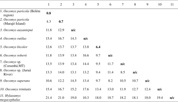
The intrapopulation genetic divergences were 0.8% for the Belém clade and 0.7% for the Marajó Island clade, while the divergence between the populations was 4.3%. The intrageneric divergences ranged from 8.2% to 17.6%, with divergences between O. paricola
and other congeneric species varying from 10.6% to 15.4% (Table 4). Morphological and morphometric analyses
We found no morphological characters that could be used to discriminate the two Belém cytotypes, other than the smaller size of the specimens of karyotype 2n=68, NF=72 relative to those of karyotype 2n=70, NF=76 (Table 5). However, the size difference may be explained by ontogenetic variation, as the former cytotype was based on young specimens from age class 1, while the latter was based on adult and older specimens belonging to age classes 2 and 3.
white). Feet coloration and the plantar surface were also different in the two populations. In the Marajó Island specimens the feet were dark brown with a darker spot on the dorsum, while in the Belém specimens the feet were cream or light brown with a brown spot on the dorsum. The plantar surface was smooth or had several squamae near the plantar pads in specimens from Marajó Island, but was always smooth in Belém specimens. We also observed that the tail length was approximately 116% of the head and body length in Marajó Island specimens, whereas it was only about 106% in the Belém specimens (Table 5).
The skulls were also notably different between the two populations. In the Marajó Island population the nasal bone was short and did not surpass the lachrymal–frontal maxillary suture, whereas in specimens from Belém this bone was long, and aligned with or surpassing the lachrymal–frontal–maxillary suture. In the specimens from Marajó Island the supra-orbital ridges were well developed, projecting dorsally from the border of the frontal bone at the orbital fossa and extending slightly onto the parietal bone, whereas in the specimens from Belém they were poorly developed and restricted to the frontal bone. The frequency of subsquamosal fenestra, which was used by Voss et al. (2001) to distinguish O. auyantepui from O. paricola,
was highly variable in the specimens from Marajó Island. Among the 16 specimens from Belém it was bilateral in 15 specimens and unilateral in one, whereas in specimens from Marajó Island it was bilaterally absent in four specimens and bilaterally present in other four. The mandible also differed between the two populations with respect to the capsular process of the lower incisor alveoli, which varied from slightly to moderately curved in the specimens from Marajó Island, but was slightly curved in the specimens from Belém.
Hotelling's T-square test for sexual dimorphism showed that there was no statistically significant difference between the sexes (p = 0.4684) in the Belém population. The same test
was applied to evaluate morphometric differences between the Belém and Marajó Island specimens. This showed that there were no statistically significant differences between the specimens from each location (p = 0.2825), although the mean values for almost all external
measurements and all skull dimensions of the Marajó Island specimens examined were greater (Table 5).
DISCUSSION
We have provided here the first description of the karyotype of O. paricola. We found
2n=70 are new for the genus Oecomys. The differences in the diploid number and
chromosome morphology of the three karyotypes of O. paricola can be explained by
rearrangements including fusion/fission and pericentric inversions. Because of the number of chromosomes and differences on their condensation, comparative analyses of the G-banding patterns was not possible, and consequently we were not able to precisely identify the chromosomes involved in the karyotype differentiation, or the chromosome rearrangements that occurred. No hybrid karyotypes were found in the Belém sample, indicating that there has been no gene flow between the karyotypes.
Bradley and Baker (2006) assessed whether the degree of cytochrome b sequence divergence in mammals can be used for species-level differentiation within the genetic species concept. They observed that sister species recognized on the basis of morphology often have cytochrome b distance values > 5%. With respect to rodents they found that intrapopulation divergence values ranged from 0.0 to 1.4%, intraspecific divergence values ranged from 0.0 to 4.7%, and intrageneric divergence values ranged from 1.3 to 16.9%. In molecular studies of sigmodontine rodents not considered by Bradley and Baker (2006) values have been reported that range from 0 to 3.87% for intrapopulation divergence, 0 to 11.37% for intraspecific divergence, and 1.23 to 21% for intrageneric divergence (Smith and Patton, 1991, 1993, 1999; Patton et al., 2000; D’Elia, 2003; Miranda et al., 2007; D’Elia et al., 2008; Catzeflis and Tilak, 2009). For Oecomys, no intrapopulation divergence values have
been reported, but intraspecific divergence ranges from 0 to 10.3%, and intrageneric divergence ranges from 7 to 12% (Smith and Patton, 1999; Patton et al., 2000; Andrade and
Bonvincino, 2003).
The intrapopulation divergences of O. paricola from Marajó Island (0.7%) and the
Belém region (0.8%) are in agreement with those reported by Bradley and Baker (2006) for rodents in general, and for sigmodontine species (citations above). Similarly, the intraspecific evolutionary divergence we found (4.3% in O. paricola) was relatively high, close to the 5%
limit of morphologically diagnosable species of mammals stated by Bradley and Baker (2006). The intrageneric divergences (8.5 to 16.7%) we found are greater than those previously reported for Oecomys, but are comparable to those of other sigmodontine rodents.
no genetic structure within this population, even between specimens representing two of the three cytotypes described (Fig. 11).
In contrast, we found consistent morphological differences between the populations from the Belém region and Marajó Island. These differences, allied to the relatively high genetic divergence between the populations (4.3%), the genetic structure indicated by the molecular analyses (Fig. 11), and the unique karyotype of the specimens from Marajó Island (Figs 8–10) suggest that these two populations are distinct species. Morphologically, the specimens from Marajó Island are similar to specimens of O. auyantepui, with which they
share the same pattern of ventral pelage color, length of the nasal bones, and development and extension of the supraorbital ridges, despite the genetic divergence of 12.9% between these species. It is possible that the two cytotypes from Belém represent cryptic species, whereby recent speciation mediated by chromosome rearrangements has occurred, but there has been insufficient time for the fixation of molecular and morphological differences.
This shows that chromosome rearrangements can have a very important role as a postmating reproductive isolation mechanism. According to King (1987, 1993), chromosome rearrangement can be the main cause of speciation when they cause a negative heterosis effect, by reducing the fertility of heterozygous bearers. This is a postmating reproductive isolation (rearrangements that segregate badly in meiosis in structural hybrids, producing unbalanced gametes and a consequent reduction in fertility). There is some evidence of a correlation between population structure and karyotypic variability, with greater variability occurring in species that have small inbreeding populations, such as occurs in rodents (Bush
et al., 1977; Bengtsson, 1980; Maruyama and Imai, 1981), relative to species with
outbreeding populations, such as cats and whales (Bush et al., 1977). Population structure has
a crucial role in fixing rearrangements, as this only happens if the rearranged chromosome can rapidly become homozygous. In small inbreeding populations the probability of this homozygous condition arising is greater, making the rearranged chromosome more common. A similar situation was reported by Granjon and Dobigny (2003), who undertook a morphological and cytogenetic study of rodents from the Lake Chad region, Africa. They demonstrated that a significant portion of the species are cryptic (morphologically indistinguishable), and can only be distinguished by karyotype analysis. This situation occurs in many vertebrates groups in the Neotropical region. For instance, Milhomem et al. (2008)
used classical cytogenetics to describe 2 cryptic species in the species complex of Gymnotus carapo (a Neotropical electric fish); the species are morphologically identical, and have
inversions. Nagamachi etal. (2010) used chromosome painting to demonstrate that the level
of chromosome reorganization is greater than previously thought, confirming that Gymnotus carapo with different karyotypes are truly different species.
The chromosome differentiation between the Marajó Island and Belém cytotypes must have occurred some time ago, because our results indicate differences at the molecular and morphologic levels, which must have occurred since the interruption of gene flow. A similar situation has been reported in the genus Akodon (Geise et al., 2005), where A. montensis, A. cursor and A. aff cursor form a species complex with other undescribed species (Silva and
Yonenaga-Yassuda, 1998). These species are so similar that they cannot be distinguished at the morphological level (Christoff et al., 2000). However, mitochondrial (Geise et al., 2001)
and karyological data (Geise et al., 1998; Silva and Yonenaga-Yassuda, 1998) show that they
are distinct species.
The occurrence of morphologically identical taxa with different diploid and fundamental numbers is common among rodents. In Oecomys, for instance, Gardner and
Patton (1976) described the karyotype of O. bicolor as 2n=80 and FN=134−136. Patton et al.
(2000) subsequently found another karyotype in O. bicolor; this had the same diploid number,
but had three bi-armed chromosomes instead of the three one-armed chromosomes in the initial karyotype, (2n=80, FN=140; Table 1). The phylogenies described by Smith and Patton (1999), and Andrade and Bonvicino (2003) suggest that O. bicolor is a species complex. Two
karyotypes have also been described for O. roberti, one (2n=80, FN=114) having 21 one-
armed chromosome pairs and 18 bi-armed pairs (Patton et al., 2000), and the other (2n=82,
FN=106) having 27 one-armed chromosome pairs and 13 bi-armed pairs (Langguth et al.,
2005).
We conclude that O. paricola from Belém may represent two cryptic species because
two different karyotypes are present in the population in the absence of significant differences in morphology and molecular characteristics. No hybrid karyotypes were found, which indicates the absence of gene flow among the cryptic species. Additionally, the Marajó Island and Belém populations represent distinct species that have been separated for some time and are undergoing morphological and molecular differentiation following reproductive isolation at the geographical and chromosomal levels. This is possibly an example of speciation where the morphological and molecular differences are accumulating following reproductive isolation.
Belém City region appear to be an example of cryptic species with no significant differences in morphology and molecular characters, but with different karyotypes. The populations of Marajó Island and Belém are distinct species that have been separated for some time and are in the process of morphological and molecular differentiation following reproductive isolation at the geographic and chromosomal levels.
ACKNOWLEDGMENTS
The authors thank the Conselho Nacional de Desenvolvimento Científico e Tecnológico (CNPq) and the Fundação de Amparo à Pesquisa do Estado do Pará (FAPESPA) for financial support, and the Instituto Brasileiro de Meio Ambiente e dos Recursos Naturais Renováveis (IBAMA) for authorizing sample collection.
REFERENCES
Andrade, A.F.B. & Bonvicino, C.R. 2003. A new karyological variant of Oecomys (Rodentia:
Sigmodontinae) and its phylogenetic relationship based on molecular data. Genome 46: 195
203.
Avise, J.C. & Walker, D. 1999. Species realities and numbers in sexual vertebrates: perspectives from an asexually transmitted genome. PNAS 96: 992-995.
Bengtsson, B. 1980. O. Rates of karyotype evolution in placental mammals. Hereditas 92: 37-
47.
Bradley, R.D. & Baker, R.J. 2001. A test of the Genetic Species Concept: cytochrome-b sequences and mammals. Journal of Mammalogy 82(4): 960-973.
Bradley, R.D. & Baker, R.J. 2006. Speciation in mammals and the genetic species concept.
Journal of Mammalogy 87(4): 643-662.
Brandt, R.S. & Pessôa, L.M. 1994. Intrapopulational variability in cranial characters of
Oryzomys subflavus (Wagner, 1842) (Rodentia: Cricetidae), in northeastern Brazil. Zoologischer Anzeiger 233(1-2): 45-55.
Bush, G.L., Case, S.M., Wilson, A.C. & Patton, J.L. 1977. Rapid speciation and chromosomal evolution in mammals. PNAS 74: 3942-3946.
Carleton, M.D., Emmons, L.H. & Musser, G.G. 2009. A new species of the rodent genus
definitions of O. concolor (Wagner) and O. mamorae (Thomas). American Museum Novitates
3661: 1-32.
Carleton, M.D. & Musser, G.G. 1984. Muroid Rodents. In: Orders and families of recent mammals of the World (S. Anderson & J. K. Jones Jr. eds) pp: 289-379. New York. John
Wiley Publications.
Carleton, M.D. & Musser, G.G. 1989. Systematic studies of Oryzomyine rodents (Muridae, Sigmodontinae): a synopsis of Microryzomys. Bulletin of the American Museum of Natural History 191: 1-83.
Catzeflis, F. & Tilak, M. 2009. Molecular systematic of Neotropical spiny mice (Neacomys:
Sigmodontinae, Rodentia) from the Guianan Region. Mammalia 73: 239-247.
Christoff, A.U., Fagundes, V., Sbalqueiro, I.J., Mattevi, M.S. & Yonenaga-Yassuda, Y. 2000. Description of new species of Akodon (Rodentia: Sigomodontinae) from southern Brazil. Journal of Mammalogy 81: 838-851.
D’Elía, G. 2003. Phylogenetics of Sigmodontinae (Rodentia, Muroidea, Cricetidae), with special reference to the akodont group, and with additional comments on historical biogeography. Cladistics 19(4): 307-323.
D’Elía, G., Pardiñas, U.F.J., Teta, P. & Patton, J.L. 2007. Definition and diagnosis of a new
tribe of sigmodontine rodents (Cricetidae: Sigmodontinae), and a revised classification of the subfamily. Gayana (Concepc.) 71:187-194.
D’Elía, G., Pardiñas, U.F.J., Jayat, J.P. & Salazar-Bravo, J. 2008. Systematics of Necromys
(Rodentia, Cricetidae, Sigmodontinae): species limits and groups, with comments on historicalbiogeography. Journal of Mammalogy 89(3): 778-790.
Emmons, L.H. & Feer, F. 1997. Neotropical rainforest mammals: a field guide. 2nd ed.,
University of Chicago.
Ford, C.E. & Hamerton, J.L. 1956. A colchicine hypotonic citrate squash sequence for mammalian testis. Mammal. Chrom. Newsl. 20: 74.
Gardner, A.L. & Emmons, L. 1984. Species group in Proechimys (Rodentia; Echimyidae) as
indicated by karyology and bullar morphology. Journal of Mammalogy 65:10-25.
Gardner, A.L. & Patton, J.L. 1976. Karyotypic variation in chromosomal evolution in the neotropical cricetine complex. Occasional Papers of the Museum of Zoology. Louisiana State University 49: 1-48
Geise, L. , Canavez, F.C. & Seuánez, H.N. 1998. Comparative karyology in Akodon
Geise, L., De Moraes, D.A. & Da Silva, H.S. 2005. Morphometric differentiation and distributional notes of three species of Akodon (Muridae, Sigmodontinae, Akodontini) in the
Atlantic Coastal area of Brazil. Arquivos do Museu Nacional 63(1): 63-74.
Geise, L., Smith, M.F. & Patton, J.L. 2001. Diversification in the genus Akodon (Rodentia,
Sigmodontinae) in Southeastern South America: mitochondrial DNA sequence analysis.
Journal of Mammalogy 82(1): 92-101.
Granjon, L. & Dobigny, G. 2003. Chromosomally characterized murid rodents from the edges of Lake Chad: evidence for an African biogeographical crossroad, or a centre of endemism?
Mammal Review 33: 77-91.
Guindon, S. & Gascuel, O. 2003. A simple, fast, and accurate algorithm to estimate large phylogenies by maximum likelihood. Systematic Biology 52(5): 696-704.
Hall, T.A. 1999. BioEdit: a user-friendly biological sequence alignment editor and analysis program for Windows 95/98/NT. Nucleic Acids Symposium Series 41:95-98.
Hammer, O., Harper, D.A.T. & Ryan, P.D. 2001. PAST: Paleontological Statistics software package for education and data analysis. Palaeontologia Electronica 4(1): 1-9.
Hershkovitz, P. 1960. Mammals of northern Colombia, preliminary report no. 8: arboreal rice rats, a systematic revision of the subgenus Oecomys, genus Oryzomys. Proceedings of the United States National Museum 110: 513-568.
Hershkovitz, P. 1962. Evolution of Neotropical cricetine rodents (Muridae) with special reference to the phyllotine group. Fieldiana Zoology 46: 1-524.
Hershkovitz, P. 1977. Living New World monkeys (Platyrrhini). 1st volume, The University
of Chicago Press, Chicago.
Howell, W.M. & Black, D.A. 1980. Controlled silver-straining of nuclear organizer regions with protective colloidal developer: a 1-step method. Experientia 36: 1014-1015.
Johns, G.C. & Avise, J.C. 1998. A comparative summary of genetic distances in the vertebrates from the mitocondrial Cytochrome-b gene. Molecular Biology and Evolution 15:
1481-1490.
King, M. 1987. Chromosomal rearrangements, speciation and the theoretical approach.
Heredity 59:1-6.
King, M. 1993. Species Evolution. The Role of Chromosome Change. Cambridge University
Press, Cambridge.
Larkin, M.A., Blackshields, G., Brown, N.P., Chenna, R., McGettigan, P.A., McWilliam, H., Valentin, F., Wallace, I.M., Wilm, A., Lopez, R., Thompson J.D., Gibson, T.J. & Higgins D.G. 2007. ClustalW and ClustalX version 2. Bioinformatics 23(21): 2947-2948.
Maruyama, T. & Imai, H.T. 1981. Evolutionary rate of the mammalian karyotype. J. Theor. Biol. 90: 111-121.
McDowell Jr., S.B. 1958. The greater antillean insectivores. Bulletin of the American Museum of Natural History 115(3): 113-214.
Milhomem, S.S.R., Pieczarka, J.C., Crampton, W.G.R., Silva, D.S., Souza, A.C.P. de; Carvalho Jr, J.R. & Nagamachi, C.Y. 2008. Chromosomal evidence for a putative cryptic species in the Gymnotus carapo species-complex (Gymnotiformes, Gymnotidae). BMC Genetics 9: 75.
Miranda, G.B., Andrades-Miranda, J., Oliveira, L.F.B., Langguth, A. & Mattevi, M.S. 2007. Geographic patterns of genetic variation and conservation consequences in three South American rodents. Biochemical Genetics 45: 839-856.
Musser, G.G., & Carleton, M.D. 1993. Family Muridae. In: Mammals species of the World
(D.E. Wilson & D.M. Reeder eds) pp. 501–753. 2nd ed. Smithsonian Institute, Washington, D.C.
Musser, G.G. & Carleton, M.D. 2005. Superfamily Muroidea. In: Mammal Species of the World a Taxonomic and Geographic Reference. (D.E. Wilson & D.M. Reeder eds) pp.
894-1531. 3rd ed. Johns Hopkins University Press, Baltimore.
Nagamachi, C.Y., Pieczarka, J.C., Milhomem, S.S.R., O'Brien, P.C.M., Souza, A.C.P. de & Ferguson-Smith, M.A. 2010. Multiple rearrangements in cryptic species of electric knifefish,
Gymnotus carapo (Gymnotidae, Gymnotiformes) revealed by chromosome painting. BMC Genetics 11: 28.
Nylander, J.A.A. 2004. MrModeltest v2. Program distributed by the author. Evolutionary
Biology Centre, Uppsala University.
Patton, J.L. & Da Silva, M.N.F. 1995. A review of the spiny mouse genus Scolomys
(Rodentia: Muridae: Sigmodontinae) with the description of a new species from the western Amazon of Brazil. Proceedings of the Biological Society of Washington 1 108(2): 319-337.
Patton, J.L., Da Silva, M.N.F. & Malcon, J.R. 2000. Mammals of the Rio Juruá and the evolutionary and the ecological diversification of Amazonia. Bull. Am. Mus. Nat. Nat. Hist.
244: 1-306.
Patton, J.L. & Gardner, A.L. 1972. Notes on the systematics of Proechimys (Rodentia,
Pocock, R.I. 1914. On the facial vibrissae of Mammalia. Proceedings of the Zoological Society of London 1914: 889-912.
Reig, O.A. 1980. A new fossil genus of South American cricetid rodents allied to Wiedomys,
with an assessment of the Sigmodontinae. Journal of Zoology 192: 257-281.
Sambrook, J., Fritsch, E.F. & Maniatis, T. 1989. Molecular cloning: a laboratory manual.
Cold Spring Harbor Laboratory Press: New York.
Schneider, H. 2006. Métodos de análise filogenética: um guia prático. 3rd edition, Holos
Editora, Sociedade Brasileira de Genética 200 p.
Seabright, M. 1971. The use of proteolitic enzymes for the mapping of structural rearrangements of man. Chromosoma 36: 204-210.
Silva, M.J.J. & Yonenaga-Yassuda, Y. 1998. Karyotype and chromosomal polymorphism of an undescribed Akodon from Central Brazil, a species with the lowest diploid chromosome
number in rodents. Cytogenetics and Cell Genetics 81: 46-50.
Smith, M.F. & Patton, J.L. 1991. Variation in mitochondrial Cytochrome-b sequence in natural populations of South American akodontine rodents (Muridae: Sigmodontinae).
Molecular Biology and Evolution 8: 85-103.
Smith, M.F. & Patton, J.L. 1993. The diversification of South American murid rodents: evidence from mitochondrial DNA sequence data for akodontine tribe. Biological Journal of Linnean Society 50: 149-177.
Smith, M.N.F. & Patton, J.L. 1999. Phylogenetic relationships and radiation of Sigmodontinae rodents in South America: evidence from Cytochrome b. Journal of Mammalian Evolution 6: 89-128.
Steppan, S.J. 1995. Revision of the tribe Phyllotini (Rodentia: Sigmodontinae), with a phylogenetic hypothesis for the Sigmodontinae. Fieldiana Zoology 80: 1-112.
Summer, A.T. 1972. A simple technique for demonstrating centromeric heterocromatin.
Experimental Cell Research 75: 304-306.
Thomas, O. 1906. Notes on South American rodents. II. On the allocation of certain species hitherto referred respectively to Oryzomys, Thomasomys, and Rhipidomys. Annals and Magazine of Natural History 18: 442-448.
Thomas, O. 1909. New species of Oecomys and Marmosa of Amazonia. Annals and Magazine of Natural History 3: 378-380.
Voss, R.S. 1991. An introduction to the neotropical muroid rodent genus Zygodontomys. Bulletin of the American Museum of Natural History 210: 1-113.
Voss, R.S., Lunde, D.P. & Simmons, N.B. 2001. The mammal of Paracou, French Guiana: a Neotropical lowland rainforest fauna part 2. Nonvolant species. Bulletin of the American Museum of Natural History 263: 1-236.
Wahlert, J.H. 1974. The cranial foramina of Protrogomorphus rodents: an anatomical and phylogenetic study. Bulletin of the Museum of Comparative Zoology 146(8): 363-410.
Weksler, M. 2003. Phylogeny of neotropical oryzomyine rodents (Muridae: Sigmodontinae) based on the nuclear IRBP exon. Molecular Phylogenetics and Evolution 29: 331-349.
Weksler, M. 2006. Phylogenetic relattioships of oryzomine rodents (Muroidea: Sigmodontinae): separate and combined analyses of morphological and molecular data.
Bulletin of the American Museum of Natural History 296: 149 p. Weksler, M., Percequillo,
A.R. & Voss, R.S. 2006. Ten new genera of Oryzomyine Rodents (Cricetidae, Sigmodontinae). American Museum Novitates 29p.
Table 1. Chromosomal characterization of the genus Oecomys. 2n=diploid number.
FN=Fundamental number.
Species 2n FN Reference
Oecomys sp. 86 98 Gardner and Patton, 1976
Oecomys bicolor 80 134-136 Gardner and Patton 1976
Oecomys bicolor 80 140 Patton et al., 2000
Oecomys roberti 80 114 Patton et al., 2000
Oecomys roberti 82 106 Langguth et al.,2005
Oecomys superans 80 108 Gardner and Patton, 1976
Oecomys sp. 72 90 Andrade and Bonvincino, 2003
Oecomys paricola 70 76 Present work
Oecomys paricola 68 72 Present work
Oecomys concolor 60 62 Patton et al.,2000
Oecomys bahiensis 60 62 Langguth et al.,2005
Table 2 – Specimens analyzed, with localities, specimen preparation type, collection and genbank number. Kar - Karyotype; MPEG – Museu Paraense Emilio Goeldi; BAR –
Specimens from Universidade Federal do Pará (Barcarena Project).
Locality Material Collection Number Genbank Number
Skin Skull Fluid Cyt-b Kar
Barcarena (01º30'S 48º40'W)
X X BAR006
X X X BAR013
X X X BAR017
X X X BAR023
X X X BAR029
Belém, Parque Ambiental de Belém (Utinga) (01º27'S 48º29'W)
X X MPEG2477
X X MPEG2584
X X MPEG2593
X X MPEG2605
X MPEG2610
X X MPEG2614
X X MPEG2615
X MPEG2632
X MPEG2635
X X MPEG2647
X X X MPEG38659
X X X MPEG38664
X X X X MPEG39699
X X X X MPEG39701
X X X MPEG39703
X X X X MPEG39705
X X X MPEG39708
X X MPEG39709
X MPEG40732
BR-010, km 87-97 (01º40'S 47º47'W) X X MG8382
X X MG8438
Marajó Island, Chaves, Fazenda Tauarí (00º39'S 50º11'W)
X X X MPEG40842
X X X MPEG40843
X X X MPEG40844
X X X MPEG40845
X X X MPEG40846
X X X MPEG40848
X X X MPEG40849
X X MPEG40850