Universidade do Minho
Escola de Engenharia
Sofia Emanuela Soares Mendonça
Elucidation of the molecular mechanisms
underlying the cytotoxic effect of
recombinant frutalin in human tumor cells
Sofia Emanuela Soar
es Mendonça
Elucidation of t
he molecular mechanisms underlying t
he cytoto
xic ef
fect of recombinant frut
Dissertação de Mestrado
Mestrado Integrado em Engenharia Biomédica
Ramo de Engenharia Clínica
Trabalho realizado sobre a orientação da
Professora Doutora Lucília Domingues
e da
Professora Doutora Lucília Saraiva
outubro de 2013
Universidade do Minho
Escola de Engenharia
Sofia Emanuela Soares Mendonça
Elucidation of the molecular mechanisms
underlying the cytotoxic effect of
iii
Agradecimentos
No final deste trabalho não posso deixar de expressar o meu sincero agradecimento às pessoas que, direta ou indiretamente, contribuíram para a concretização deste trabalho.Quero agradecer às minhas orientadoras Doutora Lucília Domingues e Doutora Lucília Saraiva, pela oportunidade que me deram em realizar este trabalho, sobretudo pela confiança que em mim depositaram, pelo tempo que despenderam, pelo incentivo, pelo apoio, pela orientação e por tudo quanto aprendi, o meu obrigada.
Aos meus colegas da Plataforma de Biologia Molecular e Sintética do Departamento de Engenharia Biológica da Universidade do Minho pelo bom ambiente e companheirismo. Quero agradecer, de forma especial, à Carla Oliveira por toda a ajuda, pela paciência, pela disponibilidade e tempo despendido com o meu trabalho. À Sofia Costa pela disponibilidade em me ajudar sempre que da sua ajuda precisei. Quero, ainda, agradecer à Joana Cunha pela companhia e boa disposição em todas as horas passadas no laboratório.
Agradeço às minhas colegas do Laboratório de Microbiologia da Faculdade de Farmácia da Universidade do Porto, à Clara, à Joana, à Mariana, à Sara, à Cláudia, à Cláudia Maciel e à Raquel pelas conversas, pela companhia e pela boa disposição. O meu agradecimento especial à Clara por sempre me ter apoiado e por toda a ajuda que me deu em todas as fases do trabalho, à Joana e à Mariana pela ajuda e pela paciência e compreensão que sempre me demonstraram.
Quero agradecer aos meus amigos de curso pela “família” que construímos ao longos deste anos, e por todos os momentos, sem excluir nenhum, que passamos juntos, que nos fizeram crescer e amadurecer. Agradeço aos meus amigos de Lousada toda a sua amizade que, mesmo sentindo a minha ausência, a compreenderam.
Agradeço, ainda, ao Joel Braga por ser uma das minhas forças, por sempre me incentivar e apoiar.
Por último, quero agradecer à minha família, em especial à minha mãe por ser o meu braço direito e esquerdo e por sempre acreditar em mim. Sem ti nunca teria chegado até aqui, nem me teria tornado na pessoa que sou hoje. Ao meu pai por sempre me apoiar, incentivar e pela compreensão que sempre teve comigo. Quero ainda fazer um agradecimento muito especial à minha irmã, a minha gémea, a minha companheira de todas as horas, por estar sempre presente e por sempre me encorajar.
v
Abstract
Frutalin is the α-D-galactose-binding jacalin-related lectin isolated from breadfruit seeds (Artocarpus incisa). Frutalin has been previously produced in Pichia pastoris and in Escherichia coli in order to overcome the limitations associated with the extraction from its natural source. A previous study also showed that recombinant frutalin expressed in Pichia pastoris and purified by size-exclusion chromatography (SEC) induces apoptosis in HeLa tumoral cells. Nevertheless, the molecular mechanism of apoptosis induction triggered by frutalin has not been studied.Therefore, with the present work it was intended to elucidate the molecular mechanism involved in apoptosis induction by the recombinant frutalin expressed in Pichia pastoris and purified by SEC. To achieve such a goal, the modulatory effect of frutalin on main regulatory proteins of apoptosis, such as the executioner members of the caspase family (caspases-3, -6 and -7) and the p53 family members was studied using yeast-based assays. The results obtained showed that the p53 family proteins (p53, p63, p73 and DN) are not direct targets of frutalin. Moreover, using human tumor cells with (HCT116 p53+/+)
and without (HCT116 p53-/-) wild-type p53, we confirmed that frutalin induces apoptosis by a
p53-independent pathway. Concerning the procaspases-3, -6 and -7, the results obtained suggest that frutalin induces apoptosis by a caspase-dependent pathway.
In addition, different molecules of frutalin expressed in a different expression system (E. coli) and/or purified by a different methodology (hydrophobic interaction chromatography; HIC) were tested for its anti-proliferative activity in order to see if one could improve the production and purification process. Thus, the activity of frutalin produced in the bacteria Escherichia coli and in the yeast Pichia pastoris purified by HIC was also evaluated regarding its effect on the proliferation of HCT116 p53+/+ tumor cells.
The frutalin expressed in E. coli did not inhibit cell proliferation. Additionally, frutalin expressed in P. pastoris and purified by HIC, resulted in two different samples suggesting that one is partially
glycosylated and the other is non-glycosylated. The sample partially glycosylated also showed an inhibitory of proliferation of HCT116 p53+/+ tumor cells. However, this activity was less potent than the one obtained
with frutalin from P. pastoris and purified by SEC. The sample non-glycosylated had no anti-proliferative effects. The same result was obtained for frutalin expressed in E. coli, suggesting that glycosylation affects the biological activity of frutalin.
In conclusion, trials to obtain the recombinant frutalin in a more straightforward production/purification process were ineffective as alternative systems, because the anti-proliferative activity of frutalin was compromised. Frutalin produced from Pichia pastoris and purified by SEC has, nevertheless, a potent anti-proliferative effect on HCT116 tumor cells and induces apoptosis through a caspase-dependent pathway.
vii
Resumo
Frutalina é uma lectina α-D-galactose-ligante jacalina-relacionada isolada nas sementes da planta fruta-pão (Artocarpus incisa). A frutalina foi produzida, anteriormente, em Pichia pastoris e em Escherichia coli de modo a superar limitações associadas à sua extração da fonte natural. Um estudo anterior mostrou que a frutalina expressa em Pichia pastoris e purificada por cromatografia de exclusão de peso molecular (SEC) induz a apoptose em células tumorais humanas. Todavia, o mecanismo molecular de indução da apoptose pela frutalina ainda não foi estudado.Assim, com o presente trabalho pretende-se elucidar o mecanismo molecular envolvido na indução da apoptose pela frutalina expressa em P. pastoris e purificada por SEC. Para atingir este objetivo foi estudado, através da utilização de um modelo celular de levedura, o efeito modulador da frutalina nas principais proteínas reguladoras da apoptose como membros da família de caspases executoras (caspases-3, -6 and -7) e membros da família da proteína p53 (p53, p63, p73 e DN). Os resultados obtidos mostraram que os membros da família da proteína p53 não são alvos diretos da frutalina. Além disso confirmou-se, através da utilização de células tumorais humanas com (HCT116 p53+/+) e sem
(HCT116 p53-/-) a forma nativa da p53, que a frutalina induz a apoptose por uma via independente da
p53. Relativamente aos resultados obtidos para as procaspases-3, -6 e -7, estes sugerem que a frutalina induz a apoptose por uma via dependente das caspases.
Foi testada, também, a atividade anti-proliferativa de diferentes moléculas de frutalina expressa num sistema de expressão diferente (E. coli) e/ou purificada por uma metodologia diferente (cromatografia de interação hidrofóbica; HIC), a fim de se verificar se poderiam melhorar o processo de produção e purificação. Assim, foi avaliado o efeito da frutalina produzida na bactéria Escherichia coli e na levedura Pichia pastoris purificada por HIC na proliferação de células tumorais HCT116 p53+/+. A frutalina
expressa em E. coli não inibiu a proliferação celular. Adicionalmente, a frutalina expressa em Pichia pastoris e purificada por HIC resultou em duas amostras diferentes sugerindo que uma é parcialmente glicosilada e outra não é glicosilada. A amostra parcialmente glicosilada mostrou ter efeito na inibição da proliferação das células tumorais HCT116 p53+/+. No entanto, esta atividade é menos potente do que a
obtida com a frutalina expressa em P. pastoris e purificada por SEC. A amostra não glicosilada não teve efeito na inibição da proliferação celular. O mesmo resultado foi obtido para a frutalina expressa em E. coli sugerindo que a glicosilação afeta a atividade biológica da frutalina.
Em conclusão, os ensaios para obter a frutalina recombinante através de um processo de produção/purificação mais simples foram ineficazes como sistemas alternativos, uma vez que comprometeram a atividade anti-proliferativa da frutalina. A frutalina produzida por Pichia pastoris e
purificada por cromatografia de exclusão de peso molecular tem, no entanto, um potente efeito anti-proliferativo nas células tumorais HCT116 e induz a apoptose por uma via dependente das caspases.
ix
Table of contents
Agradecimentos ... iii
Abstract ... v
Resumo ... vii
List of Figures ... xiii
List of Tables ... xvii
Abbreviations ... xix
General Introduction ... 1
Chapter 1 | 1.1 Plant lectins ... 3
1.1.1 Potential biomedical applications of plant lectins ... 5
1.1.2 Jacalin-related lectins (JRLs) ... 6
1.2 The frutalin lectin ... 7
1.3 Heterologous expression of frutalin ... 8
1.3.1 Pichia pastoris and E. coli as expression system ... 8
1.3.2 Recombinant frutalin ... 10
1.3.3 Potential biomedical applications of recombinant frutalin ... 11
1.4 Plant lectin-induced apoptosis ... 12
1.5 Saccharomyces cerevisiae as a model organism for molecular and pharmacological studies of human proteins ... 15
1.6 Aims of the work ... 16
| Materials and Methods ... 17
Chapter 2 2.1 Systems of production and purification of recombinant proteins ... 19
2.1.1 Production of recombinant frutalin expressed in Pichia pastoris ... 19
2.1.2 Purification of recombinant frutalin expressed in Pichia pastoris ... 19
2.1.2.2 Hydrophobic interaction chromatography (HIC) ... 20
2.1.3 Production of recombinant frutalin expressed in E. coli ... 21
2.1.4 Purification of recombinant frutalin expressed in E. coli ... 21
2.1.5 SDS-PAGE ... 22
2.1.5.1 Coomassie blue staining method ... 23
2.1.5.2 Silver nitrate staining method ... 23
2.2 Yeast-cell based phenotypic assay ... 23
2.2.1 Plasmids ... 23
2.2.2 Yeast strain, transformation and growth conditions ... 24
2.2.3 Effects of frutalin and compounds on yeast cell growth ... 24
2.2.4 Western Blot Analysis ... 25
2.2.5 Caspase activation analysis ... 25
2.3 Assays in human tumor cell lines ... 25
2.3.1 Growth conditions of cell culture ... 25
2.3.2 Effect of frutalin on the in vitro cell growth of human tumor cells ... 26
| Results ... 27
Chapter 3 3.1 Production and purification of recombinant frutalin ... 29
3.1.1 Production and purification of frutalin expressed in Pichia pastoris ... 29
3.1.2 Production and purification of frutalin expressed in E. coli ... 31
3.2 Yeast- based assay for the elucidation of the molecular mechanism of frutalin ... 32
3.2.1 Implementation of a yeast assay to search for procaspase-3, -6 and -7 activators ... 32
3.2.2 Effect of frutalin on key apoptotic regulators using yeast assays ... 35
3.3 Studies in human tumor cell lines ... 39
| Discussion ... 41 Chapter 4
xi
References ... 51 Chapter 6 |
| Appendices ... 59 Chapter 7
7.1 Amino-acid sequence of Frutalin expressed in Pichia pastoris ... 61 7.2 Amino-acid sequence of His-Frutalin expressed in Escherichia coli ... 61 7.3 Amino-acid sequence of Fh8-Frutalin expressed in Escherichia coli ... 61
xiii
List of Figures
Figure 1. Schematic representation of merolectin, hololectin, chimerolectin and superlectin (adapted from
[4]). ... 4
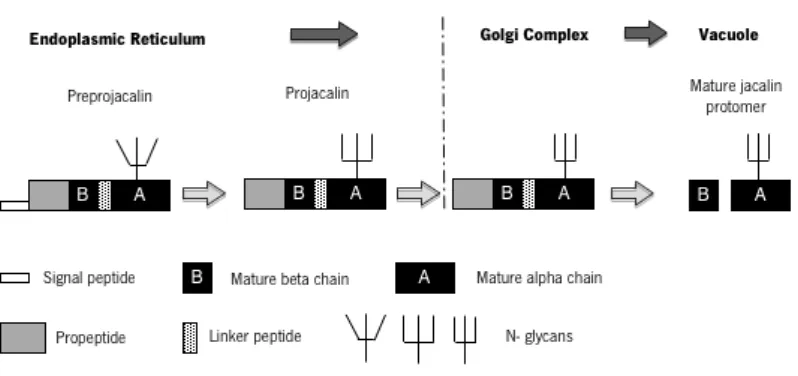
Figure 2. Schematic representation of molecular structure, biosynthesis and processing of
“galactose-specific” jacalin-related lectins (adapted from [4]). ... 7
Figure 3. Effect of different concentrations of n- frutalin and r- frutalin on HeLa cells proliferation at 24
and 48 h [33]. ... 12
Figure 4. Plant lectin-induced apoptosis was mainly mediated by intrinsic pathway (or mitochondrial
pathway) and/or extrinsic pathway (or the death receptor pathway) [46]. ... 14
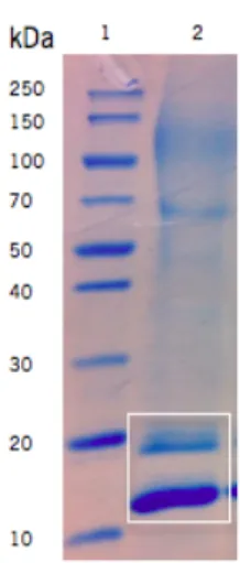
Figure 5. Expression of frutalin in P. pastoris. Analysis supernatants from methanol-induced cultures in a
12% SDS-PAGE stained with Coomassie. Legend: 1, molecular weight standards; 2, frutalin. ... 29
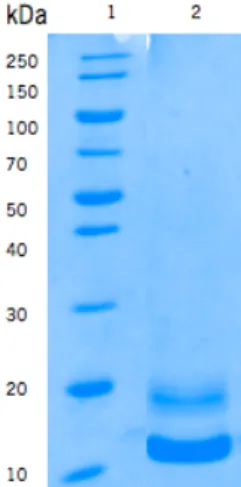
Figure 6. Purification of frutalin expressed in P. pastoris. Analyze of frutalin samples obtained from SEC
purification by Coomassie blue stained 12% SDS-PAGE. Legend: 1, molecular weight standards; 2, frutalin. ... 30
Figure 7. Analysis of recombinant frutalin samples obtained from HIC purification by Coomassie (assay 1)
and silver (assay 2) stained 12% SDS-PAGE. (A) Assay 1; Legend: 1, molecular weight standards; 2, frutalin before purification; 3, flow-through sample; 4, washing sample; 5, washing sample using PBS (diluted 1:2); 6-7, eluted sample; 8, washing column using NaOH; (B) Assay 2; Legend: 1, molecular weight standards; 2, FTL before purification; 3, FTL (dilution 1:2 with PBS with 6 M NaCl); 4, flow-through sample; 5, washing sample; 6, eluted sample with PBS with 1 M NaCl; 7-8, eluted samples with PBS with 0.5 M NaCl; 9-11, eluted samples with PBS with 0.25 M NaCl; 12-14, eluted samples with PBS; 15-18, eluted samples with 50 mM Tris (pH 10). ... 31
Figure 8. Analysis of Fh8-frutalin samples (A) obtained from nickel affinity purification (IMAC) and (B)
after dialysis process by Coomassie stained 12% SDS-PAGE. (A) Legend: 1, molecular weight standards; 2, supernatant sample; 3, flow-through sample; 4-6, washing samples; 7-12, eluted samples. (B) Legend: 1, molecular weight standards; 2-5, Fh8-frutalin samples. ... 32
Figure 9. Yeast expression of human procaspase-3 (A), procaspase-6 (B) and procaspase-7 (C) was
monoclonal antibodies, respectively; Pgk1p was used as loading control. Immunoblots were developed by enhanced chemiluminescence. ... 33
Figure 10. Growth curves for yeast expressing human procaspase-3 (A), -6 (B) and -7 (C) and control
yeast (pGALL) assessed by optical density (at 600 nm). Data represent mean ± standard error (S.E.M) of two independent experiments; values obtained not statistically different from yeast expressing control empty vector (P >0.05). ... 34
Figure 11. (A) Effect of PAC-1 on the growth of yeast expressing procaspase-3 (B) Effect of the compound
1541 on the growth of yeast expressing procaspase-6 (C) Effect of PAC-1 on the growth of yeast expressing procaspase-7. Yeast cells expressing executioner procaspases were incubated in the presence of 10, 25, 50 and 100 µM activator compound or DMSO only, for 48 h incubation. For each culture, the percentage of drug-induced growth inhibition was estimated considering 100% growth the number of CFU obtained with control yeast (pGALL). Data represent mean ± S.E.M (n=3 for procaspase-3; n=2 for procaspase-6 and n=4 for procaspase-7); values significantly different from DMSO only (*P<0.05; **P<0.01; ***P<0.001). ... 35
Figure 12. Effects of different concentrations of frutalin on the growth of control yeast and yeast
expressing p53, p63, p73 or DN. For each culture, the percentage of frutalin-induced growth inhibition was estimated considering 100% growth the number of CFU obtained with PBS only. Data represent mean ± S.E.M of three independent experiments; values obtained not statistically different (P> 0.05). ... 36
Figure 13. Effects of different concentrations of frutalin on the growth of control yeast (empty vector) and
yeast expressing procaspase-3, procaspase -6 and procaspase-7, for 42 h treatment. For each culture, the percentage of frutalin-induced growth inhibition was estimated considering 100% growth the number of CFU obtained with PBS only. Data represent mean ± S.E.M of three independent experiments; values obtained with yeast expressing procaspase-3 treated with frutalin 0.1 µM and frutalin 0.1 µM significantly different from PBS (*P< 0.05; ** P<0.01). ... 37
Figure 14. Procaspase activity in yeast after treatment with frutalin. Flow cytometric analysis using the
FL1 detection of FITC fluorescence of cells treated with PBS (control) and cells treated with 0.01 µM of frutalin. Data represent one experiment with duplicates. Legend: (A) Empty-vector (B) Procaspase-3 (C) Procaspase-6 (D) Procaspase-7. ... 38
xv
Figure 15. Effect of different concentrations of frutalin on proliferation of HCT116 p53+/+ and HCT116
p53-/- cells at 48 h. Data represent mean ± S.E.M of two independent experiments; values obtained
xvii
List of Tables
Table 1. Major historic events of plant lectins (adapted from [5]) ... 3
Table 2. Advantages and disadvantages of E.coli and P. pastoris expression systems (adapted from [1]) 10
Table 3. Buffers recipe for HIC purification assays ... 20 Table 4. Composition of SDS polyacrylamide gels ... 22
xix
Abbreviations
BPH Benign prostate hyperplasia
BMM Minimal methanol medium
BMG Minimal glycerol medium
BSA Bovine serum albumin
CFU Colony-Forming Unit
Cyt c Cytochrome c
Con A Concanavalin A
CV Column volumes
DISC Death inducing signalling complex
DNA Deoxyribonucleic acid
DR Death receptor
EDTA Ethylenediamine tetraacetic acid
FBS Fetal bovine serum
Fh8 Fasciola hepatica 8-kDa protein
FITC-VAD-fmk Fluorescein isothiocyanate conjugate of z-VAD-fmk
FTL Frutalin
gJRLs Galactose-specific jacalin-related lectins
HeLa Human cervical cancer cells
HIC Hydrophobic interaction chromatography
His Histidine
IMAC-Ni Immobilized nickel ion affinity chromatography
IPTG Isopropyl β-D-1-thiogalactopyranoside
kDa kilodalton
KML-C Korean mistletoe
LB Luria-Bertani broth medium
MEC Mucoepidermoid carcinoma
Met Methionine
mJRLs Mannose-specific jacalin-related lectins
MOMP Mitochondrial outer membrane permeabilization
N Asparagine
OD Optical density
PBS Phosphate Buffered Saline
PCL Polygonatum cyrtonema lectin
PMSF Phenylmethanesulfonyl fluoride
POL Polygonatum odoratum lectin
ROS Reactive oxygen species
S Serine
SDS-PAGE Sodium dodecyl sulfate polyacrylamide gel electrophoresis
SEC Size-exclusion chromatography
SFL Lectin from Sophora flavescens
S.E.M. Standard error
SRB Sulfurhodamine B
T Threonine
TBS Tris- buffered saline
TCA Trichloroacetic acid
UEA-I Ulex europeans I
1
Chapter 1
Chapter 1|General Introduction
3
1.1 Plant lectins
Lectins are a heterogeneous group of proteins with at least one non-catalytic domain that selectively recognizes, and reversibly binds to, specific free sugars or glycans present on glycoproteins and glycolipids without altering the structure of the carbohydrates [1]. These are distributed in nature namely in plants, viruses, animals, bacteria, vertebrates and invertebrates [1, 2]. The first description of plant lectins dates back to the late nineteenth century when Peter Stillmark initiated the study of proteins derived from plants that have demonstrated the ability to agglutinate erythrocytes [3, 4]. In 1988, in his doctoral thesis, he described the properties of agglutination of proteins that had been extracted and partially purified from castor seeds (Ricinus communis) denominated ricin. Table 1 describes the main events, over time, in relation to plant lectins.
Table 1. Major historic events of plant lectins (adapted from [5])
Year Description
1888 Detection of erythrocyte agglutination by a toxic protein fraction from castor seeds (termed ricin) (P. Stillmark) 1891 Toxic plant agglutinins applied as model antigens (P. Ehrlich)
1898 Introduction of the term “haemagglutinin” or “phytohaemagglutinin” for plant proteins that agglutinate red blood cells (M. Elfstrand) 1907/1909 Detection of non-toxic agglutinins in plants, of their nature as proteins and of “deagglutination” of erythrocytes by hog gastric mucin (K. Landsteiner, H. Raubitschek)
1919 Crystallization of a globulin from jack bean, concanavalin A (J. B. Sumner) 1935/1936 Concanavalin A identified as jack bean haemagglutinin (J. B. Sumner)
1947/1948 Detection of plant agglutinins specific for the human histo-blood group A (W. C. Boyd; K. O. Renkonen) 1952 Use of lectins and glycosidases to prove that blood group antigens are sugars and to deduce the structures of the antigens (W. M. Watkins, W. T. J. Morgan) 1954 Introduction of the term “lectin” for plant agglutinins (W. C. Boyd)
1960 Detection of the mitogenic potency of lectins toward lymphocytes (P. C. Nowell)
1963 Introduction of affinity chromatography for the isolation of lectins (I. J. Goldstein, B. B. L. Agrawal) 1972 Determination of the amino acid sequence and the three-dimensional structure of a lectin, concanavalin A (G. M. Edelman, K. O. Hardman, C. F. Ainsworth et al.) 1983 Detection of the insecticidal action of a plant lectin (L. L. Murdock)
1984 Isolation of lectins from tumors (H.-J. Gabius; R. Lotan, A. Raz) 1989 Detection of the fungicidal action of a plant lectin (W. J. Peumans)
1995 Structural analysis of a lectin-ligand complex in solution by NMR spectroscopy (J. Barbero and colleagues) 2001-2005 Development of glycan/lectin microarrays for specificity analysis of lectins/ structural analysis of glycans and glycoproteomics (various laboratories worldwide)
Chapter 1|General Introduction
The proteins that have ability to agglutinate other cells began to be called agglutinin and phytohemagglutinins [4, 6]. Later, in 1954, Boyd proposed the name lectin (derived from the Latin “legere”, which means "to select", "who chooses") due to its ability to agglutinate erythrocytes [2, 3, 5]. Although nowadays the term lectins is the most commonly used, the term hemagglutinins and agglutinins can also be used to denominate them. This denomination is considered by some authors as the most accurate, since it refers to the ability of the proteins to agglutinate erythrocytes and other cell types [3, 4].
Lectins were initially defined as a group of “carbohydrate-binding proteins of non-immune origin that agglutinate cells and/or precipitate glycoconjugates”. This definition has been accepted, albeit with limitations, by the Nomenclature Committee of the International Union of Biochemistry [4]. One limitation of this definition is that excludes many lectins. This is because this definition only refers to multivalent carbohydrate-binding proteins excluding proteins which have a single carbohydrate-binding domain, which, do not have the ability to agglutinate cells or precipitate glycoconjugates [4]. In 1995, Peumans and Van Damme proposed a new definition for lectins: “all plant proteins that possess at least one noncatalytic domain that binds reversibly to a specific mono- or oligosaccharide” [3]. The definition that has been considered the most appropriated is that lectins are heterogeneous group of proteins with at least one non-catalytic domain that selectively recognizes, and reversibly binds to, specific free sugars or glycans present on glycoproteins and glycolipids without altering the structure of the carbohydrates [1].
Thereafter it was proposed a subdivision of lectins on “merolectins”, “hololectins”, “chimerolectins” and “superlectins” (Figure 1) according to the overall structure of the mature lectins [4].
Figure 1. Schematic representation of merolectin, hololectin, chimerolectin and superlectin (adapted from [4]).
Merolectins only have a single domain binding to carbohydrates. Therefore, they are unable to agglutinate cells and precipitate glycoconjugates. Hololectins have two or more carbohydrate-binding
Chapter 1|General Introduction
5
or with a similar structure. Hololectins are di- or multivalent and unlike merolectins have the ability to agglutinate cells and/or precipitated glycoconjugates. Chimerolectins exhibit one or more carbohydrate binding domain(s) tandemly arrayed to an independent domain that can have a well-defined enzyme activity or other biological activity. This activity should be independent of carbohydrate binding domains, and the number of these domains determines the behavior of these lectins. Thus, if they only have one binding domain, they behave as merolectins, whereas when they have more than one domain they behave as hololectins. Finally, superlectins are formed as hololectins with at least two carbohydrate binding domains. However, unlike hololectins these domains are structurally and functionally different, and can recognize different sugars structures [3, 4].
Lectins widely differ from each other relatively to their biochemical/physicochemical properties, affinity for carbohydrate, molecular structure and biological activities [3]. Thus, Peumans and Van Damme [3] classified lectins into seven distinct families in accordance with the molecular structure and specificity of the carbohydrate. Therefore, lectins of a given family share common features between them. Within the seven families of lectins, four are numerous, including the legume lectins, the monocot mannose-binding lectins, the chitin-binding lectins, and the type 2 ribosome-inactivating proteins (RIPs). The jacalin-related lectins, the amaranthin lectin family and the Cucurbitaceae phloem lectins are considered small families [4]. Lastly, plant lectins have been divided into twelve different families based on carbohydrate binding domain (e.g. jacalins, proteins with legume lectins domains and ricin-B family) and according to molecular structures and amino-acid sequences [1].
1.1.1 Potential biomedical applications of plant lectins
As mentioned before, most lectins have the ability to agglutinate other cells. This effect is directly related to their ability to recognize and to bind reversibly to specific sugar structures, establishing a connection protein-carbohydrate. This feature allows lectins mediate a variety of biological processes, such as cell-cell communication, differentiation and cancer metastasis, as well as innate immune responses [1, 2].
Lectins are used as research tools in many scientific areas, namely in biochemistry, immunology, cell biology and even in the diagnosis and treatment of cancer [7].
The use of lectins in the diagnosis and treatment of cancer, a major disease of the XXI century, has contributed to the growing interest in the study of these proteins [8]. Lectins have contributed to the
Chapter 1|General Introduction
recognition of surface markers, mitogenic stimulation, cell adhesion and localization, increase of host immune defense, cytotoxicity and apoptosis [9–11].
Several studies have indicated that cancer cells have an aberrant glycosylation [12]. Once lectins bind to specific carbohydrates, they can be used to detect aberrant glycosylation. One study showed that the lectins Concanavalin A (Con A) and Ulex europeus I (UEA-I) can be used as histochemical biomarkers of mucoepidermoid carcinoma (MEC) in the most common malignant salivary tumor, which can be classified as low, intermediate or high grade. Con A binds to the neoplastic cells of the three degrees of severity. UEA-I is connected more strongly to cells of intermediate grade and more weakly to the other degrees [13].
Some lectins have showed the ability to induce cell recruitment, lymphocyte proliferation, and cytokine production and to possess immunomodulatory activity. Lectins (KML-C) from Korean mistletoe (Viscum album coloratum) showed differentially modulated macrophage-mediated immune responses. Moreover, they increased expression of various cytokines (IL-3, IL-23 and TNF-a) and reactive oxygen species (ROS) production [14].
1.1.2 Jacalin-related lectins (JRLs)
Jacalin-related lectins are considered a small family, when compared with other existing families of lectins. JRLs are divided into two subfamilies according to their specificity to carbohydrate. This family is subdivided into lectins which have an affinity to bind to the galactose (gJRLs) and mannose/maltose (mJRLs) [4, 15]. The “galactose-specific” and “mannose-specific” jacalin-related lectins beyond the differences on the specificity to carbohydrate, they also differ in their biosynthesis, processing, topogenesis and intracellular location [16].
Concerning to the biosynthesis differences between gJRLs and mJRLs, in the case of gJRLs it undergo proteolytic cleavage of the precursor polypeptide, resulting in two polypeptide chains, a heavy chain (α) and a light chain (β) of 133 and 20 amino acids, respectively [4, 15, 16]. Comparatively, the mJRLs does not undergo proteolytic modification, consisting only by uncleaved protomers with approximately 150 amino acids [15, 16].
There are evidence that the mannose-specific jacalin-related proteins are located in the cytoplasm, where they are synthesized [16]. These are not subject to any post-translational modifications and the mature polypeptides of the mJRLs correspond to the entire open reading frame of their corresponding
Chapter 1|General Introduction
7
genes [4, 16]. As a cleavage of protomer has not been found, the mJRLs have an extra loop, which makes the binding site inaccessible to galactose [17].
Jacalin, a “galactose-specific” jacalin-related lectin is synthesized in the endoplasmic reticulum [16] as preprojacalin consisting of a signal peptide of 21 amino acid residues followed by a propeptide of 39 amino acid residues, aβ chain with 20 amino acid residues, a linker tetrapeptide (TSSN), and lastly, a α chain with 133 amino acid residues. Its co-translational processing involves the removal of a signal peptide and its partial glycosylation, given rise to the propeptide that is transported to the Golgi complex (Figure 2) [4, 16, 17]. Subsequently, the propeptide is cleaved at three different locations, with the removal of the terminal propeptide and of the linker tetrapeptide. Thus, the mature jacalin is constituted by ta N-terminalβchain and a C-terminalαchain with 20 and 133 amino acid residues, respectively, being located in the vacuole. It is possible that its processing be similar to other jacalin lectins from Artocarpus plant, as well as from plant Maclura pomifera [4, 16].
Figure 2. Schematic representation of molecular structure, biosynthesis and processing of “galactose-specific”
jacalin-related lectins (adapted from [4]).
1.2 The frutalin lectin
Frutalin is a lectin isolated from the seeds of the Artocarpus incisa plant, commonly known as breadfruit. This plant belongs to the Moracae family, and it can be found in areas with a humid tropical climate. In Brazil, it is possible to find two different types of this plant, the apyrena (non-seminífera), without seeds, and the one that has seeds (seminífera) [18].
The frutalin is a tetrameric partially Nglycosylated protein which specifically recognizes αD -galactose residues [18]. It belongs to the jacalin-related lectins family, specifically to the subfamily “galactose-specific” jacalin-related lectins. Frutalin has many structural and functional similarities with
Chapter 1|General Introduction
jacalin, found in Artocarpus integrifolia plant that belongs to the same family as Artocarpus Incisa, Moracae family [18, 19]. As previously described, frutalin is a tetrameric molecule of approximately 48-49 kDa, consisting of four monomers linked by non-covalent bonds. Each monomer consists on a heavy chain (α) of 133 amino acids non-covalently bound to a light chain (β) of 20 amino acids. Frutalin presents predominantly a β sheet conformation and contains four binding sites for D-galactose, being therefore considered, according to its structure, a "hololectin" [17, 18, 20].
Immunologic studies showed that frutalin is a potent mitogenic activator of human lymphocytes and it has the ability to induce in vivo and in vitro neutrophil migration [19, 21].
1.3 Heterologous expression of frutalin
The use of recombinant lectins as potential therapeutic and diagnostic agents makes important to reduce the heterogeneity of properties resulting from the different lectins isolated from natural sources. In fact, lectins may have different isoforms that can lead to different specificities to carbohydrates. The heterologous expression and production of recombinant proteins reveals to be an important to overcome the limitations mentioned above. Additionally, the purification of lectins from its natural source can be a time-consuming process and with a low yield. This is also other limitation that can be overcome by the expression and production of these recombinant proteins. Additionally, through the heterologous expression and production of lectins is possible to obtain larger amounts of lectins with high levels of purity and with defined amino acid sequence, in a shorter period of time, when compared to the native lectins system. Therefore the properties of lectins are also more controlled [1, 22]. Thus, the use of heterologous expression systems have considerable advantages for the production of proteins of pharmaceutical interest [23], particularly for the study of cellular processes [24].
1.3.1
Pichia pastoris
andE. coli
as expression systemThe ideal system should be able to express proteins with the lowest possible cost and as authentic as possible [25]. The expression system using bacterium E. coli and the yeast P. pastoris are, currently, the two most used expression systems [26, 27].
Initially, E. coli was the most used production system, due to extensive knowledge on its genetics, easy of manipulation, low generation time and high product yields [23, 28]. However, have significant limitations to the expression of recombinant protein using E. coli as an expression host. E. coli is unable to
Chapter 1|General Introduction
9
modifications can affect the bioactivity, function, structure, solubility, stability, half-life, protease resistance, and compartmentalization of the functional proteins [27]. Despite of this, many proteins (e.g. IFN -α, -β and -γ, interleukin-2) that are non-glycosylated after expression in E. coli (glycosylated in their natural human forms) retain their biological activity. Another disadvantage of E. coli as an expression system is that most of proteins are produced in inclusion bodies and are often inactive, insoluble and require refolding [26].
New strategies have been developed in order to overcome some limitations, such as the solubility of the proteins expressed in E. coli. One example of a reliant strategy was the fusion proteins technology. Fusion partners (e.g. Fh8 tag), when fused with the protein of interest, can increase production yields, protein solubility and can be used (e.g. His tag) in the purification process [27, 29]. These limitations, in association with the high number and complexity of recombinant proteins to be expressed, led to the use of other hosts besides the E. coli in heterologous expression systems. Although the limitations exposed above E. coli remains one of the preferred hosts (host for recombinant protein expression) [30].
Pichia pastoris, the methylotrophic yeast, can overcome some of the limitations presented by the E. coli expression system. It is capable of post-translational modifications, namely to carry out proteins glycosylation’s and to produce disulfide bonds [26]. These are major advantages of the P. pastoris over E.coli. Furthermore, production yields can be higher than E. coli. However, it may be necessary to optimize the growth conditions [26, 27]. Another advantage of the methylotrophic P. pastoris when compared to other organisms including E. coli, is its ability to secrete proteins efficiently. In spite of this, as a host for heterologous expression, Pichia pastoris also present some disadvantages. For example, P. pastoris can not produce proteins that require the assistance of folding chaperones. [26]. Table 2 describes the main advantages and disadvantages of the heterologous expression system E. coli and P. pastoris.
Chapter 1|General Introduction
Table 2. Advantages and disadvantages of E. coli and P. pastoris expression systems (adapted from [1])
Host system Advantages Disadvantages
E. coli Rapid expression High yields
Genetically manipulate Inexpensive
Mass production fast and cost-effective
Proteins with disulfide bonds difficult to express
Produce unglycosylated proteins Proteins produced with endotoxins Proteins produced as inclusion bodies, are inactive and usually require refolding. P. pastoris High yield
Stable production strains Durability
Cost effective High density growth High productivity Stability proteins
Rapid growth in chemically defined media
Product processing similar to mammalian cells
Can handle S–S rich proteins, assist protein folding and glycosylate proteins
Fermentation require for very high yield Growth conditions may require optimization
Refolding may be required
1.3.2 Recombinant frutalin
Recombinant frutalin was expressed in a recombinant form in yeast Pichia pastoris and in bacteria Escherichia coli [29, 31, 32]. However, recombinant frutalin expressed in P. pastoris and E. coli exhibits differences between them. One major difference between them is the glycosylation. The frutalin expressed in Pichia pastoris is partially N-glycosylated whereas the frutalin expressed in E. coli is non-glycosylated. Furthermore, frutalin in E. coli showed ability to agglutinate rabbit erythrocytes contrary to frutalin in P.
Chapter 1|General Introduction
11
Additionally, differences between recombinant and native frutalin can also be found. In fact, during the processing in P. pastoris and in E.coli, the cleavage of linker tetrapeptide (T-S-S-N) does not occur in recombinant frutalin. Therefore, in recombinant frutalin, the α and β chains are not independent, being expressed as a single chain protein. Although the differences between recombinant and native frutalin, the ability to bind to Me-α–galactose was retained by the recombinant frutalin but with lesser affinity than native frutalin [31, 32].
Recombinant frutalin expressed in E. coli was mainly produced as insoluble protein [32] and in order to increase the amount of frutalin, a fusion partners was used to increase production yields, promote its solubility and help on its purification. The fusion of frutalin with the Fh8 tag resulted in an increase of soluble expression and His6 tag was used for help on its purification [29, 32].
1.3.3 Potential biomedical applications of recombinant frutalin
Oliveira et al. [12, 33] demonstrated the potentiality of recombinant frutalin expressed in P. pastoris as agent for the treatment and diagnosis of cancer.
Several studies showed that lectins have a promising capacity as tumor biomarkers. Recently, through immunohistochemical assays, frutalin was tested in human tissues of prostate carcinoma and benign prostate hyperplasia (BPH) as potential tumor biomarker. This study compared if native frutalin (from Artocarpus incisa) and recombinant frutalin were able to detect alterations in human prostate tissues. The native frutalin (n-frutalin) bound to all cases studied of prostate carcinoma and to benign prostate hyperplasia (BPH), 20 and 25 cases, respectively. Regarding to recombinant frutalin (r-frutalin), it bound to 14 of the 20 studied cases of prostate carcinoma and did not bind to none of the 25 studied cases of BPH. Recombinant frutalin showed that recognize specifically malignant cells. These differences can be due to distinct carbohydrate binding affinity. Thus, Oliveira et al. [12] concluded that native and recombinant frutalin could be used as histochemical biomarkers for the prostate cancer.
In another study by Oliveira et al. [33], it was found a cytotoxic effect of frutalin (native and recombinant) in HeLa cervical cancer cells (Figure 3). In fact, recombinant and native frutalin had similar effects in inhibiting the proliferation of HeLa cells (GI50 for 24 h ≈ 100 µg/mL), an effect that showed to be time- and dose-dependent.
Chapter 1|General Introduction
Figure 3. Effect of different concentrations of n- frutalin and r- frutalin on HeLa cells proliferation at 24 and 48 h
[33].
This same study showed that the inhibition of proliferation occur due to apoptotic cell death. It was also analysed the cellular localization of each lectin studied. Interestingly, it was found that the recombinant frutalin was mainly located in the nucleus, whereas native frutalin was mainly located in the perinuclear region. This study therefore revealed a potential therapeutic application of native and recombinant frutalin against cancer [33].
1.4 Plant lectin-induced apoptosis
Apoptosis is a programmed cell death process, involved in the control of cellular proliferation and DNA damage [11]. This is characterized by typical morphological cell changes like nuclear fragmentation, chromosomal fragmentation, membrane blebbing and nuclear condensation [24, 34].
Apoptosis can be induced through several molecular pathways. The extrinsic and intrinsic pathways are the most relevant pathways in apoptosis [35]. The induction of these two apoptotic signaling pathways leads to the activation of the executioner caspases that results in the cleavage of a subset of proteins resulting in the biochemical and cellular changes typical of apoptosis [36, 37]. Caspases are a family of cysteine- dependent aspartate-specific proteases that can be divided in pro-apoptotic and pro-inflammatory subfamilies. The pro-apoptotic subfamily can be divided in activator or initiator caspases (caspase-2, -8, -9, -10 and -12) and executioner or effector caspases (caspase-3, -6 and -7) that are activated by the initiator caspases. These are synthetized as inactive preforms and stored as procaspases that under certain conditions are activated by proteolysis and become able to cleave a large set of substrates [38, 39].
Chapter 1|General Introduction
13
KILLER/DR5. The activation of receptors through binding of specific ligand leads to their trimerization and consequent clustering of the intracellular death domain that results in recruitment of FAS associated with a death domain (FADD), which in turn recruits caspase-8 allowing the formation of the death-inducing-signaling-complex (DISC) [36, 37].
The intrinsic pathway (or mitochondrial pathway) involves the mitochondrial outer membrane permeabilization (MOMP), which allows a release to cytosol of pro-apoptotic proteins such as cytochrome C (cyt c) and apoptosis-inducing factor (AIF). The cytochrome c plays an important role in mitochondria-dependent apoptotic death. Upon its release it combines to the apoptosis protease-activating factor 1 (APAF-1) and the initiator caspase-9 inducing the formation of a large complex, the apoptosome, which promotes the proteolytic maturation of caspase-9. With active caspase-9, the effector caspases (caspase-3, -6 and -7) are cleaved and activated, which in turn leads to the apoptosis [36, 37].
The p53 family protein, has an important role in induction of apoptosis, namely in the intrinsic and extrinsic pathway. The p53 family protein may up-regulate several proteins involved in the intrinsic pathway. The Bcl-2 family of proteins are involved in the release of cytochrome c from the mitochondria [35]. The pro-apoptotic proteins from Bcl-2 family (e.g. Bax, Bid, Noxa, Puma) may up-regulate by p53 that results in MOMP, which in turn activate caspases leading to apoptosis [24, 36, 37]. The p53 and p73 induce the expression of p53AIP1 (p53-apoptosis inducing protein 1), which localizes to the mitochondria where it interacts with Bcl-2 to facilitate the release of cyt c and the consequent apoptosis induction through the intrinsic pathway. The p53 can also directly induce the expression of caspase-6 and Apaf-1. The p53 can upregulating two cell death receptors, KILLER/DR5 and Fas and the ligand for Fas, FasL and thus, induce the apoptosis through extrinsic pathway [35, 40].
Some of the proteins mentioned above that are involved in apoptosis such as initiator or effector caspases and p53 are lost in many cancers by inactivation or mutation of these proteins [41]. As such, induction of apoptosis by activation of these proteins is a defense against cancer and therefore an important target for cancer therapy [42–44]. Moreover, it is an important cellular homeostasis mechanism that ensures the correct development and function of multicellular organisms [45].
Different lectins induce apoptosis in different human tumor cells. The effects of plant lectins in human tumor cells are dependent of the sort of lectin and cells lineage. Their effect is time-dosage dependent. For example, Polygonatum odoratum lectin (POL), a mannose-binding lectin induces apoptosis
Chapter 1|General Introduction
through the death-receptor apoptotic pathway by increasing the levels of FasL and Fas-Associated protein with death domain (FADD) proteins that leads to the caspase -8 activation. Moreover, POL treatment leads to cytochrome c release and subsequent activations of caspase -9 and caspase -3. Thus, POL induces apoptosis in a caspase-dependent manner [46]. Polygonatum cyrtonema lectin (PCL), a mannose/sialic acid-binding lectin induces, simultaneously, apoptosis and autophagy in human melanoma A375 cells. PCL binds to the mannose-containing receptor of human melanoma cell surface and it is internalized and localized on the mitochondria. PCL induced mitochondria to generate massiveROS production promotes the release of cytochrome c and activates the p38 and p53. Thus, PCL induces apoptosis via a mitochondrial ROS-p38-p53 pathway [47]. SFL, a mannose-binding lectin from Sophora flavescens, was also shown to induce apoptosis in human cervical cancer in a typical caspase-dependent manner by death receptor pathway [42]. Likewise, the lectin ricin induces apoptosis through activation of caspase-8 and subsequent activation of caspase -3 and -7 [48]. The Figure 4 demonstrates the plant lectin-induced apoptotic mechanisms implicated in intrinsic and extrinsic pathways.
Figure 4. Plant lectin-induced apoptosis was mainly mediated by intrinsic pathway (or mitochondrial pathway)
Chapter 1|General Introduction
15
1.5
Saccharomyces cerevisiae
as a model organism for molecular and
pharmacological studies of human proteins
Model organisms have widely contributed to the knowledge of human cellular processes and proteins [24].
In fact, the use of Saccharomyces cerevisiae as a model organism has greatly contributed to the study of fields as diverse as cell metabolism, DNA replication, recombination, cell cycle, cell death, protein folding, trafficking, and organelle biogenesis [24]. S. cerevisiae was the first eukaryote to have its genome fully sequenced [49]. These data are easily accessible on the online dataset for yeast with genetic interactions, transcriptional changes, protein interactions, and localization information [24, 50, 51]. Additionally, it can be easily genetically manipulated, and there is a lot of information about their molecular biology and physiology. Another advantage of this yeast as a model organism is its short generation time, widely easy to cultivate and maintain [23].
Several cellular processes present in S. cerevisiae, such as cell cycle progression, protein secretion and apoptosis, are similar to those present in humans, thus allowing the understanding of the biological mechanism involved in human diseases. In fact, many of knowledge obtained from yeast has been transposed to the human cells [49, 52].
To establish these models for the study of heterologous proteins different approaches can be used. If the gene encoding the human protein is conserved in yeast, it is possible to study directly the function of the protein in this organism. However, if yeast does not possess orthologous of the human gene, it is necessary its heterologous expression in this organism (called "humanized yeast"). The heterologous expression in yeast of proteins involved in human diseases, has provided important information regarding the functions of these proteins (e.g. p53 and caspase family members) and about the pathobiology underlying human diseases, such as cancer [24].
Chapter 1|General Introduction
1.6 Aims of the work
In a previous study, recombinant frutalin expressed in Pichia pastoris and purified by size-exclusion chromatography showed a strong anti-proliferative effect on HeLa cervical tumor cells. Moreover, it was shown that this effect was due to the induction of cell dead by apoptosis. However, the molecular mechanism involved in apoptosis induction triggered by this recombinant frutalin was not studied.
Thus, the main goal of this work was to elucidate the molecular mechanism involved in apoptosis induction by the recombinant frutalin expressed in Pichia pastoris and purified by size-exclusion chromatography.
Furthermore, it was also aim of this work to test other frutalin molecules obtained with another expression system (Escherichia coli) or/and another purification methodology (hydrophobic interaction chromatography). These other recombinant forms of frutalin were produced with a more straightforward production and purification process but their anti-proliferative activity in human tumor cell lines was never tested before.
Based on this, the specific aims of this work were to:
• Study the modulatory effect of frutalin expressed in P. pastoris and purified by size-exclusion chromatography on main regulatory proteins of apoptosis, such as the executioner members of the caspase family (caspases-3, -6 and -7) and the p53 family members using yeast- based assays;
• Study the effect of frutalin expressed in P. pastoris and purified by hydrophobic interaction chromatographyin the proliferation of human tumor cell lines;
• Study the effect of recombinant frutalin obtained from E. coli and purified using affinity chromatography with nickel in the proliferation of human tumor cell lines.
17
Chapter 2
Chapter 2|Materials and Methods
19
2.1 Systems of production and purification of recombinant proteins
2.1.1 Production of recombinant frutalin expressed in
Pichia pastoris
Pichia pastoris KM71H/ pPICZα/ frutalin cells [31], were grown in 100 mL of buffered minimal glycerol medium (BMG) with 1% glycerol (AppliChem), 100 mM potassium phosphate (pH 6.0; AppliChem), 1.34% yeast nitrogen base with ammonium sulfate and without amino acids (Difco) and 4 × 10 -5 biotin (Sigma), overnight, at 30 °C, with continuous shaking (200 rpm). The next day, cells were
harvested by centrifugation at 4000 g for 10 minutes at room temperature and the resulting cell pellets were resuspended in 50 mL of fresh buffered minimal methanol medium (BMM). BMM medium has the same composition of BMG medium but glycerol is replaced with 0.5% (w/v) methanol (Biochemicals). The induction of cultures was carried out in 500 mL baffled shake flask and covered with two layers of sterile gauze. Inducible cultures were incubated at 15 °C, with continuous shaking (200 rpm) and fresh methanol was added daily, during the 4 days of induction, to a final concentration of 0.5% (v/v). After the induction period, the supernatants were separated from cell pellets by centrifugation at 4000 g, 4 °C for 10 minutes. To precipitate salts, the pH of the supernatants was increased to 7.5 by adding 10 N NaOH and removed by centrifugation twice at 4000 g, 10 minutes at 4 °C.
2.1.2 Purification of recombinant frutalin expressed in
Pichia pastoris
The supernatants were filtered through filters of 0.2 µm, concentrated and washed with Phosphate Buffered Saline (PBS; 137 mM NaCl, 2.7 mM KCl, 2 mM KH2PO4, 100 mM Na2HPO4) in 10 kDa Amicon
filters (Millipore) to a final volume of 1–1.5 mL. To confirm the presence of recombinant frutalin in the samples, these were analyzed by sodium dodecyl sulfate polyacrylamide gel electrophoresis (SDS-PAGE) and stained with Coomassie (described in section 2.1.5/2.1.5.1). After this, samples were purified by size-exclusion chromatography (SEC) or hydrophobic interaction chromatography (HIC). At the end of purification, samples were quantified by absorbance 280 nm, using NanoDrop 1000 (Thermo Scientific) based on molar extinction coefficient of frutalin (εfrutalin = 27390 M-1 cm-1).
2.1.2.1 Size-exclusion chromatography (SEC)
SEC is a chromatography technique that separates biomolecules according to differences in their size. In this work, a SephacrylTM S-200 HR column (GE Healthcare) was used in FPLC system (Pharmacia
Chapter 2|Materials and Methods
sample was filtered through 0.2 µm pore size filters and applied on the column at a flow rate of 1 mL/ min and elution occurs under the same conditions. Purified samples were collected and aliquot to be analyzed by SDS-PAGE (described in section 2.1.5/2.1.5.1). The samples containing pure recombinant frutalin were concentrated in 10 kDa Amicon filters (Millipore) and then stored at –20 °C.
2.1.2.2 Hydrophobic interaction chromatography (HIC)
HIC separates biomolecules according to the differences in their hydrophobicity. In this work, a prepacked Phenyl SepharoseTM 6 fast flow High Sub column (GE Healthcare) was used operated by a
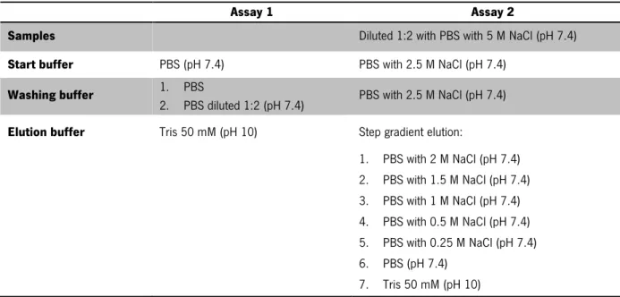
peristaltic pump (Pharmacia LKB pump) and the whole procedure of purification was performed at 4 °C. Table 3 presents the description of buffers used in this purification in order to optimize the elution conditions. In the first step, column was equilibrated with 8 column volumes (CV) of buffer (start buffer) with moderately high salt concentrations. Then, the sample was filtered through 0.2 µm pore size filters and applied on the column at a flow rate of 0.5 mL/min. Then, column was washed with 8 CV of washing buffer in order to remove all unbound proteins with a flow rate of 1 mL/min. For elution of proteins, an elution buffer with a flow rate of 0.5 mL/min was used. During purification, aliquots were collected and analyzed by SDS-PAGE (described in section 2.1.5/2.1.5.2). Finally, the column used in the procedure was cleaned with 2 CV of EDTA 100 mM, following with 2 CV of water, 2 CV of 6M Guanidinium in PBS solution and at least 2 CV of water. To store the column at 4 °C, 2 CV of EtOH 20% was used.
Table 3. Buffers recipe for HIC purification assays
Assay 1 Assay 2
Samples Diluted 1:2 with PBS with 5 M NaCl (pH 7.4)
Start buffer PBS (pH 7.4) PBS with 2.5 M NaCl (pH 7.4)
Washing buffer 1. PBS
2. PBS diluted 1:2 (pH 7.4) PBS with 2.5 M NaCl (pH 7.4)
Elution buffer Tris 50 mM (pH 10) Step gradient elution:
1. PBS with 2 M NaCl (pH 7.4) 2. PBS with 1.5 M NaCl (pH 7.4) 3. PBS with 1 M NaCl (pH 7.4) 4. PBS with 0.5 M NaCl (pH 7.4) 5. PBS with 0.25 M NaCl (pH 7.4) 6. PBS (pH 7.4) 7. Tris 50 mM (pH 10)
Chapter 2|Materials and Methods
21
2.5 mL of the sample was applied on the column and eluted with 3.5 mL PBS. After, samples were stored at –20 °C and the column was washed with water and stored in EtOH 20%.
2.1.3 Production of recombinant frutalin expressed in
E. coli
E. coli Rosseta (DE3) cells, transforming with pETM11 or pETMFh8 encoding His6-frutalin or
Fh8-frutalin [29], respectively, were grown overnight at 37 °C with continuous shaking (180 rpm) in25 mL LB medium (Applichem) supplemented with 30 mg/mL kanamycin (Sigma) and 10 mg/mL chloramphenicol (Applichem). The next day, four erlenmeyers were prepared with 250 mL of LB medium supplemented with 30 mg/mL kanamycin and 10 mg/mL chloramphenicol to make up 1 L culture. Thus, each of these erlenmeyers was inoculated with pre-culture so that the initial 0.02 optical density measured at 600 nm (OD600). After inoculation, cell cultures were incubated at 37 °C with continuous shaking (180 rpm) until
the OD600 reached a value between 0.4 - 0.6. After achieving this OD, the cell culture was maintained under stirring but at 18 °C. After 30 minutes, the expression of frutalin was induced by the addition of IPTG (Bio-Rad) to a final concentration of 0.2 mM and incubated overnight, at 18 °C with continuous shaking (180 rpm). Thereafter, cell pellet was recovered by centrifugation at 4500 rpm, for 15 min, at 4 °C and supernatant was discarded. Cell pellets were stored at –20 °C. Before and after the induction aliquots were recovered for analyze through SDS-PAGE (described in section 2.1.5/2.1.5.1).
Before the cells being purified (described in section 2.1.4) they were resuspended in 25 mL of lysis buffer (50 mM Tris pH 8.0 (JTBacker), 150 mM NaCl (AppliChem), 20 mM Imidazole (Sigma)) with 1 mM of PMSF (Sigma), 5 µL/mL DNase (Sigma) and 10 µL/mL lysozyme (Sigma). After this, cells were sonicated (Branson Sonifier 450, 6 minutes, duty cycle 50%, output 5) and then, the supernatant was harvested by centrifugation at 4 °C, 13000 g for 30 min. Supernatant was filtered through 0.45 µm pore size filters. Aliquots of total lysates and supernatant were collected for analyses by SDS-PAGE (described in section 2.1.5/2.1.5.1).
2.1.4 Purification of recombinant frutalin expressed in
E. coli
To purify His6-FTL and Fh8-FTL proteins immobilized nickel ion affinity chromatography (IMAC-Ni)
was performed. Therefore, HisTrap column (GE Healthcare) was used operated with peristaltic pump and all the procedure was performed at 4 °C. The column was equilibrated with 4-6 CV with a binding buffer (50 mM Tris pH 8.0, 150 mM NaCl, 20 mM Imidazole) with a flow rate of 1 mL/min. Then, the samples were applied to the column with a flow rate of 0.5 mL/min (in this stage the proteins with histidines bind to
Chapter 2|Materials and Methods
ligands, nickel ions). After this, the column was washed with 5-10 CV of washing buffer (50 mM Tris pH 8.0, 150 mM NaCl, 40 mM Imidazole) to remove unbound material with a flow rate of 1 mL/min. Then, for elution of the target protein an elution buffer with 50 mM Tris pH 8.0, 150 mM NaCl and 300 mM Imidazole was used. The purified samples of 5 mL were collected in 15 mL tubes and posteriorly analyzed by SDS-PAGE. To clean the column 2 CV of 70% EtOH, following 2 CV of distillate water were applied. Then, it was washed with 2 CV of 1M NaOH and at least 2 CV water. To store the column at 4 °C, 2 CV of 20% EtOH was applied.
After purification, it was necessary to perform the dialysis. Thus, samples were dialyzed in PBS buffer (pH 7.4), overnight, at 4 °C. These proteins were quantified by the Bradford assay with Bio-Rad protein reagent (Bio-Rad). Therefore, in 96-well plates, it was loaded 10 µL/well of protein sample with 200 µL of BioRad Protein reagent diluted 1:5. After 5-10 minutes, the absorbance at 595 nm was measured using the Biotech Synergy HT Microplate Reader. Bovine serum albumin (BSA) was used as standard.
2.1.5 SDS-PAGE
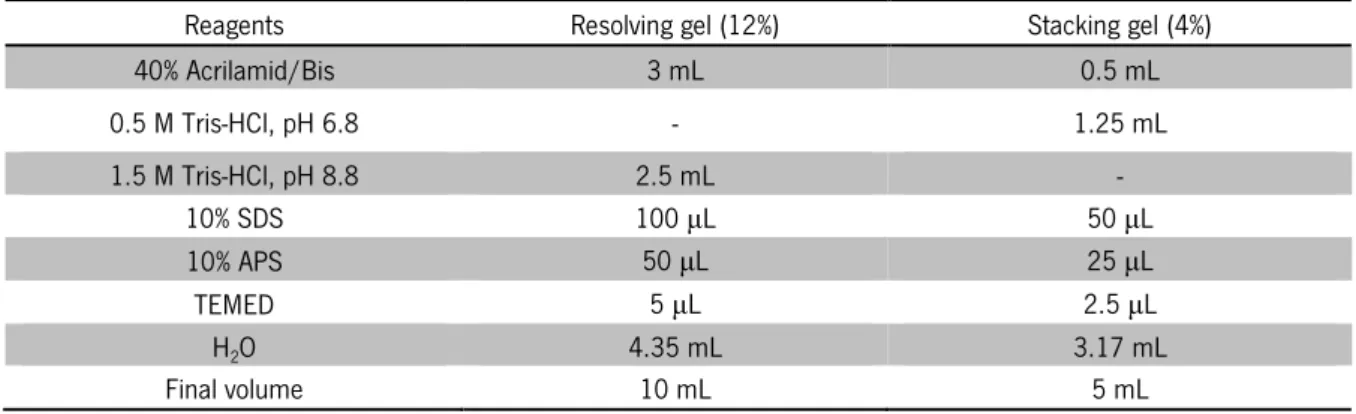
The electrophoresis under denaturing conditions, sodium dodecyl sulfate polyacrylamide gel electrophoresis (SDS-PAGE) was based on the Laemmli system [53]. The proteins were analyzed, according to this methodology, before, during and after the purification process. This system uses polyacrylamide gel with different acrylamide percentages to separate proteins based on the molecular weight. In this work, proteins were separated on 12% SDS polyacrylamide gels (SDS-PAGE). The protein molecular weight marker used in SDS-PAGE electrophoresis was PageRulerTM Unstained Broad Range Protein Ladder (5-250 kDa) from Fermentas. The composition of SDS-polyacrylamide gels is present in Table 4.
Table 4. Composition of SDS polyacrylamide gels
Reagents Resolving gel (12%) Stacking gel (4%)
40% Acrilamid/Bis 3 mL 0.5 mL 0.5 M Tris-HCl, pH 6.8 - 1.25 mL 1.5 M Tris-HCl, pH 8.8 2.5 mL - 10% SDS 100 µL 50 µL 10% APS 50 µL 25 µL TEMED 5 µL 2.5 µL HO 4.35 mL 3.17 mL
Chapter 2|Materials and Methods
23
The samples were treated with the loading sample buffer (0.15 mM Tris-HCl pH 6.8, 10% (w/v) SDS, 50% (v/v) Glycerol, 25% (w/v) β-mercaptoethanol and 0.01% (w/v) bromophenol blue) and heated at 100 °C for 5-10 min. The electrophoresis run was performed at constant voltage 120 V, using the running buffer (17.7 mM Tris, 0.25 % (w/v) SDS and 0.2 M Glycine).
2.1.5.1 Coomassie blue staining method
After electrophoresis, Coomassie Blue staining method was used to stain gels. Gels were placed in distilled water and heated in microwave for 30 seconds, then placed under constant agitation during 3 minutes. This step is repeated twice with fresh distilled water. Following this, gels were placed in Coomassie Blue solution, heated in microwave for 30 seconds and then placed under constant agitation during 30 minutes. Finally, Coomassie Blue solution was removed and distilled water was added for destaining of the gel. Coomassie Blue solution was prepared dissolving 60-80 mg the Coomassie Brilliant Blue G250 in 1 L of distilled water. This solution was stirring with a magnet for 2 to 4 hours. After this, add 3 mL of concentrated HCL 37% and solution is stored at environment temperature and protected from light.
2.1.5.2 Silver nitrate staining method
The polyacrylamide gels resulting of SDS-PAGE were stained with silver nitrate to detect proteins with lower concentrations. Thus, the gels were washed with 20% ethanol for 10 minutes and washed with distilled water for 10 minutes. Then, gels were sensitized with 0.2 g/L sodium thiosulfate for 2-5 minutes. After this, the gels were washed twice, for 20 seconds with distilled water. The gels were stained with 2.0 g/L nitrate prate solution for 30 minutes and then washed with distilled water five times for 10 seconds. To develop the color, a solution with 0.07% formaldehyde (37% formaldehyde), 30 g/L potassium carbonate anhydrous and 10 mg/L sodium thiosulfate was used. To stop the staining, gels were placed in a solution stop (50 g/L tris base and 2.5% acetic acid) and stored in distilled water.
2.2 Yeast-cell based phenotypic assay
2.2.1 Plasmids
The yeast expression vectors pLS89-(TRP1) encoding human wild-type (wt) p53, pRS314-(TRP1) encoding human TAp63α, ΔNp63α and pRS314-(TRP1) encoding human TAp73α with GAL1-10 inducible
Chapter 2|Materials and Methods
promoters, were used. Yeast expression vectors pGALL-(LEU2) encoding human procaspase-3, procaspase-6 and procaspase-7 under a GAL1-10 promoter were used.
2.2.2 Yeast strain, transformation and growth conditions
Saccharomyces cerevisiae strain CG379 (α ade5 his7-2leu2-112 trp1-289α ura3-52 [Kil-O], Yeast Genetic Stock Center, University of California, USA) [54] was transformed using the standard lithium acetate method [55]. Yeast cells were routinely grown in minimal selective medium with 2% (w/w) glucose (Sigma), 0.7 % (w/w) yeast nitrogen base without amino acids (Difco), and all the amino acids required for yeast growth (50 µg/mL; Sigma) except tryptophan (for wt p53, TAp63α, ΔNp63α and TAp73α), or except leucine (for procaspase-3, -6 and -7). For expression of human proteins, cells were diluted to 0.05 optical density (OD600) in selective induction medium, in which glucose was replaced by 2% (w/w) galactose (Sigma) and 2% (w/w) raffinose (Sigma). Yeast cells were then incubated at 30 °C, under continuous orbital shaking (200 rpm) for approximately 30 h (for wt p53, TAp63α, ΔNp63α and TAp73α) or 42 h (for procaspase-3, -6 and -7), corresponding to the time required by control yeast (transformed with the empty vectors pLS89, pRS314 or pGALL) to achieve 0.5 OD600. For growth curves experiments of yeast cells
expressing procaspase-3, procaspase-6 and procaspase-7, the growth of cultures was analyzed by OD600 up to 50 h.
2.2.3 Effects of frutalin and compounds on yeast cell growth
To analyze the effect of frutalin and of the known activator of caspase-3 and -7, PAC-1 [56] , and of the known activator of caspase-6, 1541 compound [57], on yeast cell growth, transformed cells were incubated in selective induction medium, as described in section 2.2.2, with different concentrations of recombinant frutalin expressed in P. pastoris and purified by SEC (0.01 µM, 0.1 µM, 1 µM and 2.5 µM) or PBS, or with 10, 25, 50 and 100 µM activator or with DMSO only. The time of incubation was the same described in section 2.2.2 according to the protein studied. Yeast cell growth was analyzed after 2 days incubation at 30 °C on Sabouraud Dextrose Agar plates (Liofilchem), by counting the number of colony-forming units (CFU). The percentage of growth was estimated considering as 100 % growth the number of CFU obtained with the control yeast (transformed with the empty vector).
Chapter 2|Materials and Methods
25
2.2.4 Western Blot Analysis
To analyze protein expression in yeast, samples were lysed in CellyticTM Y Cell Lysis Reagent (Sigma) in the presence of EDTA-free protease inhibitor cocktail (Boehringer Mannheim). Proteins were quantified with Bio-Rad Protein assay (Bio-Rad) and thereafter were electrophoresed on 12% SDS-PAGE (described in section 2.1.5). After this, proteins were transferred to a nitrocellulose membrane (GE Healthcare). Membranes were blocked with Tris- buffered saline (TBS), pH 7.4 (20 mM Tris-base, 150 mM NaCl,) containing 0.05% (v/v) Tween 20 and 5% (w/v) non-fat milk at room temperature for 2 h. For detection of procaspase-3, -6 and -7, membranes were probed with anti-procaspase-3 (1:2000, H-277, Santa Cruz Biotechnology), anti-procaspase-6 (1:200, Cell Signaling) and anti-procaspase-7 (1:500, Santa Cruz Biotechnology) monoclonal antibodies, respectively. For loading control, membranes were stripped and incubated with a mouse monoclonal anti-yeast phosphoglycerate kinase (Pgk1p) antibody (1:5000; Molecular Probes). Immunoblots were developed by enhanced chemiluminescence.
2.2.5 Caspase activation analysis
To evaluate the caspase activation by frutalin the fluorescent caspase inhibitor was used (CaspACE, FITC-VAD-FMK In Situ Marker; Promega). Yeast cells growth of control yeast and yeast expressing human procaspase-3, -6 and -7 was performed as described in 2.2.3. When the control yeast achieved 0.4 OD600, cells were harvested by centrifugation at 4000 rpm for 5 minutes and washed once
with 500 µL PBS. Then, to 1×10⁶ cells 500 µL PBS was added and collected by centrifugation at 7000
rpm for 7 minutes. Cells were resuspended in 100 µL PBS-solution containing 12.5 µM FITC-VAD-FMK and incubated for 1 h, at 30 °C in the dark (200 rpm). After this, 400 µL PBS was added to the suspension and the cells were harvested by centrifugation, washed and resuspended in 500 µL PBS. Fluorescence of twenty thousand cells per sample was analyzed using FACSCalibur flow cytometer (BD Biosciences), FL1 channel (Excitation/Emission=488/525 nm) and the CellQuest software (BD Biosciences).
2.3 Assays in human tumor cell lines
2.3.1 Growth conditions of cell culture
In assays with human tumor cell lines the human colon adenocarcinoma HCT116 cell line harboring a wt p53 form (HCT116 p53+/+) and its isogenic derivative, in which p53 has been knocked out
![Table 1. Major historic events of plant lectins (adapted from [5]) Year Description](https://thumb-eu.123doks.com/thumbv2/123dok_br/17767262.836472/24.892.110.822.519.1111/table-major-historic-events-plant-lectins-adapted-description.webp)
![Figure 1. Schematic representation of merolectin, hololectin, chimerolectin and superlectin (adapted from [4])](https://thumb-eu.123doks.com/thumbv2/123dok_br/17767262.836472/25.892.285.584.791.987/figure-schematic-representation-merolectin-hololectin-chimerolectin-superlectin-adapted.webp)
![Table 2. Advantages and disadvantages of E. coli and P. pastoris expression systems (adapted from [1])](https://thumb-eu.123doks.com/thumbv2/123dok_br/17767262.836472/31.892.99.803.159.866/table-advantages-disadvantages-coli-pastoris-expression-systems-adapted.webp)
![Figure 3. Effect of different concentrations of n- frutalin and r- frutalin on HeLa cells proliferation at 24 and 48 h [33]](https://thumb-eu.123doks.com/thumbv2/123dok_br/17767262.836472/33.892.298.568.130.343/figure-effect-different-concentrations-frutalin-frutalin-hela-proliferation.webp)
![Figure 4. Plant lectin-induced apoptosis was mainly mediated by intrinsic pathway (or mitochondrial pathway) and/or extrinsic pathway (or the death receptor pathway) [46]](https://thumb-eu.123doks.com/thumbv2/123dok_br/17767262.836472/35.892.192.688.645.972/figure-induced-apoptosis-mediated-intrinsic-mitochondrial-extrinsic-receptor.webp)