UNIVERSIDADE DO ALGARVE
Molecular structure and functional analysis of
runx3 in zebrafish
Brigite Sandra Nunes Simões Rodrigues
Tese para a obtenção do grau de Doutor em Ciências Biomédicas
Trabalho efectuado sob a orientação de: Professora Doutora M. Leonor Cancela
Professor Doutor Robert Kelsh Doutora Natércia Conceição
UNIVERSIDADE DO ALGARVE
Molecular structure and functional analysis of
runx3 in zebrafish
Brigite Sandra Nunes Simões Rodrigues
PhD fellowship SFRH/BD/38083/2007
Tese para a obtenção do grau de Doutor em Ciências Biomédicas
Trabalho efectuado sob a orientação de: Professora Doutora M. Leonor Cancela
Professor Doutor Robert Kelsh Doutora Natércia Conceição
runx3 in zebrafish
Declaração de autoria de trabalho
Declaro ser o autor deste trabalho, que é original e inédito. Autores e trabalhos consultados estão devidamente citados no texto e constam da listagem de referências incluída.
(Brigite Sandra Nunes Simões Rodrigues)
©Copyright
A Universidade do Algarve tem o direito, perpétuo e sem limites geográficos, de arquivar e publicar esta dissertação através de exemplares impressos reproduzidos em papel ou de forma digital, ou por qualquer outro meio conhecido ou que venha a ser inventado, e de a divulgar através de repositórios científicos e de admitir a sua cópia e distribuição com objetivos educacionais ou de investigação, não comerciais, desde que seja dado crédito ao autor e editor.
“Tudo o que um sonho precisa para ser realizado é alguém que acredite que ele possa ser realizado.”
Roberto Shinyashiki
I
Agradecimentos/Acknowledgments
I would like to send my sincerest gratitude to following people for their contribution to this work in different ways.
First, I want to thank to Professor Leonor Cancela and Professor Robert Kelsh, for accepting me as their PhD student, because without that first “yes” this project could not have been done. Thank you all the support and encouragement and essentially for always believed that I could do it, even when sometimes I didn’t believe it myself.
Second, I want to express my profound gratitude to Dr Natércia Conceição for accept to be my co-supervisor. More important than that, any words would never be enough to say THANK YOU for the long long long after dinner hours spending at my “side”, giving me the precious push that enable me to finish this project. A big thank you to Sofia and Gonçalo for sharing with me, for so long, their mum’s attention.
I would also like to thank all Lab members (present and past, in Portugal and in the UK) that have helped – directly and indirectly – to the execution of this project either with technical support or just with the right words in the right time, I am thankful to them all.
I want to say a special thanks to Cátia Marques, Cindy Fazenda, Íris Silva and Andreia Adrião for being the best bench colleagues ever!!! Also a special thanks to Iris, for her precious help with some figures presented here, to Cátia for her precious words and to Cindy that will always be my “little” girl.
Very special thanks to Marta Rafael, for always listening to my screams and to always been at a distance of a click.
Um muito obrigado à minha família pelo apoio incondicional, em especial ao meu marido e à minha filha que são maravilhosos e por sempre aceitarem da melhor forma possível todos os “nãos” que ouviram no decorrer deste trabalho.
Não menos importante, quero por fim agradecer à Fundação para a Ciência e Tecnologia, pelo financiamento, através da atribuição da bolsa de doutoramento com a referência SFRH/BD/38083/2007, que permitiu a realização deste trabalho.
III
Abstract
In mammals, runt-related family of transcription factors is encoded by the three distinct genes, RUNX1, RUNX2 and RUNX3 that share an evolutionarily conserved 128 amino acid Runt domain, which is responsible for the dual function of DNA binding and hetero-dimerization with the co-factor CBFβ. Gene ablation and gain of function experiments established all three proteins as key regulators of lineage-specific gene expression in major developmental pathways. Mutations in RUNX genes have been frequently associated with human diseases. Despite many studies to unveil mechanisms of RUNX3 action, available data is insufficient to fully understand its physiological role, particularly the importance of each isoform. We cloned for the first time a variety of transcript variants for both zebrafish runx3 and cbfβ genes, identified their temporal expression by qPCR and localized runx3 sites of expression by in situ hybridization in adult tissues and during early embryonic development. As runx3-P1 and runx3-P2 transcripts were found to be differentially expressed, we used a promoter in silico comparative approach for both P1 and P2 runx3 gene promoter regions and in vitro and in vivo promoter analysis to identify regulatory regions and conserved transcription factor binding sites, allowing us to select, from an extensive list of putative transcription binding sites, the best candidates to regulate runx3 promoter regions. Furthermore, through in vitro analysis, we have examined the possible cross- and auto-regulation of runx3 promoters by Runx2 and Runx3 isoforms, respectively. In conclusion, we have used computational and molecular approaches to improve our understanding of the complexity of Runx and Cbfβ variants and their implication for function, using zebrafish as a model. Altogether, our structural and functional data provide further support to the assumption that the expression of runx3 variants is tightly regulated, leading to a highly specific spatial-temporal expression pattern.
Keywords: Runx3, alternative promoters, splicing variants, Cbfβ, transcriptional promoter analysis, expression pattern, zebrafish.
V
Resumo
O termo CBF (“core binding factor”) corresponde a um complexo heterodimérico de factores de transcrição, composto por duas subunidades, designadas de subunidade α e β. A subunidade α é caracterizada por possuir uma região de 128 aminoácidos no seu N-terminal, designada de domínio Runt, que se manteve bastante conservada ao longo da evolução. O domínio Runt possui uma dupla função, sendo essencial não só para a ligação da subunidade α ao ADN, que reconhece a sequência específica PyGPyGGT (sendo Py uma pirimidina), mas também para a heterodimerização com a subunidade β. A subunidade β não possui capacidade de ligação ao ADN, mas actua como um co-factor que se liga à subunidade α, sendo essencial para aumentar a sua afinidade na ligação às regiões promotoras dos genes alvo, e também para a regulação do seu “turnover”, protegendo o complexo da degradação mediada por proteossomas via proteínas ubiquitinadas.
A subunidade α do complex CBF pode ser qualquer uma das três proteínas da família RUNX. Os genes RUNX surgiram muito cedo na evolução e mantiveram um elevado grau de semelhança nos vertebrados. Os metazoários primitivos, tais como o ouriço-do-mar e C. elegans parecem conter apenas um gene da família RUNX, enquanto que, até à data, foram já descritos três genes RUNX em mamíferos, quatro em insectos, no fugu e no peixe zebra, embora neste último caso seja por possuir dois genes runx2, runx2a e runx2b, para além do runx1 e runx3. Os diversos genes runx sugerem a necessidade de uma rigorosa e elaborada regulação da actividade das proteínas RUNX ao especificar eventos complexos de desenvolvimento em organismos superiores. No entanto, a subunidade β, designada CBFβ, parece ser codificada apenas por um gene, excepto no caso dos insectos, em que até à data foram descritos dois genes. Os factores de transcrição RUNX têm sido comprovados por vários estudos como sendo determinantes na regulação de vários processos biológicos, de modo a orquestrar a correcta diferenciação celular durante o desenvolvimento embrionário, e são ainda responsáveis por uma diversidade de patologias.
Todos os membros da família RUNX possuem semelhanças estruturais, no entanto têm sido implicados em actividades biológicas distintas. A existência de uma variedade de transcritos para todos os membros da família RUNX foi anteriormente descrita. Esta diversidade de transcritos deve-se em parte ao facto dos genes Runx serem transcritos através de dois
VI
promotores alternativos (P1 e P2), que se encontram separados por vários milhares de pares de bases (pb), mas sendo essa diversidade aumentada pelo grande número de “splicing” alternativo verificado nesses transcritos, assim como a presença de locais de poliadenilação alternativos. Cada gene Runx codifica para duas isoformas proteicas principais, que diferem na sua sequência N-terminal, dependendo se são codificadas a partir de transcritos derivados do promotor P1 ou do promotor P2. As proteínas que são geradas a partir de transcritos derivados do promotor P2 contêm uma sequência de cinco aminoácidos específicos no seu N-terminal – o pentapeptídeo M(R/H)IPV. Por sua vez, as proteínas que são geradas a partir de transcritos derivados do promotor P1 contêm uma sequência de 19 aminoácidos específicos no seu N-terminal, originados por “splicing” alternativo do exão 1 para um local the “splicing” conservado no exão 2, localizado 16 pb a montante do codão de iniciação (ATG) do transcrito P2. Estas duas isoformas são geralmente designadas como isoforma RUNX3-MA(S/D)NS e isoforma RUNX3-M(R/H)IPV, dependendo se são traduzidas de transcritos derivados do promotor P1 ou P2, respectivamente. Em termos de estrutura proteica, todos os membros da família RUNX possuem os mesmos domínios e motivos característicos: domínio Runt (RD), domínio de transactivação (TAD), domínio de inibição (ID), sinal de localização nuclear (NLS), motivo PY (ou PPxY) e o motivo VWRPY, que corresponde aos últimos cinco aminoácidos do C-terminal. A proteína RUNX2 apresenta ainda um domínio extra, apenas observado neste membro da família, o domínio QA, que é composto por um fragmento de resíduos Q e A.
Embora as proteínas RUNX sejam tradicionalmente descritas como sendo factores de transcrição, que podem actuar como homodímeros ou heterodímeros na ligação ao promotor dos genes alvo, foram reconhecidas mais recentemente como sendo moléculas multifacetadas que se podem associar com uma extensa variedade de proteínas, dependente do contexto celular. O resultado final da regulação da transcrição de determinado gene alvo parece ser afectado pela interação entre as proteínas RUNX e co-factores específicos de cada ambiente celular, dependendo da aproximação do local de ligação entre os factores ou da sua disponibilidade no núcleo. É evidente que cada uma das três proteínas RUNX tem funções diferentes, mas o facto de por vezes serem co-expressas no mesmo tecido indica que algumas dessas funções poderão actuar sinergisticamente ou corresponder a funções complementares que actuam em diferentes etapas temporais.
VII
Enquanto o RUNX1 tem sido repetidamente comprovado como sendo um dos alvos mais frequentes de alterações genéticas associadas à leucemia, o RUNX2 é descrito como indispensável na formação normal do osso e na diferenciação de osteoblastos, sendo fundamental no desenvolvimento do esqueleto em mamíferos. O RUNX3, por sua vez, é essencial para a regulação de um tipo celular específico de neurónios proprioceptivos TrkC+ nos gânglios das raízes nervosas dorsais (“dorsal root ganglia”, DRGs) e para a maturação dos condrócitos durante a esqueletogénese. Está ainda envolvido na regulação da proliferação e da sobrevivência de vários tipos celulares, incluindo células epiteliais gástricas, desenvolvimento das células T e diferenciação de células imunes, incluindo as células “natural killer”, células dendríticas e células B.
Dos três genes que pertencem à família dos factores de transcrição RUNX, o RUNX3 é o mais pequeno em termos de sequência nucleotídica e o que apresenta menor número de exões. No entando, a proteína apresenta todos os domínios característicos dos membros desta família. Estudos filogenéticos usando sequências dos vários Runxs de uma diversidade de espécies indicam que a evolução destes genes foi possivelmente originada a partir de um gene runx3 em invertebrados progredindo para a existência de múltiplos genes em espécies superiores. Embora o RUNX3 esteja predominantemente localizado no núcleo, onde exerce a sua função como factor de transcrição, a sua localização foi também observada no citoplasma em diferentes células cancerígenas, evidenciando a importância da localização celular das proteínas RUNX no exercício da sua função. O RUNX3 é expresso numa variedade de tecidos, incluindo tecidos moles e cartilagíneos. A análise da expressão em termos de cada variante mostrou que diferentes transcritos do RUNX3 são expressos de modo distinto nos vários tecidos; os mesmos autores mostraram ainda que os transcritos do RUNX3 são co-expressos em alguns tecidos, juntamente com os transcritos dos outros membros da família RUNX. Apesar de nos últimos anos ter sido publicado um grande número de artigos focando o estudo do RUNX3 em diferentes aspectos da sua função, a informação recolhida continua a não ser suficiente para descrever exactamente quais os seus mecanismos de acção e qual a sua importância para o desenvolvimento, nomeadamente no que diz respeito à função de cada uma das múltiplas isoformas existentes para todas as proteínas desta família.
VIII
O peixe zebra foi escolhido como modelo de estudo para a realização deste projecto. Considera-se que os peixes teleósteos, grupo ao qual pertence o peixe zebra, tenham sido o primeiro grupo a desenvolver um esqueleto ósseo e, simultaneamente, toda a maquinaria necessária para a sua formação e manutenção. A combinação da genética e da embriologia estabeleceu o peixe zebra como sendo um organismo modelo importante para análises de desenvolvimento, fisiologia e comportamento em vertebrados. O desenvolvimento de técnicas especiais de clonagem, mutagénese e transgénese permitiu a identificação de um número importante de mutantes nesta espécie. A compreensão da inter-relação entre os genomas do peixe zebra e humano poderá ajudar na identificação da função de genes humanos a partir de mutações existentes nos genes correspondentes (ortólogos) do peixe zebra, tornando-o um bom modelo de estudo para certas doenças bem como para testes que resultem na identificação de novos agentes terapêuticos. Outras das vantagens da utilização deste modelo são, por exemplo, o seu desenvolvimento externo, a possibilidade de análise in vivo do desenvolvimento devido à transparência dos embriões, uma prole numerosa (200-300 ovos por fêmea) e um desenvolvimento rápido (em 48 a 72 horas evolui do estado de zigótico para larva e torna-se adulto aos 3 meses de vida) são atributos que favorecem a utilização deste modelo na investigação de inúmeras doenças humanas.
Para melhor conhecer e caracterizar as funções do gene runx3, a primeira etapa deste trabalho foi a clonagem, em peixe zebra, dos diferentes transcritos derivados de cada um dos promotores, P1 e P2, e a utilização dessas sequências para a realização de uma análise genómica comparativa. O resultado do alinhamento múltiplo entre as proteínas RUNX3 de várias espécies revelou que o domínio Runt, constituído por 128 aminoácidos, descrito como essencial para a dupla função de ligação ao ADN nas regiões reguladoras dos genes alvo e de heterodimerização com o co-factor CBFβ, evidencia um elevado grau de conservação ao longo da evolução das espécies. Verificou-se também que os restantes domínios da proteína são igualmente conservados entre as várias espécies analisadas.
Durante a clonagem dos diferentes transcritos, derivados do promotor P1 e do promotor P2 do gene runx3, foram obtidas dez novas variantes, aqui descritas pela primeira vez, estando de acordo com o previamente descrito para os outros genes desta família, para os quais
IX
diversas variantes foram também identificadas, algumas com um papel fundamental na sua regulação.
O padrão de expressão do gene runx3 foi determinado pela análise da expressão temporal específica dos transcritos derivados dos promotores P1 e P2, por hibridação in situ e por PCR quantitativo (qPCR), ao longo dos estadios de desenvolvimento embrionário e larvar e em tecidos do animal adulto. Os resultados da análise de qPCR demonstram que, embora os transcritos do runx3 derivados dos promotores P1 e P2 sejam expressos numa variedade de tecidos, incluindo tecidos moles e tecidos mineralizados, e também nos diversos estadios embrionários e larvares testados, estes apresentam um padrão de expressão diferente. Enquanto os transcritos derivados do promotor P2 foram detectados em todos os tecidos estudados, no caso dos transcritos derivados do promotor P1 não se detectou expressão, ou a expressão relativa foi muito baixa em alguns tecidos não cartilagíneos. No entanto, para ambos os transcritos, os valores relativos de expressão foram mais acentuados nos tecídos cartilagíneos. É Interessante notar que os transcritos derivados do promotor P1 são expressos em estadios anteriores ao início da expressão zigótica, indicando a sua herança maternal, contrariamente aos derivados do promotor P2 que não foram detectados nestes estadios. Pela análise dos resultados da hibridação in situ usando uma sonda de ARN que reconhece uma região comum a ambos os transcritos, a expressão do runx3 foi determinada nos gânglios trigerminais, neurónios sensoriais designados “Rohon-Beards” e em tecidos cartilagíneos craniofaciais. Usando uma sonda específica para os transcriptos do promotor P2, observou-se que a expressão coincide com a expressão observada quando uma sonda que detecta a região comum do runx3 foi utilizada. No entanto, utilizando uma sonda específica para os transcritos derivados do promotor P1 não foi possível identificar qualquer sinal específico, provavelmente devido à sensibilidade do método utilizado.
As regiões reguladoras P1 e P2 do runx3 permanecem até ao momento pouco estudadas, e portanto a regulação deste gene constitui ainda um tema de estudo em aberto e de importante significado. Fragmentos das regiões promotoras P1 e P2 do runx3 de peixe zebra foram identificados e clonados, sendo depois sujeitos a uma análise comparativa in silico para identificação de possíveis motivos de ligação a factores de transcrição reguladores. Ambos os fragmentos dos promotores P1 e P2 analisados foram capazes de mediar a
X
transcrição da luciferase, usada como gene repórter, tanto in vitro em linhas celulares ósseas e não-ósseas, como in vivo em embriões de peixe zebra, verificando-se que ambos os promotores são activos nas condições testadas. Posteriormente, delecções de ambos os fragmentos foram testadas nas diferentes linhas celulares, permitindo a identificação de regiões reguladoras positivas e negativas em ambos os promotores do runx3, assim como a identificação de potenciais motivos de ligação a factores de transcrição reguladores nessas regiões, anteriormente determinados na nossa análise in silico.
Sabendo que o CBFβ é um co-factor que se liga ao domínio Runt de todos os membros da família RUNX, formando um heterodímero com maior afinidade de ligação ao ADN, decidimos amplificar o transcrito que codifica para esta proteína, para posteriormente introduzir num vector de expressão, de modo a testar a sua actividade em ensaios de transfecção. No decorrer dessa tarefa, 10 novas variantes, geradas por “splicing” alternativo, foram obtidas e analisadas em termos da sequência por comparação com variantes do CBFβ descritas noutras espécies. Após a análise de todas as variantes, quatro destas isoformas (isoformas 1-4) foram escolhidas como potencialmente interessantes para a análise da sua actividade in vitro. As isoformas 1 e 3 possuem uma região C-terminal distinta enquanto as isoformas 2 e 4 correspondem a variantes das isoformas 1 e 3, respectivamente. Para testar a capacidade de ligação e indução de cada uma dessas isoformas, foi usado um fragmento do ColXα1 previamente descrito como sendo regulado pela isoforma MASN-Runx2. Os resultados obtidos mostram que as isoformas 1 e 2 do Cbfβ possuem uma maior capacidade de indução do promotor do ColXα1 pela isoforma MASN-Runx2, comparado com o efeito observado pelas isoformas 3 e 4.
Foi ainda testada a hipótese do runx3 ser regulado pelo Runx2 ou por si próprio (auto-regulação). Para isso, analisou-ses o efeito das isoformas do Runx2 e do Runx3, na presença ou ausência do CBFβ, na regulação dos promotores P1 e P2 do gene runx3. Os resultados preliminares obtidos indicam a existência de um efeito na regulação dos promotores do runx3 pelas isoformas do Runx2 e do Runx3, na presença e na ausência da isoforma do Cbfβ testada, dependendo do promotor e das isoformas co-transfectadas. No entanto estes resultados precisam ainda de ser confirmados.
XI
Por fim, foi testada a capacidade de cada um dos fragmentos dos promotores P1 e P2 do runx3 reproduzir a expressão endógena em linhas trangénicas de peixe zebra. Para isso, foram construídos dois plasmídeos (Runx3-P1:GFP e Runx3-P2:GFP) contendo a sequência que codifica para a GFP (“green fluorescent protein”) sob o controlo de cada um dos fragmentos do promotor do runx3 a analisar. Estas construções foram injectadas em embriões de peixe zebra no estadio embrionário de 1 célula e a expressão da GFP analisada in vivo durante os estadios iniciais do desenvolvimento. Embora apenas se tenham analisado estes resultados transientemente, uma vez que no decorrer deste trabalho não se chegou a identificar nenhum portador do transgene, observou-se que nos peixes injectados com a contrução Runx3-P1:GFP a expressão da GFP foi muito reduzida, em contraste com a expressão observada nos embriões injectados com a contrução Runx3-P2:GFP, em que parte da expressão endógena do runx3 obtida por análise de hibridação in situ foi reproduzida. Com este trabalho pretendeu-se estudar a regulação do gene runx3 de peixe zebra e a importância das suas diferentes isoformas para a respectiva função. Foram determinados padrões de expressão das isoformas do runx3 tanto nos estadios iniciais de desenvolvimento, como em vários tecidos adultos, observando-se que as isoformas são diferencialmente expressas dependendo se derivam do promotor P1 ou promotor P2. Foi ainda realizada uma extensa análise das regiões reguladores dos promotores P1 e P2 do runx3, in silico, in vitro and in vivo, permitindo a identificação das regiões reponsáveis pela sua actividade basal, assim como a identificação de potenciais regiões reguladoras deste gene, tanto positivas como negativas, e a identificação de potenciais motivos de ligação de factores de transcrição que possam ser responsáveis pela regulação dessas regiões.
Palavras-chave: Runx3, promotores alternativos, análise da regulação de promotores, “splicing” alternativo, Cbfβ, padrões de expressão, peixe zebra.
XIII
Abreviations & Acronyms
aa amino acid
AML acute myeloid leukemia
bp base pair
Co-IP Co-immunoprecipitation CBFα core binding factor alpha
cDNA complementary DNA
DBA DNA block aligner
DMEM Dulbecco’s modified eagle’s medium DNA deoxyribonucleic acid
dNTP deoxyribonucleotide triphosphate dpf days post-fertilization
DRG dorsal root ganglion
EDTA ethylenediaminotetracetate
FBS fetal bovine serum
hpf hours post-fertilization
ISH in situ hybridization
kb kilobase pairs
kDa kilodaltons
mRNA messenger RNA
MW molecular weight
ORF open reading frame
OSF2 osteoblast-specific factor 2 PBS phosphate buffered saline PCR polymerase chain reaction
PEBP polyomavirus enhancer-binding protein PWM position weight matrices
qPCR quantitative real-time PCR RNA ribonucleic acid
RUNX runt homology domain transcription factor Taq Thermus aquaticus DNA polymerase TFBS transcription factor binding site TFs transcription factors
TSS transcription start site
uATG upstream ATG
uORF upstream ORF
XV Index
Agradecimentos/Acknowledgments ... I Abstract... III Resumo ... V Abreviations & Acronyms... XIII Figure Index ...1 Table Index ...4 Chapter 1 - General Introduction ...5 1.1 Transcription in Eukaryotes - the first step in gene expression...6 1.2 Transcription Factors...7 1.3 The core binding factor (CBF) transcription complex ...8 1.4 The β-subunit of the CBF complex – CBFβ protein ...9 1.5 The α-subunit of the CBF complex - runx transcription factors...9 1.6 Runx Aliases - Nomenclature for runt-related (RUNX) proteins ... 11 1.7 RUNX gene and protein structures ... 13 1.8 Functional domains of RUNX proteins ... 15 1.9 Runt domain characteristics ... 16 1.10 Biological roles of RUNX proteins ... 18
1.10.1 The runt-related transcription factor 1 (RUNX1) ... 18 1.10.2 The runt-related transcription factor 2 (RUNX2) ... 20 1.10.3 The runt-related transcription factor 3 (RUNX3) ... 21 1.10.3.1 Subcellular distribution of RUNX3 protein………22 1.10.3.2 Runx3 expression pattern……….22 1.10.3.2.1 Tissue distribution of Runx3 specific isoforms………..23 1.10.3.3 Functional studies to access RUNX3 biological functions………..26 1.10.3.4 Runx3 involvement in disease………27 1.11 Using zebrafish as a model organism to study gene regulation and function... 29 1.12 Main objectives ... 30 1.13 References ... 31 Chapter 2 - Cloning of Runt-related transcription factor 3 (runx3) transcript variants in zebrafish and their expression patterns during zebrafish development. ... 47 2.1 Abstract ... 48 2.2 Introduction ... 49
XVI
2.3 Material and Methods... 51 2.3.1. Ethics statement... 51 2.3.2. Zebrafish maintenance ... 51 2.3.3 Sequence alignment and analysis ... 52 2.3.4 RNA preparation... 52 2.3.5 Construction of cDNA library ... 53 2.3.6. cDNA Cloning ... 53 2.3.6.1. Rapid amplification of cDNA ends (RACE)………..53 2.3.6.2. Amplification of full-length cDNA……….54 2.3.7 Measurement of relative gene expression by quantitative real-time PCR... 54 2.3.8 Probe synthesis and whole mount ISH staining ... 55 2.4 Results ... 56
2.4.1 Molecular characterization of zebrafish runx3 gene ... 56 2.4.2 in silico analysis of Runx3 protein isoforms ... 58 2.4.3. Cloning of new zebrafish runx3 transcript variants ... 61 2.4.4 Molecular structure of zebrafish runx3 5’ UTR regions ... 63 2.4.5 qPCR analysis of runx3 P1 and P2 transcript variants ... 65 2.4.6 Expression patterns of zebrafish runx3 gene expression ... 66 2.5 Discussion ... 70 2.6 Supplementary Tables ... 76 2.7 Supplementary Figures... 78 2.8 References ... 80 Chapter 3 - Identification of cis-regulatory elements in the upstream regions of zebrafish runx3 gene through an in silico analysis: implications for function ... 93 3.1 Abstract ... 94 3.2 Introduction ... 95 3.3 Materials and Methods ... 97
3.3.1 Cell culture, transient transfection and luciferase assay ... 97 3.3.2 Sequence collection... 97 3.3.3 Comparative promoter TFBSs analysis ... 98 3.4 Results ... 99
3.4.1 Promoter activity analysis and prediction of transcription factor binding sites in zebrafish
runx3… ... 99
3.4.2 Zebrafish and fugu runx3 promoter regions comparison ... 100 3.4.3 Analysis of regulatory elements using MatInspector ... 102
XVII
3.5 Discussion ... 107 3.6 Supplementary Figures... 111 3.7 References ... 119 Chapter 4 - Molecular characterization of CBFβ gene and identification of new transcription
variants: Implications for function ... 125 4.1 Abstract ... 126 4.2 Introduction ... 126 4.3 Material and Methods... 129
4.3.1 Zebrafish RNA extraction and RNA reverse transcription ... 129 4.3.3 Sequence alignment and analysis ... 130 4.3.4 Genomic structure of zebrafish and human CBFβ gene ... 131 4.3.5 Assessment of conserved synteny ... 131 4.3.6 Isoform expression profile ... 131 4.3.7 Plasmid construction ... 132 4.3.8 Cell transfection and luciferase assays ... 132 4.3.9 Co-immunoprecipitation (Co-IP) Assay ... 133 4.3.10 Western Blot Assay... 133 4.3.11 Statistical analysis ... 133 4.4 Results ... 134
4.4.1 Molecular cloning of novel spliced variants of zebrafish cbfβ ... 134 4.4.2 Translation potential of the cbfβ spliced variants ... 138 4.4.3 Chromosomal localization and structural organization of the zebrafish cbfβ gene and cDNA…... ... 139 4.4.4 Protein sequence alignment between zebrafish and orthologs ... 140 4.4.5 Conserved gene synteny of zebrafish cbfβ gene... 143 4.4.6 Expression profiles of zebrafish cbfβ variants... 144 4.4.7 Functional analysis of the different cbfβ splicing variants ... 146 4.4.8 Co-immunoprecipitation of Cbfβ splicing variants and runx2 ... 147 4.5 Discussion ... 148 4.6 Supplementary Tables ... 153 4.7 Supplementary Figures... 155 4.8 References ... 159 Chapter 5 - Identification of regulatory regions in the two zebrafish runx3 promoters by in silico, in vitro and in vivo functional analysis ... 165 5.1 Abstract ... 166
XVIII
5.2 Introduction ... 167 5.3 Materials and Methods ... 168
5.3.1. Ethics statement... 168 5.3.2. Zebrafish maintenance ... 168 5.3.3. In silico sequence analysis ... 169 5.3.4. Genomic DNA library preparation ... 169 5.3.5 Cloning of the 5’ upstream regions (P1 and P2) of zfrunx3 ... 169 5.3.6 Plasmid construction for promoter functional analysis assays ... 171 5.3.7 Cell culture and growth conditions ... 172 5.3.8 Runx3 amplification from cell lines using RT-PCR ... 173 5.3.9 Transient transfection and functional promoter analysis assays... 173 5.3.10 Functional promoter analysis in vivo ... 174 5.3.10.1 Transient luciferase assays in vivo………174 5.3.10.2 Generation of transgenic animals……….174 5.3.11 Statistical analysis ... 175 5.4. Results ... 175
5.4.1 In silico identification of transcriptional regulators in runx3 P1 and P2 promoter regions….175 5.4.2 Characterization of the 5’ regions of the zebrafish runx3 gene ... 176
5.4.2.1 Identification of regulatory regions in runx3 P1 promoter in HEK293 cell line……178 5.4.2.2 Identification of regulatory regions in runx3 P2 promoter in HEK293 cell line……180 5.4.3 Functional characterization of Runx2/3 and Cbfβ interaction to regulate runx3
promoters……... 181 5.4.4 in vitro analysis of runx3 promoters in different cell lines ... 183 5.4.4.1. Analysis of runx3-P1 constructs activity in the different cell lines………..186 5.4.4.2. Analysis of runx3-P2 constructs activity in the different cell lines………..187 5.4.5 Tools for in vivo temporal and spatial analysis of zebrafish runx3 gene expression ... 190 5.4 Discussion ... 193 5.5 Supplementary Figures... 200 5.6 Supplementary Tables ... 204 5.7 References ... 206 Chapter 6 - General conclusions and future perspectives... 209 6.1 General conclusions and future perspectives ... 210 6.2 References ... 215
1
Figure Index
Figure 1.1 Complex metazoan transcriptional control modules. 8 Figure 1.2 RUNX/CBFβ complexes in the determination of cell fate. 11 Figure 1.3 Phylogenetic illustration of RUNX genes showing the gene
number and promoter usage in different species.
13
Figure 1.4 Genomic organization of the human RUNX genes. 14 Figure 1.5 Schematic representation of functional domains of RUNX3
protein and examples of its interacting proteins.
15
Figure 1.6 Schematic representation of the RUNX factors as organizers of transcription.
16
Figure 1.7 DRG TrkC neuron-specific enhancers are located hundred kb upstream of the P1 promoter/transcriptional start site.
25
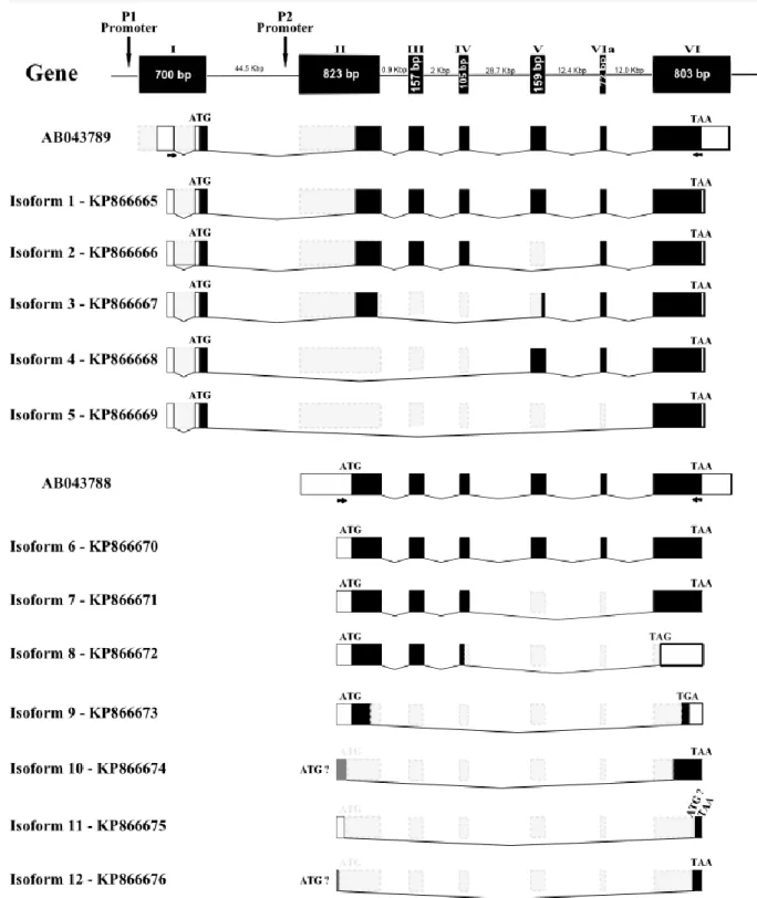
Figure 2.1 Schematic representations of the zebrafish runx3 gene, the cDNA splicing variants and corresponding protein structure.
57
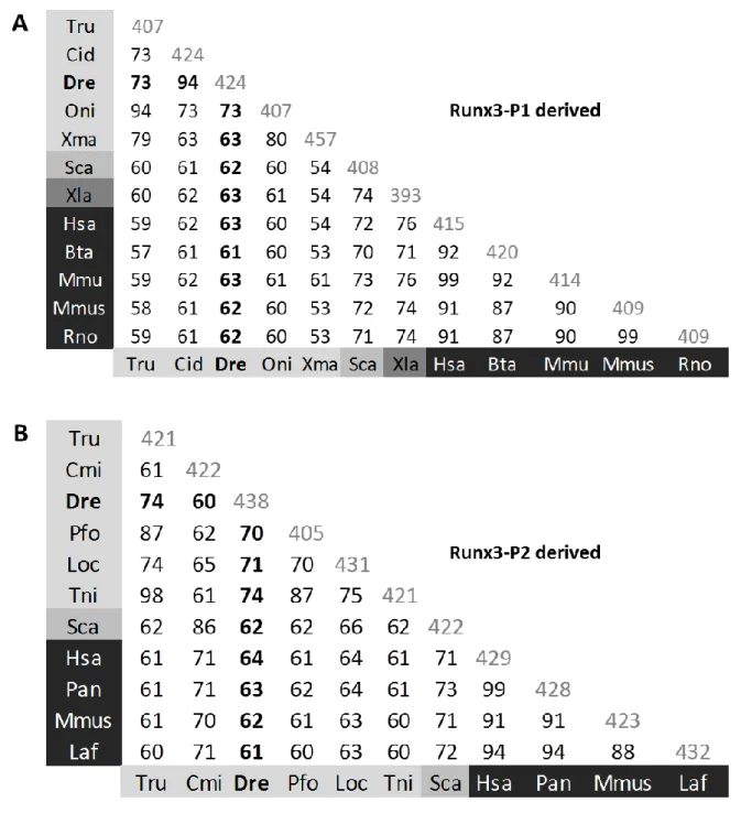
Figure 2.2 Pairwise percent identities among RUNX3 (A) P1-derived (MASN) and (B) P2-derived (M(H/R)IPV) isoforms.
59
Figure 2.3 Schematic representations of the conservation in RUNX3 protein domains.
60
Figure 2.4 Schematic representation of zebrafish alternatively spliced runx3 cDNA transcripts.
62
Figure 2.5 Schematic representations of the splicing events within the runx3 exon 1 affecting the 5’ UTR of the P1-derived transcripts.
64
Figure 2.6 Levels of runx3-P1 and runx3-P2 derived transcripts in adult zebrafish tissues.
65
Figure 2.7 Levels of runx3-P1 and runx3-P2 derived transcripts in embryonic zebrafish development.
66
Figure 2.8 Analysis of runx3 transcripts localization during early zebrafish developmental stages by ISH.
68
Figure 2.S1 Both runx3-P1 and runx3-P2 5’ UTR regions show the presence of multiple ATG nucleotide triplets upstream the ATG codon that initiates translation of the Runx3 proteins.
2
Figure 3.1 Relative transcriptional activity of zebrafish runx3 promoter constructs in U2OS and C6 cell lines.
99
Figure 3.2 Distribution of percentage of base-pairs located in block A, B, C, or D located in zebrafish and fugu promoters.
101
Figure 3.3 Retention of putative TFBSs after comparative analysis. 103 Figure 3.4 Representation of a DBA block obtained from the alignment of
P1 (a) and P2 (a’) runx3 promoter regions between zebrafish and fugu and overview of TFBS families detected by DiAlignTF on the conserved blocks analysed for P1 (b) and P2 (b’) promoters.
106
Figure 3.S1 TFBS families detected by DiAlignTF that are common in all conserved blocks obtained from the alignment of runx3 P1 (a) and P2 (b) promoter regions between zebrafish and fugu.
111
Figure 4.1 Schematic representations of zebrafish cbfβ transcripts. 135 Figure 4.2 Alignment analysis of zebrafish cbfβ protein isoform
sequences.
139
Figure 4.3 Schematic representation of zebrafish cbfβ gene, isoform 1 and protein structures.
140
Figure 4.4 Protein sequences comparison for CBFβ C-terminal. 141 Figure 4.5 Comparison of genomic environment and gene positional
order in zebrafish and human chromosomes containing CBFβ.
144
Figure 4.6 Identification of the expression profile of zebrafish cbfβ splicing variants (isoform 1-4).
145
Figure 4.7 Transcriptional co-activation of collagen type X promoter by Runx2-MASN/Cbfβ.
147
Figure 4.S1 Preparation of fusion proteins. 155
Figure 4.S2 Runx2 binds to isof1, isof2 and isof4 but not to isof3 of Cbfβ. 155 Figure 4.S3 Alignment analysis of human CBFβ protein isoform sequences. 156 Figure 4.S4 Schematic representation of human CBFβ gene and
corresponding transcripts.
156
3
Figure 5.1 In silico analysis of 1 kb of runx3 P1 and P2 upstream regions. 177 Figure 5.2 Relative transcriptional activity of zebrafish (A) runx3-P1 and
(B) runx3-P2 promoter constructs.
178
Figure 5.3 The transcriptional regulation of runx3 promoter regions are affected by co-transfection with Runx2 and Runx3 isoforms.
182
Figure 5.4 Basal activity of zebrafish (A) runx3-P1 and (B) runx3-P2 promoter regions in various runx3 expressing cells.
184
Figure 5.5 Transient injected embryos express GFP under control of runx3 PP(-3930/-554):EGFP promoter construct.
192
Figure 5.S1 Both runx3-P1 and runx3-P2 luciferase promoter constructs are functional in vivo.
200
Figure 5.S2 Qualitative analysis of Runx3 amplification by RT-PCR in different cell lines shows that Runx3 is expressed in all cells tested, except in HEK293 cell line.
201
Figure 5.S3 Basal activity of zebrafish runx3-P1 and runx3-P2 promoter regions in various runx3 expressing cells.
201
Figure 5.S4 Transfection of runx3-P1 and runx3-P2 promoter constructs driving EGFP results is GFP expressing cells.
202
Figure 5.S5 TFBS families detected by DiAlignTF software that are conserved in the alignment of the runx3-P1 promoter regions between zebrafish and human.
4
Table Index
Table 1.1 Aliases for the Runt-domain class of transcription. 12
Table 1.2 Common synonyms for each Runx protein. 12
Table 1.3 Summary of the expression data for Runx transcripts. 24
Table 2.1 Information for ISH probe synthesis. 56
Table 2.S1 PCR primers used in this chapter. 76
Table 2.S2 Accession numbers of all Runx3 protein sequences used for the in silico analysis.
77
Table 3.1 Transcription factor families conserved in each block obtained from the alignment of P1 and P2 runx3 promoter regions between zebrafish and fugu.
104
Table 4.1 Oligonucleotides used for PCR amplification. 129 Table 4.2 Splice code usages of the partial exon-skipping in cbfβ mRNA. 138 Table 4.S1 Pairwise per cent identities among CBFβ sequences. 153
Table 4.S2 Zebrafish-human ortholog gene pairs. 154
Table 5.1A Activating and repressing regions identified in runx3-P1 promoter in different cell lines.
188
Table 5.1B Activating and repressing regions identified in runx3-P2 promoter in different cell lines.
188
Table 5.2 Identification of the putative TFBSs, obtained by promoter comparative analysis, in the predicted regulatory regions of runx3-P1 and runx3-P2 promoters.
189
Table 5.S1 PCR primers used to amplify the zebrafish runx3 promoter regions.
204
Table 5.S2 Genes and respective set of primers used for the RT-PCR performed in the cell lines.
5
Chapter 1
6
1.1 Transcription in Eukaryotes - the first step in gene expression
The completion of “The Human Genome Project” in 2003 has revealed that the human genome encodes between 20.000 and 25.000 protein-coding genes, a number much lower than the initially predicted. The protein-coding sequences correspond to just 1.5% of the human genome, so all the remaining 98.5% correspond to non-coding DNA sequences. One fundamental point in Biology is to understand how the different cell types are specified in a multicellular organism. In order to our bodies to be formed from a unique cell and develop into a broad range of very diverse cell types, sharing the same genetic information, there has to be a complex mechanism for controlling gene expression in a very precise way. In some cells, certain genes are “turned off” while in other cells they are “turned on” in order to be transcribed and translated into proteins. Eukaryotes have developed diverse chemical and physical mechanisms including chromatin condensation, DNA methylation, transcriptional initiation, alternative splicing of RNA and mRNA stability to control gene expression. Failure to do so can lead to dire consequences, so a tight controlled gene expression is essential to insure that the right gene is activated in the right cell at the right time during the development (Lodish et al, 2000).
One of the most important and highly regulated gene expression processes is the transcription of the DNA template to generate a RNA molecule. There are different elements in the transcription-control regions of the gene that regulate the transcription. In eukaryotes, transcription is carried out by three nuclear RNA polymerases (Patikoglou and Burley, 1997), RNA Polymerase I, II, and III, that transcribe different classes of genes. RNA polymerase I transcribes rRNA (ribosomal RNA), RNA polymerase II transcribes mRNA (messenger RNA), microRNAs and most snRNAs (small nuclear RNAs), and RNA polymerase III transcribes tRNA (transfer RNA), rRNA 5S and other small RNAs (Carter and Drouin, 2009). The vast majority of the genes in eukaryotic genomes encode functional proteins and contain a promoter region upstream from the gene, with certain specific motifs (nucleotide sequences) that recruit RNA Polymerase II (Zwan et al, 2003). The promoter region usually contains a highly conserved sequence called the TATA box (frequently at ≈25-35 base pairs upstream from the transcription start site) or an alternative promoter element called initiator that, unlike the TATA box, has an extremely degenerated consensus sequence
7
(Lodish et al, 2000). However, in some cases, genes do not contain a TATA box or an initiator but may contain a GC-rich sequence (20-50 nucleotides within ≈100 base pairs upstream of the transcription start site). The TATA-binding protein (TBP) initiates the formation of the basal transcription complex along with multiple TBP-associated proteins and multiple additional general transcription factors. Thus, TBP together with its TBP-associated proteins was originally identified as a fraction called TFIID and this group of proteins is required to build a basal transcription complex from TATA-containing or TATA-less promoters (Pugh and Tjian, 1991). The transcription by RNA polymerase II can be regulated by activators or repressors, called transcription factors (TFs) that bind to promoter elements and enhancers. These functional regulatory elements are generally found within several hundred bases of the transcription start site of the gene to which they are linked, but they can occasionally be located tens of kilobases upstream or downstream from a promoter, within an intron, or downstream from the final exon of a gene (Lodish et al, 2000; Fickett and Wasserman, 2000). The fundamental logic of transcriptional regulation in eukaryotes is that it is combinatorial: each gene has a particular combination of regulatory elements, where the nature, number, and spatial arrangement of which determine the gene unique pattern of expression. These promoter or enhancer elements control in which cell types the gene is expressed, the times during development in which it is expressed, and the level at which it is expressed in adults (Collingwood et al, 1999).
1.2 Transcription Factors
In eukaryotes, genes are usually in a default "off" state, so TFs serve mainly to turn gene expression "on". The TFs can bind alone or attract other TFs creating a complex that can either facilitate or repress the binding by RNA polymerase II to specific target genes, thus beginning or shutting the process of transcription (Roeder, 1996; Lee and Young, 2000; Levine and Tjian, 2003) (Figure 1.1). Because TFs are crucial to the regulation of gene expression, understanding their action of mechanisms is a major area of ongoing research in cell and molecular biology.
8
Figure 1.1 Complex metazoan transcriptional control modules. A complex arrangement of multiple clustered enhancer modules interspersed with silencer and insulator elements which can be located 10-50 kilobases (kb) either upstream or downstream of a composite core promoter containing TATA box (TATA), Initiator sequences (INR), and downstream promoter elements (DPE). Figure adapted from Levine and Tjian, 2003.
The control of gene expression in mammalian cells requires interactions among many combinations of transcription regulatory proteins (reviewed in Levine and Tjian, 2003). These interactions must be both versatile to enable individual TFs to interact with a variety of structurally unrelated proteins, as well as selective to enable different members of the same transcription factor family to have differential effects on gene expression (Hu et al, 2002).
1.3 The core binding factor (CBF) transcription complex
The core binding factor (CBF) is a heterodimeric transcription factor composed by two subunits, an α-subunit and a β-subunit. The α-subunit is characterized by containing a heterodimerization domain, responsible for the binding to the DNA as well as binding to the β-subunit (Kanno et al, 1998). The β-subunit does not bind the DNA itself, but acts as a co-factor, increasing the affinity of the α-subunit to the DNA of the target genes (Hart and Foroni, 2002).
9
1.4 The β-subunit of the CBF complex – CBFβ protein
CBFβ is the non-DNA binding subunit of the heterodimeric core binding factor (CBF). It is able to dimerize with its DNA-binding subunit, the product of each of the three identified RUNX genes, mediating a variety of biological responses according to the RUNX protein (1, 2 or 3) associated into the complex (Lund and van Lohuizen, 2002). One model for CBFβ function is that this protein helps to stabilize a conformation of the runt domain that has the highest affinity for DNA. CBFβ may have developmental and clinical significance beyond its association with the runt domain. Furthermore, the CBFβ protein is more widely distributed than either the mammalian runt domain proteins that have been identified (Ogawa et al, 1993; Wang et al, 1993). This suggests that CBFβ may participate in a wide variety of developmental pathways, possibly by interacting with other types of DNA-binding proteins (Kagoshima et al, 1993).
1.5 The α-subunit of the CBF complex - runx transcription factors
The α-subunit of the CBF complex corresponds to the runt-related transcription factors (RUNX) that comprise a small family of transcription factors identified by their highly conserved DNA binding domain, designated the runt domain. The first characterized member of this family was the Drosophila regulatory gene runt, discovered in a genetic screen (Nüsslein-Volhard and Wieschaus, 1980) and named for its mutant phenotype, which reflects its role as a “primary pair rule” gene in establishing the pattern of segments in the embryo (Gergen and Butler, 1988). Subsequently, the gene was found to have additional genetic functions in Drosophila, being implicated in sex determination and neurogenesis (Duffy and Gergen, 1991; Duffy et al, 1991). Due to its localization in the nucleus, it was predicted that the RUNX protein product could act as a transcription factor regulating the transcription of other genes (Kania et al, 1990). Some years later, a second Drosophila runt-related gene, lozenge, was identified, and shown to be required in cell patterning in the eye and developing antenna, as well as in cell fate specification during hematopoiesis (Gupta and Rodrigues, 1995; Daga et al, 1996; Flores et al, 1998; Canon and Banerjee, 2000; Bataillé et al, 2005). More recently, a Drosophila genome project revealed two additional runt-related
10
genes, CG42267 and CG34145 (Rennert et al, 2003; Bao and Friedrich, 2008), being described so far four runx genes in Drosophila.
Given the importance of RUNX genes, which play vital roles in the development of various species and human diseases, in the last years a large number of groups have focus their research in characterizing orthologues of RUNX genes from phylogenetically diverse species, trying to achieve a better understanding of their origin and functions. The evolution of RUNX genes begins with one more similar to runx3 in invertebrates and then progress to multiple RUNX genes in higher species (Cohen, 2009). While primitive metazoans such as sea urchin and C. elegans appear to possess a single RUNX family member, so far, three have been described in mammals and four in insects, zebrafish and fugu. This can indicate (i) evolutionary conserved roles in metazoans and (ii) the necessity for a tight regulation of RUNX activity in specifying complex developmental events in higher organisms (Chuang et al, 2013).
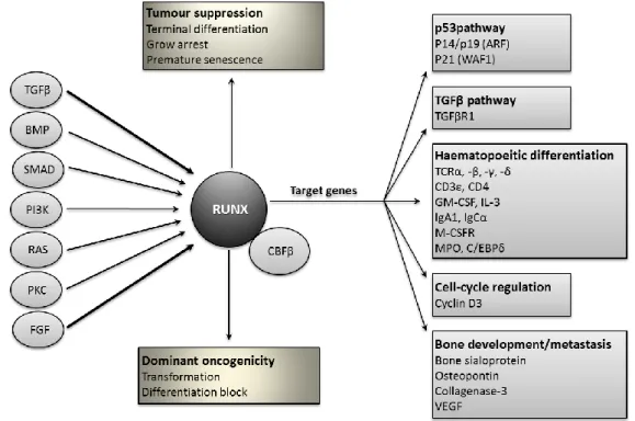
RUNX proteins are crucial transcription factors that regulate a wide range of biological processes to orchestrate proper cell fate determination during the development of metazoans (Coffman, 2003). Traditionally described as a DNA-binding protein, RUNX is now viewed as a multifaceted protein that associates with diverse proteins to direct biological outcomes in a context-dependent manner (Figure 1.2) (Blyth et al, 2005; Chuang et al, 2013).
11
Figure 1.2 RUNX/CBFβ complexes in the determination of cell fate. This diagram illustrates the central role of RUNX/CBFβ complexes in the orchestration of cell fate in response to exogenous factors and environmental signals (circles in the left side). A subset of the known RUNX target genes is depicted (grey boxes), and these targets have been selected for their potential relevance to cancer. Figure adapted from Blyth et al, 2005.
1.6 Runx Aliases - Nomenclature for runt-related (RUNX) proteins
Since the first runt-related gene has been cloned and studied in Drosophila, there was an explosion in the number of independent laboratories focusing their studies in this family of transcription factors. So, in the literature, a diversity of family names can be found for these genes (Table 1.1 and 1.2) depending on the context of their study (van Wijnen et al, 2004).
12
Table 1.1 – Aliases for the runt-domain class of transcription factors.
Table adapted from (van Wijnen et al, 2004).
Table 1.2 - Common synonyms for each Runx protein.
Table adapted from (http://www.genecards.org/)
Due to the existence of a wild diversity of names for the runt-related family of transcription factors, it was difficult to find in the literature all the references existing for this subject. So, in 1999, the Nomenclature Committee of the Human Genome Organization (HUGO) adopted
Abbreviation Gene Name Specie described
Runt Runt Drosophila
Lz Lozenge Drosophila
AML Acute myelogenous leukemia Human
PEBP2alpha Polyomavirus enhancer-binding protein 2 alpha subunit
Mouse CBFalpha Enhancer core-binding factor α of murine
leukemia viruses ( i.e SL3-3, AKV and Moloney MLV)
Mouse
PEA2 Polyoma enhancer A-binding factor 2 Mouse
SEF1 SL3-3 enhancer factor 1 Mouse
S/A-CBF SL3-3 and AKV core-binding factor Mouse
NF-deltaE3A Nuclear factor delta E3A Human
MyNF1 Myeloid nuclear factor 1 Mouse
NMP2 Nuclear matrix protein 2 Rat
OBSC Osteoblast-specific complex Rat
OSF2 Osteoblast-specific factor 2 Mouse
til-1 T-cell tumor integration locus 1 protein Mouse
run Runt domain-encoding gene Caenorhabditis elegans
Runt-Related Transcription Factor
1
RUNX1; AML1; CBFα2; CBFA2; PEBP2αB; PEBP2A2; PEA2αB; SL3-3 enhancer factor 1αB subunit; Oncogene AML1; SL3/AKV core binding factor αB subunit; AML1-EVI-1; AMLCR1; EVI-1
Runt-Related Transcription Factor
2
RUNX2; AML3; CBFα1; CBFA1; PEBP2αA; PEBP2A1; PEA2αA; SL3-3 Enhancer Factor 1αA Subunit; Oncogene AML3; SL3/AKV core-binding factor αA Subunit; Osteoblast-specific
transcription factor 2 (OSF-2); CCD
Runt-Related Transcription Factor
3
RUNX3; AML2; CBFα3; CBFA3; PEBP2αC; PEBP2A3; PEA2αC; SL3-3 Enhancer Factor 1αC Subunit; Oncogene AML2; SL3/AKV Core-Binding Factor αC Subunit
13
the use of the term ‘RUNX’ to refer to genes encoding the runt-related proteins. It was agreed by the investigators of this field, that any designations can be used to refer to the runt-related proteins, but they should be referred as RUNX proteins at least once in the Abstract and/or Title of new manuscripts submitted for publication (van Wijnen et al, 2004).
1.7 RUNX gene and protein structures
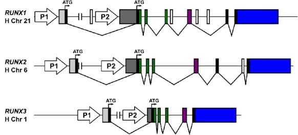
There are three RUNX genes described in mammals and all RUNX gene products share many structural similarities, but have distinct biological activities. A variety of transcripts have been described for all RUNX members, originated from the regulation of transcription from two alternative and distantly located promoters (P1 and P2), due to a high number of alternative splicing events and the presence of alternative polyadenylation (Miyoshi et al, 1995; Ahn et al, 1996; Levanon et al, 1996; Geoffroy et al, 1998). The RUNX gene structure is well conserved among all vertebrate species analysed (Figure 1.3) (Levanon and Groner, 2004).
Figure 1.3 Phylogenetic illustration of RUNX genes showing the gene number and promoter usage in different species. The three lower non-designated branches in the phylogenetic tree represent the animal groups (from bottom up) sponges, cnidarians (e.g. jellyfish) and acoelomates (e.g. flatworms). The more primitive animals contain one gene regulated by the P2 promoter. Figure adapted from Levanon and Groner, 2004.
14
There are two major protein isoforms for each RUNX gene, with a different N-terminal sequence depending if it is encoded from the P1-derived or from the P2-derived transcript. The gene structure of the three human RUNX genes is represented in Figure 1.4.
Figure 1.4 Genomic organization of the human RUNX genes. The two promoters P1 and P2 and initiator codons (ATG) are indicated. The P1-derived and P2-derived 5’ UTRs are indicated in light and dark grey, respectively, common coding exons are shown in similar color and 3’ UTRs are represented in blue. Figure adapted from Levanon and Groner, 2004.
The RUNX proteins that originate from the P2-derived transcripts contain a specific N-terminal region of five amino acids – the penta-peptide MRIPV. On the other hand, the RUNX isoforms that originate from the P1-derived transcripts contain a specific N-terminal region of 19 amino acids, originated by an alternative splicing from exon 1 onto the coding region of P2-derived transcript through a conserved in-exon splicing site located 16 bp downstream of the P2-ATG codon. This 19 amino acids long specific N-terminal is conserved in all P1-derived RUNX proteins, with the highest sequence similarity found between RUNX1 and RUNX3 (Bangsow et al, 2001). The isoforms are usually designated as isoform RUNX3-MA(S/D)NS and RUNX3-M(R/H)IPV, depending if they are encoded from the P1-derived or P2-derived transcripts, respectively.
15
1.8 Functional domains of RUNX proteins
Ito (2004) and, more recently, another report from the same group (Chuang et al, 2013), have described the common structure of the RUNX proteins by comparison of the amino acid sequences from the three RUNX proteins. They confirmed that all RUNX proteins share the same characteristic domains and motifs: runt domain (RD), transactivation domain (TAD), inhibitory domain (ID), nuclear localization signal (NLS), PY (or PPxY) motif and the VWRPY motif that corresponds to the last five amino acids of the RUNX proteins (Figure 1.5). RUNX2 possesses a unique QA domain composed of a stretch of Q and A residues (Ito and Miyazono, 2003). The N-terminal part of the molecules comprises the RD, a 128 amino acids long domain that is highly conserved between species and has an S-type immunoglobulin fold (Warren et al, 2000). The C-terminal part of the RUNX molecule contains the other domains (TAD and ID) that play a role in transcription regulation (reviewed in Downing, 1999; Ito, 1999), and a conserved PY motif, consisting of is a proline-rich peptide, that interacts with proteins harbouring the WW domain (Chuang et al, 2013) and a VWRPY motif at the end of the C-terminus of RUNX proteins, which mediate the interaction with the co-repressor Groucho/TLE (Liu et al, 2006; reviewed in Cohen, 2009) (Figure 1.5).
Figure 1.5 Schematic representation of functional domains of RUNX3 protein and examples of its interacting proteins. The domains represented are conserved among all RUNX proteins. The numbers indicate amino acid positions. TAD, ID, C-term and NLS refer to transactivation domain, inhibitory domain, C-terminus and nuclear localization signal, respectively. Figure adapted from Chuang et al, 2013.
Traditionally described as a DNA-binding protein that can act as a homodimer or heterodimer (with its partner CBFβ) to bind to the promoter of the target genes, RUNX is now viewed as a multifaceted protein that associates with a wide variety of proteins to
16
direct biological outcomes in a context-dependent manner. Those proteins can either be co-activators or repressors. Examples of some co-factors previously described to interact with RUNX proteins (Blyth et al, 2005; Chuang et al, 2013) are represented in Figures 1.5 and 1.6.
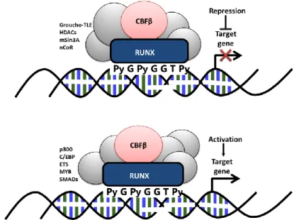
Figure 1.6 Schematic representation of the RUNX factors as organizers of transcription. RUNX factors bind to their consensus sequence (PyGPyGGTPy) in the promoter region of the target genes as a heterodimer with their binding partner CBFβ. A complex including other co-factors is generally formed, determining the outcome of the RUNX regulation on transcription of the target genes. CBFβ, core-binding factor-β; C/EBP, CCAAT/enhancer-binding protein; HDACs, histone deacytylases; nCoR, nuclear receptor corepressor; TLE, transducin-like enhancer of split. Figure adapted from Blyth et al, 2005.
The factors affecting the outcome of this interaction on transcription of the target gene seem to involve promoter-specific features such as the proximity of binding sites for co-activators or repressors as well as the availability of such cofactors in the nucleus.
1.9 Runt domain characteristics
All RUNX proteins have the characteristic runt domain that is known to have the dual function to bind to the promoter of target genes and also to bind to CBFβ protein forming the CBFα/β complex. This complex increases allosterically the DNA-binding affinity (Ogawa et al, 1993; Miller et al, 2002) and regulates the RUNX proteins turnover by protecting them
17
from ubiquitin-proteasome-mediated degradation (Huang et al, 2001). All RUNX proteins heterodimerize with CBFβ and all bind to the same consensus recognition sequence (5'-PyGPyGGT-3') (Py is any pyrimidine) found in a number of enhancers and promoters. The biological activity of the CBF complex is specified by the α-subunit according to the RUNX protein (1, 2 or 3) associated into the complex (Tahirov et al, 2001; Bäckström et al, 2002; Lund and van Lohuizen, 2002; Horsfield et al, 2007). The degree of homology between invertebrate and vertebrate runt domains is quite high, and this may reflect the conservation of dual functions within that domain, as described above.
There are two types of heterodimeric factors either (i) both subunits bind to specific DNA sequences (e.g. Jun/Fos and Myc/Max) or (ii) one subunit binds to DNA and one does not (e.g. RUNX/CBFβ). The molecular mechanisms of the DNA binding for the first type of heterodimeric factors was already described (Glover and Harrison, 1995), but for the second type it is still not well understood how non-DNA binding subunit contribute to DNA binding and whether the mechanism by which a non DNA binding subunit stimulates DNA binding is shared with different transcription factors (Tahirov et al, 2001).
Due to the important function of the heterodimeric RUNX/CBFβ complex in different developmental processes, regulating different promoters and enhancers of a variety of genes in a cell specific manner, different groups have used X-ray crystallography and nuclear magnetic resonance (NMR) to determine the tertiary structures of the runt domain and the runt/CBFβ heterodimeric complex, and the molecular mechanisms of their interaction (Nagata et al, 1999; Tahirov et al, 2001; Bravo et al, 2001; Bäckström et al, 2002). These studies revealed that the runt domain adopts the immunoglobulin (Ig)-like fold β-sandwich, which is homologous to other DNA-binding domains such as those found in p53, nuclear factor-kappa B (NF-kB), nuclear factor of activated T cells (NFAT) and other proteins (Nomura et al, 2013).
Determination of the tertiary structures together with mutational analysis was crucial to the identification of regions that are essential to the binding affinity between the runt domain and the DNA consensus recognition sequence. One example was the recent identification of the three guanines in the DNA consensus recognition sequence (5'-PyGPyGGT-3'), which are directly recognized by three arginine residues of the runt domain, as being important for the
18
efficient binding between the runt domain and the recognition element in the DNA (Nomura et al, 2013).
1.10 Biological roles of RUNX proteins
The proteins from the RUNX family show versatile functions being involved in several diseases and acting on target genes in a variety of tissues, and exhibiting both transcriptional activation and repression activities (Otto et al, 2003). These transcription factors are tissue-restricted and cancer-related, regulating cell proliferation and growth, as well as differentiation (Coffman, 2003; Miyazono et al, 2004; Stein et al, 2004; Young et al, 2007a, 2007b; Kagoshima et al, 2007). It is clear that each of the three RUNX proteins has different roles (Rennert et al, 2003), but since they were found to be co-expressed in some tissues, some of their functions may overlap, either synergistically or at different time points (Cohen, 2009).
1.10.1 The runt-related transcription factor 1 (RUNX1)
The human RUNX1 was originally cloned from the breakpoint of chromosome 21 in t(8;21)(q22;q22) by Miyoshi and collaborators (Miyoshi et al, 1991). This translocation is frequently found in patients with acute myeloid leukemia (AML) with maturation (M2 subtype) (Miyoshi et al, 1991) and has subsequently been shown to be one of the most frequent targets of leukemic-associated gene aberrations (Reviewed in Okuda et al, 2001). RUNX1 has been proved to regulate, acting as a context-dependent transcriptional activator or repressor, a number of target genes, e.g. granulocyte macrophage colony-stimulating factor (GM-CSF) (Oakford et al, 2010), receptor for macrophage colony-stimulating factor (CSF-1R) (Sauter et al, 2013), tissue inhibitor of metalloproteinase 1 (TIMP1) (Bertrand-Philippe et al, 2004), early B-cell factor 1 (Ebf1) (Seo et al, 2012), cyclin D3 (Bernardin-Fried et al, 2004), CD11 integrin (Puig-Kröger et al, 2000), complement receptor type 1 (CR1) (Kim et al, 1999), insulin-like growth factor-binding protein 3 (IGFBP-3) (Iwatsuki et al, 2005), type B leukemogenic virus (TBLV) enhancer (Mertz et al, 2001), among others.
19
RUNX1 is expressed in a number of tissues at specific time windows during embryogenesis. In the mouse embryo, expression of Runx1 is first detected in definitive hematopoietic stem cells (HSC) and in endothelial cells at HSC emergence sites (North et al, 1999; Cai et al, 2000; North et al, 2002) and later, Runx1 is highly expressed in several hematopoietic lineages including myeloid, B- and T-lymphoid cells (Lorsbach et al, 2004). Runx1 is also highly expressed in cranial and dorsal root ganglia (DRG), in the small-diameter nociceptive TrkA neurons (Simeone et al, 1995; Levanon et al, 2001, 2002), in the thymus (Satake et al, 1995; Komori et al, 1997; Levanon et al, 2001) and in cells of the chondrocyte lineage of the developing mouse skeleton (Sato el at, 2008). Tissue sections of the fetal liver or on smear preparations of peripheral blood of mutated mice embryos showed an absence of hematopoietic elements of definitive origin and analysing the yolk sac the erythropoiesis was minimally affected. So, this indicates that the gene targets of RUNX1 are essential for the development of all cell lineages of definitive hematopoiesis, but not required for erythropoiesis (Okuda et al, 2001). In zebrafish and Xenopus, the expression pattern of runx1 is well conserved compared to the mammalian pattern, and was shown to be expressed in blood progenitors (Tracey et al, 1998; Kalev-Zylinska et al, 2002; Burns et al, 2005). In these species, runx1 is also expressed in a few scattered cells within craniofacial cartilages (Kalev-Zylinska et al, 2003; Flores et al, 2006; Park and Saint-Jeannet, 2010) and in Rohon-Beard sensory neurons, that are a population of specialized neurons with mechanoreceptive properties only found in fish and amphibians and contribute importantly to the embryonic nervous system of these species (Rossi et al, 2009).
Knockout studies, using different Runx1 deficient mice lines, show that the lack of functional Runx1 protein production causes necrosis and haemorrhaging in the central nervous system and blocks definitive hematopoiesis resulting in death of the mice during the mid-gestational period (Okuda et al, 1996; Wang et al, 1996a). The importance of the heterodimerization of CBFβ with RUNX1 is supported by analysis of the disruption of CBFβ that causes a similar phenotype as the one obtained for the Runx1 knockout (Wang et al, 1996b). Consistent with the Runx1 knockout mice, the zebrafish mutant embryos show normal primitive hematopoiesis, but a blockage of the definitive hematopoiesis (Jin et al, 2009). However, the RUNX1 role in the generation and maintenance of HSCs during adult hematopoiesis remains poorly understood. Recently, Sood and colleagues (2010) reported that a zebrafish