GDANSK UNIVERSITY OF TECHNOLOGY
CHEMICAL FACULTY
DEPARTMENT OF ANALYTICAL CHEMISTRY
MASTER THESIS
DETERMINATION OF CYCLOPHOSPHAMIDE IN
HUMAN URINE BY HPLC COUPLED TO TANDEM
MASS SPECTROMETRY
Student:
Promoter:
Anu Marahatta Agata Kot Wasik, Ph.D., Eng
Supervisor:
ACKONOWLDEGEMENT
I would like express my deep and sincere gratitude to my supervisor, Ph.D Agata Kot Wasik and Ph.D. Andrzej Wasik for their stimulating suggestions and encouragement helped me in all the time of research. Throughout my thesis writing period they provided encouragement sound advice and lots of good idea.
I owe my most sincere gratitude to Professor Jacek Namiesnik who gave me opportunity to work with them in Department of Chemistry at Gdansk University of Technology. Also my sincere gratitude to Professor Bogdan Zygmunt for providing me moral support during my stay at GUT.
I would like to thank Monika Janicka and Agata Zygler for their help, all advices and making the working atmosphere pleasant.
TABLE OF CONTENTS LIST OF ABBREVIATIONS ... 4 UNITS ... 5 LIST OF TABLES ... 6 LIST OF FIGURES ... 7 1. INTRODUCTION ... 8
2. AIM AND OBJECTIVE OF THESIS ... 9
3. THEORITICAL PART ... 10
3.1. Cyclophosphamide ... 10
3.1.1. Physical and chemical properties of CP ... 10
3.1.2. Metabolism of CP ... 11
3.1.3. Absorption, distribution and elimination of CP ... 12
3.1.4. Mechanism of action of CP ... 12
3.1.5. Cyclophosphamide use ... 13
3.2. Solid Phase extraction ... 14
3.3. Literature review ... 15
4. EXPERIMENTAL PART ... 18
4.1. Chemicals and reagents ... 18
4.2. Apparatus and Laboratory ... 18
4.3. Analytical procedure ... 19
4.3.1. Chromatographic condition ... 19
4.3.2. Mass spectrometry parameter ... 19
4.3.3. Preparation of stock and working standard solutions ... 20
4.3.4. Urine collection ... 20
4.3.5. Comparison of LC-MS and LC-MS/MS in term of sensitivity ... 21
4.3.6. SPE extraction ... 21
5. RESULT AND DISCUSSION ... 25 5.1. Mass spectrometry ... 25 5.2. Liquid Chromatography ... 27 5.3. Linearity ... 27 5.4. Sensitivity ... 27 5.5. Carry over ... 27 5.6. Recovery ... 27
5.7. Influence of solvent used to prepare CP standard solution ... 28
5.8. Matrix effect ... 30
5.9. Influence of time of evaporation on recovery ... 31
5.10. Sorbent types ... 32
5.11. Influence of different eluent solvent on recovery ... 33
5.12. Stability study of CP solutions ... 34
6. APPLICATION OF THE METHOD ... 36
7. CONCLUSION. ... 37
8. SUMMARY ... 38
LIST OF ABBREVIATIONS
CP Cyclophosphamide CUR Curtain gas
CE Collision energy CEPM Carboxyethylphosphoramide CarboxyCP Carboxycyclophosphamide DP Declusturing potential DCM Dichloromethane DCCP Dechloroethylcyclophosphamide ESI Electrospray ionization
HPLC High performance liquid chromatography i.d. Internal diameter
IF Ifosfamide
ISV Ion spray voltages
KH2PO4 Potassium dihydrogen phosphate
K2HPO4 Potassium hydrogen phosphate
4-Keto CP 4- Ketocyclophosphamide LC Liquid chromatography LLE Liquid liquid extraction LOD Limit of detection LOQ Limit of quantification MS/MS Tandem mass spectrometry MRM Multiple reaction monitoring
m/z Mass/Charge MeOH Methanol N2 Nitrogen
4-OHCP 4- Hydroxycyclophosphamide pKa Dissociation constant
UNITS
Unit English term Å angstrom °C Celsius degree cm centimeter h hour
L/h Liter per hour µg microgram
µg/L microgram per liter µg/mL microgram per milliliter µm micrometer
mg milligram
mg/L milligram per liter mg/mL milligram per microliter min. minute
mL milliliter
mL/min milliliter per minute mm millimeter
msec millisecond ng/L nanogram per liter psi Pound per square inch s second
LISTS OF TABLE
Table 1 Physical and chemical properties of CP ... 10
Table 2 Literature review presenting different methods of extraction and analysis of CP ... 15
Table 3 Cartridges applied for SPE during CP isolation from urine sample ... 19
Table 4 Characteristics of health workers ... 20
Table 5 Sensitivity comparison of LC-MS and LC-MS/MS ... 21
Table 6 MRM parameters ... 25
Table 7 Influence of time of evaporation on recovery ... 31
LISTS OF FIGURES
Figure 1 Metabolism of cyclophosphamide ... 11
Figure 2 Schematically presentation of SPE procedure ... 23
Figure 3 Calibration curve of CP diluted in methanol: water (1:1) ... 24
Figure 4 Calibration curve of urine sample spike with CP ... 24
Figure 5 The HPLC/MRM chromatogram of cyclophosphamide and proposed fragmentation ... 25
Figure 6 The HPLC/MRM chromatograms a) for a blank urine sample and b) for a blank urine sample spiked with 0.936 µg/mL of CP. ... 26
Figure 7 Influence of solvent used to prepare CP standard solution ... 28
Figure 8 CP diluted in a) methanol b) methanol: water (1:1) c) mixture urine: water (1:1) d) urine: buffer pH=3 mixture e) buffer pH=3 ... 29
Figure 9 Influence of sample preparation of urine before SPE on recovery of CP ... 30
Figure 10 Influence of sorbent on CP recovery ... 32
Figure 11 Influence of different eluent solvent on CP recovery ... 33
Figure 12 Stability study of extract urine with buffer pH=3 ... 35
Figure 13 Stability study of extract urine with PBS buffer pH=7 ... 35
1. INTRODUCTION
Antineoplastic drugs are commonly used to treat cancers which are generally referred to as chemotherapy. The majority of antineoplastic drugs are non- selective in their action which exhibits their effects in both cancerous and non-cancerous cells in most organs and body tissues. Chemotherapy agents are known to be mutagens and carcinogens in animals, humans and in patients treated with therapeutic dose [1]. Cyclophosphamide (CP) is known to be human carcinogenic according to International agency for research on cancer (IARC Group I).
Medical professionals were unaware of the risk from exposure to antineoplastic agents during performance of their daily routine. The routes of exposure are typically inhalation, dermal or oral. Workers may be exposed by inhalation via droplets, particulates and vapors when they create aerosols, generate dust by crushing tablets and clean up spills. Dermal exposure may occur when workers touch contaminated surfaces during preparation, administration or disposal of hazardous drugs and oral exposure may occur from hand to mouth contact. In most paper air sampling for antineoplastic drugs have little to no air borne contamination. Drugs particulates can become airborne after the drying of contaminated areas. Vaporization of antineoplastic agents has also been reported with various drugs such as carmustine, ifosfamide, thiotepa and CP [2, 3]. When food or beverages are prepared, stored or consumed in work areas, they may easily become contaminated with airborne particles of antineoplastic drugs. A potential source of exposure is direct skin contact when a spill or lack occurs and large volume of drug is released to the environment. Antineoplastic drugs can have chronic effects on reproduction, causing abortion, stillbirth, infertility and congenital abnormalities and other minor health effect include hair loss, headache, acute irritation and hypersensitive [4, 5].
Few methods have been described for the determination of CP in urine till now. The method used for the determination of CP is gas chromatography separation with nitrogen phosphorous detection or mass spectrometric detection. Other method used for analysis of CP is HPLC [6]. UV detection is not however, selective and sensitive enough to enable low exposure levels to be quantified because CP has weak chromophores [7, 8]. Polarographic
2. AIMS AND OBJECTIVE OF THE THESIS
The main objective of this study was
a) To optimize SPE method for isolation of organic compound such as CP from urine sample. Optimization is achieved by using different sorbent, matrix, washing solvent and elution solvent. Biological samples such as plasma and urine are much more complex due to presence of proteins, salts, acids, bases and various organic compounds with similar chemistry to analytes of interest. As a result, the extraction methods for biological samples have been complicated. If unsuitable sample preparation methods have been employed before injection, the entire analytical process can be wasted. The main task is to remove maximum interfering substance from urine matrix and conversion of analytes into a more suitable form for injection. b) To optimize the separation conditions of the HPLC-MS/MS analysis to achieve the
optimal (short retention time, no interferences) cyclophosphamide separation. Optimization is achieved by changing different parameter such as
Mobile phase and its composition Flow rate
Scan type
c) After developing suitable analytical method, this SPE-LC-MS/MS method is applied for the analysis of urine samples of health workers from Medical University of Gdansk, who were exposed with CP during their work in pharmacy. Hospital workers are exposed to antineoplastic drugs via dermal, inhalation route during preparing and handling.
3. THEORITICAL PART
CP general information
3.1. Cyclophosphamide
CP (2-[bis(2-chloroethyl)amino] tetrahydro-2H-1,3,2-oxazaphosphorine 2-oxide) is an alkylating agent that is frequently used as an antineoplastic drug. CP is a prodrug that requires activation by the cytochrome P450 enzyme system to form its pharmacologically active metabolite 4- hydroxycyclophosphamide [10]. The metabolites are genotoxic due to their ability to cross-link DNA and thereby cause DNA damages. CP is classified as carcinogenic to humans [11]. It has very narrow therapeutic index [12]. CP is rapidly converted to number of mutagenic and carcinogenic products, some of which have been isolated from urine [13].
3.1.1. Physical and chemical properties of CP
Basic physical and chemical properties of CP [11, 14-17] are summarized below in Table 1.
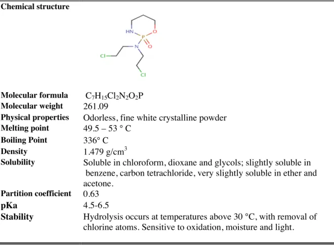
Table 1 Physical and chemical properties of CP Chemical structure
Molecular formula C7H15Cl2N2O2P Molecular weight 261.09
Physical properties Odorless, fine white crystalline powder
Melting point 49.5 – 53 ° C Boiling Point 336° C
Density 1.479 g/cm3
Solubility Soluble in chloroform, dioxane and glycols; slightly soluble in benzene, carbon tetrachloride, very slightly soluble in ether and acetone.
Partition coefficient 0.63
3.1.2. Metabolism of CP
CP is a prodrug which metabolized to both active and inactive metabolilites, as shown in figure 1. CP is activated by hepatic microsomal mixed function oxidases cytochrome P450 to form 4–hydroxycyclophosphamide (4-OHCP), which exists in equilibrium with its tautomer aldophosphamide (AldoCP). 4-OHCP is very unstable, readily diffuses in to cells and spontaneously decomposes into phosphoramide mustard (PM) by ß elimination of acrolein [18]. PM is an active alkylating species which is responsible for alkylating effect of CP [19]. Acrolein is an unwanted by product which may enhance CP-induce cell damage, possibly by depletion of cellular glutathione by conjugation [20]. The aldehyde dehydrogenase enzymes are involved in formation of carboxyphosphamide (CarboxyCP) from aldophosphamide [21]. 4- Ketocyclophosphamide (4-KetoCP) are formed due to oxidation of 4-OHCP by alcohol dehydrogenase. Other inactive metabolite includes 2-dechloroethylcyclophosphamide (DCCP) produced by separate oxidative N-dealkylation reaction [22].
3.1.3. Absorption, distribution and elimination of CP
CP is well absorbed orally, with peak concentrations occurring after 1-3 hours and has bioavailability of 85-100% [23]. The drug is rapidly absorbed from the blood after IV injection [11].
CP is distributed with a volume of distribution of 30 -50 L, which approximates to the total body water [10]. Penetration of CP and its metabolites into body fluids is limited. Metabolites like CarboxyCP and DCCP were not found in cerebrospinal fluid whereas PM and CP could be detected [24, 25].
CP and its metabolites are eliminated by urine in 24 hours after the start of treatment. The major function of CP in the body is eliminated by hepatic metabolism, but small fraction is eliminated by renal excretion of unchanged drug in urine. Less than 20 % of the administered dose is eliminated unchanged in the urine. Between 30-60% of the total CP dose is eliminated renally as CP or metabolites. CarboxyCP is major metabolites found appearing in urine [26, 27]. Small fraction of CP dose is eliminated via feces and expired air. In children and young adults plasma half life is shorter than adults. The elimination half life of CP ranges between 5-9 hours over a large concentration range. Total systemic clearance of CP ranges from 4-5 L/h of which the greater part is non-renal clearance. The plasma half life of CP is approximately 5 h in human during the first day after the dose [29].
3.1.4. Mechanism of action of CP
First CP is converted by the liver into two chemicals acrolein and phosphoramide. Acrolein and phosphoramides are the active compounds that prevent cell division by cross-linking DNA strands and decreasing DNA synthesis. These drugs can’t distinguish between normal and cancerous cells. CP also possesses potent immunosuppressive activity [30].
3.1.5. Cyclophosphamide use:
CP has been widely in different disease treatments: Breast cancer [31-33] Ovarian cancer [34] Prostate cancer [35-38] Lung cancer [39,40] Lupus erythematosus [41,42] Rheumatoid arthritis [43,44] Multiple Sclerosis [45,46]
Idiopathic pulmonary fibrosis [47-48] Wegener granulomatosis [49-51] Hodgkin’s Lymphoma [52-54] Thrombocytopenic purpura [55-58] Polyarteritis nodosa [59]
Analytic of CP
3.2. Solid Phase Extraction (SPE):
SPE is one of the techniques available to an analyst to bridge the gap that exists between the sample collection and analysis step. In SPE, extraction is performed by absorbing the analyte from the matrix onto a solid sorbent. The solid phase is then separated from the solution and other solvents added. The first solvent is usually a wash to remove possible adsorbed matrix components; eventually an eluting solvent is brought into contact with sorbent to selectively desorb the analyte.
Today SPE is the most popular sample preparation method. SPE is choice of method when handling biological samples for liquid chromatography (LC), a sample is mandatory to limit column clogging, presence of co-eluting substances as well as matrix effects frequently encounter with mass spectrometric detection. SPE allow good sample clean up.
It is a very active area in the field of separation science [62-65]. Liquid-liquid extraction (LLE) has remained the preferred technique for the preparation of the liquid samples for several years especially in the environmental field. LLE is labor intensive, difficult to automate, and is frequently plagued by practical problems, such as emulsion formation. In addition, it consumes relatively large volumes of high-purity solvents with expensive disposal requirements [66]. The increased development of SPE has occurred during the past five or six years; with many improvements in formats, automation and introduction of new phases. One reason was the pressure to decrease organic solvent usage in laboratories which has encouraged the requirement for solvent free procedures and has greatly contributed to the growth of SPE at the expense of LLE procedure [67]. Other reasons for the growing interest in SPE techniques are the large choice of sorbents with the capability for new ones of trapping analytes, shorter processing time and simpler procedure [68].
The general procedure of SPE is to load a solution onto the SPE phase, wash away undesired components and then wash off the desired analytes with another solvent into a collection tube.
3.3. Literature review
Literature review concerning CP extraction and analyses has been summarized in table 2.
Table 2 Literature review presenting different methods of extraction and analysis of CP.
Drugs Matrix Sample
Preparation
HPLC Parameter Mobile phase MS Reference
CP Urine Liquid-Liquid Hypersil BDS C8 150 4.6 mm, 5 µm Solvent A: Methanol Solvent B: 0.02 M ammonium acetate, pH 4.5 Triple quadrupole mass spectrometer [69] CP and metabolites (DCL-CP, 4KetoCP, CarboxyCP ) - Beta Basic C8 100 3 mm, 5 µm Solvent A:
Water with 0.1% formic acid Solvent B:
Methanol
MS/MS [70]
CP and other drugs (IF, Doxorubicin, Epirubicin Daunorubicin) SPE Hypersil BDS C8 150 4.6 mm, 5 µm Solvent A: Water + 0.1 % formic acid
Solvent B:
Acetonitrile +0.1 % formic acid
Triple quadrupole mass spectrometer [12] Metotrexate CP
SPE Max RP Capillary column 0.5 50 mm, 4 µm
Solvent A:
20 mM ammonium acetate, pH 4 with acetic acid
Solvent B: Methanol Triple quadrupole mass spectrometer [71] CP LLE GC- Methyl siloxen capillary column 30 m 0.25 mm, film thickness0.25µm - GC-MS [72] CP Urine Plasma Centrifuged the sample for 5 min. Genesis C18 50 2.1mm, 4 µm Solvent A: 0.5 % acetic acid Solvent B:
Methanol with 0.5 % acetic acid
Triple quadrupole
mass spectrometer
CP and other drugs (Doxorubicin, Doxorubicinol) Plasma SPE Symmetry C18 30 2.1 mm Solvent A: 5 mM acetate buffer, pH 3.5 Solvent B: Methanol Triple quadrupole mass spectrometer [74] CP and metabolites (4-OHCP) Centrifused for 15 min. Zorbax Extend C18 analytical column 150 2.1 mm, 5 µm Solvent A: 1 mM ammonium hydroxide in water , pH 10 Solvent B: Acetronitrile Triple quadrupole mass spectrometer [75] CP and metabolites (4- OHCP, CEPM) Plasma Centrifused for 5 min. Zorbax Extend 50 2.1 mm, 5µm Solvent A: Water: methanol Solvent B: 1o mM ammonium acetate pH 8.5 Single quadrupole [86] CP and metabolites (4KetoCP CarboxyCP 3- Dechloroethylifosfamide) SPE Ultrasep ES 100 Pharm RP18 ( 125 2 mm, 5µm Solvent A: Methanol Solvent B:
20 mM ammonium acetate with 1 % acetic acid. Single quadrupole [77] CP and metabolites (4KetoCP CEPM 4-OHCP DCCP) Centrifused Zorbax SB-C8 15cm 4.6 mm Solvent A: Methanol Solvent B: 10 mM ammonium acetate ,pH 4 Micromass Quattro II tandem quadrupole mass spectrometer [78]
CP and other drug IF SPE LLE Supelcosil LC-18 150 4.6 mm, 5µm Solvent A 10 mM phosphate buffer pH6 Solvent B Acetonitrile (75:25) - [79]
CP and other drug IF Urine Wipe samples Liquid- Liquid extraction Hypersil C8 BDS 15 cm 4.6 mm, 5 µm Solvent A: Methanol Solvent B: 0.02 M acetate buffer pH 4.0 Triple quadrupole mass spectrometer [83] CP - GC - GC-MS/MS [81] CP - GC - GC-MS/MS [82]
CP and other drugs Methotrexate 5-Fluorouracil Wipe sample - Synergi 4u Max RP capillary column 0.5 50 mm, 5µm Solvent A: 20 mM ammonium acetate pH 4 Solvent B: Methanol Triple quadrupole mass spectrometer [76]
CP and other drugs Taxol Gemcitabine
SPE Solvent A:
0.1 % HCOOH in distilled water/ acetronitrile (80:20v/v) Solvent B: 0.1 % HCOOH in distilled water/ACN (30:70 v/v) Triple quadrupole mass spectrometer [80] CP and metabolite (OH-CP) Mouse plasma and tissue Centrifused for 5 min. YMC C18 basic column 50 2 mm, 5µm Solvent A: ACN Solvent B: 0.1 % formic acid LC-MS/MS [85] CP Water Lyophilization extraction Nucleosil C18 0.2 10 cm, 3 µm Solvent A:
Ammonium formate buffer pH 5.7 Solvent B
Methanol
4. EXPERIMENTAL PART 4.1. Chemicals and reagents
Ammonia ( POCH, Poland )
Cyclophosphamide (Baxter, Poland )
Deionized water ( 0.22 µm, Hydro lab HLP 5, Poland) Dichloromethane ( POCH, Poland )
Ethyl acetate ( POCH, Poland ) Formic acid 80 % ( POCH, Poland ) Hydrochloric acid ( POCH, Poland )
Methanol ( J.T. Baker, Holland ) HPLC grade, LC-MS grade, HPLC gradient grade Nitrogen gas ( Oxygen S.C., Poland )
Potassium dihydrogen phosphate (KH2PO4 ) ( POCH, Poland )
Potassium hydrogen phosphate (K2HPO4 ) ( POCH, Poland )
Sodium hydroxide ( POCH, Poland )
4.2. Apparatus and Laboratory equipment
Disposable pipette tips ( Eppendorf, Germany )
Laboratory glassware (volumetric flask, measuring cylinder, beaker) Microliter Syringe 10 µL, 50 µL ( Hamilton, USA )
Magnetic stirrer
pH meter Basic 20+ ( Crison, Spain)
Pipettes model (P10, P100, P2500) ( Eppendorf, Germany )
4000 Q Trap mass spectrometer (Applied Biosystems MDS SCIEX ) Refrigerator (Siemens, USA)
Sonicator ( Labart Sp. z o.o, Poland )
Series 1200 HPLC ( Agilent Technologies, Palo Alto, CA, USA) Syringe filter device
Weighing balance ( Mettler Toledo, USA)
SPE Cartridges from different manufacturers are summarized in Table 3.
Table 3 Cartridges applied for SPE during CP isolation from urine sample Sorbent Manufacturer Mass/
Volume (mg/mL) Porosity (Å) Particle diameter (µm) End-capping % Carbon
Chromabond easy Machery–Nagel 200/6 50 80 n.a n.a Chromabond C18 ec Machery–Nagel 1000/6 60 45 Yes 14 %
Lichrolut RP-select B Merck 200/3 n.a 40-63 n.a n.a Octadecyl J.T. Baker 500/3 47-60 n.a Yes 17-18 Oasis HLB Waters 150/6 80 30 n.a n.a Strata- X Phenomene 200/3 85 33 n.a n.a Zorbax C18 Agilent 200/3 80 50 No 11
4.3. Analytical procedure
4.3.1. Chromatographic conditions
HPLC analysis was performed by injection volume 50 µL of the sample onto the Lichrospher® 100 RP-18 column (125 4 mm) with particle size of 5 µm (Merck, Germany). The column outlet was coupled with to an ESI 4000 Q Trap mass spectrometer (Applied Biosystems MDS SCIEX) equipped with an electrospray ionization.
The flow rate was 1000 µL/min. The mobile phase, under gradient conditions, was as follows: mobile phase A, methanol; mobile phase B, 10 mM ammonia and formic acid buffer adjusted to pH 3; time program, 0, min 20 % A/80 % B; 7 min, 80% A/20% B; 9 min, 80% A/20% B; 10 min, 20% A80% B and this mobile phase composition was maintained for 4 minutes. Total run time was 15 min.
4.3.2. Mass spectrometry parameters
MS/MS analysis was performed on the 4000 Q Trap triple quadrupole mass spectrometer equipped with an electrospray ionization (ESI) source operating in positive ion mode. Analyst software (Applied Biosystems/ MDS Sciex, Analyst software version 1.5) was used for the instrument control and data collection. The instrument was operated in multiple reaction monitoring (MRM) mode and the following ion transition (Precursor product) were monitored: m/z 261.00 140.00. The elctrospray voltage
was 5000 V and dwell time was 200 msec. Curtain gas (CUR) 40 psi; collision energy (CE) 31 V; declustering potential (DP) 80 V; collision cell exist potentials (CEX) 22 V were used for the analysis of CP.
4.3.3. Preparation of stock and working standard solutions
Stock solution was prepared by dissolving 25.0 mg of pure CP drug in 25.0 mL of methanol to give concentration of 1.0 g/L. Stock solution was stored at refrigerator (4°C). Working standard solution was prepared weekly by diluting 10 µL of stock solution in 10 ml of volumetric flask using mixture MeOH: Water (1:1) as a solvent, it was kept at 4°C. Final concentration of CP was 1 µg/mL.
4.3.4. Urine collection:
Urine samples from fifteen health workers from Medical university of Gdansk who handle CP were collected in 100 mL of bottles. Two blank samples were collected from six year old boy and 26 year old girl. Table 4 shows the characteristics of health workers. Collected urine samples were labeled and stores at – 80°C until sample preparation. Sample preparation was done after bringing the urine sample in ambient temperature. No drug spillage or accidents were reported in connection with the urine sampling.
Table 4: Characteristics of health workers
Person Sex Person Sex
1 F 8 F 2 F 9 F 3 F 10 F 4 F 11 F 5 F 12 F 6 F 13 F 7 F 14 F 15 M
4.3.5 Comparison of LC-MS and LC-MS/MS in term of sensitivity
The limit of detection (LOD) and limit of quantification (LOQ) were determined by analyzing blank urine spiked with working standard solution. LOQ was determined by comparing the measured signals of the samples with known low concentrations of analytes to those of blank samples, thereby establishing the minimum amount of compounds that could be reliably quantified with signal to noise ratio 10:1. LOD and LOQ were, respectively, defined as three and ten times the standard deviation of the LC-MS/MS and LC-MS peak areas detected at the retention times of the analytes of interest.
Table 5 Sensitivity comparison of LC-MS and LC-MS/MS
Linearity LC-MS LC-MS/MS LOD 9.36 µg/mL 0.27 ng/mL LOQ 93.6 µg/mL 0.54 ng/mL
For CP detection limits, 1000 times lower sensitivity with LC-MS operating in the SIM mode than with the triple quadrupole were achieved. In terms of sensitivity and selectivity LC-MS/MS is markedly superior.
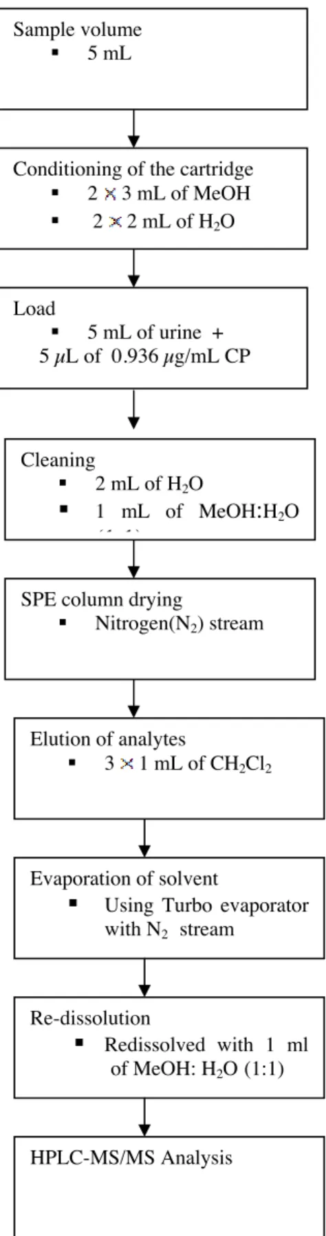
4.3.6. SPE extraction
The most important parameters in SPE are the selection of the type and amount of the sorbent, the determination of the sample volume that can be applied without loss in recovery, the composition and volume of the washing solution that can be applied without loss of the analytes, and finally the composition and volume of the elution.
For optimizing SPE methods different parameters were changed such as the cartridge stationary adsorbent, pH, solvent used for cleaning, solvent used for elution and elution volume. SPE methods were carried out using different cartridge such as Octadecyl from J.T. Baker (500 mg/3mL), Chromabond C18 ec from Machery- Nagel (1000 mg/6mL),
Strata- X from phenomene (200 mg/3mL), Lichrolut RP- select B from Merck (200 mg/3mL), Oasis HLB from Waters (150 mg/3mL), Zorbax C18 from Agilent (200 mg/mL),
Chromabond Easy from Machery- Nagel (200 mg/mL). Among them recovery values were higher with Oasis HLB and Strata- X. So further analysis were carried out using Strata-X.
Buffer PBS pH 7, buffer pH 3, urine adjusted to pH 3, normal urine were used to see the recoveries. Normal urine has high recovery so analysis was carried out with it.
For cleaning different solvents were used such as water, buffer PBS pH 7, buffer pH 3 and methanol: water (1:1). In order to remove interference from the analyte mixture of methanol: water (1:1) was used.
Whereas for elution dichloromethane, methanol were checked. To see the impact different elution volume (1ml, 2ml, 3ml, 4ml, 5ml, 6ml, 7ml, and 8ml) were used and it was found that 3ml was enough to elute the analyte.
SPE procedure used for CP determination in urine samples has been shown on figure 2.
Figure 2 Schematically presentation of SPE procedure
Cleaning
2 mL of H2O
1 mL of MeOH:H2O (1:1)
SPE column drying
Nitrogen(N2) stream
Elution of analytes
3 1 mL of CH2Cl2
Evaporation of solvent
Using Turbo evaporator with N2 stream Re-dissolution Redissolved with 1 ml of MeOH: H2O (1:1) HPLC-MS/MS Analysis Sample volume 5 mL
Conditioning of the cartridge 2 3 mL of MeOH
2 2 mL of H2O Load 5 mL of urine + 5 µL of 0.936 µg/mL CP
4.3.7. Calibration
Calibration curves were prepared with known concentrations of CP: 1) diluted in methanol: water (1:1)
2) spiked in 5ml of blank urine
Five calibrations standards were made by adding working solution of CP to 1 mL methanol: water (1:1) and directly analyzed. The CP concentrations of the standard were 0.608, 1.17, 2.34, 4.68, 9.36 ng/mL.
Figure 3 Calibration curve of CP diluted in methanol: water (1:1).
Calibrations urine samples were prepared by spiking human urine samples from unexposed subject (blank urine) with CP, in order to obtain five concentration (0.187, 0.374, 0.936, 1.4, 1.87 ng/mL). Calibration samples were extracted following the above described procedure and analyzed by HPLC-MS/MS.
5. RESULTS AND DISCUSSION 5.1. Mass spectrometry
In ESI positive ion mode the compounds yielded abundant [M+H]+ ions. This protonated molecule at m/z 261 for CP was selected as the precursors ions for the MS/MS fragmentation of the analyte. The most abundant fragment was detected at m/z 140 and proposed fragmentation is shown in figure 6.
Cleavage of the nitrogen-phosphorus bond gave fragment ion at m/z 140, resulting in the loss of di(2-chloroethyl) amine. The transition from m/z 261 to 140 was used in MRM analysis. The MRM parameter and precursor- product ion combinations used are summarized in table 6.
Table 6 MRM parameters
Compound Retention time Precursor ion Product ion CP 6.75 min 261.0 140
Figure 5 HPLC-MS/MS working in MRM mode obtained for CP standard solution. Proportion of CP fragmentation has been included.
Figure 6 (a) shows result for blank urine sample and (b) shows a typical MRM chromatogram corresponding to a blank urine sample spiked with 4.68 ng/mL of CP. No interfering peaks were presented at the retention time of CP.
a)
5.2. Liquid Chromatography:
The elution gradient and influence of mobile phase pH were studied in order to optimize the analytical performance. A short analytical column and elution gradient with 10 mM formic acid with ammonia buffer pH 3 and methanol were chose as the best compromise between retention time and ionization of analyte. Under these chromatographic conditions, CP was separated in 15 min in a flow rate 1 mL/min.
5.3. Linearity
Calibration curves showed high linearity over the concentration range 0.187 -1.87 ng/mL with correlation coefficient greater than 0.998.
5.4. Sensitivity
The limits of detection (LOD) and LOQ were determined by analyzing blank urine samples spiked with the working standard CP. LOD and LOQ were found to be 0.27 ng/mL and 0.54 ng/mL. These values allowed the analysis of CP in human urine sample at trace level.
5.5 Carry over
To determine the carry over, blank samples were injected after a sample. No carry over from analytes were seen in blank sample.
5.6 Recovery
The recovery was determined using urine sample spiked with CP (5 µL of 0.936 µg/mL in 5 mL urine). Final concentration in urine was 4.68 µg/mL. The obtained peak areas of the spiked urine standards at these levels after sample preparation were compared with the peak areas resulting from aqueous standard solutions at the same concentrations without sample preparation.
5.7. Influence of solvent used to prepare CP standard solution:
CP at concentration 4.68 ng/ml was prepared in five different solvent: a) Methanol
b) Methanol: Water (1:1) mixture c) Urine: Water ( pH=3) mixture (1:1) d) Urine: Buffer (pH=3) mixture (1:1) e) Buffer pH=3
Data obtained from CP diluted in MeOH: Water (1:1) mixture have been treated as 100 %, therefore CP diluted in all the other solutions gives different recoveries, as shown in figure 7.
Figure 7 Influence of solvent used to prepare CP standard solution.
Analysis of these CP solutions gave different chromatograms, as it has been presented in figure 8. It was found that methanol: water (1:1) mixture give highest peak area whereas those dilutions of CP with urine sample show very low recovery. Also baseline and peak shape of CP are not good probably due to presence of interferences.
Figure 8 CP diluted in a) methanol b) methanol: water (1:1) c) mixture urine: water (1:1) pH=3 d) urine: buffer pH=3 mixture e) buffer pH=3.
5.8. Matrix effects
Urine is a complex biological material, which contains urea, creatinine, uric acid, hormones, pigments and mucins. Some of them can influence extraction process using SPE (deactivation of active points at the surface of sorbent, sorption, blocking of flow channels). Therefore, the way the urine sample is treated before sorption in SPE can be crucial for recovery of CP. Urine samples, after spiking with CP were prepared in four different ways:
a) 5 mL urine + 2 mL PBS buffer (pH=7) b) 5 mL urine + 2 mL buffer (pH=3)
c) 5 mL urine acidified with ammonia to pH=3. d) Urine
CP was isolated from samples using SPE and recovery of CP has been shown in figure 9.
Figure 9 Influence of sample preparation of urine before SPE on recovery of CP.
No significant difference has been observed between urine samples prepared before SPE in different way, especially in case of Oasis HLB, Lichrolut RP-select B, Chromabond C18 ec and Chromabond Easy sorbent. For SPE, urine sample without pretreatment were used for carrying the experiment.
5.9. Influence of time of evaporation on recovery
A set of six different CP solutions in MeOH has been prepared. The content of CP was constant (4.68 ng/mL) but amount of MeOH was different (1 mL, 2 mL, 3mL). Solvent was removed using Turbo evaporator. Temperature was set on 40° C and gas pressure was 4 psi. Differences in solvent amount should result in differences in evaporation time. Dry residue was re-dissolved in the mobile phase (mixture MeOH: water 1:1, v/v) and analyzed. Results, in the form of recovery calculated according to CP standard solution without evaporation treated as 100 % are shown in Table 7.
Table 7 Influence of time of evaporation on recovery
Time required for sample evaporation
Recovery % (R1)
Recovery % (R2) Approx. 20 min. (MeOH 1 mL) 91 93 Approx. 40 min. (MeOH 2 mL) 89 93 Approx. 60 min ( MeOH 3 mL) 94 137
Sample having only 1 ml of methanol evaporates faster than sample containing 2 ml and 3 ml of solvent. Each 1 mL of MeOH needs approximately 20 minutes for complete evaporation. From table 7 we can conclude that there is no influence of time of evaporation on CP recovery. Recovery ranged from 89 to 94 % except in one sample where recovery is 137 %, this is due to human error.
5.10. Sorbent types
To check best sorbent seven different type of cartridge such as J.T baker ( Octadecyl ), Chromabond C18 ( Octadecyl), Strata-X from Phenomena ( Polymeric surface modified styrene-
divinylobenzene), Merck Lichrolut RP-select, Agilent Zorbax C18 (monomeric bonding, carbon loading 11 %), Waters Oasis HLB cartridge (hydrophilic-lipophilic balance sorbent reversed phase sorbent polymeric water- wettable reversed phase sorbent), Chromabond Easy ( polar modified polystyrene- divinylbenzene copolymer with weak anion exchanger) were used.
Figure 10 Influence of sorbent on CP recovery
Oasis, Strata, Merck and Zorbax give high recovery. So, analyses were carried out using Strata-X.
5.11.Influence of different eluent solvent on recovery:
Methanol, dichloromethane, methanol: water (1:1), methanol and ethyl acetate solvents were used for eluting the analyte in order to find suitable elution solvent. Elution was performed by adding 3 mL aliquots of different eluting solvent as mention above except methanol: water (1:1) mixture, passing them through under vacuum and collecting them separately, dried and analyzed. Whereas in case of methanol: water (1:1) elution was performed by using 1 ml and analyzed directly.
Figure 11 Influence of different eluent solvent on CP recovery.
Using methanol: water (1:1) solvent, analyte was not eluted. Whereas methanol, dichloromethane, methanol and ethyl acetate were good solvent for eluting the CP from cartridge. Among three, dichloromethane show high recovery of CP in Stata –X catridge. Thus, dichloromethane was found suitable eluting solvent for CP analyte.
5.12. Stability study of CP solutions
In order to observe the degradation process of CP in respect to time extract from urine samples were kept at ambient temperature and after the degradation process started they were moved to refrigerator at 4 °C. All extracts and CP standard solution were dissolved in the mixture MeOH: H2O (1:1) and were kept in transparent vials. All the samples were analyzed
after every three days and recoveries were calculated (Table 8) according to results obtained the very first day of analysis (100 %).
Table 8 Degradation of CP in time
Sorbent used
Recovery [ %]
Day 1 Day 3 Day 6 Day 9
Extract from urine sample with buffer pH=3
Extract from Chromabond C18 ec 100 71 46
Extract from Chromabond easy 100 73 60 53 Extract from Octadecyl 100 50 37 33 Extract from oasis HLB 100 51 37 29 Extract from Lichrolut RP- select 100 48 37 33 Extract from Strata- X 100 48 36 32 Extract from Zorbax C18 100 38 29 26
Extract from urine sample with PBS buffer pH=7
Extract from Chromabond C18 ec 100 92 77 69 Extract from Chromabond easy 100 78 70 59 Extract from oasis HLB 100 54 43 38 Extract from Lichrolut RP- select B 100 - - -
Extract from Strata- X 100 50 39 35 Extract from Zorbax C18 100 59 48 43 Recovery of CP was found almost half reduced after keeping the extract at ambient temperature which represented in Figure 13 and 14. These results indicated an approximately deterioration of 50 percent in day 3 and approximately 67 percent over day 9. This information supports that both standard and samples should be analyzed as soon as possible after the samples are collected to avoid error due to loss of potency. Depending on the environmental conditions in
degradation processes which can naturally happen. Photo degradation, as a chemical reaction that occurs under the influence of photons or light, could take place and cause decrease of CP content in the extract. We believe that bacteria presented in urine samples were removed during SPE. However, we do not have scientific proof because it was not the subject of the investigations.
Figure 12 Stability study of extract urine (urine samples diluted with buffer pH=3).
6. APPLICATION OF THE METHOD
To assess the feasibility and effectiveness of this SPE- LC-MS/MS method, urine samples of health workers were collected and analyzed. Fifteen samples were collected and store at – 80 °C until analysis. Two blank samples were collected from six year boy and twenty six year old girl. Spiked sample was prepared and analyze. Chromatogram is shown below (Figure 14). The CP results were below the analytical detection limits in all samples.
7. CONCLUSION
According to IARC, CP is human genotoxic carcinogen [1, 11]. Several reports have discussed exposure to CP during occupational activities and concluded that it should be avoided because any detectable level is considered to be hazardous. In order to obtain accurate measurements of CP in urine samples SPE methods was optimized. Optimization of SPE method was carried out by changing different parameter such as different type and amount of sorbent, composition and volume of elution, washing solvent. SPE was effective with no interfering peaks from matrix compound and recovery was above 75%.
After optimizing SPE method, CP separation conditions of the HPLC-MS/MS analysis was achieve by changing different parameter such as suitable mobile phase and its composition, flow rate, scan type, polarity. LC/MS/MS detection was performed using triple quadrupole mass spectrometer working in MRM mode. The samples were prepared using SPE and analyzed using a gradient separation. Total analysis time was 15 minute. Calibration curve were made to see the instrument performance and SPE procedures which show good linearity.
A sensitive, specific and accurate HPLC/MS/MS has been developed for the analysis of CP in human urine. This SPE-LC-MS/MS method was applied for the analysis of urine samples of health workers. Fifteen samples urine was collected from Medical University of Gdansk, who were exposed with CP during their work in pharmacy. Two blank samples were collected from six year boy and twenty six year girl. In all sample CP was found below LOD.
The stability of the CP was studied. Analyses were conducted on the day prepared (day 1) and days 3, 6 and 9. These results indicated an approximately deterioration of 50 percent in day 3 and approximately 67 percent over day 9. This information supports that both standard and samples should be analyzed as soon as possible after the samples are collected to avoid error due to loss of potency.
8. SUMMARY
Biological samples urine are much more complex due to presence of proteins, salts, acids, bases and various organic compounds with similar chemistry to analytes of interest. As a result, the extraction methods for biological samples have been difficult. The main task was to remove maximum interfering substance from urine matrix and conversion of analytes into a more suitable form for injection. Therefore, optimization of SPE method was done by changing different parameter such as different type and amount of sorbent, composition and volume of elution, washing solvent. The use of 24 well SPE plate for matrix purification significantly allows the use of small urine and solvent volumes, reduces the sample preparation time and ensures a high throughput, thus allowing the routine biological monitoring of CP as indices of human exposures. In order to investigate the effectiveness of the SPE method tests were repeated on Strata-X cartridge and method showed good recovery (above 75 %) of CP.
After optimizing SPE method, CP separation conditions of the HPLC-MS/MS analysis was achieve by changing different parameter such as suitable mobile phase and its composition, flow rate, scan type, polarity. The LC separation was performed on Lichrocart® 100 RP-18 column (125 4 mm, particle size of 5 µm) with 1 mM formic acid with ammonia buffer pH 3 and methanol using gradient program at a flow rate 1 mL/min with total run time 15 min. A reversed phase HPLC system was interfaced with an ESI source coupled to tandem mass spectrometry. The triple quadrupole mass spectrometer was operated in positive ion mode and MRM was used for analysis of CP. LOD and LOQ were 0.27 ng/mL and 0.54 ng/mL. A sensitive, specific and accurate HPLC/MS/MS has been developed for monitoring CP in urine samples. The use of reversed phase HPLC coupled to tandem mass spectrometry has facilitated the analysis of CP in short retention time.
Fifteen urine samples were collected from medical personnel of Medical University of Gdansk, who were exposed with CP during their work in pharmacy. SPE-HPLC-MS/MS method was applied for the analysis of health workers and all urine samples from university workers has CP below analytical limit of detection.
9. REFERENCES
1. IARC. Monographs on the evaluation of the carcinogen risk of chemicals to humans. No. 4, IARC. Lyon, France, 1982.
2. K.T. Kiffmeyer, C. Kube, S. Opiolka, G.K. Schmidt, G. Schoppe, J.M.P. Sessink, Vapour pressures, evaporation behavior and airborne concentrations of hazards drugs: implications for occupational safety, Pharmeaceut J, 268(2002)331-337. 3. T. H. Connor, M. Shults, M.P. Fraser, Determination of the vaporization of
solutions of mutagenic antineoplastic agents at 23 and 37 C using a desiccators technique, Mutat Res.,470(2000)85-92.
4. B.G. Valanis, W.M. Vollmer, K.T. Labuhn, A.G. Glass, Association of antineoplastic drug handling with acute adverse effects in pharmacy personnel, Am.
J. Hosp. Pharm., 50 (1993)455-462.
5. B. Valanis, W.M. Voller, P. Steele, Occupational exposure to antineoplastic agents: Self reported miscarriages and stillbirths among nurses and pharmacists, J. Occup.
Environ. Med., 41(1999)632-638.
6. T.T. Kensler, R. J. Behme, D. Brooke, High performance liquid chromatographic analysis of cyclophosphamide, J. Pharm. Sci., 68(1979)172.
7. L.C. Burton, C.A. James, Rapid method for the determination of ifosfamide and cyclophosphamide in plasma by high performance liquid chromatography with solid phase extraction, J. Chromatogr., 431(1988)450-454.
8. A.M. Rustum, N.E. Hoffman, Determination of cyclophosphamide in whole blood and plasma by reversed phase high performance liquid chromatography, J.
Chromatogr., 422(1987)125-134
9. M.M. Ellaithy, M. F. El-Tarras, N.B. Tadros, M. M. Amer, J. Assoc. Anal. Chem. 67(1984)679.
10. M.E. De Jounge, A.D.R. Huitemia, S. Rodenhuis, J. H. Beijnen, Clinical pharmacokinetics of cyclophosphamide, clin. Pharmacokinet. 44(2005)1135 11. IARC. Monographs on the evaluation of the carcinogenic risk of chemicals to man.
Geneva: World health organization, IARC, 1972-present (Multivolume work), 26(1981)166.
12. C.Sottani, P. Rinaldi, E. Leoni, G. Poggi, C. Teragni, A. Delmonte, C. Minoia, Simultaneous determination of cyclophosphamide, ifosfamide, doxorubicin,
epirubicin and daunorubicin in human urine using high performance liquid chromatography/ electrospray ionization tandem mass spectrometry: bioanalytical method validation, Rapid Commun. Mass Spectrom., 22(2008)2645-2659.
13. M. Hirst, D.G. Millis, Tse S. Levin and D. F. White, Occupational exposure to cyclophosphamide, Lancet, 1(1984)186.
14. L. Wang, C. Albansi, V. Faucet- Marquis, A. Pfohl-Leszkowicz, C. Dorandeu, B. Marion, C. Causserand, Cyclophosphamide removal from water by nanaofiltration and reverse osmosis membrane, Water Res, 43(2009)4115-4122.
15. J. E. F. Reynolds, A. B. Prasad. (eds.) Matrindale- The extra pharmacopoeia. 28th ed. London: The Pharmaceutical Press, 1982, p. 199.
16. R. J. Lewis, Sr (Ed.). Hawley’s Condensed Chemical Dictionary. 12th ed. New York, NY: Van Norstrand Rheinhold Co., 1993p. 341.
17. IARC. Monographs on the evaluation of the carcinogenic risk of chemicals to man. Geneva: World health organization, IARC, 1972-present (Multivolume work), 9(1975) 136.
18. T. K. H. Chang, G. F. Weber, C. L. Crespi, D. J. Waxman, Differential activation of cyclophosphamide and ifosphamide by cytochromes P-450 2B and 3 A in human liver microsomes, Cancer Res., 53(1993)5629-5637.
19. R. F. Struck, M. C. Kirk, M. H. Witt, W. L. Russell, Isolation and mass spectral identification of blood metabolites of cyclophosphamide: Evidence for phosphoramide mustard as the biologically active metabolites, Biomed. Mass
Spectrom., 2(1975)46-52.
20. H. Blomgren, M. Hallstrom, Possible role of acrelein in 4-hydroperoxycyclophosphamide induced cell damage in vitro, Methods Find Exp
Clin Pharmacol, 13(1991)11-4
21. D. L. Hill, W. R. Laster, R. F. Struck, Enzymatic metabolism of cyclophosphamide and nicotine and production of toxic cyclophosphamide metabolite, Cancer Res., 32(1972)658-665.
37(1988)301-24. J. Z. Fuks, M.J. Egorin, J. Aisner, S. S. Ostrow, M. E. Klein, N. R. Bachur, M. Colvin, P. H. Wiernik, Cyclophosphamide and dimethysulfoxide in the treatment of squamous carcinoma of the drug, Cancer Chemother and Pharmacol, 6(1981) 117-120
25. I. Jardine, C. Fenselau, M. Appler, M-N. Kan, R. B. Brundrett, M. Colvin, Quantitation by gas chromatography- chemical ionization mass spectrometry of cyclophosphamide, phosphoramide mustard and nornitrogen mustard in the plasma and urine of patients receiving cyclophosphamide therapy, Cancer Res., 38(1978)408-415.
26. M. J. Tasso, A. V. Boddy, L. Price, R. A. Wyllie, A. D.J. Pearson, J. R. Idle, Pharmacokinetics and metabolism of cyclophosphamide in pediatric patients,
Cancer Chemother and Pharmacol, 30(1992)207-211.
27. S. M. Yule, A. V. Boddy, M. Cole, L. Price, R. Wyllie, M. J. Tasso, A. D. J. Pearson, J. R. Idle, Cyclophosphamide metabolism in children, Cancer Res., 55(1995)803-809.
28. S. M. Yule, L. Price, A. D. J. Pearson, A. V. Boddy, Cyclophosphamide and ifosfamide metabolites in the cerebrospinal fluid of children, Clin. Cancer Res., 3(1997)1985-1992.
29. D. Busse, F. W. Busch, F. Bohnenstengel,M. Eichelbaum, P. Fischer, J. Opalinska, K. Schweizer, H. K. Kromer, Dose escalation of cyclophosphamide in patients with breast cancer: consequences for pharmacokinetics and metabolism, J. Clin. Oncol., 15(1997)1885-1896.
30. http://www.medicinenet.com/cyclophosphamide/article.htm
Accessed date: 10 january 2010.
31. M. Kimura, T. Tominaga, Y. Takatsuka, M. Toi, R. Abe, H. Koyama, S. Takashima, Y. Nomura, S. Miura, I. Kimijima, H. Tashiro, Y. Ohashi, Randomized trial of cyclophosphamide, epirubicin and fluorouracil chemotherapy compared with cyclophosphamide, methotrexate and fluorouracil with node positive breast cancer in Japan, Breast Cancer, online published 3 July 2009.
32. M. Colleoni, A. Rocca, M. T. Sandri, L. Zorzino, G. Masci, F. Nole, G. Peruzzotti, C. Robertson, L. Orlando, S. Cinieri, F de Braud, G. Viale, A. Goldhirch, Low dose
oral methotrexate and cyclophosphamide in metastic breast cancer: antitumor activity and correlation with vascular endothelial growth factor levels, Annals of
Onco., 13(2002)73-80
33. S.E. Jones, B.G.M. Durie, E. Salmon, Combination chemotherapy with adriamycin and cyclophosphamide for advanced breast cancer, CA Cancer J Clin,36(1975)90-97.
34. J.M. J. Garcia, A. Sanchez, B. Pajares, E. Perez, L. Alonso, E. Alba, Combined oral cyclophosphamide and bevacizumab in heavily pre-treated ovarian cancer, Clin
Transl Oncol,10(2008)583-586.
35. D. Raghavan, K. Cox, B.S. Pearson, G. J. Coorey, J. Rogers, W. H. Watt, A.s. Coates, E. McNeil, J.J. Grygiel, oral cyclophosphamide for the management of hormone refractory prostate cancer, British J. Urology, 72(2008)625-628.
36. L. Brandes, S. Bracken, E. Ramsey, N,-diethy-2-[4-(phenylmethyl) phenoxy ethanamine in combination with cyclophosphamide: an active, low-toxicity regimen for metastatic hormonally unresponsive prostate cancer, J. Clin Oncol, 13(1995) 1398-1403.
37. A. Nicolini, P. Mancini, P. Ferrari, L. Anselmi, G. Tartarelli, V. Bonazzi, A. Carpi, R. Giardino, Oral low dose cyclophosphamide in metastatic hormone refractory prostate cancer, Biomed and Pharmacother, 58 (2004) 447-450.
38. S. Bracarda, M. Tonato, P. Rosi, V. De Angelis, E. Mearini, S. Cesaroni, P. Fornetti, M. Porena, Oral estramustine and cyclophosphamide in patients with metastatic hormone refractory carcinoma , CA Cancer J Clin, 88(2000) 1438-1444. 39. Jr. PA. Bunn, FA. Greco, L. Einhorn, Cyclophosphamide, doxorubicin and
etoposide as first line therapy in the treatment of small cell lung cancer, Semin
Oncol, 3(1986)45-53.
40. R. Arriagada, T.L. Chevalier, J.P. Pignon, A. Riviere, I. Monnet, P. Chomy, C. Tuchais, M. Tarayre, P. Ruffie, Initial chemotherapeutic doses and survival in patients with limited small cell lung cancer, N Engl J Med, 329(1993)1848-1852. 41. D.T. Boumpas, H.A. Austin, J.E. Balow, E. M. Vaughan, C. H. Yarboro, J.H.
42. M. F. Gourley, H. A. Austin, D. Scott, C. H. Yarboro, E. M. Vaughan, J. Muir, D. T. Boumpas, J. H. Klippel, J. E. Balow, A. D. Steinberg, Methylprednisolone and cyclophosphamide alone or in combination, in patients with lupus nephritis: A randomized, control trial, Ann of Intern med, 125(1996) 549-557.
43. C.A. Wallace, D.D Sherry, Trial of intravenous pulse cyclophosphamide and methyprednisolone in the treatment of severe systemic-onset juvenile rheumatoid arthritis, Arthritis care and research, 40(2005)40.
44. H. J. Williams, J. C. Reading, J. R. Ward, W.M. O’Brien, Comparison of high low dose cyclophosphamide therapy in rheumatoid arthritis, Arthritis care and research, 23(2005) 521-527.
45. D. E. Gladstone, K. W. Zamkoff, L. Krupp, R. Peyster, P. Sibony, C. Christodoulou, E. locher, P. K. Coyle, High dose cyclophosphamide for moderate to severe refractory multiple sclerosis, Arch Neurol., 63(2006)1388-1393.
46. B. W. Guttman, R. P. Kinkel, J. A. Cohen, R. M. Ransohoff, K. Schwetz, D. Gogol, M. Namey, R. A. Rudick, Treatment of fulminant multiple sclerosis with intravenous cyclophosphamide, The Neurologist, 3(1997) 186.
47. R.P. Baughman, E. E. Lower, Use of intermittent, intravenous cyclophosphamide for idiopathic pulmonary fibrosis, Chest, 102(1992) 1090-1094.
48. D. A. Zisman, J. P. Lynch III, G. B. Toews, E. A. Kazerooni, A. Flint, F. J. Martinez, Cyclophosphamide in the treatment of idiopathic pulmonary fibrosis: A prospective study in patients who failed to respond to corticosteroids, Chest, 117(2000)1619-1626.
49. L. Guillevin, J. F. Cordier, F. Lhote, P. Cohen, B. Jarrouse, I. Royer, P. Lesavre, C. Jacquot, P. Bindi, P. Bielefeld, J. F. Desso, F. Detree, A. Dubois, E. Hachulia, B. Hoen, D. Jacomy, C. Seigneuric, D. Lauqque, M. Stern, M. L. Boursier, Arthritis
and rheumatism, 40(1997)2187-2198.
50. M. Haubitz, U. Frei, U. Rother, R. Brunkhorst, K. M. Koch, Cyclophosphamide pulse therapy in Wegener granu;omatosis, Neprol Dial Transplant 6(1991)531-535. 51. M. J. Reza, L. Dornfeld, L. S. Goldberg, R. Bluestone, C. M. Pearson, Wegener
granulomatosis, Arthritis care and research, 18(2005)501-506.
52. A. Fabbri, M. Lenoci, A. Gozzetti, I. Chitarrelli, F. Olcese, D. Raspadori, M. Gobbi, F. Lauria, Low dose oral fludarabine plus cyclophosphamide as first line treatment
in elderly patients with indolent non- Hodgkin lymphomas, Brit J Haematol, 139(2007)90-93.
53. R. I. Fisher, E. R. Gaynor, S. Dahlberg, M. M. Oken, T. M. Grogan, E. M. Mize, J. H. Glick, C. A. Coltman, T. P. Miller, Comparison of a standard regimen with three intensive chemotherapy regimens for advanced non- hodgkin’s lymphoma, Annals
of Internal medicine, 328 (1993)1002- 1006.
54. P. Sonneveld, M. de Ridder, H. van der Lelie, K. Nieuwenhius, H. Schouten, A. Mulder, I van Reijswould, W. Hop, B. Lowenberg, Comparison of doxorubicin and mitoxantrone in the treatment of elderly patients with advance diffuse non – hodgkin’s lymphoma using CHOP versus CNOP chemotherapy, J. Clinic. Oncol., 13() 2530-2539
55. A. Reiner, T. Gernsheimer, S. Slichter, Pulse cyclophosphamide therapy for refractory autoimmune thrombocytopenic purpura, 85(1995) 351- 358.
56. M. Verlin, R. K. Laros, J. A. Penner, Treatment of refractory thrombocytopenic purpura with cyclophosphamide, American Journal of Hematology 1(2006)97-104. 57. R.K. Laros, J. A. Penner, Refractory thrombocytopenic purpura treated successfully
with cyclophosphamide, JAMA, 215(1971)445-449.
58. P. Sanchez, J. Anguita, T. Pintado, Use of cyclophosphamide in the treatment of thrombotic thrombocytopenic purpura complicating systemic lupus erythematosus: report of two cases, Annals of Hematology, 78( 1999)285-287.
59. A. Fauci, J. Doppman, S. Wolff, Cyclophosphamide induced remissions in advanced polyarteritis nodosa, Am J Med., 64(1978) 890-894.
60. V. M. Moyo, D. Smith, I. Brodsky, P. Crilley, R. J. Jones, R. A. Brodsky, High dose cyclophosphamide for refractory autoimmune hemolytic anemia, Blood, 100(2002) 704-706.
61. R. A. Brodsky, A. R. Chen, I. Brodsky, R. J. Jones, High dose cyclophosphamide as salvage therapy for severe aplastic anemia, Experimental Hematology, 32( 2004) 435-440.
64. N.J.K. Simpson, Solid Phase Extraction – Principles Strategies and Applications, Marcel Dekker, New York 1998.
65. R.E. Majors, New designs and formats in solid phase extraction sample preparation,
LC/GC Int. September (1998) 8–16.
66. C.F. Poole, New trends in solid phase extraction, Tr in Anal. Chem., 6(2003)362-373.
67. M.C. Hennion, C. Cau-Dit-Coumes, V. Pichon, Trace analysis of polar organic
pollutants in aqueous samples: tools for the rapid prediction and optimization of the solid phase extraction parameters, J. Chromatogr. A, 823 (1998) 147.
68. M.C. Hennion, Solid phase extraction: method development, sorbents and coupling with liquid chromatography, J. Chromatogr. A, 856(1999)3-54.
69. C. Sottani, R. Turci, L. Perbellini, C. Minoia, Liquid- Liquid extraction procedure for trace determination of cyclophosphamide in human urine by high performance liquid chromatography tandem mass spectrometry, Rapid Commun. Mass
Spectrom., 12(1998)1063-1068.
70. D. Kasel, A. Jetter, S. Harlfinger, W. Gebhardt, U. Fuhr, Quantification of cyclophosphamide and its metabolites in urine using liquid chromatography/ tandem mass spectrometry, Rapid Commun. Mass Spectrom., 18(2004) 1472-1478. 71. A. Barbieri, L. Sabatini, P. Indiveri, R. Bonfiglioli, V. Lodi, F. s. Violante,
Simultaneous determination of low levels of methotraxate and cyclophosphamide in human urine by micro liquid chromatography/ electrospray ionization tandem mass spectrometry, Rapid Commun. Mass Spectrom., 20(2006)1889-1893.
72. B. Karahalil, K.I. Akkoyunlu, Determination of urinary cyclophosphamide in oncology nurses handling antineoplastic drugs by gas chromatography-mass spectrometry, J. Pharm. Sci., 28 (2003)125-130
73. M. Hedmer, P. Hoglund, E.Cavallin- Stahl, M. Ablin, A. G. Jonsson; Validation of urinary excretion of cyclophosphamide as biomarker of exposure by studying its renal clearance at high and low plasma concentrations in cancer patients; Int. Arch.
Occup. Environ. Health; 81 (2008)285-293.
74. R. Difrancesco, J.J. Griggs, J. Donnelly, R. Dicenzo, Simultaneous analysis of cyclophosphamide, doxorubicin and doxorubicinol by liquid chromatography coupled to tandem mass spectrometry, J. Chromatogr. B, 852(2007)545-553.
75. C. Ekhart, A. Gebretensae, H. Rosing, S. Rodenhuis, Jos H. Beijnen, A.D.R. Huitema, Simultaneous quantification of cyclophosphamide and its active metabolite 4- hydroxycyclophosphamide in human plasma by high performance liquid chromatography coupled with electrospray ionization tandem mass spectrometry, J. Chromatogr. B, 854(2007)345-349.
76. L. Sabatani, A. Barbieri, M. Tosi, F.S. Violante, A new high performance liquid chromatographic/ electrospray ionization tandem mass spectrometric method for the simultaneous determination of cyclophosphamide, methotrexate and 5- fluorouracil as markers of surface contamination for occupational exposure monitoring, J. Mass
spectrum., 40(2005)669-674.
77. F. Baumann, C. Lorenz, U. Jaehde, R. Preiss, Determination of cyclophosphamide and its metabolites in human plasma by high performance liquid chromatography- mass spectrometry, J. Chromatogr. B, 729(1999)297-305.
78. T.F.Kalhorn, S. Ren, W.N. Howald, R.F. Lawrence,J. T. Slattery, Analysis of cyclophosphamide and five metabolites from human plasma using liquid
chromatography- mass spectrometry and gas chromatography nitrogen phosphorous detection, J. Chromatogr. B, 732(1999)287-298.
79. I. Martins, J. O. Souza, A. L. Sanson, E. P. Vieira, A. G. Paiva, Simultaneous determination of cyclophosphamide and ifosfamide in plasma using SPE-HPLC_UV methods, Lat. Am. J. Pharm, 28(2009)41-6
80. C. Sottani, R. Turci, R. Schierl, R. Gaggeri, A. Barbieri, F. S. Violante, C. Mionia, Simultaneous determination of gemcitabine, taxol, cyclophosphamide and
ifosfamide in wipe samples by high performance liquid chromatography/ tandem mass spectrometry: protocol of validation and uncertainty of measurement, Rapid
Commun. Mass Spectrom., 21(2007)1289-1296.
81. M. Tanimura, K. Yamada, S. Sugiura, K. Mori, H. Nagata, K. Tadokora, T. Miyake, Y. Hamaguchi, P. Sessink, T. Nabeshima, An environmental and biological study of occupational exposure to cyclophosphamide in the pharmacy of a Japanese
82. W. Fransma, S. Peelen, S. Hilhorst, N. Roeleveld, D. Heederik, H. Kromhout, A polled analysis to study trends in exposure to antineoplastic drugs among nurses,
Ann. Occup. Hyg., 51(2007)231-239.
83. C. Minoia, R. Turci, C. Sottani, A. Sehiavi, L. Perbellini, S. Angeleri, F. Draicchio, P. Apostoli, Application of high performance liquid chromatography/ tandem mass spectrometry in the environmental and biological monitoring of health care
personnel occupationally exposed to cyclophosphamide and ifosfamide, Rapid
Commun. Mass Spectrom., 12(1998)1485-1493.
84. C. Sottani, R. Turci, R. Schierl, R. Gaggeri, A. Barbieri, F. S. Violante, C. Mionia, Simultaneous determination of gemcitabine, taxol, cyclophosphamide and
ifosfamide in wipe samples by high –performance liquid chromatography/ tandem mass spectrometry: protocol of validation and uncertainty of measurement, Rapid
Commun. Mass Spectrom., 21(2007)1289-1296.
85. N. Sadagopan, L. Cohen, B. Roberts, W. Collard, C. Omer, Liquid chromatography-tandem mass spectrometric quantitation of cyclophosphamide and its hydroxyl metabolite in plasma and tissue for determination of tissue distribution, J.
Chromatogr. B, 759(2001)277-284.
86. T.F. Kalhorn, W.N. Howald, S. Cole, B. Phillips, J. Wang, J. T. Slattery, J.S. McCune, Rapid quantitation of cyclophosphamide metabolites in plasma by liquid chromatography-mass spectrometry, J. Chromatogr.,835(2006)105-113.
This document was created with Win2PDF available at http://www.win2pdf.com.
The unregistered version of Win2PDF is for evaluation or non-commercial use only. This page will not be added after purchasing Win2PDF.