UNIVERSIDADE DE LISBOA
FACULDADE DE MEDICINA DENTÁRIA
COMPARATIVE ANALYSIS OF SIMULATED
ROOT CANAL ANATOMY AFTER
DIFFERENT PREPARATION SYSTEMS
SUSANA COELHO
DISSERTAÇÃO
MESTRADO INTEGRADO EM MEDICINA DENTÁRIA
UNIVERSIDADE DE LISBOA
FACULDADE DE MEDICINA DENTÁRIA
COMPARATIVE ANALYSIS OF SIMULATED
ROOT CANAL ANATOMY AFTER
DIFFERENT PREPARATION SYSTEMS
Susana Coelho
Dissertação orientada
Pelo Prof. Doutor António Ginjeira
MESTRADO INTEGRADO EM MEDICINA DENTÁRIA
i
AGRADECIMENTOS
Ao meu orientador, Professor Doutor António Ginjeira, pela orientação, apoio, simpatia e disponibilidade durante a elaboração desta dissertação. Por todos os ensinamentos que transmitiu.
À Mónica Amorim, pela amizade, confiança e disponibilidade sempre que foi necessário.
À Ana Paula Tavares, Isabel Pereira e Sónia Tavares. Por toda a amizade, confiança, companheirismo. Pelos ótimos momentos e imensas gargalhadas dadas sempre que nos juntamos. Convosco tudo foi mais fácil. A nossa amizade é o que de melhor levo destes anos. Espero que o que este curso uniu, a vida nunca separe. À Ana Paula Tavares por ser a melhor dupla que podia ter tido. Pelo apoio e presença constantes, nos bons momentos mas também nos menos bons. Guardarei para sempre no meu coração todos eles com muito carinho, alegria e saudade. À Isabel Pereira por toda a energia, alegria e boa disposição que transmitiu. Pela disponibilidade sempre demonstrada para ajudar a resolver qualquer situação e pela companhia de todos os momentos. À Sónia Tavares pela alegria, disponibilidade, bondade, confiança e apoio constantes ao longo destes últimos anos. Pessoas como tu são raras. Sempre disposta a ajudar com um sorriso nos lábios e sem esperar nada em troca. Muito obrigada!
Ao Nuno Martins, por todo o carinho, amor, paciência, compreensão, ajuda e apoio incondicionais. Conseguiste estar sempre perto, mesmo estando longe. Que daqui em diante a distância acabe e possamos caminhar na mesma direção juntos.
À minha avó Alice, uma das pessoas mais importantes da minha vida, por toda a ternura, carinho, segurança e apoio que desde sempre me tem transmitido. Por seres sempre um dos meus portos de abrigo. Estás sempre no meu coração.
iii
Aos meus fiéis amigos de 4 patas, aos que estão presentes e à que já partiu. Por todo o carinho, companhia, descontração, momentos de pura felicidade e diversão. Convosco este percurso tornou-se mais fácil.
Por último, e mais importante, aos meus pais e irmão. Às pessoas mais importantes da minha vida. Àquelas que estiveram presentes sempre que necessitei e que aconteça o que acontecer, sei que estarão lá para me apoiar. O que hoje sou, quem sou, onde cheguei e o que conquistei, a vós vos devo. Agradeço por todo o amor, carinho, compreensão, confiança, ajuda e força. Por sempre acreditarem em mim, nunca me deixarem cair, por me darem asas para voar e fazerem de tudo para eu conseguir atingir os meus objetivos. As palavras nunca vão ser suficientes para vos agradecer. Prometo que nunca vos vou desiludir.
v
RESUMO
INTRODUÇÃO: Um dos principais objetivos da preparação do canal radicular é seguir a sua anatomia original, mantendo a curvatura do mesmo e a relação espacial do foramen apical com os tecidos periapicais e a superfície radicular. À medida que a curvatura do canal radicular aumenta, mais difícil é ter uma preparação adequada do canal, pelo que a instrumentação mecânica continua a ser uma das tarefas mais difíceis do tratamento endodôntico.
A evolução dos instrumentos na área da endodontia tem ocorrido ao longo do tempo. Tradicionalmente a preparação do canal radicular é efetuada com limas manuais de aço inoxidável,aumentado o tempo de tratamento e o risco de fratura do instrumento assim como outros acidentes iatrogénicos durante a instrumentação. Para tentar solucionar estas questões, a instrumentação mecanizada em rotação contínua com o uso de limas de níquel-titânio (NiTi) foi introduzida em 1992 pelo Dr. John T. McSpadden. Devido à constante evolução nesta área, em 2008, surgiu um novo conceito de instrumentação mecanizada com movimento reciprocanteou alternado, apresentado por Ghassan Yared, com a utilização de uma lima única de NiTi com o objetivo de diminuir a quantidade de instrumentos rotatórios necessários para a preparação do canal, simplificar a técnica e, consequentemente reduzir o tempo de trabalho e possibilidade de acidentes iatrogénicos. Neste movimento reciprocante, o instrumento gira no sentido anti-horário cortando a dentina e horário, desprendendo-se da mesma. Assim, verifica-se um avanço automático do instrumento através do canal ao fim de cada ciclo, verifica-sendo necessária uma mínima pressão no sentido apical.
Com o intuito de melhorar as propriedades mecânicas dos instrumentos endodônticos, particularmente a flexibilidade e a resistência à fratura, surgem também tratamentos térmicos, nomeadamente o tratamento M-wire ™ (Tulsa Dentsply em 2007) e, mais tarde, uma nova geração de instrumentos com NiTi Blue-Wire que experimentam um tratamento térmico e mecânico complexo, resultando numa camada visível de óxido de titânio na superfície do instrumento.
OBJETIVO: Comparar a eficiência na preparação canalar em blocos de acrílico com curvatura em forma de S, analisando a quantidade de material removido e consequentemente a manutenção original do canal radicular, de dois sistemas de limas:
vi
Reciproc® blue, lima única de NiTi com movimento reciprocante e iRace, um sistema de limas com movimento rotativo contínuo.
MATERIAIS E MÉTODOS: Foi utilizada uma amostra total de 20 blocos de resina com canais curvos em forma de S, aleatoriamente divididos em 2 grupos (n=10), cada uma preparada até um calibre de 0.25mm e a um comprimento de 15,5mm: grupo A, Reciproc® blue e grupo B, sistema de limas iRace. Em cada amostra pré-instrumentada, o interior do canal foi preenchido com tinta de água castanha e nas amostras pós-instrumentadas com tinta de água laranja, com o intuito de se visualizar melhor o canal. Numa plataforma específica e numa posição pré-determinada os blocos foram posicionados e fotografados com uma câmara digital, antes e após a preparação mecânica. O protocolo utilizado para instrumentar a amostra dos dois grupos seguiu uma sequência baseada nas instruções fornecidas pelos fabricantes, para que os resultados sejam mais precisos, aumentando a validade do estudo. As imagens foram sobrepostas no programa Pixlr Editor e posteriormente analisadas. Os parâmetros analisados incluíram quantidade total de material removido ao nível das paredes interna e externa das curvaturas coronal e apical do canal radicular simulado. Para determinar os limites da curvatura coronal e apical foi utilizado o programa Rhinoceros® software, recorrendo ao método de Pruett, sendo neste caso, traçadas 3 linhas; a primeira corresponde ao longo eixo da porção coronal do canal e a segunda ao longo do eixo da primeira curvatura (formando um ponto de encontro no desvio do canal), e seguidamente são marcados dois pontos que definem o início e o fim da curvatura coronal. A curvatura apical é definida através da segunda linha traçada na curvatura coronal e uma terceira linha traçada segundo o longo eixo da curvatura apical, sendo depois marcados também dois pontos que correspondem ao início e fim da curvatura apical. Definidas as curvaturas, serão analisados o transporte interno e externo de cada curvatura. No programa ImageJ®foram realizadas as medições do transporte de resina na imagem com a sobreposição do canal pré e pós-instrumentado. As medições foram efetuadas tendo como limites a margem do canal pré-instrumentado e a margem do canal pós-instrumentado, ao nível da curvatura coronal e apical.
Foi realizada também uma avaliação qualitativa da manutenção ou não, da curvatura original do canal, tendo sido escolhidos seis examinadores (dois especialistas em endodontia, dois médicos dentistas inexperientes e dois alunos do curso de medicina dentária) que avaliaram seis imagens escolhidas aleatoriamente, três para cada grupo de instrumentos.
vii
A análise estatística foi executada com o programa de estatística IBM SPSS versão 23.0, com recurso ao teste de normalidade Shapiro-Wilk e ao teste Mann-Whitney U uma vez que se tratavam de comparações entre grupos de dimensão reduzida e em que se verificou rejeição ao teste de normalidade. Este último teste foi utilizado para efectuar a comparação do transporte ocorrido entre os grupos. Foram considerados valores estatisticamente significativos com p <0.05.
RESULTADOS: Relativamente ao transporte total, não houve diferença estatisticamente significativa na quantidade total de material removido entre os dois grupos. Em relação ao transporte ocorrido na curvatura coronal e apical houve diferenças estatisticamente significativas entre os dois grupos. Verificou-se que com o sistema Reciproc® blue houve uma quantidade significativamente maior de material removido ao nível da curvatura coronal, enquanto que o sistema de limas iRace/iRace Plus demonstrou uma quantidade significativamente maior de material removido na curvatura apical, causando assim maior modificação das respetivas curvaturas. A diferença entre os dois sistemas de limas no que diz respeito ao transporte ocorrido na parede interna foi estatisticamente significativa, apenas na curvatura coronal, observando-se mais transporte com o sistema Reciproc® blue. Relativamente à parede externa, em ambas as curvaturas, coronal e apical, se registaram diferenças estatisticamente significativas entre os grupos. Na parede externa da curvatura coronal, foi o sistema de limas Reciproc® blue que removeu maior quantidade de material, e na mesma parede da curvatura apical foi o sistema de limas iRace/iRace Plus que demonstrou uma quantidade significativamente maior de material removido.
Relativamente à avaliação efetuada pelos examinadores verificou-se que a experiência do clínico causou mais diferenças na avaliação das imagens no grupo do sistema Reciproc® blue, mas a grande maioria das avaliações indica que existe manutenção da forma original do canal ou poucas alterações na mesma, tendo em conta as curvaturas coronal e apical. Quanto ao grupo do sistema iRace/iRace Plus as avaliações registadas indicam a manutenção da forma original do canal, considerando ambas as curvaturas.
DISCUSSÃO E CONCLUSÃO: A instrumentação do canal radicular é uma das etapas mais importantes do tratamento endodôntico. É essencial para a eficácia de todos os procedimentos subsequentes, incluindo a desinfeção química e obturação do canal
viii
radicular. A preparação de um canal curvo, especialmente um canal de curvatura dupla (em forma de S) é um dos procedimentos mais desafiadores. A análise das modificações na curvatura do canal após a instrumentação tem sido amplamente utilizada para avaliar a eficácia de uma técnica, ou das propriedades mecânicas de um instrumento, na manutenção da anatomia do canal original.
Neste estudo, para comparar a capacidade de moldagem do canal pelos diferentes instrumentos e avaliar a manutenção da anatomia original foi utilizada a técnica de sobreposição de fotografias pré e pós-instrumentação dos blocos de resina em forma de S. Esta técnica não fornece a informação tridimensional gerada pela microtomografia computorizada, mas é reprodutível e permite a comparação visual direta dos resultados. Para avaliar a instrumentação dos canais radiculares em forma de S foram utilizados blocos de resina, que são uma alternativa aos canais radiculares de dentes humanos extraídos. Nesta investigação, o diâmetro apical final foi estabelecido utilizando limas com um diâmetro na ponta equivalente ao tamanho 25, no entanto, a conicidade não é a mesma.
É importante salientar que não foi encontrada literatura referente à lima Reciproc® blue e foi encontrada literatura limitada referente ao sistema de limas iRace, no âmbito do presente artigo.
Neste estudo in vitro, cujo procedimento experimental foi executado por um operador sem experiência, a lima Reciproc® blue promoveu a remoção de maior quantidade de material ao nível da curvatura coronal e o sistema de limas iRace/iRace Plus ao nível de curvatura apical, todavia relativamente ao transporte total, não há diferenças estatisticamente significativas entre os dois sistemas de limas. Avaliando a manutenção da anatomia original, o sistema iRace/iRace Plus foi o que melhor manteve a anatomia original do canal em forma de S.
PALAVRAS-CHAVE:
Reciproc® blue; iRace; limas de níquel-titânio; instrumentação canalar; endodontia.
ix
ABSTRACT
INTRODUCTION: Evolution of endodontic shaping instruments has occurred over time, leading to improved materials, reduced procedural errors and preparation techniques with only minor alterations to the canal morphology. Although these new methods and instruments improvements, maintain the original canal anatomy still is a challenge.
OBJECTIVES: The purpose of this study is to compare the shaping abilities of two different system files: Reciproc® blue, a reciprocating NiTi single file and iRace/iRace Plus, a continuous rotary file system.
MATERIAL AND METHODS: Twenty simulated root canals were prepared and randomly divided into 2 groups (n=10): group A, Reciproc® blue and group B, iRace/iRace Plus file system. Standardized pre and postoperative images were taken using a digital camera, superimposed and then recorded. Transportation at coronal and apical curvatures was measured. A qualitative analysis with blinded examiners was done. The statistical analysis was obtained using Shapiro-Wilk test and Mann-Whitney U test, with a significance of p<0,05.
RESULTS: Reciproc® blue causes greater resin material removal at the level of coronal curvature, and iRace/iRace Plus system at the apical curvature level (statistically significant differences). Reciproc® blue causes more transportation on the inner and outer margins of coronal curvature and iRace/iRace Plus system is responsible for a greater transportation on the outer margin of the apical curvature (statistically significant differences). The clinician’s expertise caused more evaluation differences in Reciproc® blue system but the great majority indicates that exist a maintenance or few changes of the original shape of the canal, and iRace/iRace Plus system group presents maintenance of the original shape of the canal.
CONCLUSION: Under the limitations of this study and based on the results obtained, although Reciproc® blue caused greater resin material removal at the level of coronal curvature and iRace/iRace Plus system at the apical curvature level, there is no statistically significant difference between the two files systems for total transportation. Evaluating the maintance of the original anatomy, iRace/iRace Plus system was the file system which best maintained the original anatomy of the S-shaped canal.
KEYWORDS: Reciproc® blue; iRace system; nickel-titanium files; root canal
xi
INDEX
AGRADECIMENTOS ... i
RESUMO ... v
ABSTRACT ... ix
SYMBOLS, ABBREVIATIONS AND UNITS...xiii
FIGURES INDEX ... xiv
TABLES INDEX ... xv
GRAPHS INDEX ... xvi
1. INTRODUCTION ... 1
1.1. Definition and aims of endodontics treatment ... 1
1.2. NiTi endodontic instruments – Evolution ... 1
1.2.1. Blue-wire NiTi alloy... 2
1.2.2. Single-file reciprocating system ... 3
1.3. Reciproc®blue system ... 4
1.4. iRace/iRace Plus System ... 5
2. AIMS ... 7
3. MATERIALS AND METHODS ... 8
3.1. Simulated Root Canal ... 8
3.2. Working Length ... 9 3.3. Canal Instrumentation ... 9 3.3.1. Sequence of Instrumentation ... 11 3.4. Image Analysis ... 13 3.5. Statistical Analysis ... 15 4. RESULTS ... 16 4.1. Quantitative Analysis ... 16 4.2. Qualitative Analysis ... 20
xii
5. DISCUSSION ... 24 6. CONCLUSIONS ... 28 REFERENCES ... xxix
xiii
SYMBOLS, ABBREVIATIONS AND UNITS
Symbols: % - Percentage p – Significance ® - Registered Trademark ™ - Trademark Abbreviations: NiTi – Nickel-Titanium CCW – counterclockwise CW – clockwise RB – Reciproc® blue iR – iRace/iRace Plus WL - Working length K File – Kerr File
Units:
mm – Millimeter
xiv
FIGURES INDEX
Figure 1 – Reciproc® blue system composed by three single files (VDW,
Munich, Germany) (Reciproc® blue. User guide)………...……4
Figure 2 – A – iRace system composed by a sequence of three files; B – iRace
Plus system (complementary kit) composed by a sequence of two files (FKG, La Chaux-de-Fonds, Switzerland) (FKG, iRace, instructions for use)………6
Figure 3 – S-shaped curvature in clear resin blocks (ISO 15,
Endo-Training-Bloc-S .02 Taper; Dentsply Maillefer, Ballaigues, Switzerlan)………8
Figure 4 - Root canal within brown ink (Higgins, Leeds, MA) in simulated
s-shaped root canal………...8
Figure 5 - Reproduction table (Kaiser Fototechnik GmbH & Co.KG)………..9 Figure 6 – Sterilized Reciproc® blue file (25.08) (VDW, Munique, Germany)...9
Figure 7 – iRace and iRace Plus sequence files (FKG, La Chaux-de-Fonds, Switzerland)……….10
Figure 8 - WaveOne™ endo motor (Dentsply Maillefer, Switzerland)………...10
Figure 9 - 10k stainless-steal file………10 Figure 10 – Sequence of instrumentation with Reciproc®blue file (Reciproc® blue. User guide)………..11
Figure 11 – Sequence of instrumentation with iRace / iRace Plus system (FKG,
iRace, instructions for use)………..12
Figure 12 – Sequence of five images to defined coronal and apical curvature
based on Pruett’s methods. Figure 13 A, B and C: correspond to coronal curvature. Figure 13 D and E, to apical curvature………14
Figure 13 – Resin blocks. A) Pre-instrumentation; B) Post-instrumentation; C)
Superimposed image………14
Figure 14 - Representative images of simulated canals instrumented with A -
xv
TABLES INDEX
Table 1 – Group A – Reciproc®blue. Measurements of inner and outer coronal and apical curvatures………...16
Table 2 – Group B – iRace /iRace Plus. Measurements of inner and outer coronal and apical curvatures………..17
Table 3 – Mean values and standard deviation of each group relatively of total
transportation………...17
Table 4 – Mean values and standard deviation of each group relatively of
transportation in coronal and apical curvature………18
Table 5 – Mean values and standard deviation of each group relatively of
transportation in coronal and apical curvature considering inner and outer sides of the root canal……….19
xvi
GRAPHS INDEX
Graph 1 – Total transportation distribution by group………18 Graph 2 – Coronal and apical transportation distribution by group…………..19 Graph 3 – Coronal and apical (inner and outer) transportation distribution by
group………....20
Graph 4 – Evaluation of the coronal curvature prepared by Reciproc® blue…21 Graph 5 – Evaluation of the coronal curvature prepared by iRace/iRace Plus
system...21
Graph 6 – Evaluation of the apical curvature prepared by Reciproc® blue....22 Graph 7 – Evaluation of the apical curvature prepared by iRace/iRace Plus
1
1. INTRODUCTION
1.1. Definition and aims of endodontics treatment
According to European Society of Endodontology (2006), endodontology is a discipline that studies not only the form, function, health and diseases of the dental pulp and periradicular region but also their prevention and treatment. The etiology and diagnosis of dental pain and diseases are an essential part of the endodontic practice.
Root canal treatment is indicated when the pulp is non vital, with irreversible inflammation or to prevent or treat apical periodontitis. Preparation of the root canal system is recognized as being one of the most important stages in root canal treatment (Schilder, 1974; Ruddle, 2002). It includes the removal of vital and necrotic tissues from the root canal system, along with infected root dentine and, in cases of retreatment, the removal of metallic and non-metallic obstacles. It aims to prepare the canal space to facilitate disinfection by irrigants and medicaments and to do an adequate obturation (Hülsmann et al, 2005). One of the primary goals of root canal preparation is following the original anatomy of the canal, maintaining root canal curvature and spatial relationship of the apical foramen to periapical tissues and root surface (Kumar & Shruthi, 2012). The perfect preparation for the root canal is a tapered funnel shaped form with increasing diameters from the end-point to the canal orifice. However, as root canal curvature increases, more difficult it is to have an adequate canal preparation (Shäfer et al, 1996). Thus, mechanical instrumentation remains one of the most difficult tasks in endodontic therapy (Hülsmann et al, 2005).
1.2. NiTi endodontic instruments – Evolution
Evolution of endodontic shaping instruments has occurred over time, leading to improved materials and reduced procedural errors. Stainless steel hand files and H and K‑files were the conventional shaping method, having been replaced by rotary systems because of their troublesome use when shaping curved canals and owing to several disadvantages, including both rigidity that may cause many iatrogenic errors like transportations, ledges or zipping and the tendency to time-consuming treatments
2
(Peters, 2004; Yared, 2012; Alsilani et al, 2016). Furthermore, instrumentation of narrow and curved root canals is not easy leading also to many errors (Peters, 2004)
Therefore, to try to solve these problems, Nickel-titanium (NiTi) continuous rotary techniques have been introduced in 1992 by Dr. John T. McSpadden. This alloy has unique properties, is resilient, tough, and has a low elastic modulus, improving both the morphological characteristics and safety of canal shaping (Thompson, 2000). NiTi alloy is the material of choice for root canal instruments because of their superelasticity and shape memory property, which makes it able to maintain the original canal shape (Alapati et al, 2009).
However, despite their numerous advantages, the NiTi rotary instruments present risk of fracture when rotating in curved canals due to repeated tensile‑compressive forces being applied to the file in maximum curved areas, leading to cyclic fatigue (Ankrum et al, 2004; Arias et al, 2012). Thermal treatments of NiTi alloys has been successfully used to improve the mechanical properties of endodontic instruments arising M-wire™ NiTi files (Tulsa Dentsply in 2007) and later, a new generation of instruments with Blue-wire NiTi that experience a complex heating-cooling proprietary treatment that results in a visible titanium oxide layer in the surface of the instrument (Lopes et al, 2013; Pereira et al, 2013; Plotino et al, 2014).
On the other hand, the use of NiTi continuous rotary instruments takes a lot of clinical time because they may require multiple exchanges of file sizes and some of these files need prior glide path preparation with hand files. So, in addition to the thermal treatment modifications emerged a new generation of NiTi instruments, the single‑file NiTi reciprocating systems (Yared, 2012).
In this study, it will be used Reciproc® blue, a NiTi single-file reciprocating system and iRace/iRace Plus, a continuous rotary NiTi system.
1.2.1. Blue-wire NiTi alloy
A new NiTi alloy, Blue-Wire, was recently developed by Tulsa Dental Product Specialties using a proprietary thermomechanical process. This alloy undergoes a complex heating-cooling proprietary treatment that results in a visible titanium oxide layer in the surface of the instrument. This treatment controls the transition temperatures, creating a shape memory alloy, which is claimed by the manufacturer to result in superior mechanical properties and performance of the NiTi instruments
3
(Plotino et al, 2014; De-Deus et al, 2017). According to the study by De-Deus et al, Blue NiTi shows overall improved performances when compared with conventional M-Wire NiTi, presenting improved flexibility and fatigue resistance, and reduced micro hardness (De-Deus et al, 2017). Reciproc®blue file, used in this study, is submitted to this thermomechanical treatment.
1.2.2. Single-file reciprocating system
The fourth generation of NiTi endodontic instruments was marked by the deviation from this rotary motion with the introduction of NiTi instruments that are designed to be used in a reciprocation style of motion (Haapasalo & Shen, 2013).
The concept of reciprocating motion was introduced by Ghassan Yared, who used the single ProTaper F2 instrument (Tulsa Dentsply, Tulsa, OK, USA) in reciprocating motion to shape root canals (Yared, 2007).
Reciprocation involves the file rotating in both counter-clockwise and clockwise directions before completing a full 360° rotation cycle: essentially a form of mechanized ‘balanced force’. One movement is counter‑clock wise, which engages and cuts dentin, and the other is clockwise, which disengages the file from the dentin to avoid taper lock and relieves stress on the file. This action reduces the cyclic fatigue and subsequent file fracture and requires less working time during root canal preparation phase. This type of movement reduces file breakage and increases its resistance to both cyclic and torsional fatigue (De-Deus et al, 2010; Varela-Patino et al, 2010; Gavini et al, 2012; Saber et al, 2015).
The employment of reciprocating motion instead of the conventional continuous rotation method was suggested as an advantage for the preparation of curved canals with the use of one single NiTi file (De-Deus et al, 2010; Franco et al, 2011; You et al, 2011). The single-file system suggests that the instrument designs can complete shaping of the root canal with single file instrumentation. Thus, only one instrument is required to prepare a root canal, what is interesting because the learning curve is considerably reduced and it is more cost-effective than the conventional multifile NiTi rotary systems, which is highly beneficial both for the clinician and for the patient (Yoo & Cho, 2012; Plotino et al, 2012; Dhingra et al, 2015).
4
1.3. Reciproc®blue system
Reciproc® blue is an improved version of the original Reciproc® instrument, introduced in 2016 by VDW Dental. Due to a thermomechanical treatment that changes the molecular structure of the NiTi, the Reciproc® blue file generation combines the ease of the original Reciproc® one file endo concept with a greater resistance to cyclic fatigue (2.3 times more) and higher flexibility (40% plus), as well as its characteristic blue color. (VDW, Reciproc® blue, User guide)
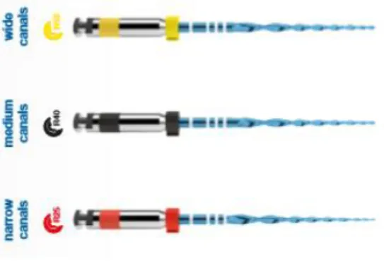
Reciproc® blue system includes three files (R25, R40 and R50) (Figure 1) in three lengths (21, 25 and 31mm): R25 (diameter of 0.25mm at the tip and an 8% taper over the first 3mm from the tip) for narrow canals, R40 (diameter of 0.40mm at the tip, 6% taper over the first 3mm from the tip) for medium canals, and R50 (diameter of 0.50mm at the tip, a 5% taper over the first 3mm from the tip) for wide canals (Altunbas et al, 2015; Berutti et al, 2012; Plotino et al, 2012). Reciproc®blue instruments have a short shaft of 11 mm, enabling better access to molars compared to many other instruments which have a shaft of 13 mm or longer (Goel et al, 2015). These files present a s-shaped cross section, variable taper, non-cutting tip, can be used without glide path management in the majority of cases and have been specifically designed for use in reciprocation (150° CCW and 30° CW rotation). As the rotation in the cutting direction is larger than reverse direction, it results in movement towards apex. (Dhingra et al, 2015; VDW, Reciproc® blue, User guide). Only one instrument is used for the canal preparation depending on the initial size of the canal, being single-file and also single-use (VDW, Reciproc® blue, User guide).
Figure 1 – Reciproc® blue system composed by three single files (VDW, Munich,
5
1.4. iRace/iRace Plus System
iRace/iRace Plus (FKG, La Chaux-de-Fonds, Switzerland) NiTi continuous rotary files have been introduced as a simplified sequence of the Race system in 2011 and are made of conventional NiTi wire (Saber et al, 2015). It is believed that these instruments are advantageous to use in curved canals because of their constant and reduced taper, a triangular cross-section with a sharp edge, an alternating cutting edge and a rounded safety tip. They also present an electrochemical polishing that offer an enhanced resistance against fatigue and corrosion (Hiran et al, 2016). This file system has an easy identification of ISO sizes (large ring) and taper (thin ring, yellow: 2%, red: 4%, blue: 6%) and a SafetyMemoDisc (SMD) to master fatigue and number of uses. It is claimed by the manufacturer that this new sequence provides a quick, safe and effective protocol for preparation of curved root canals (FKG, iRace, instructions for use).
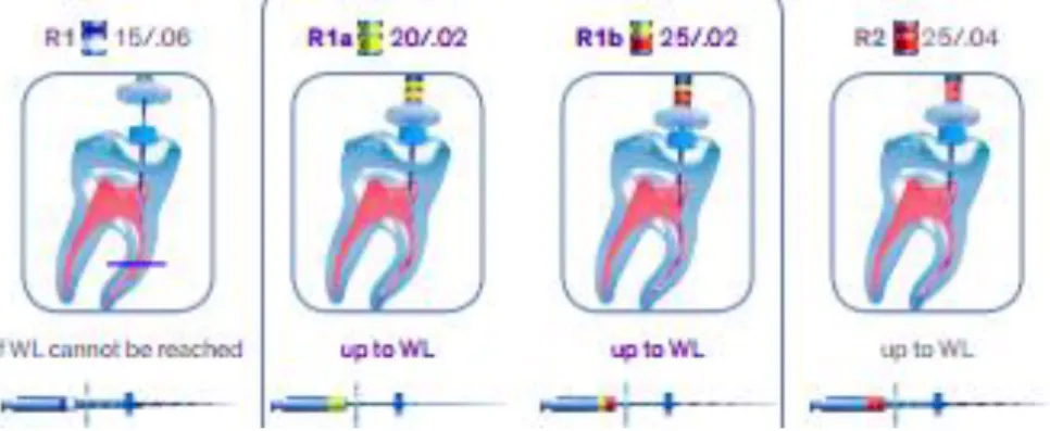
iRacesystem is composed by a sequence of three files to treat the majority of cases (straight, slightly curved or wide canals), all in different lengths (21, 25 and 31mm): R1 (size 15 tip and 6% taper), R2 (size 25 tip and 4% taper) and R3 (size 30 tip and 4% taper). In addition to this basic sequence, there is a complementary kit, iRace Plus, that is constituted by two highly flexible instruments that allow treatment of more difficult cases (highly curved, narrow or calcified canals): R1a (size 20 tip and 2% taper) and R1b (size 25 tip and 2% taper). This complementary kit is used between R1 file (when it does not reach the working length) and R2 file, continuing after with R3 file (Figure 2) (FKG, iRace, instructions for use).
In this study we used the sequence of iRaceand iRacePlus files only until R2 file (R3 file was excluded of the study).
6
Figure 2 – A – iRace system composed by a sequence of three files; B – iRacePlus system (complementary kit) composed by a sequence of two files (FKG, La
7
2. AIMS
The purpose of this study is to compare the shaping ability with focus on the maintenance of original anatomy in simulated S-shaped root canals, of two different system files: Reciproc® blue, a reciprocating NiTi single file and iRace/iRace Plus, a continuous rotary file system.
Specific goals:
1 - To compare transportation of coronal curvature of the two files after instrumentation.
H0 - Coronal curvature transportation is alike in all instruments.
H1 - Coronal curvature transportation is different between instruments.
2 - To compare transportation of apical curvature of the two files after instrumentation.
H0 - Apical curvature transportation is alike in all instruments. H1 - Apical curvature transportation is different between instruments.
3 - To compare maintenance of the original root canal anatomy. H0 - Root canal anatomy maintained in all instruments.
8
3. MATERIALS AND METHODS
3.1. Simulated Root Canal
A total of 20 simulated canal with an S-shaped curvature in clear resin blocks (ISO 15, Endo-Training-Bloc-S .02 Taper; Dentsply-Maillefer, Ballaigues, Switzerlan) (Figure 3) were prepared by two different Ni-Ti rotary files system, using the technique recommended by the manufacturer: Reciproc® blue (VDW, Munique, Germany) and iRace/iRace Plus(FKG, La Chaux-de-Fonds, Switzerland).
The resin blocks were randomly numbered from 1 to 20 and then randomly assigned to two groups (n= 10): Group A - 10 simulated canal resin blocks, prepared with Reciproc® blue (RB); Group B - 10 simulated canal resin blocks, prepared with iRace and with iRace Plus (iR). Each canal had a mean canal length of 16mm. Brown water ink (Higgins, Leeds, MA) was injected into the canal space within each resin block with a disposable syringe (Injekt®) (Figure 4). A specific platform allowed to take pictures of the canals before and after instrumentation using a precise camera (Olympus Digital Camera E500) and a repositioning of the resin blocks (Figure 5).
Figure 3 – S-shaped curvature in clear resin blocks (ISO 15, Endo-Training-Bloc-S .02
Taper; Dentsply Maillefer, Ballaigues, Switzerlan)
9
Figure 5 - Reproduction table (Kaiser Fototechnik GmbH & Co.KG) and digital camera
(Olympus Digital Camera E500)
3.2. Working Length
Working length was stablished by advancing a 10K stainless-steal hand file (Dentsply-Maillefer, Ballaigues, Switzerland) into the canal until the apical terminus of the resin block. Adjusted the stop to the top of the orifice of the canal and the value of the working length was the measurement value of that length minus 0.5mm. The WL was determined to be 15,5mm.
3.3. Canal Instrumentation
Reciproc® blueand iRace/iRace Plus systems were selected to prepare the resin blocks. Reciproc® blue file, R25 (Figure 6), tip size 25, with a taper of 0.08 over the first apical millimeters, has a progressively taper from D1 to D16. iRace and iRace Plus
file system (Figure 7) used in this study until the R2 file, following the sequence determined by the manufacture: R1 file, tip size 15, with a taper of 0.06; R1a file, tip size 20, with a taper of 0.02; R1b file, tip size 25, with a taper of 0.02; R2 file, tip size 25, with a taper of 0.04. All files operated with WaveOne™ endo motor (Dentsply Maillefer, Ballaigues, Switzerland) with their respective recommended settings: on Group A with “RECIPROC ALL” mode and on Group B files were used at 600 rpm and at a torque of 1.5 Ncm (Figure 8).
10
Figure 7 – iRace and iRace Plus sequence files (FKG, La Chaux-de-Fonds, Switzerland)
Figure 8 - WaveOne™ endo motor (Dentsply Maillefer, Ballaigues, Switzerland)
All canals were prepared by the same operator, who had no experience using these files.
The following preparation sequences were made after all canals were scouted up to the working length with a 10K stainless-steal file (Figure 9). The instruments were used in a slow and-out pecking motion, the blades were cleaned after three/four in-and-out movements using gauze soaked with water and copious irrigation with water was performed throughout the entire preparation sequence for all samples, using a disposable syringe (Injekt®) and 27-gauge irrigation needle (BD Microlance™). A 10 K-file was used to remove debris. Each instrument was discarded after use in 3 resin blocks.
11
3.3.1. Sequence of Instrumentation
The sequence below was the recommended by the manufacturer at a working length of 15,5mm:
Group A – Reciproc®blue
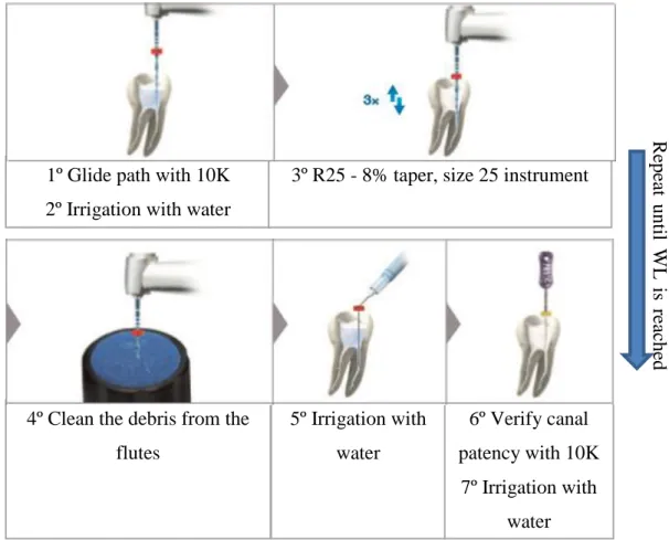
With a stainless steel size 10K hand file create a glide path before using RB files. Instrumentation starts in the presence of an irrigant. With an in and out movement (pecks), passively advance the file and remove after three/four pecks. Clean the debris from the flutes, irrigate and verify canal patency with an SS 10K. Irrigate again. Repeat in small increments until working length is reached (Figure 10):
Figure 10 – Sequence of instrumentation with Reciproc®blue file (Reciproc® blue. User guide)
1º Glide path with 10K 2º Irrigation with water
R epe at unti l W L is re ac he d
3º R25 - 8% taper, size 25 instrument
4º Clean the debris from the flutes 5º Irrigation with water 6º Verify canal patency with 10K 7º Irrigation with water
12
Group B – iRace/ iRacePlus
With a stainless steel size 10K hand file create a glide path before using iR files. Instrumentation starts in the presence of an irrigant. With an in and out movement (pecks), passively advance the file and remove after three/four pecks. Clean the debris from the flutes, irrigate and verify canal patency with a stainless steel size 10K. Irrigate again. Repeat in small increments until working length is reached and following this sequence (Figure 11):
1º Glide path with 10K 2º Irrigation with water
3º R1 - 6% taper, size 15 instrument (This does not reach the WL) 4º R1a – 2% taper, size 20 instrument (Until WL is reached) 5º R1b – 2% taper, size 25 instrument (Until WL is reached) 6º R2 – 4% taper, size 25 instrument (Until WL is reached)
After three/four pecks of each instrument: clean the debris from the flutes, irrigate with water, verify canal patency with 10K and irrigate again with water.
Figure 11 – Sequence of instrumentation with iRace/iRace Plus system (FKG, iRace,
13 3.4. Image Analysis
To allowbetter visualization and analysis of canal anatomy, resin blocks were colored with brown water ink (pre-instrumentation) and orange water ink (post-instrumentation) injected by a disposable syringe (Injekt®) only for shooting. A specific table (Kaiser Fototechnik GmbH & Co.KG) was used to take pictures of the canals before and after shaping, was set-up to allowed precise camera and resin blocks repositioning. The footage was standardized: a landmark was made in each sample as a reference and the samples were all shot at the same distance and placed in the same position using a graph paper. Digital images were recorded using an Olympus Digital Camera E500 with a 35 mm macro lens and saved as .jpeg format files.
The shaping effects of the instrumentation systems were analyzed using Rhinoceros® software (version 5.0; Robert McNell & Associates, Seattle, WA), Pixlr Editor (Autodesk, Incorporated, San Rafael, California, USA) and ImageJ® 1.5.
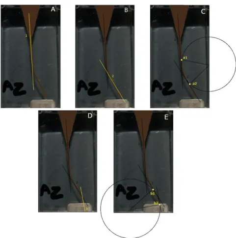
To do a precise measurement, it is crucial to define the area corresponding to coronal and apical curvature. Based on that, for this study Pruett’s method was used (Pruett et al, 1997) (Figure 12). Pruett states that the curvature is defined by two parameters, angle of curvature and radius of curvature, Rhinoceros Software was the programme used to define coronal and apical curvatures. A straight line was traced along the long axis of the coronal straight portion of the canal (Figure 12A). A second line (Figure 12B) was traced along the long axis of the first curvature of the canal. There is a point (a1 and a2) on each of these lines at which the canal deviates to begin or end the canal curvature (Figure 12C). Line 1 and 2 defined the coronal curvature. A third line (Figure 12D) was traced along the long axis of the apical straight portion of the canal. Line 2 and 3 defined the apical curvature (Figure 12E). This process was made in each one of the twenty samples.
14
Figure 12 – Sequence of five images to defined coronal and apical curvature based on
Pruett’s methods. Figure 12 A, B and C: correspond to coronal curvature. Figure 12 D and E, to apical curvature
After this procedure, the pre-instrumentation digital images and the post-instrumentation images were superimposed and standardized, accomplished by reducing the opacity of the post-instrumentation images, using online image editor Pixlr Editor (Figure 13).
Figure 13 – Resin blocks. A) Pre-instrumentation; B) Post-instrumentation; C)
15
On ImageJ® 1.5 program, the scale was calibrated and set to mm for measuring the respective areas. With “Freehand line” tool, the area corresponding to the difference between the margin of the pre and post-instrumented canal of the coronal and apical curvatures pre-determined was delimited. Measurements were automatically made and saved in excel files.
To compare maintenance of the original root canal anatomy, a qualitative analysis was done, asking to six blinded examiners with different levels of clinical practice (two endodontic specialists, two inexpert clinicians and two graduation students) if the original coronal and apical curvature were maintained, if less significant straightening occurred or if significant straightening occurred in these curvatures. The examiners evaluated three superimposed images, randomly chosen, from each group.
3.5. Statistical Analysis
The statistical analysis was obtained using the IBM SPSS® Statistics version 23.0.0 software. Descriptive statistical analysis was performed to each group (A and B). In each experimental group mean and standard deviation were calculated for the inner and outer of coronal and apical curvatures values. The Shapiro-Wilk test was used to evaluate the data normality. Since comparisons were made between groups of reduced size and because there was a rejection of the normality test, the Mann-Whitney U test was used to analyze the results and to compare the transportation occurred between groups. Differences were considered statistically significant when p<0,05.
16
4. RESULTS
4.1. Quantitative Analysis
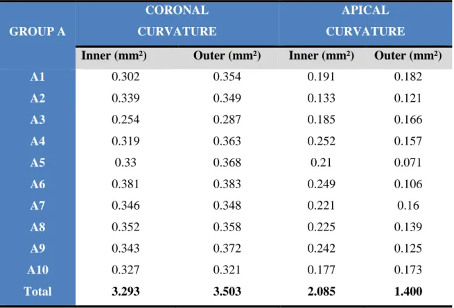
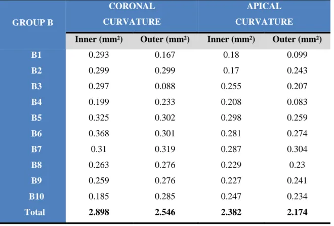
The result of the total amount of material removed was established by measure the distance between the pre and post-instrumentation margins, inner and outer of both curvatures. This procedure was repeated for the group A (Table 1) and group B (Table 2). GROUP A CORONAL CURVATURE APICAL CURVATURE
Inner (mm²) Outer (mm²) Inner (mm²) Outer (mm²)
A1 A2 A3 A4 A5 A6 A7 A8 A9 A10 Total 0.302 0.339 0.254 0.319 0.33 0.381 0.346 0.352 0.343 0.327 3.293 0.354 0.349 0.287 0.363 0.368 0.383 0.348 0.358 0.372 0.321 3.503 0.191 0.133 0.185 0.252 0.21 0.249 0.221 0.225 0.242 0.177 2.085 0.182 0.121 0.166 0.157 0.071 0.106 0.16 0.139 0.125 0.173 1.400
Table 1 – Group A – Reciproc®blue. Measurements of inner and outer coronal and apical curvatures
17 GROUP B CORONAL CURVATURE APICAL CURVATURE
Inner (mm²) Outer (mm²) Inner (mm²) Outer (mm²)
B1 B2 B3 B4 B5 B6 B7 B8 B9 B10 Total 0.293 0.299 0.297 0.199 0.325 0.368 0.31 0.263 0.259 0.185 2.898 0.167 0.299 0.088 0.233 0.302 0.301 0.319 0.276 0.276 0.285 2.546 0.18 0.17 0.255 0.208 0.298 0.281 0.287 0.229 0.227 0.247 2.382 0.099 0.243 0.207 0.083 0.259 0.274 0.304 0.23 0.241 0.234 2.174
Table 2 – Group B – iRace/iRacePlus. Measurements of inner and outer coronal and apical curvatures
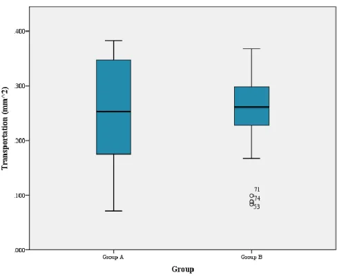
On total transportation, there were no statistically significant difference in the total amount of material removed between the two groups (p=0.491) (Table 3).
GROUP TOTAL TRANSPORTATION
A – RB 0.257 ± 0.093
B – iR 0.250 ± 0.064
p = 0.491
Table 3 – Mean values and standard deviation of each group relatively of total
18
Graph 1 – Total transportation distribution by group
Concerning to transportation of coronal and apical curvature of each group, there were statistically significant differences between coronal transportation (p <0.001) and apical transportation (p = 0.002) of groups A and B (Table 4). Group A (RB) presented significantly greater amount of resin material removed from coronal curvature while group B (iR) demonstrated significantly greater amount of resin material removed from apical curvature.
GROUP CORONAL CURVATURE APICAL CURVATURE
A – RB 0.340 ± 0.032 0.174 ± 0.050
B - iR 0.272 ± 0.063 0.230 ± 0.059
p < 0.001 p = 0.002
Table 4 – Mean values and standard deviation of each group relatively of
19
Graph 2 – Coronal and apical transportation distribution by group
Differences between the two systems files on the inner side were statistically significant only on coronal curvature (p = 0.023), verifying in the group A - Reciproc® blue, more transportation. Considering the outer side, both curvatures, coronal and apical, presented statistically significant differences between the groups (p < 0.001 and p = 0.015, respectively). On the outer side of the coronal curvature, group A registered more transportation and in the same side of the apical curvature were Group B - iRace/iRace Plus, that demonstrated significantly greater amount of resin material removed (Table 5).
GROUP CORONAL CURVATURE APICAL CURVATURE
Inner Outer Inner Outer
A – RB 0.329±0.034 0.350±0.028 0.209±0.037 0.140±0.035
B – iR 0.290±0.048 0.255±0.073 0.238±0.044 0.217±0.071
p = 0.023 p < 0.001 p = 0.165 p = 0.015
Table 5 – Mean values and standard deviation of each group relatively of transportation
20
Graph 3 – Coronal and apical (inner and outer) transportation distribution by group
4.2. Qualitative Analysis
Considering each blinded examiners evaluation, the next graphics shows this evaluation taking into account the presence or absence of rectifications in the coronal and apical curvatures for each system file. Each examiner evaluated three superimposed images, randomly chosen, from each group.
21 0 1 2 3 4 5 6 Curvature Maintenance Less Significant Straightening Significant Straightening Coronal Curvature Students Inexpert Clinicians Endodontists
Graph 4 – Evaluation of the coronal curvature prepared by Reciproc® blue
Graph 5 – Evaluation of the coronal curvature prepared by iRace/iRace Plus system
0 1 2 3 4 Curvature Maintenance Less Significant Straightening Significant Straightening Coronal Curvature Students Inexpert Clinicians Endodontists
22 0 1 2 3 4 5 Curvature Maintenance Less Significant Straightening Significant Straightening Apical Curvature Students Inexpert Clinicians Endodontists
Graph 6 – Evaluation of the apical curvature prepared by Reciproc® blue
Graph 7 – Evaluation of the apical curvature prepared by iRace/iRace Plus system
0 1 2 3 4 Curvature Maintenance Less Significant Straightening Significant Straightening Apical Curvature Students Inexpert Clinicians Endodontists
23
With the evaluation made by the examiners the following results were obtained:
A - Reciproc® blue group
The clinician’s expertise caused more evaluation differences in this group but the great majority indicates that there is maintenance or few changes of the original shape of the canal, considering the coronal and apical curvatures;
B – iRace/iRace Plus system group
Maintenance of the original shape of the canal, considering the coronal and apical curvatures.
Figure 14 - Representative images of simulated canals instrumented with A -
Reciproc® blue, B – iRace/iRace Plus system
B A
24
5. DISCUSSION
Shaping of the root canal is one of the most important steps in root canal treatment (Peters, 2004). It is essential for the efficacy of all subsequent procedures, including chemical disinfection and root canal filling (Hulsmann et al, 2005). The preparation of a curved canal, especially a double curved (S-shaped) canal is one of the most challenging procedures in root canal treatment (Hiran et al, 2016). Analysis of modifications in canal curvature after instrumentation has been widely used to evaluate the tendency of a technique, or mechanical properties of an instrument, to maintain the original canal anatomy or to straighten the curves (Berutti et al, 2009).
The purpose of this study is to compare the shaping ability of two instrumentation systems with different rotary movements and manufacturing processes, using simulated S-shaped root canals: Reciproc® blue and iRace System (iRace and iRace Plus). These system files were selected because they are commonly used in clinical practice and to investigate which instrumentation movement, reciprocating or continuous rotary, is the most indicated to shape severely curved root canals.
In this study, to compare the shaping effects of instruments and to evaluate the maintenance of the original shape of the canal, the technique of superimposing the pre and post-operative photographs of s-shaped resin blocks was employed. This method has been used in a number of studies investigating the shaping ability of endodontic files (Yoshimine et al, 2005; Bonaccorso et al, 2009; Burroughs et al, 2012; Neto & Ginjeira, 2016). This technique does not provide the three-dimensional information generated by micro-computed tomography but it does provide reproducibility and direct visual comparison of the results what is improved with the injection of water ink into the pre and post-operative resin blocks. This procedure creates a clear visualization of the root canal outline when the images are superimposed (Hiran et al, 2016).
To assess the instrumentation of s-shaped root canals, clear resin blocks were used in this study. These resin blocks are an alternative to root canals in extracted human teeth. Although the use of real teeth provides conditions that are similar to the clinical situation, it has large variations in the root canal morphology (Schäfer & Vlassis, 2004). Resin blocks enables the standardization of the canal morphology, in terms of angle, radius of curvature, diameter, length and as the conditions are identical for the different instruments, allows direct comparison between them (Lim & Webber, 1985; Schäfer et al, 1995). The disadvantages of using rotary instruments in resin blocks
25
is the different hardness between resin and dentin and the heat generated, that which might distort the canal, reduce the cutting efficiency and lead to separation of the instrument. Furthermore, the cross-sections differ from natural teeth (Zhang et al, 2008).
In this investigation, the final apical diameter was carried out using instruments with a tip diameter equivalent to size 25, however the tapers were not congruent. Reciproc® blue R25 (single-file system) and iRace/iRace Plus sequence until R2 file (multi-file system) were selected in accordance with the recommendations of the manufacturer as these file or sequence of files are designated for narrow or curved canals.
The first stage of the study comprised a quantitative analysis through observation of changes in root canal anatomy between pre-instrumentation and post-instrumentation images followed by a qualitative observation made by examiners to compare the maintenance of the original root canal anatomy, concerning the presence of straightening curves.
It is important to emphasize that no studies with Reciproc® blue system and few with iRace system could be found in the literature review, so it is not possible to directly compare the results of this study with others. The comparison was done with the anterior version of Reciproc® blue system and iRace system, Reciproc® and RaCe system files.
Based on the results obtained with the quantitative analysis, the null hypothesis was rejected. Within the limitations of an in vitro study, whose experimental procedure was executed by an operator without experience, there were statistically significant differences between transportation of coronal and apical curvature of each system file. Reciproc® blue produced more transportation in coronal curvature and iRace/iRace Plus system in apical curvature. Considering the outer side, both curvatures, coronal and apical, presented statistically significant differences between the system files. Reciproc® blue caused more transportation on the outer side of the coronal curvature and iRace/iRace Plus system in the outer side of the apical curvature. Differences between the two systems files on the inner side were statistically significant only on coronal curvature, with Reciproc® blue producing more transportation.
In the study of Altunbas et al, that compares the shaping ability of Reciproc® with continuous rotary files system, Reciproc® provided the widest instrumentation in the total length of the canal and removed more resin from the inner and outer sides of the curve. This agrees with previous studies that showed that Reciproc® instruments
2 4
26
removed more dentin along the canal (Capar et al, 2014; Gergi et al, 2015). A sharp double cutting edge S‑shaped geometry and a smaller cross‑sectional area may explain the greater cutting ability of Reciproc® instruments. The final taper might have influenced the material removal. In a recent study, the shaping ability of four single file systems with different tapers has been compared and the study reported that more tapered instruments removed more resin compared with less tapered instruments (Saleh et al, 2015). Hence, differences between the resin removal of the instruments can be attributed to their common features such as the cross‑section, working motion, manufacturing method, and taper (Altunbas et al, 2015). In the present study, the final taper was 0.08 at the apical 3 mm for Reciproc® blue system and 0.04 for iRace/iRace Plus. Although there were no statistically significant difference in the total amount of material removed between Reciproc® blue system and iRace/iRace Plus, the first system file caused more total transportation, according to the studies above.
RaCe instruments allowed preparation of curved root canals to apical diameters larger than those normally achieved when using other rotary NiTi instruments with only minimal canal transportation and adequate centering ability (Pasternak-Junior et al, 2009). In the present study, iRaCe/iRace Plus instruments produced more transportation in apical curvature. The shaping ability of this system files can be explained by their small cross-sectional area, which increases their flexibility and gives more space for debris removal, and the design of the working part with alternating cutting edges, that is claimed to prevent the screwing in effect thus reducing intra-operative torque values (Paqué e et al. 2005; Saber et al, 2015).
The second stage of the study comprised a qualitative analysis where endodontists, inexpert clinicians and students evaluated the maintenance of the original root canal anatomy, with the presence or absence, of the coronal and apical curvatures rectification. The differences registered are due to clinical experience and different levels of endodontic knowledge. Taking into account most of the evaluations made, a few changes on the original shape of the coronal curvature and the maintenance of the apical curvature with Reciproc® blue system file are consistent with the quantitative results. The maintenance of the original shape of the canal, considering the coronal and apical curvatures, was also registered with iRace/iRace Plus system group. The evaluations carried out by the different examiners that were divided between the curvature maintenance and less significant straightening or that are not consistent with the quantitative results, can demonstrate that although statistically significant
2 4
2 5
27
differences regarding canal transportation were obtained, from a clinical point of view, these differences are of limited importance.
Additional studies comparing endodontic files with different instrumentation movements, assessing other parameters and with a larger sample size are needed to understand which system file is the most indicated to shape severely curved root canals.
28
6. CONCLUSIONS
Instrumentation of narrow and severely curved canals is not easy and may cause canal transportation and undesirable iatrogenic accidents. So, in clinical procedures it is important to choose an appropriate instrument system to each case, to reduce the errors and aiming to achieve optimum cleaning and shaping.
Under the limitations of this study and based on the results obtained, although Reciproc® blue caused greater resin material removal at the level of coronal curvature and iRace/iRace Plus system at the apical curvature level, there is no statistically significant difference between the two files systems for total transportation. Evaluating the maintenance of the original anatomy, iRace/iRace Plus system was the file system that best maintained the original anatomy of the S-shaped canal.
xxix
REFERENCES
1. Alapati SB, Brantley WA, Iijima M, Clark WA, Kovarik L, Buie C, et al. Metallurgical characterization of a new nickel-titanium wire for rotary endodontic instruments. J Endod. 2009;35:1589-93.
2. Alsilani R, Jadu F, Bogari DF, Jan AM, Alhazzazi TY. Single file reciprocating systems: A systematic review and meta-analysis of the literature: Comparison of reciproc and WaveOne. J Int Soc Pent Communit Dent. 2016;6:402-9. 3. Altunbas D, Kutuk B, Kustarci A. Shaping ability of reciprocating single-file and
full-sequence rotary instrumentation systems in simulated curved canals. Eur J Dent. 2015 Jul-Sep;9(3):346-51.
4. Ankrum MT, Hartwell GR, Truitt JE. K3 Endo, ProTaper and ProFile systems: breakage and distortion in severely curved roots of molars. J Endod. 2004;30:234-7.
5. Arias A, Perez‑Higueras JJ, de la Macorra JC. Differences in cyclic fatigue resistance at apical and coronal levels of Reciproc and WaveOne new files. J Endod. 2012;38:1244‑8.
6. Berutti E, Paolino DS, Chiandussi G, Alovisi M, Cantatore G, Castellucci A, Pasqualini D. Root canal anatomy preservation of WaveOne reciprocating files with or without glide path. J Endod. 2012 Jan;38(1):101-4.
7. Berutti E, Cantatore G, Castellucci A, Chiandussi G, Pera F, Migliaretti G, Pasqualini D. Use of nickel-titanium rotary PathFile to create the glide path: comparison with manual preflaring in simulated root canals. J Endod. 2009 Mar;35(3):408-12.
8. Bonaccorso A, Cantatore G, Condorelli GG, Schafer E, Tripi TR. Shaping ability of four nickel-titanium rotary instruments in simulated S-shaped canals. J Endod 2009; 35 (6): 883–6.
9. Burroughs JR, Bergeron BE, Roberts MD, Hagan JL, Himel VT. Shaping ability of three nickel-titanium endodontic file systems in simulated S-shaped root canals. J Endod 2012; 38 (12): 1618–21.
xxx
10. Capar ID, Ertas H, Ok E, Arslan H, Ertas ET. Comparative study of different novel nickel-titanium rotary systems for root canal preparation in severely curved root canals. J Endod 2014;40:852-6.
11. De-Deus G, Silva EJ, Vieira VT, Belladonna FG, Elias CN, Plotino G, Grande NM. Blue Thermomechanical Treatment Optimizes Fatigue Resistance and Flexibility of the Reciproc Files. J Endod. 2017 Mar;43(3):462-466.
12. De-Deus G, Moreira EJL, Lopes HP, Elias CN. Extended cyclic fatigue life of F2 ProTaper instrument used in reciprocating movement. Int Endod J. 2010;43:1063–8.
13. Dhingra A, Ruhal N, Miglani A. Evaluation of single file systems Reciproc, Oneshape, and WaveOne using Cone Beam Computed Tomography – An in vitro study. Journal of Clinical and Diagnostic Research. 2015 Apr;9(4):ZC30-ZC34. 14. Franco V, Fabiani C, Taschieri S, Malentacca A, Bortolin M, Del Fabbro M.
Investigation on the shaping ability of nickel–titanium files when used with a reciprocating motion. J Endod. 2011;37:1398–401.
15. Gavini G, Caldeira CL, Akisue E, Candeiro GT, Kawakami DA. Resistance to fl exural fatigue of Reciproc R25 files under continuous rotation and reciprocating movement. J Endod. 2012;38:684-7
16. Gergi R, Osta N, Bourbouze G, Zgheib C, Arbab-Chirani R, Naaman A. Effects of three nickel titanium instrument systems on root canal geometry assessed by micro-computed tomography. Int Endod J 2015;48:162-70.
17. Goel A, Rastogi R, Rajkumar B, Choudary TM, Boruah L, Gupta V. Arora R, Bhatt A. An Overview of Modern Endodontic NiTi Systems. International Journal of Science and Research (IJSR). 2015: 4(4):595-7.
18. Haapasalo M, Shen Y. Evolution of nickel–titanium instruments: From past to future. Endodontic topics. 2013;29(1):3-17.
19. Hiran S, Pimkhaokham S, Sawasdichai J, Ebihara A, Suda H. Shaping ability of ProTaper NEXT, ProTaper Universal and iRace files in simulated S-shaped canals. Aust Endod J. 2016; 42: 32–36.
20. Hülsmann M, Peters OA, Dummer P. Mechanical preparation of root canals: shaping goals, techniques and means. Endodontic Topics. 2005:10: 30–76.
21. iRace® Brochure. [Available from: http://www.fkg.ch/sites/default/files/201705_fkg_irace_brochure_en.pdf]
xxxi
22. Kumar RV, Shruthi C. Evaluation of the sealing ability of resin cement used as a root canal sealer: An in vitro study. Journal of Conservative Dentistry. 2012. 23. Lim KC, Webber J. The validity of simulated root canals for the investigation of
the prepared root canal shape. Int Endod J 1985 Oct;18(4):240-246.
24. Lopes HP, Gambarra-Soares T, Elias CN, Siqueira JF, Inojosa IF, Lopes WS, Vieira VT. Comparison of the mechanical properties of rotary instruments made of conventional nickel-titanium wire, M-wire, or nickel-titanium alloy in R-phase. J Endod. 2013 Apr;39(4):516-20.
25. Neto F, Ginjeira A. Comparative analysis of simulated root canals shaping, using ProTaper Universal, Next and Gold. Rev port estomatol med dent cir maxilofac. 2016;57(2):82–86.
26. Paqué F, Musch U, Hulsmann M. Comparison of root canal preparation using RaCe and ProTaper rotary Ni-Ti instruments. International Endodontic Journal 2005; 38,8 –16.
27. Pasternak-Junior B, Sousa-Neto MD, Silva RG. Canal transportation and centring ability of RaCe rotary instruments. International Endodontic Journal 2009; 42, 499– 506.
28. Pereira ES, Gomes RO, Leroy AM, Singh R, Peters OA, Bahia MG, Buono VT. Mechanical behavior of M-Wire and conventional NiTi wire used to manufacture rotary endodontic instruments. Dent Mater. 2013 Dec;29(12):e318-24.
29. Peters OA. Current challenges and concepts in the preparation of root canal systems: A review. J Endod. 2004;30:559‑67.
30. Plotino G, Grande NM, Cotti E, Testarelli L, Gambarini G. Blue treatment enhances cyclic fatigue resistance of vortex nickel-titanium rotary files. J Endod. 2014 Sep;40(9):1451-3.
31. Plotino G, Grande NM, Testarelli L, Gambarini G. Cyclic fatigue of Reciproc and WaveOne reciprocating instruments. International Endodontic Journal 2012. 32. Pruett JP, Clement DJ, Carnes DL. Cyclic fatigue testing of nickel titanium
endodontic instruments. J Endod 1997;23:77–85.
33. Reciproc® blue. Product information. [Available from: https://www.vdw-dental.com/en/products/detail/reciprocr-blue-instruments]
34. Reciproc® blue. User guide. [Available from: https://www.vdw-dental.com/en/products/detail/reciprocr-blue-instruments]
xxxii
35. Ruddle C. Cleaning and shaping the root canal system. In: Cohen S, Burns R, eds. Pathways of the Pulp, 8th ed. St Louis, MO: Mosby, 2002: 231–292.
36. Saber SE, Nagy MM, Schafer E. Comparative evaluation of the shaping ability of WaveOne, Reciproc and OneShape single‑file systems in severely curved root canals of extracted teeth. Int Endod J. 2015;48:109‑14.
37. Saber SE, Nagy MM, Schafer E. Comparative evaluation of the shaping ability of ProTaper Next, iRaCe and Hyflex CM rotary NiTi files in severely curved root canals. International Endodontic Journal. 2015;48:131–36.
38. Saleh AM, Vakili Gilani P, Tavanafar S, Schäfer E. Shaping ability of 4 different single-file systems in simulated S-shaped canals. J Endod 2015;41:548-52.
39. Schäfer E. Effects of four instrumentation techniques on curved canals: A comparison study. Journal of endodontics. 1996 Dec; 22(12): 685-689.
40. Schäfer E, Tepel J, Hoppe W. Properties of endodontic hand instruments used in rotary motion. Part2. Instrumentation of curved canals. J Endod 1995;21:493497. 41. Schäfer E, Vlassis M. Comparative investigation of two rotary nickel-titanium
instruments: ProTaper versus RaCe. Part 1. Shaping ability in simulated curved canals. Int Endod J 2004;37:229-238.
42. Schilder H. Cleaning and shaping the root canal. Dent Clin North Am. 1974: 18: 269–296. 2.
43. Thompson SA. An overview of nickel-titanium alloys used in dentistry. Int Endod J. 2000;33:297-310.
44. Varela-Patino P, Ibanez-Parraga A, Rivas-Mundina B, Cantatore G, Otero XL, MartinBiedma B. Alternating versus continuous rotation: a comparative study of the effect on instrument life. J Endod. 2010;36:157–9.
45. Yared G. Canal preparation with only one reciprocating
instrument without prior hand filing: A new concept. Int Dent. 2012;2:78‑87.
46. Yared G. Canal preparation using only one Ni-Ti rotary instrument: preliminary observations. Int Endod J. 2007;41:339-344
47. Yoo YS, Cho YB. A comparison of the shaping ability of reciprocating NiTi instruments in simulated curved canals. Restor Dent Endod. 2012 Nov;37(4):220-7.
xxxiii
48. Yoshimine Y, Ono M, Akamine A. The shaping effects of three nickel-titanium rotary instruments in simulated S-shaped canals. J Endod 2005; 31 (5): 373–5. 49. You SY, Kim HC, Bae KS, Baek SH, Kum KY, Lee W. Shaping ability of
reciprocating motion in curved root canals: a comparative study with micro-computed tomography. J Endod. 2011;37:1296–300.
50. Zhang L, Luo HX, Zhou XD, Tan H, Huang DM. The shaping effect of the combination of two rotary nickel-titanium instruments in simulated S-shaped canals. J Endod 2008 Apr;34(4):456-458.