RESUMO.- [Pseudoalergia relacionada à ativação do complemento em cães após administração intraveno-sa de uma formulação lipossomal de antimoniato de meglumina.] O crescente uso das nanotecnologias nas te-rapias avançadas tem permitido a observação de reações adversas específicas relacionadas às nanoestruturas. A to -xicidade de uma nova formulação lipossomal de antimonia-to de meglumina após dose única foi avaliada em cães com leishmaniose visceral. Grupos de 12 animais receberam por via intravenosa uma dose única de antimoniato de meglu-mina lipossomal (grupo I [GI], 6,5 mg Sb/kg), lipossomas vazios (GII) ou solução salina isotônica (GIII). A avaliação de parâmetros hematológicos e bioquímicos não revelou
Complement activation-related pseudoallergy in dogs following
intravenous administration of a liposomal formulation of
meglumine antimoniate
1Raul R. Ribeiro2*, Eliane P. Moura3, Weverton M. Sampaio3†, Sydnei M. Silva4, Gustavo
O. Fulgêncio5, Wagner L. Tafuri3, Marilene S.M. Michalick4 and Frédéric Frézard6
ABSTRACT.- Ribeiro R.R., Moura E.P., Sampaio W.M., Silva S.M., Fulgêncio G.O., Tafuri W.L., Michalick M.S.M. & Frézard F. 2013. Complement activation-related pseudoal-lergy in dogs following intravenous administration of a liposomal formulation of meglumine antimoniate. Pesquisa Veterinária Brasileira 33(8):1016-1020. Centro de Ciências Agrárias, Ambientais e Biológicas, Universidade Federal do Recôncavo da Bahia, Campus Universitário Cruz das Almas s/n, Cruz das Almas, BA 44380-000, Brazil. E-mail: [email protected]
The increasing use of nanotechnologies in advanced therapies has allowed the observa -tion of specific adverse reac-tions related to nanostructures. The toxicity of a novel liposo -me formulation of -meglumine antimoniate in dogs with visceral leishmaniasis after single dose has been investigated. Groups of 12 animals received by the intravenous route a single dose of liposomal meglumine antimoniate (group I [GI], 6.5 mg Sb/kg), empty liposomes (GII) or isotonic saline (GIII). Evaluation of hematological and biochemical parameters sho -wed no significant changes 4 days after administration. No undesired effects were regis -tered in the GIII. However, adverse reactions were observed in 67.7% of dogs from both groups that received liposomal formulations. The side effects began moments after bolus administration and disappeared during the first 15 minutes after treatment. Prostation, sialorrhea and defecation were the most frequent clinical signs, registered in 33.3% and 41.6 % of animals from the groups GI and GII, respectively. Tachypnea, mydriasis, miosis, vomiting and cyanosis were also registered in both groups. The adverse reactions observed in this study were attributed to the activation of the complement system by lipid vesicles in a phenomenon known as Complement Activation-Related Pseudoallergy (CARPA). The in -fluence of the physical-chemical characteristics of liposomal formulation in the triggering of CARPA is discussed.
INDEX TERMS: Hypersensitivity reactions, Complement Activation-Related Pseudoallergy (CARPA), liposomes, complement activation, canine leishmaniasis.
1 Received on October 12, 2012.
Accepted for publication on February 15, 2013.
2 Centro de Ciências Agrárias, Ambientais e Biológicas, Universidade Fede
-ral do Recôncavo da Bahia. Campus Universitário Cruz das Almas s/n, Cruz das Almas, BA 44380-000, Brazil. *Corresponding author: [email protected]
3 Departamento de Patologia, Instituto de Ciências Biológicas, Universi -dade Federal de Minas Gerais (UFMG), Av. Antônio Carlos 6627, Belo Hori -zonte, MG 31270-901, Brazil.
4 Departamento de Parasitologia, Instituto de Ciências Biológicas, UFMG, Belo Horizonte, MG.
5 Departamento de Clínica e Cirurgia Veterinárias, Escola de Veterinária,
UFMG, Belo Horizonte, MG.
alterações significativas quatro dias após a administração. Nenhum efeito indesejável foi registrado no GIII. No entan -to, reações adversas foram observadas em 67,7% dos cães de ambos os grupos que receberam formulações liposso-mais. Os efeitos colaterais iniciaram momentos após a ad -ministração em “bolus” e desapareceram no decurso dos primeiros 15 minutos após o tratamento. Prostração, sia -lorréia e defecação foram os sinais clínicos mais frequen -tes, registrados em 33,3% e 41,6% dos animais dos grupos GI e GII, respectivamente. Taquipnéia, midríase, miose, vômitos e cianose também foram registrados em ambos os grupos. As reações adversas observadas neste trabalho foram atribuídas à ativação do sistema complemento pelas vesículas lipídicas em fenômeno conhecido como Pseudo -alergia Relacionada à Ativação do Complemento (PARAC). A influência das características físico-químicas da formula -ção lipossomal no desencadeamento de PARAC é abordada.
TERMOS DE INDEXAÇÃO: Reações de hipersensibilidade, Pseudo
-alergia Relacionada à Ativação do Complemento (PARAC), lipos
-somas, ativação do complemento, leishmaniose canina.
INTRODUCTION
Conventional therapies for numerous diseases in human and animals may be further improved through the inno -vative strategies of nanomedicine. According to the United States’ National Institutes of Health, this new area can be defined as “an offshoot of nanotechnology, which refers to highly specific medical interventions at the molecular sca -le for curing disease or repairing damaged tissues, such as bone, muscle, or nerve” (Webster 2006). In this context, the development of various types of drug-carrier nanodevices offers new strategies for targeted drug delivery, minimizing the secondary effects and the toxicity associated to drug widespread to healthy organs or cells (Irache et al. 2011).
The therapeutic approach for intracellular diseases is a real example of the benefits that can provide the nanote -chnology revolution in the pharmaceutical area. The intra -cellular nature of some pathogens often requires long-term therapy and combination of drugs, since the host cell mem -branes may protect them from a variety of drugs and host immune responses (Armstead & Li 2011). In that sense, the use of drug delivers, e.g. liposomes, provides satisfac -tory therapeutic responses in canine visceral leishmaniasis (CVL) (Valladares et al. 2001, Frézard & Demicheli 2010). The properties of drug sustained release and targeting pro -mote high concentrations of the encapsulated drug in the major sites of parasite infection, improving the drug effecti -veness. When compared to conventional antimonial drugs such as meglumine antimoniate (MA), liposomal formula -tions were hundreds of times more effective against visce -ral leishmaniasis in mice, hamsters and dogs (Chapman et al. 1984, Alving 1986, Croft 1986). Despite the increased leishmanicidal activity of antimonial drugs, no liposomal formulation of Sb has reached the market so far. Further -more, some experimental formulations produced acute to -xicity in dogs, including chills and diarrhea, which cleared by 24 h postdosing, and hepatic dysfunction at 24 h postdo -sing (Nieto et al. 2003).
Although the biocompatibility is an important feature of any drug delivery system, the nanodevices carry an incre -ased risk for hypersensitivity reactions. Acute immune to -xicity was reported after administration of liposomal dru -gs (Ambisome®, Doxil® and Dauno Xome®), radiocontrast media and micellar solvents containing amphiphilic lipids (Szebeni 2005). There is substantial evidence suggesting that these acute allergic reactions are not mediated by pre --existing IgE antibodies (Hypersensitivity Type I), but by complement activation (Szebeni 2005), in a phenomenon aptly called Complement Activation-Related Pseudoallergy (CARPA). It was also established that vesicle structural fac -tors of liposomes (e.g. phospholipid composition, vesicle size and surface charge) could modulate complement acti -vation and CARPA (Szebeni et al. 2000, Szebeni et al. 2012). As part of our continuous efforts to characterization and development of a novel liposomal formulation (Brazi -lian Patent Pending INPI/2640), this study determined the profile of adverse reactions in dogs naturally infected with
Leishmaniainfantum after single dose of liposomal MA and
discussed its possible mechanism taking into account the physico-chemical characteristics of therapeutic formulation.
MATERIALS AND METHODS Animals
Thirty-six mongrel dogs (weighing 8-15 kg) naturally infected
with L. infantum, exhibiting different clinical forms of CVL, were
identified and captured during an epidemiological survey carried out by Control Zoonosis Center in Santa Luzia City Hall (Minas Gerais state, Brazil Southeast). The serological diagnosis was established by the Serology Laboratory of the Institute of Biological Sciences, Federal University of Minas Gerais (UFMG) by indirect immuno -fluorescence assay (IFAT) and enzyme-linked immunosorbent assay (ELISA). All animals were found to be positive by IFAT (>1:40 di-lutions) and ELISA (optical density >0.100; >1:400 dilutions). In addition, parasitological diagnosis was performed by observation of parasite forms in both cytological examinations and/or cultures of bone marrow aspirates in Novy-Nicolle-McNeal (NNN) enriched
with minimum essential medium (a-MEM). Prior to treatment, the
animals were maintained in quarantine in kennels and were treated for intestinal helminthic infections (Canex composto®, Vetbrands Health Animal), ectoparasites infestations (Front Line®, Merial) and immunized against viral infections (Defensor® and Vanguard® HTLP 5/CV-L, vaccine Pfizer, Brazil). During the whole experimental pe -riod, the dogs were housed in a screened kennel and received drinking
water and a balanced feed ad libitum (Pedigree Champ®, Effem).
The present research adhered to the Principles of Laboratory Animal Care (NIH publication #85-23, revised in 1985) and received appro -val from the Ethical Committee for the use of Experimental Animals (CETEA) of the UFMG (Brazil) protocol n° 123/05.
Materials
Cholesterol (CHOL) and dicetylphosphate (DCP) were pur
-chased from Sigma Co. (St. Louis, MO, USA). Distearoylphospha
-tidylcholine (DSPC) was obtained from Avanti Polar Lipids Inc. (Alabaster, AL, USA). N-methyl-D-glucamine and antimony penta
-chloride (SbCl5, 99%) were obtained from Aldrich Chemical Co. (Milwaukee, Wis, USA).
Preparation of meglumine antimoniate
and pentavalent antimony oxyhydrated in water. The resulting product contained approximately 29% of antimony by weight, as determined by plasma emission spectroscopy (ICP-OES) using a Perkin-Elmer Optima 3000 plasma emission spectrometer.
Preparation and characterization of meglumine antimonia-te-containing liposomes
MA-containing liposomes with reduced size were prepared as described previously (Schettini et al. 2006). Briefly, small unila
-mellar vesicles (SUVs) were obtained by ultrasonication of a sus
-pension of multilamellar vesicles in de-ionized water, made from DSPC, CHOL and DCP (molar ratio of 5:4:1) at the final lipid con
-centration of 55 g/L. After filtration through sterile 0.22 μm mem
-brane, the SUVs suspension was mixed with sucrose at a sugar/ lipid mass ratio of 3:1 and a final sugar concentration of 0.3 M. The resulting mixture was immediately frozen in liquid nitrogen and subsequently dried (freeze-dryer, 4.5 L, Labconco, UK). Rehydration of the dried powder was performed with a MA aqueous solution (antimony concentration of 0.65 M) and phosphate-buffered saline (PBS: 0.15 M NaCl, 0.01 M phosphate, pH 7.2) as follows: 40 percent of the original SUVs volume of MA solution was added to the lyophi
-lized powder and the mixture was vortexed and incubated for 30 min at 55°C; the same volume of PBS was then added and the mixtu
-re was vortexed and incubated for 30 min at 55°C. Drug-containing liposomes were separated from non-encapsulated drug by centri
-fugation (14,000Xg, 30 min). The liposome pellet was then washed twice and finally resuspended in isotonic saline at a final antimony concentration of about 10 g/L. The concentration of encapsulated antimony and the phospholipid concentration were determined in the resulting liposome suspension, using previously described colo
-rimetric assays (Schettini et al. 2006). The mean hydrodynamic dia
-meter of the vesicles was 400 nm, with a mean polydispersity factor of 0.3. Empty liposomes with a comparable mean hydrodynamic diameter were obtained using the same method as that described above, except that the meglumine antimoniate solution was repla
-ced by a 0.65 M N-methyl-D-glucamine aqueous solution at pH 7.2.
Treatment protocol
Thirty-six dogs were stratified by weight, sex and clinical forms and randomly distributed into three groups, each group containing initially four asymptomatic, four oligosymptomatic and four symptomatic dogs (Mancianti et al. 1988). Group I was treated with a single dose of liposomal MA at 6.5 mg Sb/kg/dose. Group II received a single dose of antimony-free liposomes given at the same lipid dose as in group I. Group III received a single dose of isotonic saline given at the same volume as in group I.
Toxicity studies
The animals from groups I, II, and III were evaluated for clini
-cal and behavioral changes as well as for changes in some hema
-tological and biochemical markers of hematopoietic, hepatic, and renal functions before and 4 days after treatment. Temperature and food and water intake were registered, and heart and respira
-tory frequencies were measured. Blood samples were taken from the cephalic vein contralateral to that used for infusion for analy
-sis of hemogram (red blood cells, packed cell volume, hemoglobin, white blood cells, neutrophils, lymphocytes, monocytes, eosino
-phils, and platelets), renal profile (urea and creatinine), and hepa
-tic profile (aspartate aminotransferase, alanine aminotransferase, alkaline phosphatase, and total bilirubins).
RESULTS
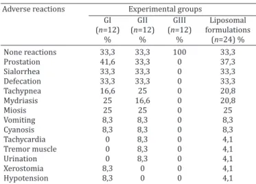
Table 1 shows the frequency of adverse reactions in dogs with leishmaniasis after treatment with different liposomal
formulations. In general, a single intravenous dose of lipo -somal formulations, either containing MA (n=12) or empty,
resulted in similar profiles and intensities of adverse reac -tions. The clinical reactions began moments after bolus ad -ministration and disappeared during the first 15 minutes, involving 67.7% of dogs from both groups that received li -posomal formulations. No adverse effect was observed in the control group (saline).
Prostation, sialorrhea, and defecation were the most fre -quent signs, affecting between 33.3 and 41.6 % of animals from GI and GII groups (Table 1). Tachypnea, mydriasis and miosis were observed in three (16.6 %) or four (25 %) animals per group. Tremor muscle, tachycardia, urination and cyanosis were observed once (8.3%) only in GII. On the other hand, two dogs from GI only each showed xerostomia (8.3%) and hypotension (8.3%). Vomiting was registered in one animal of each group, resulting in 8.3% of frequency. The clinical condition of infected animals apparently did not influence the toxicity of liposomal formulations (Fig.1).
Table 1. Frequency of adverse reactions observed in dogs naturally infected with Leishmania infantum after
intravenous administration of single dose of different liposomal formulations
Adverse reactions Experimental groups
GI GII GIII Liposomal (n=12) (n=12) (n=12) formulations
% % % (n=24) %
None reactions 33,3 33,3 100 33,3 Prostation 41,6 33,3 0 37,3 Sialorrhea 33,3 33,3 0 33,3 Defecation 33,3 33,3 0 33,3 Tachypnea 16,6 25 0 20,8 Mydriasis 25 16,6 0 20,8
Miosis 25 25 0 25
Vomiting 8,3 8,3 0 8,3 Cyanosis 8,3 8,3 0 8,3 Tachycardia 0 8,3 0 4,1 Tremor muscle 0 8,3 0 4,1 Urination 0 8,3 0 4,1 Xerostomia 8,3 0 0 4,1 Hypotension 8,3 0 0 4,1 Data are given as percentage of the dogs that showed different adverse
reactions in accordance with each experimental group. GI: single intra -venous dose of liposomal meglumine antimoniate (400nm) at 6.5mg de Sb/kg; GII: single dose of empty liposomes at the same lipid dose as in GI; GIII: single dose of isotonic saline at the same volume as GI and GII; Liposomal Formulations: considering the animals from de group GI and GII, that is dogs who received treatment with liposomal formulation.
Fig.1. Frequency of adverse reactions observed in dogs classified ac -cording to clinical forms of leishmaniasis, after administration of single dose of liposomal formulations. GI: liposomal meglumi -ne antimoniate (400nm) by i.v. route at 6.5mg Sb/kg/dose; GII: antimony-free liposomes by i.v. route at the same lipid dose as in
No hematologic, hepatic and renal laboratory toxicity were registered (data not shown).
DISCUSSION
One of the main objectives of the Brazilian Group for Rese -arch and Development of Leishmanicidal Drugs has been the design and development of liposomal formulations of MA (Brazilian Patent Pending INPI/2640). Since the first assays of liposomal formulations in canine model, our group has registered side effects (Costa Val 2004, Ribeiro et al. 2008). However, the assessment of the specific profile of reactions after single dose in dogs with different clinical forms of leishmaniasis and the discussion of the triggered mechanism and its relation with the vesicle characteristics have not been made.
The equal participation of different clinical conditions in the groups was important, not only because it involves the diverse realities that are encountered in clinical veterinary practice, but also because the development of the disease may enhance the acute toxicity of formulations (Amusate -gui 1998). Interestingly, the clinical forms of leishmaniasis have no significant influence on the profile and intensity of side effects.
The side effects registered in this study could not be attributed to Sb, because there was no difference betwe -en the groups treated with liposomal MA (GI) and empty liposomes (GII). Furthermore, the toxic effects of the me -tal, including arthralgia, myalgia, diarrhea, anorexia and inflammation at the site of inoculation (Alvar et al. 2004), are expected only upon chronic treatment and should not disappear spontaneously.
The increasing use of advanced therapies based on nanotechnology have allowed the observation of signs of acute immune toxicity with distinct characteristics of hypersensitivity reactions groups traditionally accepted and described by Coombs and Gell in 1968 (Szebeni 2005). In these cases, the allergen led to activation of the comple -ment system, a phenomenon that has been named CARPA. Thus, it is likely that the acute clinical adverse reactions observed in this work were consequence of the action of anaphylatoxins (C3a and C5a) released after activation of the complement cascade by the lipid vesicles. There is ex -tensive literature on complement system activation by li -posomes (Devine et al. 1994, Liu et al. 1995, Szebeni 2005). Natural antibodies against phospholipids and cholesterol are present in all animal species (Wassef et al. 1989) and the binding of these proteins to lipids of liposomes would allow the activation of complement through the classic rou -te (Liu et al. 1995). There is also the possibility of activating the complement cascade by the alternative route (Funato et al. 1992). Probably, the increase of the pulmonary arte -rial pressure, the reduction of the cardiac debit (Szebeni et al. 2000) and the increase of pulmonary and peripheral vascular resistance, generated by the products of the com -plement, provided a transitional circulatory collapse that resulted in tissue hypoxia. Consequently the sympathetic nervous system was stimulated and resulted in some the clinical signs observed: tachycardia, tachypnea, mydriasis
and xerostomia. As a compensatory mechanism, the pa -rasympathetic nervous system was also stimulated provi -ding the reactions of miosis, sialorrhea, vomiting, urination and defecation.
The occurrence of clinical effects just after the contact with allergen is a common symptom between CARPA and classical hypersensitivity type I (IgE-mediated), but the high reaction rate (66.6% of animals) and spontaneous re -solution of adverse reactions are features exclusive of the activation of complement (Szebeni 2005). In CARPA, the toxicity reaches a peak between one and five minutes after the administration of the formulation (Szebeni 2005) and, in the present study, the side effects have always been in -cluded in the first fifteen minutes after treatment. Indeed, the presence of DCP in the lipid composition of liposomes is probably critical, since it ensures anionic character of the vesicles, which is expected to accelerate protein binding onto the surface of the vesicles (Szebeni et al. 2000).
Costa Val (2004) described occurrence of similar adver -se reactions in dogs treated with empty liposome. However, no effect was observed in the groups that received saline, free MA and liposomal MA. Although the lipid composition used was the same, the mean hydrodynamic diameter of the vesicles (1200 nm) was greater than that used in this study (400 nm). In theory, the binding of antibodies to lipo -somes and subsequent activation of the complement sys -tem should be proportional to the total vesicular surface area exposed to plasma (Szebeni et al. 2000). Comparing the results of Costa Val (2004) and ours, it is likely that the smaller surface area of contact in micrometric vesicles and the lowest dose of lipid applied provided a less effective ac -tivation of the complement system.
No hematologic, hepatic and renal laboratory toxicity were observed 96 hours after administration of formula -tions. Although the activation of complement appears to be an intrinsic property of lipid bilayers formed by choles -terol and electrically charged phospholipids (Devine et al. 1994), laboratory changes associated of systems carriers of drugs can be observed in the absence of clinical effects (Valladares et al. 2001).
Since the speed of entry of liposomes in the vascular system is considered critical factor for activation of the complement (Szebeni et al. 2000), the intravenous admi -nistration of this formulation by continuous infusion in fluid therapy should reduce or eliminate some or all of the side effects observed, without changing the characteris -tics of the product. Another proposal for reducing the fre -quency of CARPA would be the premedication of patients with antihistamines and corticosteroids.
In future studies, in vitro and in vivo tests for comple-ment activation may be useful to determine the mechanism of the immunotoxicity of the formulation, in accordance with the US Food and Drug Administration (FDA) recom -mendations.
Acknowledgements.- This research was supported by Laboratório de So
REFERENCES
Alvar J., Cañavate C., Molina R., Moreno J. & Nieto J. 2004. Canine leishma -niasis. Adv. Parasitol. 57:1-88.
Alving C.R. 1986. Liposomes as drug carriers in leishmaniasis and malaria. Parasitol. Today 2:101-107.
Amusategui I. 1998. Tratamiento de la leishmaniosis canina: valoración, caracterización y comparación de la respuesta a distintos protocolos a base de antimoniato de meglumina asociado o no al alopurinol. Tesis de Doctorado en patologia y clinica equina, Facultad de Veterinaria, Univer -sidad Complutense de Madrid, Madrid. 315p.
Armstead A.L. & Li B. 2011. Nanomedicine as an emerging approach against intracellular pathogens. Int. J. Nanomed. 6:3281-3293.
Brazilian Patent Pending INPI/2640 - Frézard F., Demicheli C., Schettini D.A., Ribeiro R.R., Melo M.N. & Michalick M.S.M. November 2004.
Chapman W.L., Hanson W.L., Alving C.R. & Hendricks L.D. 1984. Antileish -manial activity of liposome-encapsulated meglumine antimonate in the dog. Am. J. Vet. Res. 45:1028-1032.
Costa Val A.P. 2004. Tratamento da leishmaniose visceral canina com an -timonial pentavalente encapsulado em lipossomas. Tese de Doutorado em Ciência Animal, Escola de Veterinária, Universidade Federal de Mi -nas Gerais, Belo Horizonte, MG. 125p.
Croft S.L. 1986. Liposomes in the treatment of parasitic diseases. Pharm. Int. 7:229-233.
Demicheli C., Ochoa R., Lula I.S., Gozzo F.C., Eberlin M.N. & Frézard F. 2003. Pentavalent organoantimonial derivatives: two simple and efficient synthetic methods for meglumine antimonate. Appl. Organomet. Chem. 17:226-231.
Devine D.V., Wong K., Serrano K., Chonn A. & Cullis P.R. 1994. Liposome --complement interactions in rat serum: implications for liposome survi -val studies. Biochem. Biophys. Acta 1191:43-51.
Frézard F. & Demicheli C. 2010. New delivery strategies for the old penta -valent antimonial drugs. Expert. Opin. Drug Deliv. 12:1343-1358. Funato K., Yoda R. & Kiwada H. 1992. Contribution of complement system
on destabilization of liposomes composed hydrogenated egg phospha -tidylcholine in rat fresh plasma. Biochim. Biophys. Acta 1103:198-204. Irache J.M., Esparza I., Gamazo C., Agüeros M. & Espuelas S. 2011. Nano
-medicine: Novel approaches in human and veterinary therapeutics. Vet. Parasitol. 180:47-71.
Liu D., Song Y.K. & Liu F. 1995. Antobody dependent complement mediated liver uptake of liposomes containing GM1. Pharm. Res. 12:1775-1780.
Mancianti F., Gramiccia M., Gradoni L. & Pieri S. 1988. Studies on canine leishmaniais control. I. Evolution of infection of different clinical forms of canine leishmaniasis following antimonial treatment. Trans. R. Soc. Trop. Med. Hyg. 82:566-567.
Nieto J., Alvar J., Mullen A.B., Carter K.C., Rodrígues C., San Andrés M.I., San Andrés M.D., Baillie A.J. & González F. 2003. Pharmacokinetics, toxicities, and efficacies of sodium stibogluconate formulations after intravenous administration in animals. Antimicrob. Agentes Chemother. 47:2781-2787.
Ribeiro R.R., Moura E.P., Pimentel V.M., Sampaio W.M., Silva S.M., Schettini D.A., Alves C.F., Melo F.A., Tafuri W.L., Demicheli C., Melo M.N., Frézard F. & Michalick M.S. 2008. Reduced tissue parasitic load and infectivity to sand flies in dogs naturally infected by Leishmania (Leishmania) chagasi
following treatment with a liposome formulation of meglumine antimo -niate. Antimicrob. Agentes Chemother. 57:2564-2572.
Schettini D.A., Ribeiro R.R., Demicheli C., Rocha O.G.F., Melo M.N., Michalick M.S.M. & Frézard F. 2006. Improved targeting of antimony to the bone marrow of dogs using liposomes of reduced size. Int. J. Pharm. 315:140-147.
Szebeni J. 2005. Complement activation-related pseudoallergy: A new class of drug-induced acute immune toxicity. Toxicology 216:106-121.
Szebeni J., Baranyi L., Savay S., Bodo M., Morse D.S., Basta M., Stahl G.L., Bunger R. & Alving C.R. 2000. Liposome-induced pulmonary hyperten -sion: properties and mechanism of a complement-mediated pseudoal -lergic reaction. Am. J. Physiol. Heart Circ. Physiol. 279:1319-1328. Szebeni J., Bedőcs P., Rozsnyay Z., Weiszhár Z., Urbanics R., Rosivall L.,
Cohen R., Garbuzenko O., Báthori G., Tóth M., Bünger R. & Barenholz Y. 2012. Liposome-induced complement activation and related cardiopul -monary distress in pigs: factors promoting reactogenicity of Doxil and AmBisome. Nanomedicine 8:176-184.
Valladares J.E., Riera C., González-Ensenyat P., Diaz-Cascon A., Ramos G., Solano-Gallego L., Gállego M., Portús M., Arboix M. & Albertola J. 2001. Long term improvement in the treatment of canine leishmaniasis using an antimony lipossomal formulation. Vet. Parasitol.97:15-20.
Wassef N.M., Johnson S.H., Graeber G.M., Swartz G.M., Schultz C.L., Hailey J.R., Johnson A.J., Taylor D.G., Ridway R.L. & Alving C.R. 1989. Anaphylac -toid reactions mediated by autoantibodies to cholesterol in miniature pigs. J. Immunol. 143:2990-2995.