A
An
ANDRÉ
álise co
pro
UNIVER FA PROGÉA DOLO
omparati
tocolos
SERVIÇO MINISTÉR RSIDADE F ACULDAD GRAMA D ODORES C
va de co
de reab
endod
Uberlândia
1 PÚBLICO RIO DA ED FEDERAL DE DE OD DE PÓS-G DONTOLO
CORREI
omporta
bilitação
donticam
a, Fevereir O FEDERA DUCAÇÃO L DE UBER ONTOLOG RADUAÇÃ OGIAA MIRA
amento
i
de dent
mente.
Tese apr de Odont Federal requisito p Título de na Área Clínica Odro de 2016 AL O RLÂNDIA GIA ÃO EM
ANDA VA
in vivo
e
tes trata
resentada tologia da de Uberl parcial para Doutor em de Conc dontológica 6ALDIVIA
e
in vitro
ados
à Faculd Universid lândia, co ra obtenção m Odontol centração a IntegradaA
o
de
2
ANDRÉA DOLORES CORREIA MIRANDA VALDIVIA
Análise comparativa de comportamento
in vivo
e
in vitro
de
protocolos de reabilitação de dentes tratados
endodonticamente.
Tese apresentada à Faculdade de Odontologia da Universidade Federal de Uberlândia, como requisito parcial para obtenção do Título de Doutor em Odontologia na Área de concentração de Clínica Odontológica Integrada.
Banca Examinadora:
Orientador: Prof. Dr. Carlos José Soares, UFU Prof. Dr. Alfredo Júlio Fernandes Neto, UFU
Profa. Dra. Veridiana Resende Novais Simamoto, UFU Prof. Dr. Cesar Penazzo Lepri, UNIUBE
Prof. Dr. Roberto Sales e Pessoa, UNITRI Suplentes
Prof. Dr. Manoel Damião de Sousa Neto, FORP,USP Prof. Dr. Paulo Sérgio Quagliatto, UFU
Dados Internacionais de Catalogação na Publicação (CIP) Sistema de Bibliotecas da UFU, MG, Brasil.
V146a 2016
Valdivia, Andréa Dolores Correia Miranda, 1980
Análise comparativa de comportamento in vivo e in vitro de protocolos de reabilitação de dentes tratados endodonticamente / Andréa Dolores Correia Miranda Valdivia. - 2016.
113 p. : il.
Orientador: Carlos José Soares.
Tese (doutorado) - Universidade Federal de Uberlândia, Programa de Pós-Graduação em Odontologia.
Inclui bibliografia.
1. Odontologia - Teses. 2. Endodontia - Tratamento - Teses. 3. Pinos dentários - Teses. 4. Coroas (Odontologia) - Teses. I. Soares, Carlos José. II. Universidade Federal de Uberlândia. Programa de Pós-Graduação em Odontologia. III. Título.
4
DEDICATÓRIA
A Deus
Agradeço por t udo que acont ece em nossas vidas, nunca sabem os o que Deus t em pra nos dar, m as Ele conhece nossos corações, nossos
m edos e nossas necessidades.
À m inha fam ília,
Especialm ent e ao m eu filho Alej andro, o m aior present e de Deus na m inha vida e à m inha avó Maria Dolores, pelas saudades et ernas.
“Sem Deus não há vida, sem família não há base
e sem amigos não há mundo colorido.”
(Verena)
5
AGRADECIMENTOS
A Deus, obrigado Senhor, por estar sempre ao meu lado, pelo amparo nas horas de dificuldade e pelas pessoas maravilhosas que colocastes em meu caminho;
A minha família, especialmente aos meus pais, meu filho, esposo e sogros; porque em vocês encontro meu porto seguro, obrigada pelas orações, pelo incentivo, pelo amor e por estarem sempre presentes nos momentos importantes da minha vida;
Ao meu orientador Prof. Carlos José Soares, professor muito obrigada por todas as oportunidades que me proporcionou e pela confiança que sempre depositou em mim. Você é um exemplo de profissionalismo, inteligência, determinação, dedicação e competência, que deve ser seguido por todos;
Aos professores da banca de defesa e aos membros suplentes, muito obrigada pela disposição em contribuir com nosso trabalho;
Aos amigos Roberto Sales, Ravel Miranda e Jean, por toda a disponibilidade, apoio, incentivo e amizade durante a execução deste trabalho;
Aos amigos do grupo de pesquisa Biaor (Biomecânica Aplicada à Odontologia Restauradora), que estiveram presente em todos os momentos de muita risada e lágrimas desse trabalho;
Aos amigos de Doutorado, Aline, Carol, Crisnicaw, Felipe, Flaviana, Karla, Luiz Fernando, Maria Antonieta, Marina, Pri, Roberta, Vanessa, pelo companheirismo durante todo o Doutorado;
À alguns amigos em especial, Aline, Camila Rosatto, Renata Afonso, Gabi, Pri, Valessa, Crisnicaw, Suely, Monise, Maria Antonieta, Profa. Gisele. Obrigada pelo apoio nas diferentes etapas desta pesquisa que cada um teve da sua maneira, e que foram essenciais;
6
À Monise, que começamos nossa amizade durante a sua iniciação científica, obrigada pelos anos de convivência, pela paciência com meu aprendizado em orientar, pela dedicação e empenho no trabalho e pelo incentivo e confiança de sempre;
Aos Professores da Katholieke Universiteit Leuven (Bélgica), em especial ao Prof. Jos Vander Sloten, Siegfried Jaecques e Bart Van Meerbeek e aos amigos de Leuven que durante meu doutorado sanduiche sempre estiveram prontos a ajudar e sem medir esforços para fazer dar certo;
Ao Prof. Dr. João César G. Henriques da Área de Diagnóstico Estomatológico, pelo apoio na aquisição das imagens tomográficas dos pacientes;
A todos os professores da FOUFU e ao Programa de Pós Graduação, pelo apoio constante à pesquisa, ensino e extensão;
A todos os funcionários da FOUFU, por toda a ajuda, colaboração, dedicação e disposição em atender, da melhor forma possível, às nossas necessidades;
Ao Hospital Odontológico da FOUFU representado pelo Prof. Dr. Marcio Teixeira;
Ao CPbio, Centro de Pesquisa Odontológico de Biomecânica, Biomateriais e Biologia Celular da FOUFU;
À CAPES pela bolsa de Doutorado Sanduiche e FAPEMIG pela bolsa de Doutorado e auxílio financeiro e desta pesquisa.
E a todas as pessoas que, de alguma forma, contribuíram para que essa etapa fosse vencida!
7
EPÍGRAFE
A lição do bambu chinês
Depois de plantada a semente deste incrível
arbusto, não se vê nada,
por aproximadamente 5 anos, exceto o lento
desabrochar de um diminuto broto,
a partir do bulbo.
Durante 5 anos, todo o crescimento é
subterrâneo, invisível a olho nu,
mas, uma maciça e fibrosa estrutura de raiz,
que se estende vertical
e horizontalmente pela terra está sendo construída.
Então no final do 5º ano, o bambu chinês,
cresce até atingir a altura de 25 metros.
Um escritor americano escreveu:
“Muitas coisas na vida pessoal e profissional
são iguais ao bambu chinês:
você trabalha, investe tempo, esforço, faz tudo
o que pode para nutrir seu crescimento,
e, às vezes não vê nada por semanas, meses, ou anos.
Mas se tiver paciência para continuar
trabalhando, persistindo e nutrindo,
o seu 5º ano chegará, e, com ele, virão um
crescimento e mudanças que você jamais
esperava...
O bambu chinês nos ensina que não devemos
facilmente desistir de nossos projetos,
de nossos sonhos... especialmente no nosso trabalho,
(que é sempre um grande projeto em nossas vidas)
E que devemos lembrar do bambu chinês,
para não desistirmos facilmente
diante das dificuldades que surgirão.
É preciso muita fibra para chegar às alturas e,
Ao mesmo tempo,
muita flexibilidade para se curvar ao chão.”
(Autor desconhecido)
8
SUMÁRIO
Resumo 9
Abstract 12
1. Introdução e Referencial Teórico 15
2. Objetivos 20
3. Capítulos 23
3.1. Capítulo 1 - Effect of Surface Treatment of Fiberglass Posts on Bond Strength to Root Dentin
24
3.2. Capítulo 2 - Patient specific finite element analysis of fiber post and ferrule design
33
3.3. Capítulo 3 - Biomechanical effect of ferrule presence on incisor teeth restored with fiberglass post and CAD/CAM ceramic crown
after thermal cycling and fatigue loading
53
3.4. Capítulo 4 - Reabilitação estética do sorriso com uso de pino de fibra de vidro associado à coroa cerâmica CAD/CAM – Aspectos clínicos e biomecânicos.
81
4. Conclusões 102
5. Referências 104
6. Anexos 108
Parecer do Comitê de ética Release para Imprensa
109 113
R
Análise endodo Gradua
R
ES
e comparativa onticamente – A ação em Odont
SUM
de comporta ANDRÉA DOLO tologia – Faculd
MO
mento in vivo ORES CORREI dade de Odonto
9 o e in vitro d
IA MIRANDA V ologia – Univers
e protocolos d VALDIVIA – Tes
idade Federal d
de reabilitação se de Doutorado de Uberlândia
o de dentes t o – Programa d
Pós-10 RESUMO
11
A
Análise endodo Gradua
A
B
e comparativa onticamente – A ação em Odont
STRA
de comporta ANDRÉA DOLO tologia – Faculd
ACT
mento in vivo ORES CORREI dade de Odonto
12 o e in vitro d
IA MIRANDA V ologia – Univers
e protocolos d VALDIVIA – Tes
idade Federal d
de reabilitação se de Doutorado de Uberlândia
o de dentes t o – Programa d
Pós-13 ABSTRACT
14
I
TE
Análise endodo Gradua
NTR
EÓRI
e comparativa onticamente – A ação em Odont
ODU
ICO
de comporta ANDRÉA DOLO tologia – Faculd
UÇÃO
mento in vivo ORES CORREI dade de Odonto
15
O E
o e in vitro d IA MIRANDA V ologia – Univers
REF
e protocolos d VALDIVIA – Tes
idade Federal d
ERE
de reabilitação se de Doutorado de Uberlândia
NCIA
o de dentes t o – Programa d
AL
Pós-16
1. INTRODUÇÃO E REFERENCIAL TEÓRICO
Dentes tratados endodonticamente frequentemente requerem retentores intrarradiculares para realização de procedimentos restauradores, devido às grandes perdas de estrutura dental causada por cárie ou acessos endodônticos à cavidade pulpar (Bateman et al., 2003). O propósito dos retentores não é reforçar a estrutura, mas reter e estabilizar os materiais restauradores, uma vez que estes podem interferir na resistência mecânica do dente, aumentando o risco de dano da estrutura dental remanescente (Santos-Filho et al., 2008). Fatores relacionados ao retentor intrarradicular, tais como: comprimento, espessura, configuração e a composição podem influenciar na biomecânica destes dentes, alterando o padrão de distribuição de tensões (Barjau-Escribano et al., 2006), resistência à fratura (Barjau-Escribano et al., 2006; Santos-Filho et al., 2008; Silva et al., 2011) e deformação da estrutura remanescente (Santos-Filho et al., 2008; Silva et al., 2011). Assim nas últimas décadas, a introdução dos pinos de fibra de vidro surgiu como alternativa aos pinos metálicos (Bateman et al., 2003), já que os pinos de fibra apresentam módulo de elasticidade similar ao da dentina (Naumann et al., 2006), reduzindo a concentração de tensões nas interfaces, capacitando o complexo restaurador a mimetizar o comportamento biomecânico de dentes hígidos (Santos-Filho et al., 2008; Silva et al., 2011). Estudos utilizando o método de elementos finitos têm se mostrado como instrumento valioso na análise da distribuição de tensões nas restaurações de dentes anteriores. O fato dos pinos de fibra exibirem propriedades biomecânicas mais similares às da dentina que os pinos metálicos (Lanza et al., 2005), tem motivado pesquisadores ao estudo cada vez mais crescente dos retentores intrarradiculares com auxilio desta metodologia (Santos-Filho et al., 2014; Veríssimo et al., 2014).
17
base de sustentação, as forças que incidem sobre o núcleo são direcionadas obliquamente, tornando a raiz mais susceptível à fratura (Pegoraro, 2000).
A existência de variedade de opções no tratamento reabilitador com retentores intrarradiculares geram dúvidas aos profissionais de qual o melhor planejamento reabilitador. Na cimentação de pinos no interior do canal, quer seja em raízes fragilizadas (Silva et al., 2011) ou não, altas tensões de contração são geradas durante o processo de polimerização do cimento resinoso (Ferrari et al., 2009, Pereira et al., 2015). O risco de fratura pode ser potencializado por falhas na interface adesiva entre o retentor e o material de cimentação e entre este e a dentina radicular. O fator de configuração cavitário (fator C), é altamente desfavorável no interior do canal, e parece contribuir com a formação de tensões de contração de polimerização na interface adesiva influenciando negativamente na qualidade de adesão, justificando a desunião do retentor como fator de falha clínica prevalente destes procedimentos restauradores (Jongsma et al., 2011).
Outro aspecto que pode contribuir para a ocorrência de falha de deslocamento do pino é a falta de completa polimerização do cimento resinoso (Pereira et al., 2015).Atualmente encontra-se a disposição do clínico pinos de fibra de vidro translúcidos, que prometem transmitir a luz até o terço apical das raízes, promovendo melhor polimerização do cimento e consequente melhora na resistência adesiva. Cimentos resinosos autoadesivos têm sido propostos para minimizar este aspecto pela simplificação do processo de cimentação adesiva (Aguiar et al., 2010). Entretanto, apesar das vantagens, falhas na adesão entre pino de fibra-cimento-dentina têm sido relatadas (de Souza Menezes et al., 2011), e estudos in vivo têm mostrado que a adesão nesta interface é crítica para o sucesso clínico deste tipo de restauração (Cagidiaco et al., 2007, Valdivia et al., 2014).
18
asperização da superfície para aumentar a retenção micro-mecânica, por meio do condicionamento ácido, jateamento ou silicatização; ou (3) associação das duas técnicas anteriores. Comparando-se estas técnicas de tratamento de superfície, o condicionamento com peróxido de hidrogênio é o que tem apresentado melhores resultados, pois além de ser eficiente, apresenta facilidade técnica de utilização (Monticelli et al., 2008), promove dissolução parcial da matriz de resina epóxica (Monticelli et al., 2008) e não causa danos as fibras do pino (Monticelli et al., 2006; Valdivia et al., 2014).
O estabelecimento de associação de metodologias que visam analisar parâmetros distintos, porém complementares se faz necessário em avaliações in vitro para aproximar a detecção de falhas mais próximo da realidade. O emprego de processo de envelhecimento que caracteriza a fadiga das estruturas envolvidas por meio de ciclos térmicos e mecânicos possibilitam a simulação de alguns desafios verificados em ambiente clínico aos quais materiais restauradores e substratos dentários são expostos (Silva et al., 2011). Os ensaios mecânicos destrutivos são importantes meios de análise do comportamento do dente e de diferentes materiais (Soares et al., 2006), no entanto, torna-se necessária a associação com metodologias não-destrutivas, como ensaio de extensometria (Santos-Filho et al., 2008; Veríssimo et al., 2014) ou computacionais como método de elementos finitos (Soares, 2003; Magne, 2007; Magne & Oganesyan, 2009; Santos-Filho et al., 2014; Veríssimo et al., 2014), favorecendo análise biomecânica sequencial e detalhada do comportamento da amostra. Por conseguinte, parece que na biomecânica um bom caminho é o emprego da associação com metodologias que se completem que possam ser retroalimentadas e que acabem envolvendo menores investimentos e com respostas mais próximas àquelas que se conseguem com os experimentos in vivo. A associação com metodologias computacionais como o método de elementos finitos (MEF) constitui mais um importante aliado na caracterização deste processo de comportamento das estruturas dentais.
19
estruturas biológicas estudadas. Pensar nesta extrapolação para modelos geometricamente similares é perfeitamente pertinente, contudo a análise de estruturas biológicas de geometrias altamente variadas ainda se pauta consideravelmente na análise da variabilidade por meio de utilização de diversas amostras para que os valores médios representem mais adequadamente o comportamento biomecânico. Inicia-se na análise biomecânica a necessidade de ampliação de amostras biológicas para que resultados provenientes de diferentes modelos possam prever melhor estes aspectos. Diante deste horizonte, nada mais válido que a associação de análise clínica prospectiva com a simulação simultânea destas estruturas em modelos computacionais e ainda experimentais para que este ciclo investigativo possa ser retroalimentado e retrovalidado.
O
Análise endodo Gradua
O
B
e comparativa onticamente – A ação em Odont
BJET
de comporta ANDRÉA DOLO tologia – Faculd
IVOS
mento in vivo ORES CORREI dade de Odonto
20
S
o e in vitro d IA MIRANDA V ologia – Univers
e protocolos d VALDIVIA – Tes
idade Federal d
de reabilitação se de Doutorado de Uberlândia
o de dentes t o – Programa d
Pós-21 2. OBJETIVOS
Objetivo Geral
Este projeto visa de forma sequencial e progressiva analisar o efeito do protocolo restaurador de dente tratado endodonticamente por meio de avaliação comparativa in vivo e simultânea in vitro, empregando ensaios mecânicos e computacionais pelo método de elementos finitos. Visa ainda validar os ensaios computacionais e ensaios experimentais laboratoriais por meio de análise comparativa com parâmetros clínicos.
Objetivos específicos
Objetivo específico 1
Capítulo 1 - Effect of Surface Treatment of Fiberglass Posts on Bond
Strength to Root Dentin
O objetivo deste estudo foi avaliar a influência do tratamento de superfície de pinos pré-fabricados na resistência de união de pino ao canal radicular, por meio de teste de micropush-out.
Objetivo específico 2
Capítulo 2 - Patient specific finite element analysis of fiber post and ferrule design
O objetivo deste estudo foi desenvolver e validar a geração de modelos tridimensionais específicos de paciente com incisivos centrais anteriores com diferente tamanho de férula, restaurados com pino de fibra de vidro e coroa em cerâmica pura CAD/CAM usando tomografia computadorizada Cone-beam (CT) e combinação de softwares específicos de elementos finitos.
Objetivo específico 3
Capítulo 3 – Biomechanical effect of ferrule presence on incisor teeth
restored with fiberglass post and CAD/CAM ceramic crown after thermal
22
O objetivo deste estudo foi avaliar a deformação antes e após ciclagem térmica e mecânica, resistência à fratura e padrão de falha de incisivos tratados endodonticamente restaurados com pino de fibra de vidro e coroa total em cerâmica pura CAD/CAM com e sem férula.
Objetivo específico 4
Capítulo 4 - Reabilitação estética do sorriso com uso de pino de fibra de
vidro associado à coroa cerâmica CAD/CAM - Aspectos clínicos e
biomecânicos.
C
Análise endodo Gradua
C
A
e comparativa onticamente – A ação em Odont
APÍTU
de comporta ANDRÉA DOLO tologia – Faculd
ULOS
mento in vivo ORES CORREI dade de Odonto
23
S
o e in vitro d IA MIRANDA V ologia – Univers
e protocolos d VALDIVIA – Tes
idade Federal d
de reabilitação se de Doutorado de Uberlândia
o de dentes t o – Programa d
Pós-24 3. CAPÍTULOS
3.1 CAPÍTULO 1
Artigo publicado no periódico Brazilian Dental Journal
Brazilian Dental Journal (2014) 25(4): 314-320 ISSN 0103-6440
http://dx.doi.org/10.1590/0103-6440201300143
Effect of surface treatment of fiberglass posts on bond strength to root dentine
Andréa Dolores Correia Miranda Valdivia1, Veridiana Resende Novais1, Murilo Sousa Menezes1, Marina Guimarães Roscoe1, Carlos Estrela2, Carlos José Soares1
1. Operative Dentistry and Dental Materials Department, School of Dentistry, Federal University of Uberlândia, Uberlândia, MG, Brazil
2. Endodontics Department, School of Dentistry, Federal University of Goiás, Goiânia, GO, Brazil
C
Análise endodo Gradua
C
A
e comparativa onticamente – A ação em Odont
APÍTU
de comporta ANDRÉA DOLO tologia – Faculd
ULOS
mento in vivo ORES CORREI dade de Odonto
32
S
o e in vitro d IA MIRANDA V ologia – Univers
e protocolos d VALDIVIA – Tes
idade Federal d
de reabilitação se de Doutorado de Uberlândia
o de dentes t o – Programa d
Pós-33 3.2 CAPÍTULO 2
Artigo a ser enviado para publicação no periódico Journal of Dental Research
Patient specific finite element analysis of fiber post and ferrule design
Andréa Dolores Correia Miranda Valdivia1 Monise de Paula Rodrigues1
Roberto Sales e Pessoa1 Crisnicaw Veríssimo1 Jos Vander Sloten2
Priscilla Barbosa Ferreira Soares1 Carlos José Soares1
1 Department of Operative Dentistry and Dental Materials, School of Dentistry, Federal University of Uberlândia, Minas Gerais, Brazil.
2
Biomechanics section – Mechanical Engineering Department, Leuven Medical Techonology Centre, Katholieke Universiteir Leuven, Belgium.
Corresponding author: Prof. Dr. Carlos José Soares - Avenida República do
34
Patient specific finite element analysis of fiber post and ferrule design
Abstract
Biomechanical effect of ferrule on anterior endodontic treated teeth has been evaluated using clinical trials, in vitro tests and finite element analysis. No studies have been performed using patient specific model with real no-uniform ferrule and no-linear antagonist biting load with clinical validation. Patient with both upper central incisors with different ferrule design that received endodontic treatment and restoration using fiber post, composite core and CAD-CAM di-silicate ceramic crowns, was selected. The bite force was measured for each central incisor and for both incisors. Strain-gauge was attached on buccal surface of both teeth to record tooth strain during bite force recording. A cone beam tomography was used to scan the teeth and the projection data were exported using the DICOM files for Mimics and 3-Matic (Materialise) and Patran (MSC) softwares for finite element patient specific model generation of the anterior maxilla with both central incisor and mandibular incisors. The bite loading was simulated in 3 methods: M1, nodal point load using individual force clinically measured (55N for right incisor; 100N for left incisor); M2, nodal load using the force measure for both teeth (155N); M3, non-linear contact load by the antagonist teeth (155N). The mechanical properties were obtained from the literature. Modified von Mises equivalent stress was used for stress evaluation. Stresses on left incisor, which had no-uniform ferrule were higher compared with the right incisor, regardless of the loading method. The bone level influence the stress distribution, higher bone limit to cavosurface preparation resulted in higher stress concentration. The stress in the roots and fiber posts for M1 and
M2 models were higher than those for M3 model. Simulate loading using antagonist result in lower stresses, being more realistic compared with clinical success of fiber post. The maintenance of uniform ferrule was more relevant than localized higher ferrule.
1. Introduction
35
et al., 2003; Silva et al., 2010). The correct choice of the post system used for teeth rehabilitation and the ferrule effect are crucial for treatment prognosis (Stankiewicz and Wilson, 2002). Fiber posts have been used clinically as an alternative to metal posts in the restoration of endodontically treated teeth (Ferrari et al., 2007; Goracci and Ferrari, 2011; Soares et al., 2012). The amount of coronal and root dentin remains after root canal instrumentation and post space preparation are correlated with the fracture resistance and plays an important role in the longevity of the tooth and restoration (Ichim et al., 2006; Silva et al., 2010, Veríssimo et al., 2014). A recent meta-analysis suggested that coronal wall absence might increase the risk of fiber post-core restoration failure, while the role of ferrule effect is still not entirely understood (Yang et al., 2015). Others studies reported that the presence of uniform ferrule surrounding remaining tooth structure enhanced fracture resistance and increase the long term success of post-endodontic of anterior teeth (Silva et al., 2010; Soares et al., 2012).
Numerous finite element studies have linked the ferrule effect on biomechanics behavior of anterior endodontic treated teeth with various factors such as remaining dentin thickness (Coelho et al., 2009; Santos Filho et al., 2014), different ferrule height and configuration (Ichim et al., 2006; Roscoe et al., 2013; Juloski et al., 2014; Zhang et al., 2015). However all these studies
have used the FEA models are created using general data of the anterior teeth, with the standardization of the ferrule design to isolate the effect of the specific factor. Other simplification on finite element analysis of fiber post is the nodal static load or time dependent gradually increased load application (Roscoe et al., 2013; Juloski et al., 2014).
36
(Adams and Askenazi, 1999; Soares et al., 2012). Nonlinear solutions require more computational interactions to converge to a final solution, being more costly in terms of computation and time. However, the nonlinear FEA is more powerful tool to predict stress within structures in comparison with conventional linear static models (Soares et al., 2012). No FEA studies of endodontic treated teeth restored with fiber post have been performed yet, on the author’s knowledge, using patient specific model that represent real no-uniform ferrule presence and nonlinear antagonist bite load application and followed by clinical validation.
Use of strain-gauge methods for finite element analysis validation on in vitro studies is usual and determines more accuracy on two-way (Bicalho et al. 2014, Santos Filho et al., 2014). Patient validation has been nowadays used to reproduce clinical conditions on finite element validation (Juloski et al., 2014). The validation and data correlation of FEA patient specific analysis might represent a powerful strategy for combination of clinical trials and finite element analysis to predict the clinical failures. Therefore the aim of this study was to develop protocol and validate the generation of three-dimensional patient specific model of anterior central incisors with different ferrule design restored with glass fiber post and CAD-CAM all-ceramic restoration using cone-beam computed tomography and specific software combination for finite elements analysis.
2. Materials and Methods
2.1. Patient rehabilitation
The subject included in this study was recruited from the Dental Hospital
from Federal University of Uberlandia. This study had the approval of the Ethic Committee (#144.423/2012). The patient presented two upper central incisors with necessity of periodontal surgery, endodontic treatment and fiber post, composite core and all-ceramic crowns rehabilitation.
37
(Dentsply, Petrópolis, Rio de Janeiro, Brazil) and calcium hydroxide-based endodontic sealer (Sealer 26, Dentsply) (Valdivia et al., 2012). Root canals were prepared using dedicated drill for conic smooth glass fiber posts (ExactoTranslucido no. 3, Angelus) with 1.0mm on apical and 2.0mm on cervical limit. The posts were immersed in 24% hydrogen peroxide (H2O2, Dinâmica, SP, Brazil) followed by silane coupling agent (Silano, Angelus) application for 1 minute (Valdivia et al., 2012). The posts were luted with self-adhesive resin cement (RelyXUnicem 2; 3M ESPE, St Paul, Minn). After 5 minutes, the resin cement was light activated on each surface for 40 seconds with an LED unit (Radii-Cal; SDI, Bayswater, Australia). The coronal tooth remaining was etched using 37% phosphoric acid (Cond AC 37, FGM, Joinville, SC, Brazil) for 15s and the etch-and-rinse 3 steps adhesive system (Scotchbond Multi-Purpose; 3M ESPE) was used, following the manufacturer’s instructions.
The composite resin cores were built incrementally using Tetric Ceram composite resin (Ivoclar/Vivadent, Ellwangen, Germany), light activating each increment for 40 seconds using an LED unit (Radii-Cal; SDI). Complete crown coverage preparations featuring 1.5 mm of axial reduction and 6 degrees of axial convergence of the walls were performed with diamond rotary burs (KG Sorensen, Barueri, SP, Brazil). Impression of the tooth preparation was taken using polyvinylsiloxane material (Express™ VPS; 3M ESPE). Lithium di-silicate glass-ceramic CAD/CAM system (e.max IPS CAD, Ivoclar/Vivadent, Ellwangen, Germany) was used for all-ceramic crowns. The internal surfaces of ceramic crowns the restorations were etched with 10% hydrofluoric acid for 20 seconds (Cond AC Porcelana, FGM) followed by silane application for 1 min. Ceramic crowns were luted using self-adhesive resin cement (RelyXUnicem 2, 3M ESPE), light activating after 5 minutes for 40 seconds on each surface using an LED unit (Radii-Cal; SDI).
2.2. Strain and bite force intraoral measurement
38
specimen was used to compensate for local environment temperature fluctuations due to electrical gauge resistance. Strain data were recorded on a computer that performed the signal transformation and data analysis (AqDados 7.02 and AqAnalysis; Lynx, SP, Brazil). At the same time of strain measurement the bite force in Newtons (N) was measured postoperatively for each restored tooth isolated and both teeth at the same time (Gnatodinamômetro Digital Especial Kratos, Brazil). The load recording was repeated for 5 times and the mean values were: for the right incisor had 55.2 ± 5.1N, for the left incisor had 102.8± 7.3N and for both teeth biting had 155± 9.5N. These biting forces values were used for finite element model input (Fig. 1).
2.3. Cone-Beam images acquisition
After the rehabilitation, the patient was positioned on cone beam tomography (i-CAT GXCB-500™ Imaging Sciences International, Hatfield, Pennsylvania) with the median sagittal plane perpendicular to the horizontal plane, and the occlusal plane parallel to the horizontal plane, was used to scan the samples at a voxel dimension of 0.125 mm. A total of 704 slices was provided with 23 seconds of acquisition and the exposure parameters were 120 kV, 3.0 to 7.0 mA. The projection data were exported using the DICOM (Digital Imaging and Communication in Medicine) file format (Fig. 2).
2.4. 3D Finite element model generation
39
Then, an advanced STL design and meshing software (3-Matic 8.0; Materialise, Leuven, Bélgica) was used to simulate the forms of treatment used in the teeth samples. The treatment of each STL was held separately followed by the merging of all parts in a single STL file called the assembly. The definitive assembly was then remeshed using the 3-matic REMESH component. Self-intersecting curves was maintained and the tolerance variation from the original data will be specified (quality of triangles does not mean tolerance variation from the original data). As in Mimics Remesh, the quality is defined as a measure of triangle height/base ratio so that the file can be imported in the FEA software package without generating errors.
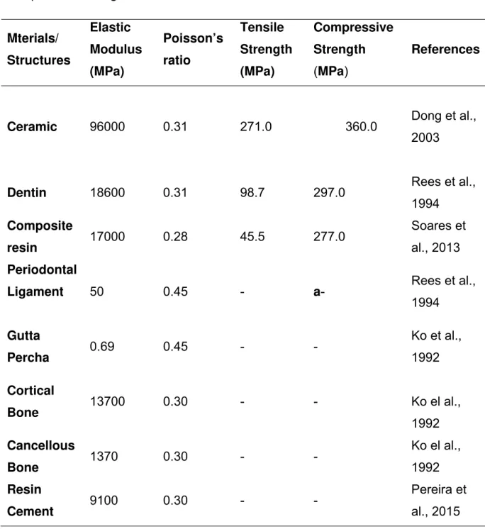
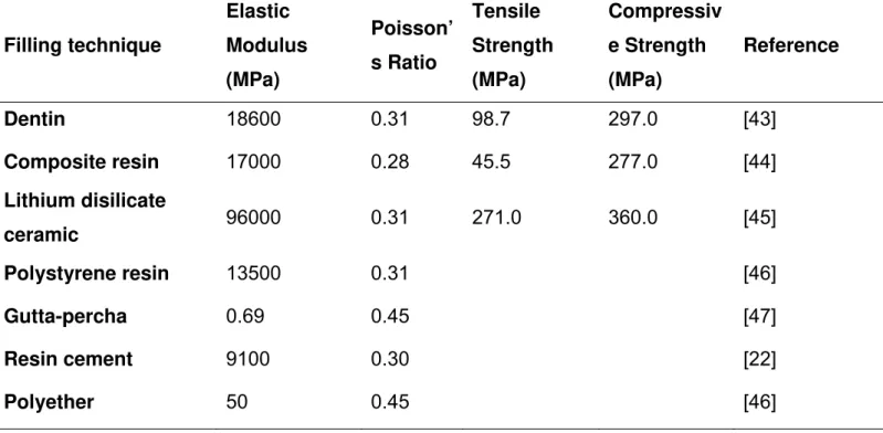
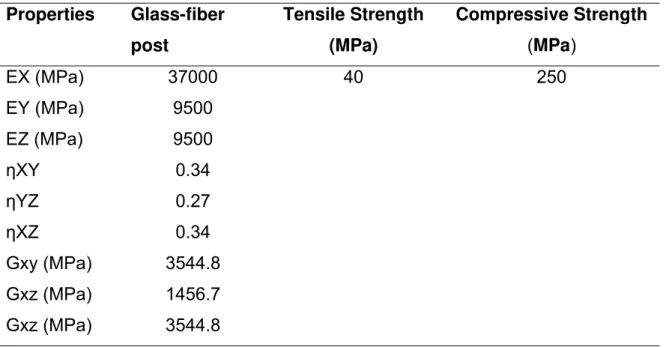
The STL models were imported to MSC.Patran® 2010r2 (MSC.Software, MSC software, Santa Ana, CA, USA) and meshed. Tetrahedral elements were used to ensure smooth contact at the interfaces. The volumetric meshes of bone and all model components were therefore generated based on the optmized surface’s standard triangulated language (STL) descriptions (Pessoa et al., 2010). During meshing process of the bone solid model, the entire volume that is contained within the outer bone surface is meshed. This means that the mesh consists of tetrahedral elements located in either cortical or trabecular bone. To discriminate between both tissues, different elastic properties can be assigned, based on the grey values in the CT images (Jaecques et al. 2004, Pessoa et al. 2010). In this way, the information in the CT images may be used not only to extract the patient’s bone geometry and but also to assign patient-specific bone mechanical properties. After that, the volumetric meshes were imported in a FEA software package (MSC.Marc/MSC.Mentat, MSC.Software, Santa Ana, CA) for the attribution of material properties to the other model components (i.e. ceramic, composite resin, dentin, resin cement, glass fiber post and periodontal ligament). The values of the Young’s modulus and Poisson’s ratio for the materials and structures were adopted from the literature and are summarized in Table 1. Glass fiber post was considered as an orthotropic material (Table 2).
40
simulated: M1, nodal point load using individual force clinically measured (55N for right incisor; 100N for left incisor); M2, nodal load using the mean force measure for both teeth (155N); M3, non-linear contact load by the antagonist teeth (155N). The load for M1 and M2, had a coronal-apical direction and an inclination of 135 degrees in relation to the tooth longitudinal axis The analysis and post-processing were performed for each model by means of the MSC.MARC/ Mentat® 2010r3 software (MSC.Software). Stress were analyzed using modified von Mises equivalent stresses, which integrate all stress components into one stress equivalent value. The compressive and tensile strength ratio used to calculate modified von mises stresses are summarized on Table I and Table II.
3. Results
The modified von Mises stress distributions for dentin structure, fiber post and resin cement are summarized on Fig. 3. Since the highest stresses were found in the root dentin and fiber post, the stresses were evaluated in these structures. Stresses on the root dentin and fiber post for left central incisor were higher compared with the right central incisor, regardless of the shape (Fig. 3). The higher stress concentration was located on distal region of left incisor, where was measured smaller ferrule. The bone level influence the stress distribution, higher distance from the bone limit to cavosurface preparation resulted in higher stress concentration (Fig. 3). The stress distributions in the
roots and fiber posts for models with individual load for each incisor and mean point load were higher than those of antagonist non-linear loading (Fig. 3).
4. Discussion
41
When loads are applied to a structure, structural strains (deformation) and stresses are generated. This is normal, and is how a structure performs its structural function. However if such stresses become excessive and exceed the elastic limit, structural failure may result (Soares et al., 2012). Stresses represent how masticatory forces are transferred through a tooth structure (Versluis and Tantbirojn, 2011). These stresses cannot be measured directly, and for failure in complex structures it is not easy to understand why and when a failure process is initiated, and how we can optimize the strength and longevity of restorative procedures. The relationship between stress and strain is expressed in constitutive equations that may be numerical calculated by using finite element. This study focused on the pre-processing phase of finite element, more specifically on model generation and boundary conditions, represented by loading method simulation. To simulate the masticatory forces, this study used point loads and by means of a simulated opposing incisal of the antagonist tooth. A point load application resulted in high stress concentrations around the loaded nodes, creating unrealistic stress concentrations. (Soares et al., 2012). The point load applications for M1 and M2 used in this study were
gradually increased in 10 increments, which represent a more realist simplification than total load applied on point load on just one increment. However, the non-linear contact method, represented by antagonist teeth loading, resulted in lower stress concentration on the root dentin, demonstrating that higher levels of stress concentration observed in several studies maybe are overestimated. (Roscoe et al., 2013; Santos Filho et al., 2014) In reality, a masticatory contact force is likely to be distributed across certain contact areas on lingual surface of the upper anterior teeth. Contact areas move depending on stiffness and thus deformation of both opposing teeth.
42
canal. The most of the in vitro study have simulate the ferrule on buccal and lingual region, however the fracture line observed is always oblique connecting the proximal area at the cavosurface angle with buccal region at the bone support level. This is worse for maxillary central incisors, which are exposed to repeated oblique stresses due to their position in the dental arch (Arunpraditkul et al., 2009). Overall, it appeared that preserving a ferrule is beneficial to increase the fracture resistance of endodontically treated teeth (Zicari et al., 2013; Soares et al., 2012). Based on the results of this study, for clinical trial is recommended to record the ferrule dimension at least on 4 regions: buccal, medial, lingual and distal. With these data is possible to correlate the future failure with more realist effect of ferrule presence and configuration. Other aspect that contribute on the stress concentration on proximal region of right incisor is the thickness of the root dentin on cervical area (1.06mm –right incisor; 1.83- left incisor). The weakened root resulted in high levels of tensile stress inside the root canal (Santos Filho et al., 2014)
The occlusal stability is mandatory factor for longevity of tooth restoration. The use of glass fiber post placed in incisors or canines had a failure rate about three times higher than that of restorations placed in premolars or molars (Naumann et al., 2012; Soares et al., 2012). These findings may be explained by the higher horizontal forces causing tensile stress on anterior teeth, compared with a more perpendicular compressive force vector for posterior teeth (Schmitter et al., 2007, Naumann et al., 2012). The intensity of the stress concentration observed on the root dentin when was simulated different loading intensity on both central incisors, demonstrated that load level related directly to the stress concentration. Based on the results of this study, stable and homogeneously occlusal contact during protrusive masticatory movement result in more homogeneous stress distribution, reducing the failure possibility of anterior endodontic restored teeth.
43
coupled with an enhanced flexural tendency, changing the stress distribution, and probably increasing the total displacement. (Roscoe et al., 2013)
The inherent problem with three-dimensional finite element models is that the geometrical input needs to be generated (Cattaneo et al., 2001). The computed tomography scanning method describe in this study was able to generate for the first time the endodontic treated teeth restored with fiber post accurate patient specific models as demonstrated with models for studies that use bone structures and implant studies (Cattaneo et al., 2001; Pessoa et al., 2010, Pessoa et al., 2014). The method developed still requires some manual input especially when applied, as in this case, the materials with very similar radiodensity like fiber post, resin cement, root dentin. This study showed a the association of clinical trials with patient specific finite element analysis that may be used for more powerful biomechanical analysis of endodontic treated teeth restored with fiber post. An in-depth understanding of the biomechanical environment of patient-specific restored tooth can be gained through the use of FEA. This increase in knowledge of stress/strain distributions and magnitudes within a specific rehabilitation systems and surrounding jawbone may give support for the optimization of the restoration designs and protocols of materials usage, as a function of the parameters beneficial to treatment long-term sucess, thereby decreasing the risks of failure. Moreover, the possibility of prediction of failure aspects in a given rehabilitation would allow to antecipate and avoid that it really happen.
Acknowledgments
This study was supported by a PhD grant from CAPES Foundation to Dr. Valdivia (Grant number 7100/13-2) and grant from FAPEMIG. Authors are indebted to Dr. Henriques J.C. (Departament of Stomatology and Radiology, Dental School, UFU – Federal University of Uberlândia, MG, Brazil) for the radiology support.
References
44
2. Arunpraditkul S, Saengsanon S, Pakviwat W. 2009. Fracture resistance of endodontically treated teeth: three walls versus four walls of remaining coronal tooth structure. J Prosthodont. 18(1):49-53.
3. Bateman G, Ricketts DN, Saunders WP. 2003. Fibre-based post systems: a review. Br Dent J. 195(1): 43-8.
4. Bicalho AA, Pereira RD, Zanatta RF, Franco SD, Tantbirojn D, Versluis A, Soares CJ. 2014. Incremental filling technique and composite material--part I: cuspal deformation, bond strength, and physical properties. Oper Dent. 39(2):71-82.
5. Cattaneo PM, Dalstra M, Frich LH. 2001. A three-dimensional finite element model from computed tomography data: a semi-automated method. Proc Inst Mech Eng [H]. 215(2):203-13.
6. Coelho CS, Biffi JC, Silva GR, Abrahão A, Campos RE, Soares CJ. 2009. Finite element analysis of weakened roots restored with composite resin and posts. Dent Mater. 28(6):671-8.
7. Dong XD, Darvell BW. 2003. Stress distribution and failure mode of dental ceramics structures under hertzian indentation. Dent Mater. 19(6):542-51.
8. Ferrari M, Cagidiaco MC, Goracci C, Vichi A, Mason PN, Radovic I. 2007. Long-term retrospective study of the clini- cal performance of fiber posts. Am J Dent 20(5):287-291.
9. Goracci C, Ferrari M. 2011. Current perspectives on post systems: a literature review. Aust Dent J. 56(1):77-83.
10. Ichim I, Kuzmanovic DV, Love RM. 2006. A finite element analysis of ferrule design on resto- ration resistance and distribution of stress within a root. Int Endod J. 39(6):443-452.
11. Jaecques SVN, Van Oosterwyck H, Muraru L, Van Cleynenbreugel T, De Smet E- Wevers, M Naert I, Vander Sloten J. 2004. Individualised, micro CT-based finite element modelling as a tool for biomechanical analysis related to tissue engineering of bone. Biomaterials. 25(9):1683–1696.
12. Juloski J, Apicella D, Ferrari M. 2014. The effect of ferrule height on stress distribution within a tooth restored with fibre posts and ceramic crown: a finite element analysis. Dent Mater. 30(12):1304-15.
45
14. Naumann M, Koelphin M, Beuer F, Meyer-Luecked H. 2012. 10-year survival evaluation for glass-fiber-supported postendodontic restoration: a prospective observational clinical stusy. J Endod. 38(4):432-435
15. Pereira RD, Bicalho AA, Franco SD, Tantbjorn D, Verluis A, Soares CJ. 2015. Effect of photo-activation timing on the mechanical properties of resin cement and bond strength of fiberglass post to root dentin. Oper Dent. 40(5):E206-21.
16. Pessoa RS, Bezerra FJ, Sousa RM, Vander Sloten J, Casati MZ, Jaecques SV. 2014. Biomechanical evaluation of platform switching: different mismatch sizes, connection types, and implant protocols. J Periodontol. 85(9):1161-71. 17. Pessoa RS, Muraru L, Júnior EM, Vaz LG, Sloten JV, Duyck J, Jaecques SV. 2010. Influence of implant connection type on the biomechanical environment of immediately placed implants - CT-based nonlinear, three-dimensional finite element analysis. Clin Implant Dent Relat Res. 12(3):219-34. 18. Rees JS, Jacobsen PH, Hickman J. 1994. The elastic modulus of dentine determined by static and dynamic methods. Clin Mater. 17(1):11-5
19. Roscoe MG, Noritomi PY, Novais VR, Soares CJ. 2013. Influence of alveolar bone loss, post type, and ferrule presence on the biomechanical behavior of endodontically treated maxillary canines: strain measurement and stress distribution. J Prosthet Dent. 110(2):116-26.
20. Santos-Filho PC, Veríssimo C, Raposo LH, Noritomi PY, Marcondes Martins LR. 2014. Influence of ferrule, post system, and length on stress distribution of weakened root-filled teeth. J Endod. 40(11):1874-8.
21. Santos-Filho PC, Veríssimo C, Soares PV, Saltarelo RC, Soares CJ, Marcondes Martins LR. 2014. Influence of ferrule, post system, and length on biomechanical behavior of endodontically treated anterior teeth. J Endod. 40(1):119-23.
22. Schmitter M, Rammelsberg P, Gabbert O, Ohlmann B. (2007). Influence of clinical baseline findings on the survival of 2 post systems: a randomized clinical trial. Int J Prosthodont. 20(2):173-8
46
24. Soares CJ, Valdivia AD, da Silva GR, Santana FR, Menezes Mde S. 2012. Longitudinal clinical evaluation of post systems: a literature review. Braz Dent J. 23(2):135-740.
25. Soares CJ, Versluis A, Valdivia ADCM, Bicalho AA, Veríssimo C, Barreto BCF, Roscoe MG. 2012. Finite Element Analysis in Dentistry - Improving the quality of oral health care. Finite Element Analysis - From Biomedical Applications to Industrial Developments. Crotia, InTech, chapter 2.
26. Soares CJ, Bicalho AA, Tantbirojn D, Versluis A. 2013. Polymerization shrinkage stresses in a premolar restored with different composite resins and different incremental techniques. J Adhes Dent.15(4):341-50.
27. Stankiewicz NR, Wilson PR. 2002. The ferrule effect: a literature review. Int Endod J. 35(7):575–581.
28. Valdivia AD, Raposo LH, Simamoto-Júnior PC, Novais VR, Soares CJ. 2012. The effect of fiber post presence and restorative technique on the biomechanical behavior of endodontically treated maxillary incisors: an in vitro study. J Prosthet Dent. 108(3):147-57.
29. Veríssimo C, Simamoto Júnior PC, Soares CJ, Noritomi PY, Santos-Filho PC. 2014. Effect of the crown, post, and remaining coronal dentin on the biomechanical behavior of endodontically treated maxillary central incisors. J Prosthet Dent. 111(3):234-46.
30. Versluis A, Tantbirojn D. Filling cavities or restoring teeth? 2011. J Tenn Dent Assoc. 91(2):36- 42; quiz 42-3.
31. Xin P, Nie P, Jiang B, Deng S, Hu G, Shen SG. 2013. Material assignment in finite element modeling: heterogeneous properties of the mandibular bone. J Craniofac Surg. 24(2):405-10.
32. Yang A, Lamichhane A, Xu C. 2015. Remaining Coronal Dentin and Risk of Fiber-Reinforced Composite Post-Core Restoration Failure: A Meta-analysis. Int J Prosthodont. 28(3):258.
47
34. Zicari F, Van Meerbeek B, Scotti R, Naert I. 2013. Effect of ferrule and post placement on fracture resistance of endodontically treated teeth after fatigue loading. J Dent. 41(3):207-15.
Table I – Mechanical isotropic properties, poisson’s ratio, tensile and
compressive strength for all Materials.
Mterials/ Structures Elastic Modulus (MPa) Poisson’s ratio Tensile Strength (MPa) Compressive Strength (MPa) References
Ceramic 96000 0.31 271.0 360.0 Dong et al., 2003
Dentin 18600 0.31 98.7 297.0 Rees et al.,
1994 Composite
resin 17000 0.28 45.5 277.0
Soares et al., 2013 Periodontal
Ligament 50 0.45 - a- Rees et al.,
1994
Gutta
Percha 0.69 0.45 - -
Ko et al., 1992
Cortical
Bone 13700 0.30 - - Ko el al.,
1992 Cancellous
Bone 1370 0.30 - -
Ko el al., 1992 Resin
Cement 9100 0.30 - -
48
Table II. Orthotropic properties, tensile Strength and Compressive Strength of
the glass-fiber post.
Properties* Glass-fiber post Tensile Strength (MPa)
Compressive Strength (MPa)
EX (MPa) 37000 40 250
EY (MPa) 9500
EZ (MPa) 9500
ηXY 0.34
ηYZ 0.27
ηXZ 0.34
Gxy (MPa) 3544.8
Gxz (MPa) 1456.7
Gxz (MPa) 3544.8
49 Figure Legends
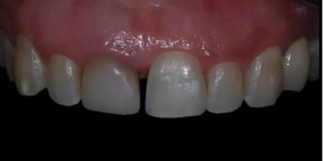
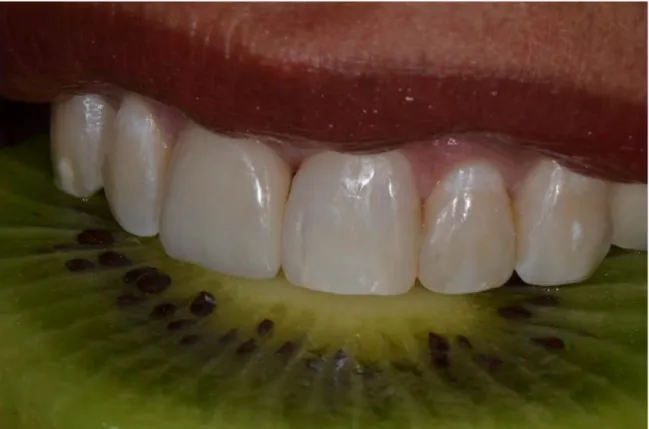
Figure 1. Rehabilitated patient; A. initial conditions of several dental structure
loss; B. tooth preparation of glass fiber post/composite core; C. strain-gauge measurement and bite force recording for isolated tooth; D. strain-gauge measurement and bite force recording for both teeth together; E. Final rehabilitation; F. Initial contact of antagonist teeth during protrusive jaw movement.
Figure 2. Finite element model generation; A. cone beam computerized
tomography; B. structures and materials segmentation on Mimics software; C. STL. mesh with no organized triangles; D. final mesh created on 3-Matic and Patran softwares; E. Final patient specific model.
Initia fiber reco reco anta Cone segm final mod al condition r post/com ording for ording for agonist teet e beam mentation o mesh cre del.
ns of seve mposite co
isolated t both teeth th during p
compute on Mimics eated on 3
eral dental ore; C. s tooth; D. h together; protrusive j erized tom software; 3-Matic an 50 structure strain-gaug strain-gau ; E. Final aw movem
mography; C. STL. m d Patran s
loss; B. to ge measu uge meas rehabilitat ment.
B. stru mesh with n
softwares;
ooth prepa urement a urement a tion; F. In
uctures a no organize E. Final p
51
C
Análise endodo Gradua
C
A
e comparativa onticamente – A ação em Odont
APÍTU
de comporta ANDRÉA DOLO tologia – Faculd
ULOS
mento in vivo ORES CORREI dade de Odonto
52
S
o e in vitro d IA MIRANDA V ologia – Univers
e protocolos d VALDIVIA – Tes
idade Federal d
de reabilitação se de Doutorado de Uberlândia
o de dentes t o – Programa d
Pós-53 3.3 CAPÍTULO 3
Artigo a ser enviado para publicação no periódico Journal of Dentistry
Title: Biomechanical effect of ferrule presence on incisor teeth restored with fiberglass post and CAD/CAM ceramic crown after thermal cycling and fatigue loading.
Short title: Biomechanical behavior of endodontic treated teeth
A.D.C.M Valdiviaa, M.P. Rodriguesa, A.A. Bicalhoa, C. Veríssimoa, B. Van Meerbeekb, J.V. Slotenc, R.S. Pessoaa, C.J. Soaresa,*
a
BIAOR, Department of Operative Dentistry and Dental Materials, Dental School, Federal University of Uberlândia, Uberlândia, Minas Gerais, Brazil. b
BIOMAT, Department of Oral Health Sciences, KU Leuven (University of Leuven), and Dentistry, University Hospitals Leuven, Leuven, Belgium.
c BMe, Department of Mechanical Engineering, KU Leuven (University of Leuven), Leuven, Belgium.
54
Biomechanical effect of ferrule presence on incisor teeth restored with fiberglass post and CAD/CAM ceramic crown after thermal cycling and fatigue loading.
ABSTRACT
Objectives. To evaluate the biomechanics of endodontically treated incisors
restored with fiberglass post and CAD-CAM ceramic crown with and without ferrule after thermal and mechanical aging.
Methods. Twenty bovine incisors were divided into two groups (n=10): Fe, with
ferrule of 2 mm and NFe, without ferrule. The teeth were endodontic treated and restored with fiberglass post (Exacto 3), composite core (Tetric Ceram), and CAD-CAM lithium disilicate ceramic crown (IPS e.max CAD). The specimens were subjected to 20,000 thermo-cycles and 2,400,000 simulated chewing cycles. Ceramic crown and root dentin strains (µS) were measured using strain-gauges (n=10) during 100N loading before and after thermal and mechanical aging, and at fracture load. Specimens were subsequently loaded to fracture (N). Stress distribution was analyzed using 3D finite element models created by micro-CT (n=3). Strain data were analyzed by two-way ANOVA and Tukey HSD tests, fracture resistance was analyzed using t-Student and fracture mode using Chi-square test (α=0.05).
Results. After aging NFe had higher root dentin deformation than Fe. Fe had
higher fracture resistance than NFe. Fe had fracture involving ceramic crown or associated with core. NFe had more root dentin fracture and post debonding. The NFe had lower ration fracture resistance/root strain than Fe. The stress levels on root dentin and fiberglass were lower for Fe.
Conclusions. Fe prevented the post detaching maintain the root dentin strain
after thermal-mechanical aging. The NFe increased root dentin strain after aging process. Fe had higher fracture resistance, lower stress concentration on root dentin and less catastrophic fractures.
Clinical Significance. Tooth structure remaining of endodontically treated
55
Keywords. Fiber post, ferrule, ceramic, strain-gauge test, fracture resistance,
finite element analysis, thermal cycling, fatigue.
1. Introduction
Endodontically treated teeth commonly requires post and core restorations for retention purposes.[1,2] Posts associated with all ceramic crowns are an option for teeth with a severe loss of coronal structure.[3] Several materials and techniques had been advocated for restoring endodontic treated teeth; for example lithium disilicate crowns CAD/CAM system have shown a good clinical performance.[4] The correct choice of the post system and the ferrule effect are crucial for treatment prognosis.[5] Fiberglass posts have been used clinically as an alternative to metal posts in the restoration of endodontically treated teeth.[6-8] The major advantage of fiberglass posts is their similar elastic modulus to dentin,[9] which may lead to a better distribution of the occlusal loads along the root.[10,11] Failures in post-retained crowns generally occur in the maxillary anterior region, where horizontal forces are greater than in other areas.[12]
The amount of coronal and root dentin remains after root canal instrumentation and post space preparation correlated with the fracture resistance and plays an important role in the biomechanical behavior of endodontic treated teeth restored with fiber post.[2,3] A recent meta-analysis suggested that coronal wall absence might increase the risk of fiber post-core restoration failure, while the role of ferrule effect is still not entirely understood.[13] Others studies reported that the presence of uniform ferrule surrounding remaining tooth structure enhanced fracture resistance and increase the fracture resistance,[2] and increase the long term success of post-endodontic of anterior teeth.[8]
56
However, this method does not provide enough information about the internal structural behavior of the complex tooth restoration against loading. Therefore, it is necessary to combine nondestructive methods such as the strain gauge methods for complete biomechanical analysis.[2,3,11,15] The strain measured during the nondestructive tests in this study can be regarded as an indication of the repetitive deformation that roots undergo during functioning, resulting in such structural fatigue.[16] To approximate the in vitro test to the clinical failures of restores tooth the artificial aging, of specimens using cyclic mechanical loading and thermal cycling represent the ideal in vitro design for a study reproducing the physiological functions of the oral environment.[17] Generally, the artificial aging of dental materials is indicated because it accelerates the degradation process, which causes a significant decrease in the mechanical properties.[18,19]
The aim of this study was to evaluate the strain before and after thermo-mechanical fatigue aging, the fracture resistance and fracture mode of endodontically treated incisors restored using a fiberglass post and CAD/CAM all ceramic crown with and without ferrule. The null hypothesis tested was that biomechanical behavior would not be affected by the amount of remaining coronal dentin.
2. Materials and Methods
2.1. Specimen Preparation
Gates-57
Glidden drills (Dentsply Maillefer, Ballaigues, Switzerland). A no. 4 Gates-Glidden drill (Dentsply Malleifer) was used in the cervical and middle thirds of the root canal. Canals were rinsed with 1.0% sodium hypochlorite (Miyako do Brasil, Guarulhos, São Paulo, Brazil) and physiological saline (Avante Pharma, Belo Horizonte, Minas Gerais, Brazil), dried with paper points, and obturated with gutta-percha (Dentsply) and calcium hydroxide-based cement (Sealer 26, Dentsply).[20] The specimens of the Fe group were prepared with a diamond rotary cutting instrument (no. 3215; KG Sorensen, Barueri, SP, Brazil) creating a 2.0-mm-high circular ferrule with rounded cervical ending. Post space was obtained initially with a heated instrument (M-Series Hand Pluggers; Dentsply Maillefer), and the residual gutta-percha was then removed with Gates-Glidden drills, standardizing the post space at 8.0 mm for the group without ferrule and 10.0 mm for the group with ferrule, preserving 5.0 mm of gutta-percha at the apex. Next, root canals were enlarged with a 1.0- to 1.6-mm-diameter conical drill (Exacto drill no. 3, Angelus Science and Technology, Londrina, Paraná, Brazil) to 8.0 mm for the group with no ferrule and 10.0 mm for the group with ferrule, generating standardized post space for the fiberglass post.[2]
58
cylinder. After polystyrene resin polymerization, the roots were removed from the cylinder, and the wax was removed from both the root surface and cylinder. Impression material (Impregum Soft; 3M ESPE) was placed into the resin cylinders, the roots were reinserted, and the excess polyether material was removed with a scalpel blade.[15]
2.2 .Post and core and crown fabrication
Prefabricated fiberglass posts (Exacto Translucido No. 3; Angelus, Londrina, Brazil) with 1.4-mm and 0.9-mm diameters in the coronal and apical portions, respectively, were cleaned with a 70% alcohol solution (Miyako do Brasil) then, the post were immersed in 24% hydrogen peroxide (H2O2, Dinâmica, SP, Brasil) for 1 minute followed by rising and drying.[15] Afterwards, the one-bottle silane coupling agent (Silano; Angelus Science and Technology) was applied for 1 minute.
59
Specimens were restored with CAD-CAM all ceramic crowns. Each sample was scanning using the CEREC 3D software (Sirona Dental Systems, Bensheim, Germany) and milled out of lithium disilicate glass ceramic block (IPS e.max CAD, size I12, Ivoclar Vivadent) according to the manufacturer’s instructions. The internal restoration surface was etched with 10% hydrofluoric acid (Condac Porcelana, FGM, Joinville, SC, Brazil) for 20 seconds followed by rising and drying. The silane agent (Silano, Ângelus, Londrina, Paraná, Brazil) was then applied for 1 minute. [23] Ceramic crowns were cemented using self-adhesive resin cement (RelyX U200; 3M ESPE) following the same protocol described for post fixation.
2.3. Strain measurement test
The specimens were submitted to the strain-gauge test before and after artificial aging. To measure the tooth deformation, 2 strain gauges with grade of 1mmX1mm (PA-06-038AA-120-LEN; Excel Sensores, Embú, São Paulo, Brazil) were placed to the root surface 2.0mm below the crown limit, one satin-gauge was placed on the buccal surface (Fig. 1C), parallel to the long axis, and the other on the lingual surface (Fig. 1D).[11] One strain gauge with grade of 4mmX2mm (PA-06-060CC-350-LEN; Excel Sensores) was attached to the buccal surface of the ceramic crown (Fig. 1C). The strain gauges were bonded with a cyanoacrylate resin adhesive (Super Bonder; Loctite) and connected to a data acquisition device (ADS0500IP; Lynx, São Paulo, São Paulo, Brazil). In addition, a control specimen, with 3 strain gauges attached but not subjected to loading, was mounted adjacent to the tested tooth to compensate for temperature fluctuations due to gauge electrical resistance or local environment.[2,3,11]
60 2.4. Thermocycling and Fatigue loading
Thermal variations were induced in a thermal cycling machine (Thermo-cycler, Willytech, Munich, Germany) between two water baths of 5 oC and 55 oC with a dwell time of 30 seconds each temperature. All specimens were subjected during 20,000 cycles. After first 10,000 thermal aging cycles, the specimens were submitted to a fatigue load of 1,200,000. Then more 10,000 thermal aging cycles and 2,400,000 cycles were performed. The fatigue loading was performed under water irrigation using a chewing simulator with sliding movement (Willytech, Munich, Germany), simulating 10 years of clinical function.[24] Load was applied at 45o at a frequency of 1.6 Hz. A sinusoidal load of 0-50N was applied with stainless-steel ball-shaped stylus in the lingual surface of the ceramic crown (Fig. 1F).[25,26] Failures under fatigue loading were recording during test by integrated LVDT displacement sensors, which able to detect displacement of 100µm and connected to a PC-software.
2.5. Strain during fracture procedure (CSt-Fr), fracture resistance and
fracture mode
61
sputter coated with gold (Bal-Tec SCD 050; Balzers, Liechtenstein) and examined under a scanning electron microscope (EVO MA 10, CARL ZEIZ, Germany). SEM images were obtained at different magnifications to illustrate the failure modes.
2.6. Residual stress calculation-finite element analysis
62
As a specific approach for better model generation, the STL models were imported to MSC.Patran® 2010r2 (MSC.Software, MSC software, Santa Ana, CA, USA) and meshed. Tetrahedral elements were used to ensure smooth contact at all the models interfaces. The volumetric meshes of all models components were therefore created based on the optimized surface’s standard triangulated language (STL) descriptions. After that, meshes were imported in a FEA software package (MSC.Marc/MSC.Mentat, MSC.Software, Santa Ana, CA) for the attribution of material properties to the other model components (i.e. bone, periodontal ligament, enamel, dentin, resin cement, gutta-percha, fiberglass post and resin composite). The elastic moduli of the restorative materials and dental structure are shown in Table 1 and Table 2. To simulate the interface among model components, precisely bonded contacts were held. The nodes on the base of the bone model structure were rigidly fixed in the x, y- and z-directions, to simulate the experimental set-up test. The loading conditions were simulated with nodal point load using individual force experimentally tested (100N applied at 45o on lingual surface of the crown). The load application had a coronal-apical direction in relation to the tooth longitudinal axis. The assessment and post-processing were performed for each model using equivalent von Mises stresses by means of the MSC.MARC/ Mentat® 2010r3 software (MSC.Software).
3. Results
3.1. Fatigue loading
Only one NFe specimen failed during fatigue loading because of post debonding after 1,200,000 cycles. All the other specimens survived after 2,400,000 fatigue cycles.
3.2. Tooth structure and ceramic strain
63
and aging presence. The NFe group had similar root dentin deformation than Fe group before aging, however after aging NFe group had higher root dentin deformation than Fe group, irrespective of dentin location. The aging process had no effect on root dentin strain for Fe group, irrespective of dentin location. However, The aging process increased significantly the root dentin strain for NFe group, irrespective of dentin location.
The values of ceramic crown, buccal root surface and lingual root surface strains (µS) and at the maximum fracture loading are shown in Table 4. The ceramic crown had lower deformation than root dentin irrespective of ferrule presence. The dentin deformation on buccal surface was always higher than on lingual surface. For ceramic crown deformation, no difference was found between FE and NFE. The NFe group had similar root buccal dentin deformation than Fe group. However the Fe had higher root lingual dentin deformation than NFe group.
3.3. Fracture resistance
The mean fracture resistance (N) and standard deviation values are shown in Table 5. Test t-Student showed that Fe had higher resistance to fracture than NFe.
64
The ratio between the maximum resistance and root dentin deformation at fracture moment is shown in Table 5. The NFe had lower ration fracture resistance/root strain than Fe groups (P < 0.001).
3.4. Finite element analysis
The von Mises stress distributions are summarized on Fig. 4, 5 and 6. The stress level on ceramic crown surface was lower than on root dentin and similar for all models (Fig. 4). The higher stress concentration was located on external surface of the root dentin (Fig. 5). The stress located on lingual root dentin surface was higher than on buccal root dentin. The higher stress concentration is located at the root dentin on NFe group interface with ceramic crown and composite core (Fig. 6). The stress distribution by Maximum Principal Stress for both groups are shown on Figure 7.
4. Discussion
The aim of this study was to evaluate the hypothesis that the presence of ferrule on anterior endodontic treated incisor restored with glass fiber post/composite core/CAD-CAM ceramic crown influence the biomechanical performance, expressed by crown and root dentin strains, fracture resistance, fracture mode and stress distribution. The results of the present study confirmed that the presence of ferrule maintain the integrity among restorative materials and root dentin with no increasing of root deformation after aging, reduced the stress concentration at root dentin, increased the fracture resistance and reduced the root fracture of endodontically treated incisors restored using a fiberglass post. Therefore the null hypothesis was rejected.