Polypoidal choroidal vasculopathy causing
cystoid macular edema and the response to
ranibizumb intravitreal treatment
Miguel Hage Amaro1, Aaron Brock Roller2, Cesar Tavares Pereira dos Santos Motta3, Mario Martins dos Santos Motta4
Recebido para publicação em: 30/11/2010 - Aceito para publicação em 22/5/2011
A
BSTRACTPurpose: To report a polypoidal vascular choroidopathy clinical case causing cystoid macular edema and the response to Ranibizumab intravitreal treatment. Methods: A 62-year old caucasian woman was referred by her comprehensive ophthalmologist for retinal avaliation. On presentation best corrected visual acuity was 20/100 in the left eye and 20/20 in right eye.Anterior segment examination was unremarkable in both eyes. Clinical examination and FA on the left eye demonstrated numerous small drusen and a cystoid macular edema due leakage from any polips in justapapilar region and from polips in the superior arcade vascular region and subretinal fluid and cystic change in the OCT.The right eye had FA normal. The patient refused to submitt a ICGV angiography. The patient was treated by intravitreal ranibizumab injections in the left eye every 4 weeks, 3 injections ,three months. Results: The patient showed resolution both of cystic change and subretinal fluid in the OCT, and the visual acuity in the six months follow-up improved to 20/25.The patient was followed by 18 months at this time and the visual acuity remained stable 20/25. Conclusion: We reported a patient case of cystoid macular edema from a polypoidal choroidal vaculopathy that responded well to ranibizumab intravitreal injection as monotheraphy with disappearance of the initial subretinal fluid and cystic change in OCT follow-up and stop the polips.
Keywords: Choroid diseases/drug therapy; Polypoidal choroidal vasculopathy /drug therapy Macular edema/etiology; Injections; Antibodies, monoclonal/therapeutic use; Case report
1MD, Instituto de Olhos e Laser de Belém (PA), Brazil;
2Department of Ophthalmology, Iowa University, Iowa city – USA;
3Resident Universidade do Rio de Janeiro – UNIRIO - Rio de Janeiro (RJ), Brazil; 4Professor, Universidade do Rio de Janeiro – UNIRIO - Rio de Janeiro (RJ), Brazil.
Study carried out with Vitreous Retina Ophthalmology Department of the Iowa University-USA
Vasculopatia Coroidiana Polipoidal causando Edema Macular
Cistóide e a resposta ao tratamento com Ranibizumab
R
ESUMOObjetivo: Relatar um caso de paciente com Vasculopatia coroidiana polipoidal com edema macular cistóide e a resposta ao tratamento com Ranibizumab intravítrea como monoterapia Métodos: Uma paciente com 62 anos foi referida por seu oftalmologista para avaliação retiniana . Apresentava acuidade visual com correção de 20/100 no olho esquerdo e 20/ 20 no olho direito. A avaliação do segmento anterior era normal em ambos os olhos. No exame de fundo de olho e retinografia fluoresceínica apresentava numerosas drussas pequenas e edema macular cistóide devido a vazamentos de alguns pólipos vasculares coroidianos na arcada vascular superior e ainda fluido sub-retiniano e alteração cística no OCT . O olho direito apresentava angiografia fluoresceínica da retina e OCT normais.A paciente recusou-se a se submeter a vídeoangiografia com indocianina verde. A paciente foi tratada com injeção intravítrea de Ranibizumab como monoterapia, sendo uma injeção a cada quatro semanas, três injeções em três meses. Resultados: A paciente apresentou resolução da alteração cística e do fluido sub-retiniano, ambos presentes no OCT prévio ao tratamento no olho esquerdo. A acuidade visual melhorou para 20/25 após 6 meses de tratamento.A paciente permanence com acuidade visual estável de 20/25 após 18 meses de acompanhamento. Conclusão: Reportamos um caso de paciente com edema macular cistóide originada da vasculopatia coroidiana polipoidal que respondeu ao tratamento com Ranibizumab intravítrea, como monoterapia com desaparecimento do fluido sub-retiniano e da alteração cística no OCT tendo cessado o vazamento dos pólipos que eram a causa do edema macular cistóde.
Descritores: Doenças da coróide/quimioterapia; Vasculopatia polipoidal coroidiana/quimioterapia; Edema macular/ etiologia; Injeções; Anticorpos monoclonais/uso terapêutico; Relato de caso
I
NTRODUÇÃOP
olypoidal choroidal vasculopathy (PCV) is a designation coined by Yannuzzi to describe a distinct exudative macular disorder causing recurrent and multiple detachments of the retinal pigment epithelium(1,2). These are typicallyserosanguineous and neurosensory retinal detachments, secondary to bleeding and leakage from polypoidal choroidal vascular lesions(1,2).
PCV is characterized by an inner choroidal vascular network of vessels, ending in an aneurysmal bulge or outward projection, visible clinically as a reddish-orange, spheroid, polyp-like(1,2).
PCV lesions have been seen in patients of many different racial backgrounds, but are known to selectively affect patients of more pigmented races(3). PCV lesions are
found in 23-55% of patients with presumed neovascular age-related macular degeneration (AMD) in Asian countries; in patients of Caucasian origin, PCV lesions have been found in roughly 8-13% of patients with presumed neovascular AMD(3). Initially, this disease was described as “posterior
uveal bleeding syndrome”(4,5) and later as “multiple recurrent
retinal pigment epithelium detachment”(6,7).
It has been proposed that PCV is a variant of type 1 neovascular AMD(8,10); true type 2 choroidal
neovascularization (CNV) is not a rare complication of PCV, and the eyes often show what appears to be classic CNV on fluorescein angiography. However, it is difficult to discriminate type 2 CNV from pure fibrinous tissue deposition before treatment, even with a detailed examination by optical coherence tomography (OCT). PCV has also been reported in association with dry AMD(11).
Vascular changes typical of PCV are evident with slit-lamp biomicroscopy unless blood or exudate overly the lesions(2,7,12,13).
Fluorescein angiography (FA) can be useful in making the diagnosis if there is no serosanguineous leakage overlying the polyps, but indocyanine green videoangiography (ICGV) is the gold standard method for visualizing polyps and the vessel network.
The treatment strategies now available for PCV include verteporfin photodynamic therapy (PDT)(14,15) and
anti-VEGF therapy with ranibizumab as a monotherapy. Results with this smaller molecular weight compound are promising, showing regression of polypoidal changes(16,17).
A combination of PDT therapy and ranibizumab could provide a good alternative, by reducing the number of injections needed relative to that used in other anti-VEGF monotherapy.There are also reports of phototrombosis of neovessels, mediated by indocyanine green(18).
Cystoid macular edema (CME) can have a variety of causes, including diabetic maculopathy, age-related macular degeneration, retinal vein occlusions, chronic uveitis, epiretinal membranes, choroidal tumors, radiation retinopathy, perifoveal retinal telangiectasis, retinitis pigmentosa, dominantly inherited CME, foveal X-linked retinoschisis, and others.
We describe in this report a case in which PCV led to cystoid macular edema and the response to Ranibizumab intravitreal treatment.
C
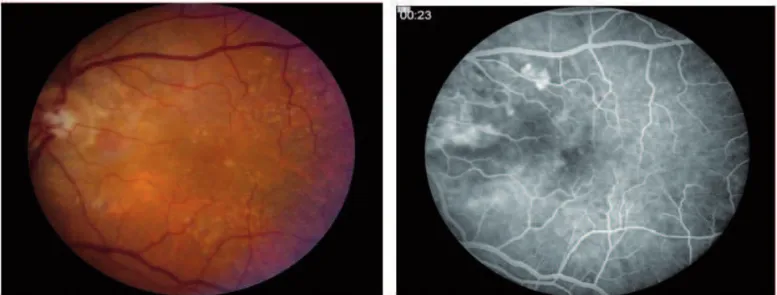
ASE REPORTpresentation, the best corrected visual acuity was 20/100 in her left eye and 20/20 in her right eye. Anterior segment examination was unremarkable in both eyes. Clinical examination and FA of the left eye demonstrated numerous small drusen and cystoid macular edema, due to leakage from polyps in the juxtapapillary region and in the superior arcade region (figures 1 and 2). Subretinal fluid and cystic change were noted by OCT(figure 3). The right eye was normal by FA. The patient refused to submit to ICGV.
The patient was treated by intravitreal ranibizumab injections in the left eye every four weeks. After three injections and six months of follow-up, the patient showed resolution of both cystic changes and subretinal fluid by OCT (figure 4), and visual acuity during the six-month follow-up improved to 20/25.
The patient has been followed for 18 months at the time of writing, and her visual acuity has remained stable at 20/25.
D
ISCUSSIONImproved visual acuity has been found in many cases to be correlated with anatomic improvement and with restoration of a more normal macular architecture; this has been confirmed by advanced imaging techniques(19).
An association between VEGF and PCV has been suggested by histopathological evidence of increased VEGF concentration in the aqueous humor in patients with PCV(20).
The PrONTO study(21) recommended that
Figure 1: Several polypoidal vascular configurations beneath the retinal pigment epithelium
patients treated for exudative macular degeneration from subfoveal CNV be followed by OCT monthly, together with visual acuity testing and possible fluorescein angiography. Our patient was followed by visual acuity testing and by OCT, and improvement in her clinical presentation and in vi-sual acuity was noted.
Verteporfin photodynamic therapy has been shown to be effective in the treatment of symptomatic patients with PCV, although the incidence of retinal pigment epithelium atrophy and of recurrence suggest the need for additional treatment options(14,15).
A study of intravitreal bevacizumab in PCV showed vascular abnormalities persisting in 10 of out 11 eyes after three months(22). This therapy might be
less efficacious for PCV due to the limited retinal penetration expected for a molecule of bevacizumab’s molecular mass. With a molecular weight of 48 kD, ranibizumab is much smaller than the full-length RhuMab VEGF antibody (bevacizumab), which has a molecular weight of 148 kD. Additional studies have reported similar poor results with the larger antibody(23,24).
Results after six months of the EVEREST study were presented during a scientific review by Novartis in Basel, Switzerland. EVEREST is the first multi-center, double-blind, indocyanine green angiography (ICGA)-guided, randomized controlled trial with an angiographic treatment outcome, designed to assess the effect of Visudyne(R) (verteporfin photodynamic therapy) alone
or in combination with Lucentis(R) (ranibizumab), as compared to that of Lucentis alone, in patients with symptomatic macular polypoidal choroidal vasculopathy (PCV). A total of 61 PCV patients of Asian ethnicity from five countries (China, Hong Kong), Taiwan, Korea, Thailand and Singapore) participated in the study; long-term results are not yet available.
There have been recent reports of studies using ranibizumab monotherapy in PCV cases. Monthly intravitreal injections of ranibizumab for three months had a short-term beneficial anatomic effect, with polyps disappearing on ICG angiography in nine out of 13 lesions (69.2%); retinal thickness had diminished significantly by OCT (p=0.02)(14). Another report of continuous anti-VEGF
treatment with ranibizumab over six months for polypoidal choroidal vasculopathy(17) stimulated us to use this model
of treatment in the case discussed here.
In conclusion, we report the case of a patient with cystoid macular edema from polypoidal choroidal vasculopathy who responded well to ranibizumab intravitreal injection as monotherapy. Treatment led to disappearance of the initial subretinal fluid and cystic change by OCT at follow-up, having halted polyp leakage.
Acknowledgements
We thank Professor James Folk, Judith Gardner and Donald Beisner, Professor of Vitreoretinal Diseases and Surgery at Iowa University, for analyzing this case with us.
Figure 3: Subretinal fluid nasal to the macula and cystic change
R
EFERENCES1. Yannuzzi LA. Idiopathic polypoidal choroidal vasculopathy. Presented at the February 1982. Macula Society Meeting, Miami, Florida, USA.
2. Yannuzzi LA, Sorenson J, Spaide RF, Lipson B. Idiopathic polypoidal choroidal vasculopathy (IPCV). Retina. 1990;10(1):1-8.
3. Sho K, Takahashi K, Yamada H, Wada M, Nagai Y, Otsuji T, et al. Polypoidal choroidal vasculopathy: incidence, demographic features, and clinical characteristics. Arch Ophthalmol. 2003;121(10):1392-6.
4. Kleiner RC, Bruckner AJ, Johnston RL. Posterior uveal bleed-ing syndrome. Ophthalmology.1984;91:110.
5. Kleiner RC, Bruckner AJ, Johnston RL. The posterior uveal bleeding syndrome. Retina. 1990;10(1):9-17.
6. Stern RM, Zalov ZN, Zegarra H, Gutman FA. Multiple recur-rent serosanguineous retinal pigment epithelial detachments in black women. Am J Ophthalmol. 1985;100(4):560-9. 7. Perkovich BT, Zakov ZN, Berlin LA, Weidenthal D, Avins
LR. An update on multiple recurrent serosanguineous retinal pigment epithelial detachments in black women. Retina. 1990;10(1):18-26.
8. Yannuzzi LA, Wong DW, Sforzolini BS, Goldbaum M, Tang KC, Spaide RF, et al. Polypoidal choroidal vasculopathy and neovascularized age-related macular degeneration. Arch. Ophthalmol. 1999;117(11):1503-10.
9. Lafaut BA, Aisenbrey S, Van den Broecke C, Bartz-Schmidt KU, Heimann K. Polypoidal choroidal vasculopathy pattern in age-related macular degeneration: a clinicopathologic cor-relation. Retina. 2000;20(6):650-4.
10. Uyama M, Wada M, Nagai Y, Matsubara T, Matsunaga H, Fukushima I, et al. Polypoidal choroidal vasculopathy: natu-ral history. Am J Ophthalmol. 2002;133(5):639-48.
11. Lois N. Idiopathic polypoidal choroidal vasculopathy in a patient with atrophic age-related macular degeneration. Br J Ophthalmol. 2001;85(8):1011-2.
12. Lijima H, Iida T, Imai M, Gohdo T, Tsukahara S. Optical coher-ence tomography of orange-red subretinal lesions in eyes with idiopathic polypoidal choroidal vasculopathy. Am J Ophthalmol. 2000;129(1):21-6.
13. Spaide RF, Yannuzzi LA, Slakter JS, Sorenson J, Orlach DA. Indocyanine green videoangiography of idiopathic polypoi-dal choroipolypoi-dal vasculopathy. Retina.1995;15(2):100-10. 14. Chan WM, Lam DS, Lai TY, Liu DT, Li KK, Yao Y, Wong TH.
Photodynamic therapy with verteporfin for symptomatic polypoidal choroidal vasculopathy: one-year results of a pro-spective case series. Ophthalmology. 2001;111(8):1576-84.
15. Eandi CM, Ober MD, Slakter JS, Yannuzzi LA. Selective pho-todynamic therapy for neovascular age-related macular de-generation with polypoidal choroidal neovascularization. Retina. 2007;27(7):825-31.
16. Reche-Frutos J, Calvo-Gonzalves C, Donate-Lopez J, Garcia-Feijoo J, Leila M, Garcia-Sanchez J. Short-term anatomic effect of ranibizumab for polypoidal choroidal vasculopathy. Eur J Ophthalmol. 2008;18(4):645-8.
17. Kokame GT, Yeung L, Lai JC. Continuous anti-VEGF treat-ment with ranibizumab for polypoidal choroidal vasculopathy: 6-month results. Br J Ophthalmol. 2010;94(3):297-301. 18. Cardillo JA, Jorge R, Costa RA, Nunes SM, Lavinsky D,
Kuppermann BD, et al. Experimental selective choriocapillaris photothrombosis using a modified indocyanine green formu-lation. Br J Ophthalmol. 2008;92(2):276-80.
19. Brown DM, Regillo CD. Anti-VEGF agents in the treatment of neovascular age-related macular degeneration: applying clinical trial results to the treatment of everyday patients. Am J Ophthalmol. 2007;144(4):627-37.
20. Matsuoka M, Ogata N, Otsuji T, Nishimura T, Takahashi K, Matsumura M. Expression of pigment epithelium derived factor and vascular endothelial growth factor in choroidal neovascular membranes and polypoidal choroidal vasculopathy. Br J Ophthalmol. 2004;88(6):809-15. 21. Lalwani GA, Rosenfeld PJ, Fung AE, Dubovy SR, Michels S,
Feuer W, et al. A variable-dosing regimen with intravitreal ranibizumab for neovascular age-related macular degenera-tion: year 2 of the PrONTO Study. Am J Ophthalmol. 2009;148(1):43-58.e1.
22. Gomi F, Sawa M, Sakaguchi H, Tsujikawa M, Oshima Y, Kamei M, Tano Y. Efficacy of intravitreal bevacizumab for polypoidal choroidal vasculopathy. Br J Ophthalmol. 2008;92(1):70-3. 23. Ghajarnia M, Kurup S, Eller A. The therapeutic effects of
intravitreal bevacizumab in a patient with recalcitrant idio-pathic polypoidal choroidal vasculopathy. Semin Ophthalmol. 2007;22(2):127-31.
24. Lai TY, Chan WM, Liu DT, Luk FO, Lam DS. Intravitreal bevacizumab (Avastin) with or without photodynamic therapy for the treatment of polypoidal choroidal vasculopathy. Br J Ophthalmol. 2008;92(5):661-6.
Corresponding author address: Miguel Hage Amaro, MD Quintino Bocaiúva, nº 516