in Dogs by Using Peptides Selected from Hypothetical Proteins
Identified by an Immunoproteomic Approach
Miguel A. Chávez-Fumagalli,aVivian T. Martins,bMiriam C. S. Testasicca,cDaniela P. Lage,dLourena E. Costa,aPaula S. Lage,a Mariana C. Duarte,aHenrique G. Ker,eTatiana G. Ribeiro,cFernando A. A. Carvalho,fWiliam C. B. Régis,gAlexandre B. dos Reis,e Carlos A. P. Tavares,bManuel Soto,hAna Paula Fernandes,iEduardo A. F. Coelhoa,d
Programa de Pós-Graduação em Ciências da Saúde, Infectologia e Medicina Tropical, Faculdade de Medicina, Universidade Federal de Minas Gerais, Belo Horizonte, Minas Gerais, Brazila
; Departamento de Bioquímica e Imunologia, Instituto de Ciências Biológicas, Universidade Federal de Minas Gerais, Belo Horizonte, Minas Gerais, Brazilb ; Programa de Pós-Graduação em Ciências Farmacêuticas, Faculdade de Farmácia, Universidade Federal de Minas Gerais, Belo Horizonte, Minas Gerais, Brazilc
; Departamento de Patologia Clínica, COLTEC, Universidade Federal de Minas Gerais, Belo Horizonte, Minas Gerais, Brazild
; Departamento de Análises Clínicas, Escola de Farmácia, Universidade Federal de Ouro Preto, Ouro Preto, Minas Gerais, Brazile
; Departamento de Bioquímica e Farmacologia, Centro de Ciências da Saúde, Universidade Federal do Piauí, Teresina, Piauí, Brazilf
; PUC Minas and Minasfungi do Brasil Ltda., Belo Horizonte, Minas Gerais, Brazilg
; Centro de Biología Molecular Severo Ochoa, CSIC-UAM, Departamento de Biología Molecular, Universidad Autónoma de Madrid, Madrid, Spainh
; Departamento de Análises Clínicas e Toxicológicas, Faculdade de Farmácia, Universidade Federal de Minas Gerais, Belo Horizonte, Minas Gerais, Brazili
In Brazil, the percentage of infected dogs living in areas where canine visceral leishmaniasis (CVL) is endemic ranges from 10 to
62%; however, the prevalence of infection in dogs is probably higher than figures reported from serological studies. In addition,
problems with the occurrence of false-positive or false-negative results in the serodiagnosis of CVL have been reported. The
present work analyzed the potential of synthetic peptides mapped from hypothetical proteins for improvement of the
serodiag-nosis of
Leishmania infantum
infection in dogs. From 26 identified leishmanial proteins, eight were selected, considering that no
homologies between these proteins and others from trypanosomatide sequence databases were encountered. The sequences of
these proteins were mapped to identify linear B-cell epitopes, and 17 peptides were synthesized and tested in enzyme-linked
im-munosorbent assays (ELISAs) for the serodiagnosis of
L. infantum
infection in dogs. Of these, three exhibited sensitivity and
specificity values higher than 75% and 90%, respectively, to differentiate
L. infantum
-infected animals from
Trypanosoma cruzi
-infected animals and healthy animals. Soluble
Leishmania
antigen (SLA) showed poor sensitivity (4%) and specificity (36%) to
differentiate
L. infantum
-infected dogs from healthy and
T. cruzi
-infected dogs. Lastly, the three selected peptides were
com-bined in different mixtures and higher sensitivity and specificity values were obtained, even when sera from
T. cruzi
-infected
dogs were used. The study’s findings suggest that these three peptides can constitute a potential tool for more sensitive and
spe-cific serodiagnosis of
L. infantum
infection in dogs.
T
he leishmaniases consist of a wide range of diseases present in
88 countries, with 12 million people infected and 350 million
at risk of infection (
1
). Zoonotic visceral leishmaniasis is a severe
disease caused by
Leishmania infantum
in the Mediterranean area,
the Middle East, Africa, Asian countries, and Latin America (
2
,
3
).
The disease also is emergent in dogs living in the United States,
Canada, northern Italy, and Germany (
4
–
6
). In Brazil, the disease
is caused by
Leishmania chagasi
(syn.
L. infantum
), with the
para-sites being transmitted by
Lutzomyia longipalpis
and
Lutzomyia
cruzi
and wild and domestic canids representing the main
reser-voirs of parasites (
7
).
Upon infection, dogs develop asymptomatic or symptomatic
clinical forms of disease (
8
–
10
). Serological tests used for
symp-tomatic canine visceral leishmaniasis (CVL) diagnosis are
facili-tated by the strong humoral response that generally accompanies
the development of acute disease (
11
,
12
). Courtenay et al. (
13
)
showed that a high percentage of asymptomatic dogs developed
symptoms after some months and that those dogs were able to
infect about 99.6% of sandflies. In this context, in areas in which
CVL is endemic, about 10 to 62% of apparently healthy and/or
seronegative dogs are positive for
Leishmania
by PCR (
14
–
17
).
Thus, asymptomatic dogs, which play a role in the transmission of
parasites, are not detected by conventional serological tests, such
as the indirect fluorescent antibody test (IFAT) and the
enzyme-linked immunosorbent assay (ELISA) (
18
). Nonetheless, the
de-tection of asymptomatic CVL might be crucial in controlling
ep-idemics and avoiding the spread of disease among dogs, as well as
between dogs and human populations (
19
,
20
).
There are areas of endemicity where transmission of
Leishma-nia
spp. and
Trypanosoma cruzi
parasites are superposed and, due
to the phylogenetic similarity between those parasites, serological
cross-reactions and/or false-positive results are quite common
(
21
,
22
). As a strategy to develop a more sensitive and specific
method for serodiagnosis of CVL, some individual
Leishmania
proteins were used as recombinant antigens (
23
,
24
). However,
due to the high variability observed in the humoral responses of
infected dogs, efficient diagnosis based on purified antigens might
require a mixture of antigens or the use of chimeric antigens
con-Received16 January 2013 Returned for modification1 February 2013 Accepted22 March 2013
Published ahead of print3 April 2013 Address correspondence to Eduardo A. F. Coelho, [email protected].
Copyright © 2013, American Society for Microbiology. All Rights Reserved.
doi:10.1128/CVI.00023-13
on October 6, 2017 by UNIVERSIDADE FEDERAL DE OURO PRETO
http://cvi.asm.org/
taining several leishmanial proteins (
25
). One alternative means
to identify sensitive and specific antigens for the diagnosis of CVL
is through the use of synthetic peptides. These antigens are
rela-tively simpler and cheaper to produce than recombinant proteins.
It also has been reported that the use of synthetic peptides
(indi-vidually or in a mixture format), in comparison with the use of
recombinant proteins, is able to increase the sensitivity and/or
specificity of immunoassays for the serodiagnosis of parasitic
diseases (
26
), such as canine and human visceral leishmaniasis
(
27
,
28
).
In an attempt to identify more-refined antigens for the
serodi-agnosis of CVL, 26 hypothetical proteins from
L. infantum
, which
were identified previously by means of an immunoproteomic
ap-proach (
29
), were evaluated in this study. Of the hypothetical
leishmanial proteins investigated, eight were identified as
Leish-mania
-specific proteins. Therefore, the present study sought to
map B-cell epitopes from these eight antigens and to use their
corresponding peptides for improvement of the sensitivity and
specificity of the serodiagnosis of
L. infantum
infection in dogs.
MATERIALS AND METHODS
Ethics statement.Experiments were performed in compliance with na-tional guidelines for instituna-tional animal care, and the Committee on the Ethical Handling of Research Animals from the Federal University of Minas Gerais approved this study (protocol number 043/2011). Serum samples were kindly provided by Alexandre Barbosa dos Reis, Maria Norma Melo (Department of Parasitology, Institute of Biological Sci-ences, Federal University of Minas Gerais, Belo Horizonte, Brazil), and Fernando Aécio de Amorim Carvalho.
Parasites.Leishmania infantum(strain MOM/BR/1970/BH46) was grown at 24°C in Schneider’s medium (Sigma, St. Louis, MO) supple-mented with 20% heat-inactivated fetal bovine serum (Sigma), 20 mM
L-glutamine, 200 U/ml penicillin, and 100g/ml streptomycin, at pH 7.4. Parasites were provided by Maria Norma Melo.
Antigen preparation.SolubleLeishmaniaantigen (SLA) extract was prepared from stationary-phase promastigotes ofL. infantum, as de-scribed previously (30). Briefly, 1⫻1010parasites were washed three times in cold sterile phosphate-buffered saline (PBS). After seven cycles of freezing (⫺196°C) and thawing (37°C), followed by ultrasonication (Ul-trasonic processor, GEX600) with five cycles of 30 s at 38 MHz, the sus-pension was centrifuged at 8,000⫻gfor 30 min at 4°C, and the superna-tant containing SLA was collected. The protein concentration was estimated by the Bradford method (31), and aliquots were stored at
⫺80°C until use.
Serum samples.Serum samples used in this study were obtained from the area of Belo Horizonte, Minas Gerais, Brazil, in which CVL is endemic. Sera of dogs with CVL were selected on the basis of two serological tests (IFAT [Bio-Manguinhos IFAT-LVC kit] and ELISA [Bio-Manguinhos EIE-LVC kit], both from Bio-Manguinhos, Fiocruz, Brazil) for Leishma-niaspp. Dogs with IFAT titers of less than 1:40 or ELISA reactivity below the cutoff value indicated by the manufacturer were considered to be seronegative. Animals with IFAT titers of more than 1:40 and ELISA val-ues over the cutoff were considered to be seropositive and infected with Leishmaniaspp. Thus, symptomatic dogs were those positive by IFAT and ELISA and also parasite positive by PCR-restriction fragment length poly-morphism (RFLP) testing in blood samples and presenting more than three clinical symptoms (weight loss, alopecia, adenopathy, onychogry-phosis, hepatomegaly, conjunctivitis, and exfoliative dermatitis on the nose, tail, and ear tips). The asymptomatic dogs were seronegative by IFAT and ELISA but were positive by PCR-RFLP assay in blood samples. Healthy dogs were selected from an area of Belo Horizonte in which CVL is endemic, were considered negative based on molecular (PCR assay in blood samples) and serological (IFAT and ELISA) testing, and were clin-ically free of symptoms. Regarding theT. cruzi-infected animals, 19
ani-mals selected in an area with endemic leishmaniasis (Minas Gerais, Brazil) were inoculated intraperitoneally with metacyclic trypomastigote forms ofT. cruzistrain Be-78 (2,000 trypomastigotes per kg body weight). Fol-lowing inoculation, samples of blood were collected daily (from day 1 to day 42) by venipuncture of the ear veins of infected dogs. The numbers of parasites in the blood samples were determined under an optical micro-scope, according to the method described by Brenner (32), and para-sitemia curves were plotted using the daily mean numbers of parasites. The production ofT. cruzi-specific IgG antibodies in the infected animals was determined by ELISA using totalT. cruziantigens. Sera ofT. cruzi -infected dogs were obtained from previous projects that evaluated the immune responses in these infected animals (33,34).
Sequence analysis and mapping of B-cell epitopes.The process ofin silicoanalysis ofL. infantumhypothetical proteins consisted of two steps, (i) the search for similarity among sequences deposited in nonredundant protein databases and (ii) comparison with the genomes of other trypano-somatids whose genomes have been sequenced completely or are in the phase of annotation, i.e.,Leishmania major,Leishmania mexicana, Leish-mania braziliensis,Trypanosoma cruzi, andTrypanosoma brucei(all avail-able atwww.genedb.org). The sequences of the 26 hypothetical proteins were mapped using the BepiPred program (http://www.cbs.dtu.dk /services/BepiPred) (35) and the algorithm described by Kolaskar and Tongaonkar to identify antigenic determinants in proteins (http://tools .immuneepitope.org/tools/bcell/iedb_input) (36). B-cell peptides that were matched by the 2 programs simultaneously were selected and syn-thesized by GenScript. The lyophilized peptides were diluted in Milli-Q water (Millipore) for use in the experiments.
ELISA.Previous titration curves were performed to determine the most appropriate antigen concentration and antibody dilution to be used. Falcon flexible microtiter immunoassay plates (Becton, Dickinson) were coated with individual peptides (each at 1.0g/well) in 50l of Milli-Q water for 18 h at 37°C or SLA (2.0g/well) in 100l of coating buffer (pH 9.6) for 18 h at 4°C. After sensitization, free binding sites were blocked for 1 h at 37°C using 200l of a solution composed of PBS and 0.05% Tween 20 (PBST) and containing 5% casein. After the plates had been washed five times using PBST, they were incubated with 100l of canine serum for 1 h at 37°C. Samples were diluted 1:100 (for peptide ELISA) or 1:200 (for SLA ELISA) in PBST containing 0.5% casein. Plates were washed seven times using PBST and were incubated with an anti-dog IgG horseradish peroxidase-conjugated antibody (1:10,000; Sigma, St. Louis, MO) for 1 h at 37°C. After seven washes with PBST, the reaction was developed through incubation with H2O2, ortho-phenylenediamine, and citrate phosphate buffer (pH 5.0) for 30 min in the dark. The reaction was stopped by adding 25l of 2 N H2SO4, and the optical density was read in an ELISA microplate spectrophotometer (SpectraMax Plus; Molecular Devices, Canada), at 492 nm.
Data analysis.All of the statistical analyses were performed using GraphPad Prism (version 5.0 for Windows). The cutoff values for assess-ing the sensitivity and specificity of synthetic antigens were determined using receiver operating characteristic (ROC) analysis, and the area under the curve (AUC) was calculated to assess the accuracy of the tests. First, ROC curves were plotted with values for healthy control dogs versus val-ues for symptomatic CVL dogs (all peptides). Cutoff valval-ues were chosen to obtain a minimum of 95% specificity. Second, ROC curves were plotted with values forT. cruzi-infected dogs versus values for symptomatic CVL and values for healthy control dogs versus values for asymptomatic CVL (selected peptides; seeTable 3). The cutoff values for each peptide were those obtained in the previous analysis (seeTable 2). Then, sensitivity, specificity, and AUC values were assessed. The same procedure was ad-opted to assess the sensitivity, specificity, and AUC values for synthetic antigen mixes.
RESULTS
Peptide identification.
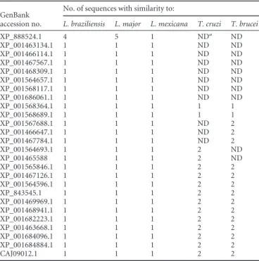
In the present work, 8 of 26 previously
identified hypothetical proteins were recognized as
Leishmania
on October 6, 2017 by UNIVERSIDADE FEDERAL DE OURO PRETO
http://cvi.asm.org/
specific antigens by database searches (
Table 1
). The other 18
pro-teins presented primary sequences similar to those of propro-teins of
T. cruzi
and/or
T. brucei
species and were not evaluated in this
study. The 8 selected proteins were mapped
in silico
to predict the
B-cell-specific epitopes, and a total of 17 peptides were
identi-fied and synthesized. The sequences of the peptides were AAVC
VAAALYAL (PepLi1), AGQSVPNSL (PepLi2), CTECDTGYSLTS
DYQCKAITT (PepLi3), FTVTRDVTMSSTSFDDYTMVLDLS
(PepLi4), SGALFSFPAGLEDASE (PepLi5), TMMPDTPSADASP
SPRITRI (PepLi6), GTSAVYERYLLLTP (PepLi7), KLLFPLPPPP
LRLPEALQELSPECH (PepLi8), VLVAAAALVIAAEQLRMPLPA
(PepLi9), SGPGAGAAL (PepLi10), QGPPPLASV (PepLi11), SVL
KGYQALKQSTAGSD (PepLi12), QEEAEEEEAAAVAGSAQPHP
(PepLi13), DMVALQEEAKSVRDRRLALEEIMR (PepLi14), DK
KQKAREERFAASLQRRLERRKA (PepLi15), PVEAVEEAVAT
(PepLi16), and QPQQPVTQQPVYQPPPPMEPV (PepLi17).
ELISA.
The synthetic peptides were then employed as antigens
in ELISA to compare their diagnostic performance, using sera
from
L. infantum
-infected dogs and healthy animals. Of the 17
peptides, 6 (namely, PepLi1, PepLi2, PepLi3, PepLi6, PepLi7, and
PepLi15) presented sensitivity and specificity values of
ⱖ
90%
and
ⱖ
95%, respectively, in identifying
L. infantum
-infected dogs
(
Table 2
). In this context, these 6 peptides were selected for the
next phase of experiments in which their sensitivity and specificity
were investigated using 20 serum samples from asymptomatic
CVL dogs and 19 serum samples from
T. cruzi
-infected animals
(
Table 3
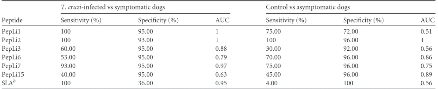
). Of these evaluated antigens, PepLi1, PepLi2, and PepLi7
presented sensitivity values of 100%, 100%, and 93% and
speci-ficity values of 95%, 93%, and 95%, respectively. In a comparison
of serum samples from asymptomatic and healthy dogs, the
sen-sitivity values were 75%, 100%, and 75% and the specificity values
were 72%, 96%, and 96% for PepLi1, PepLi2, and PepLi7,
respec-tively.
Peptide mix for the CVL diagnosis.
Finally, the 3 selected
pep-tides were combined in different mixtures to evaluate their
diag-TABLE 1Number of trypanosomatide sequences exhibiting similarityto hypothetical proteins selected fromLeishmania infantum
GenBank accession no.
No. of sequences with similarity to:
L. braziliensis L. major L. mexicana T. cruzi T. brucei
XP_888524.1 4 5 1 NDa
ND
XP_001463134.1 1 1 1 ND ND
XP_001466114.1 1 1 1 ND ND
XP_001467567.1 1 1 1 ND ND
XP_001468309.1 1 1 1 ND ND
XP_001564657.1 1 1 1 ND ND
XP_001568117.1 1 1 1 ND ND
XP_001686061.1 1 1 1 ND ND
XP_001568364.1 1 1 1 1 1
XP_001568689.1 1 1 1 1 1
XP_001567688.1 1 1 1 ND 2
XP_001466647.1 1 1 1 ND 2
XP_001467784.1 1 1 1 ND 2
XP_001564693.1 1 1 1 2 ND
XP_001465588 1 1 1 2 ND
XP_001565846.1 1 1 1 2 2
XP_001467126.1 1 1 1 2 2
XP_001564596.1 1 1 1 2 2
XP_843545.1 1 1 1 2 2
XP_001469969.1 1 1 1 2 2
XP_001468941.1 1 1 1 2 2
XP_001682223.1 1 1 1 2 2
XP_001463668.1 1 1 1 2 2
XP_001684096.1 1 1 1 2 2
XP_001684884.1 1 1 1 2 2
CAJ09012.1 1 1 1 2 2
a
ND, not detected.
TABLE 2Diagnostic performance of selected peptides using sera from symptomatic CVL dogs and serologically negative dogsa
GenBank accession no. Peptide Sensitivity (% [95% CIb]) Specificity (% [95% CI]) AUC
XP_888524.1 PepLi1 100 (69.00–100) 95.00 (75.10–99.90) 1
PepLi2 100 (69.00–100) 95.00 (74.00–99.90) 1 PepLi3 93.00 (68.00–99.80) 96.00 (79.70–99.90) 0.9307 PepLi4 67.00 (38.40–88.20) 96.00 (79.70–99.90) 0.8373 PepLi5 53.00 (26.60–78.70) 96.00 (79.70–99.90) 0.8653
XP_001463134.1 PepLi6 100 (78.20–100) 96.00 (79.70–99.90) 0.9840 PepLi7 90.00 (55.50–99.80) 95.00 (75.10–99.90) 1
XP_001466114.1 PepLi8 87.00 (59.50–98.30) 96.00 (79.70- 99.90) 0.9387 PepLi9 73.00 (44.90–92.20) 96.00 (79.70–99.90) 0.9520 PepLi10 53.00 (26.60–78.70) 96.00 (79.70–99.90) 0.9067 PepLi11 47.00 (21.30–73.40) 96.00 (79.70- 99.90) 0.9147
XP_001467567.1 PepLi12 47.00 (21.30–73.40) 96.00 (79.70–99.90) 0.6053 XP_001468309.1 PepLi13 67.00 (38.40–88.20) 96.00 (79.70–99.90) 0.9027 XP_001564657.1 PepLi14 73.00 (44.90–92.20) 96.00 (79.70–99.90) 0.9173
XP_001568117.1 PepLi15 93.00 (68.10–99.80) 96.00 (79.70–99.90) 1 PepLi16 67.00 (38.40–88.20) 96.00 (79.70–99.90) 0.8533
XP_001686061.1 PepLi17 27.00 (7.80–55.10) 96.00 (79.70–99.90) 0.7467
SLAc 100 (78.20–100) 96.00 (79.70–99.90) 1
a
Samples from symptomaticL. infantum-infected dogs (n⫽25) and animals with no clinical signs of CVL and negative parasitological and serological results forLeishmania
antigens (n⫽40) were tested in ELISA. Receiver operating characteristic (ROC) curves were used to determine sensitivity, specificity, and AUC values. b
CI, confidence interval.
cSLA, solubleLeishmania infantumantigen extract.
on October 6, 2017 by UNIVERSIDADE FEDERAL DE OURO PRETO
http://cvi.asm.org/
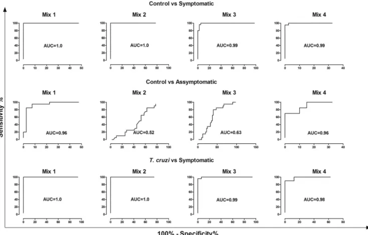
nostic performance in serological tests by ELISA. In this context,
the different mixtures were composed as follows: mix 1, PepLi
plus PepLi2; mix 2, PepLi1 plus PepLi7; mix 3, PepLi2 plus PepLi7;
mix 4, PepLi1 plus PepLi2 plus PepLi7.
Figure 1
shows the results
of the individual distribution of the serum samples in relation to
the different peptide mixtures. When the sensitivity of the
differ-ent mixtures to differdiffer-entiate among the serum samples of
symp-tomatic
L. infantum
-infected and healthy dogs was evaluated, mix
1, mix 2, mix 3, and mix 4 presented sensitivities of 100%, 100%,
80%, and 95%, respectively (
Fig. 2
). Regarding the specificity to
differentiate the serum samples of symptomatic
L. infantum
-in-fected dogs from those of
T. cruzi
-infected animals, the mixtures
presented values of 100%, 100%, 76%, and 95% for mix 1, mix 2,
mix 3, and mix 4, respectively (
Table 4
).
DISCUSSION
Serological tests are currently recommended for the laboratory
diagnosis of CVL. IFAT and ELISA are the most widespread
diag-nostic assays applicable to leishmaniasis; however, their low
sen-sitivity to detect cases with low or absent levels of
Leishmania
-TABLE 3Diagnostic performance of selected peptides using other serum samplesaPeptide
T. cruzi-infected vs symptomatic dogs Control vs asymptomatic dogs
Sensitivity (%) Specificity (%) AUC Sensitivity (%) Specificity (%) AUC
PepLi1 100 95.00 1 75.00 72.00 0.51
PepLi2 100 93.00 1 100 96.00 1
PepLi3 60.00 95.00 0.88 30.00 92.00 0.56
PepLi6 53.00 95.00 0.79 70.00 96.00 0.86
PepLi7 93.00 95.00 0.97 75.00 96.00 0.75
PepLi15 40.00 95.00 0.63 45.00 96.00 0.89
SLAb 100 36.00 0.95 4.00 100 0.56
aSamples fromT. cruzi-infected dogs (n⫽19), asymptomatic (n⫽20) and symptomatic (n⫽25)L. infantum-infected dogs, and animals with no clinical signs of CVL and negative parasitological and serological results forLeishmaniaantigens (n⫽40) were used. Receiver operating characteristic (ROC) curves were used to determine ELISA sensitivity, specificity, and AUC values.
b
SLA, solubleLeishmania infantumantigen extract.
FIG 1Evaluation of ELISA reactivity using peptide mixtures with different serum samples. ELISAs were performed using serum samples fromT. cruzi-infected (n⫽19), asymptomatic (n⫽20) and symptomatic (n⫽25)L. infantum-infected, and healthy (n⫽40) dogs. Reactions against mix 1 (A), mix 2 (B), mix 3 (C), and mix 4 (D) are shown. Mixes were as follows: mix 1, PepLi1 plus PepLi2; mix 2, PepLi1 plus PepLi7; mix 3, PepLi2 plus PepLi7; and mix 4, PepLi1 plus PepLi2 plus PepLi7. O.D., optical density.
on October 6, 2017 by UNIVERSIDADE FEDERAL DE OURO PRETO
http://cvi.asm.org/
specific antibodies and their cross-reactivity with other diseases,
including Chagas’ disease, represent important limitations for
their use in laboratory serodiagnosis (
37
). Moreover, crude
anti-gens also are limited by the difficulty of producing large quantities
in a standardized manner (
30
).
Improvements in sensitivity and specificity might be achieved
by using individual
Leishmania
proteins that are recognized by
sera from
L. infantum
-infected dogs. After database searches were
performed in this study, eight antigens were identified as
Leishma-nia
-specific proteins. Since they were recognized by sera from
L.
infantum
-infected dogs in a previous immunoproteomics study
(
29
), their primary sequences were mapped to obtain
B-cell-spe-cific epitopes, which were synthesized and subsequently evaluated
as new and more-refined antigens for a more sensitive and specific
serodiagnosis of
L. infantum
infections in dogs.
The specificity of ELISA using SLA depends heavily on antigen
preparation. Many times, false-positive results are obtained when
serum samples are collected from animals with other diseases,
such as Chagas’ disease (
22
,
38
). Recombinant leishmanial
anti-gens also have been tested with ELISA to develop a more specific
test (
25
); although their specificity is generally greater, sensitivity
is hampered, given the much lower availability of B-cell epitopes
than for SLA and the high level of heterogeneity of major
histo-compatibility complex (MHC) molecules of canine populations
(
39
).
Among the tested synthetic antigens, PepL1, PepL2, PepL3,
FIG 2Diagnostic performance of peptide mixtures with different serum samples. Samples fromT. cruzi-infected (n⫽19), symptomatic CVL (n⫽25), and healthy (n⫽40) dogs were used. Receiver operating characteristic (ROC) curves were used to determine ELISA sensitivity, specificity, and AUC values, and sensitivity and specificity values for the different mixtures with serum samples fromT. cruzi-infected, symptomatic CVL, and healthy dogs are shown. The mixes were as follows: mix 1, PepLi plus PepLi2; mix 2, PepLi1 plus PepLi7; mix 3, PepLi2 plus PepLi7; and mix 4, PepLi1 plus PepLi2 plus PepLi7.TABLE 4Diagnostic performance of peptide mixes using different serum samplesa
Mix
Control vs symptomatic dogs Control vs asymptomatic dogs T. cruzi-infected vs symptomatic dogs
Sensitivity (%) Specificity (%) AUC Sensitivity (%) Specificity (%) AUC Sensitivity (%) Specificity (%) AUC
1 100 100 1 20.00 100 0.96 100 100 1
2 100 100 1 0 95.00 0.52 100 94.00 1
3 80.00 100 1 0 95.00 0.63 76.00 100 1
4 95.00 100 1 70.00 97.50 0.96 95.00 88.00 0.98
a
Samples fromT. cruzi-infected dogs (n⫽19), symptomatic (n⫽25) and asymptomatic (n⫽20) visceral leishmaniasis dogs, and animals with no clinical signs of CVL and negative parasitological and serological results forLeishmaniaantigens (n⫽40) were used. ROC curves were used to determine ELISA sensitivity, specificity, and AUC values.
on October 6, 2017 by UNIVERSIDADE FEDERAL DE OURO PRETO
http://cvi.asm.org/
PepL6, PepL7, and PepL15 showed the highest sensitivities (100%,
100%, 93%, 100%, 90%, and 93%, respectively) and specificities
(95%, 95%, 96%, 96%, 95%, and 96%, respectively). In canine
epidemiological screening, a test with high sensitivity and
speci-ficity is desirable. The performance observed for the synthetic
an-tigens in this work is consistent with the performance of many
antigens (mainly recombinant proteins) that have been developed
in recent years for CVL diagnosis by ELISA (
23
,
24
), with the
advantages of being cheaper, simpler, reproducible, useful for
large-scale testing and, in most cases, more specific and sensitive
(
26
). Our results show an improvement in the sensitivity of the
synthetic antigen ELISA, compared to the indirect
immunofluo-rescence assay, in asymptomatic dogs, due to the fact that the IFAT
shows a lack of sensitivity for the detection of specific antibodies in
asymptomatic dogs (
20
) and in healthy dogs, due to the large
number of false-positive IFAT reactions (
18
). It should be taken
into account that the ELISA technique allows better interpretation
of the results than IFAT, because in that technique the
interpreta-tion is subjective and depends on the expertise of the operator.
Multiple-epitope chimeric antigens have been evaluated as
markers for the serodiagnosis of CVL (
25
,
40
). In an attempt to
improve sensitivity and specificity and to avoid cross-reactions,
we prepared antigenic mixtures of the synthetic antigens that
showed the best results and tested them by ELISA. All of the
mix-tures containing the synthetic antigens, i.e., mix 1, mix 2, mix 3,
and mix 4, showed high sensitivities (100%, 100%, 80%, and 95%,
respectively) and specificity (100% in all cases), as well as a perfect
accuracy (AUC
⫽
1) when tested in discriminating dogs with
symptomatic CVL from healthy animals. When tested in
discrim-inating between dogs with symptomatic visceral leishmaniasis and
T. cruzi
-infected dogs, mix 1, mix 2, and mix 4 showed high
sen-sitivities (100%, 100, and 95%, respectively), while mix 3 showed
a sensitivity of 76%. All of the mixes showed high specificities
(100%, 94%, 100%, and 88%, respectively).
The detection of asymptomatic
L. infantum
-infected dogs is
considered crucial in epidemiological studies, for laboratory
diag-nosis of the disease. A cohort study showed that the majority of
seronegative animals became positive in parasitological tests a few
months after presenting serological conversion to
Leishmania
an-tigens (
13
). In addition, in experimentally infected dogs, diagnosis
was possible as early as 45 days postinfection and before the
ani-mals become seropositive. In this context, the diagnostic
elucida-tion of asymptomatic cases would contribute to better
character-ization of the epidemiology of CVL and evaluation of control
actions. Surprisingly, mix 2 and mix 3 failed to discriminate
asymptomatic CVL dogs from healthy dogs, while mix 1 and mix
4 showed sensitivities of 20% and 70% and specificities of 100%
and 97.5%, respectively. Recently, Costa et al. (
27
) performed
ELI-SAs using single and mixed synthetic antigens from previously
evaluated proteins, which showed high sensitivity in serum
sam-ples with low (95%) and intermediate (95%) antibody titers and
high specificity (95%); the capacities of these synthetic antigen
mixtures were not assayed against asymptomatic dogs, and
cross-reactivity assays were not performed. In addition, Faria et al. (
28
)
performed ELISAs using a mixture of synthetic antigens and
ob-tained high sensitivity and specificity values (78.5% and 80%,
re-spectively), but all of the synthetic antigens and the mixture
ex-hibited high cross-reactivity with
T. cruzi
serum samples.
The serum samples used in this work did not contain samples
from dogs infected with other
Leishmania
species, such as
L.
bra-ziliensis
; the collection of sera from dogs was restricted to the
ur-ban area of Belo Horizonte (Minas Gerais, Brazil), where a low
incidence of infection with
L. braziliensis
in dogs was noted
re-cently (
41
). In addition, the sample size used in this work was
limited and follow-up evaluation of the asymptomatic dogs was
not performed. Thus, our data should be taken as a proof of
con-cept of the capacity of the proposed synthetic antigens for the
diagnosis of CVL and might serve as a reference for further assays.
Taken together, the results presented here demonstrate that
the 3 synthetic peptides obtained from previously selected
hypo-thetical proteins might be considered an interesting alternative for
a more sensitive and specific serodiagnosis of CVL, when used in
an isolated or multiple-epitope chimeric mixture format in
sero-logical testing by ELISA. This study can be considered relevant
mainly in identifying seronegative animals without clinical signs
but with positive molecular results for
L. infantum
, represented
here as asymptomatic dogs, in epidemiological studies and/or
ar-eas in which CVL is endemic.
ACKNOWLEDGMENTS
This work was supported by grants from the Pró-Reitoria de Pesquisa from UFMG (Edital 07/2012), Instituto Nacional de Ciência e Tecnologia em Nano-biofarmacêutica (INCT-NANOBIOFAR, Fundação de Amparo à Pesquisa do Estado de Minas Gerais (FAPEMIG) (CBB-APQ-02364-08, CBB-APQ-00356-10, CBB-APQ-00496-11, and CBB-APQ-00819-12), Conselho Nacional de Desenvolvimento Científico e Tecnológico (CNPq) (APQ-472090/2011-9), and the Instituto Nacional de Ciência e Tecnologia em Vacinas (INCT-V). E.A.F.C. and A.P.F. are CNPq grant recipients. M.A.C.-F. is a FAPEMIG/CAPES grant recipient. This study was supported in Spain, in part, by grants from the Ministerio de Ciencia e Innovación (FIS/PI1100095).
REFERENCES
1.World Health Organization. 2009. Leishmaniasis: the disease and its impact.http://who.int/emc/disease/leish/index.html.
2.Grimaldi G, Jr, Tesh RB.1993. Leishmaniases of the New World: current concepts and implications for future research. Clin. Microbiol. Rev.
6:230 –250.
3.Gramiccia M, Gradoni L.2005. The current status of zoonotic leishman-iases and approaches to disease control. Int. J. Parasitol.35:1169 –1180. 4.Petersen CA.2009. Leishmaniasis, an emerging disease found in
compan-ion animals in the United States. Top. Compancompan-ion Anim. Med.24:182– 188.
5.World Health Organization.2010. Control of the leishmaniases: report of a meeting of the WHO Expert Committee on the Control of Leishman-iases, Geneva, 22–26 March 2010. WHO technical report series, no. 949. WHO, Geneva, Switzerland.
6.Ready PD.2010. Leishmaniasis emergence in Europe. Euro Surveill.15: 19505.http://www.eurosurveillance.org/ViewArticle.aspx?Articleld⫽19505. 7.Missawa NA, Veloso MAE, Maciel GBML, Michalsky EM, Dias ES.
2011. Evidence of transmission of visceral leishmaniasis byLutzomyia cruziin the municipality of Jaciara, State of Mato Grosso, Brazil. Rev. Soc. Bras. Med. Trop.44:76 –78.
8.Baneth G, Koutinas AF, Solano-Gallego L, Bourdeau P, Ferrer L.2008. Canine leishmaniosis: new concepts and insights on an expanding zoono-sis: part one. Trends Parasitol.24:324 –330.
9.Barbiéri CL.2006. Immunology of canine leishmaniasis. Parasite Immu-nol.28:329 –337.
10. Ciaramella P, Oliva G, Luna RD, Gradoni L, Ambrosio R, Cortese L, Scalone A, Persechino A.1997. A retrospective clinical study of canine leishmaniasis in 150 dogs naturally infected byLeishmania infantum. Vet. Rec.141:539 –543.
11. Maia C, Campino L.2008. Methods for diagnosis of canine leishmaniasis and immune response to infection. Vet. Parasitol.158:274 –287. 12. Porrozzi R, Costa MV, Teva A, Falqueto A, Ferreira A.2007.
Compar-ative evaluation of enzyme-linked immunosorbent assays based on crude and recombinant leishmanial antigens for serodiagnosis of symptomatic
on October 6, 2017 by UNIVERSIDADE FEDERAL DE OURO PRETO
http://cvi.asm.org/
and asymptomaticLeishmania infantumvisceral infections in dogs. Clin. Vaccine Immunol.14:544 –548.
13. Courtenay O, Quinnell RJ, Garcez LM, Shaw JJ, Dye C.2002. Infec-tiousness in a cohort of Brazilian dogs: why culling fails to control visceral leishmaniasis in areas of high transmission. J. Infect. Dis.186:1314 –1320. 14. de Andrade HM, Reis AB, dos Santos SL, Volpini AC, Marques MJ, Romanha AJ.2006. Use of PCR-RFLP to identifyLeishmaniaspecies in naturally-infected dogs. Vet. Parasitol.140:231–238.
15. Lachaud L, Chabbert E, Dubessay P, Dereure J, Lamothe J, Dedet JP, Bastien P.2002. Value of two PCR methods for the diagnosis of canine visceral leishmaniasis and the detection of asymptomatic carriers. Parasi-tology125:197–207.
16. Martin-Sanchez J, Lopez-Lopez MC, Acedo-Sanchez C, Castro-Fajardo JJ, Pineda JA, Morillas-Marquez F.2001. Diagnosis of infections with Leishmania infantumusing PCR-ELISA. Parasitology122:607– 615. 17. Solano-Gallego L, Morell P, Arboix M, Alberola J, Ferrer L. 2001.
Prevalence ofLeishmania infantuminfection in dogs living in an area of canine leishmaniasis endemicity using PCR on several tissues and serol-ogy. J. Clin. Microbiol.39:560 –563.
18. Figueiredo FB, Madeira MF, Nascimento LD, Abrantes TR, Mouta-confort E, Passos SRL, Schubach TMP.2010. Canine visceral leishman-iasis: study of methods for the detection of IgG in serum and eluate sam-ples. Rev. Inst. Med. Trop. Sao Paulo52:193–196.
19. Scalone A, Luna R, Oliva G, Baldi L, Satta G, Vesco G, Mignone W, Turilli C, Mondesire RR, Simpson D, Donoghue AR, Frank GR, Gra-doni L.2002. Evaluation of theLeishmaniarecombinant K39 antigen as a diagnostic marker for canine leishmaniasis and validation of a standard-ized enzyme-linked immunosorbent assay. Vet. Parasitol.104:275–285. 20. Mettler M, Grimm F, Capelli G, Camp H, Deplazes D.2005. Evaluation
of enzyme-linked immunosorbent assays, an immunofluorescent-antibody test, and two rapid tests (immunochromatographic-dipstick and gel tests) for serological diagnosis of symptomatic and asymptomatic Leishmaniainfections in dogs. J. Clin. Microbiol.43:5515–5519. 21. Troncarelli MZ, Camargo JB, Machado JG, Lucheis SB, Langoni H.
2009.Leishmaniaspp. and/orTrypanosoma cruzidiagnosis in dogs from endemic and nonendemic areas for canine visceral leishmaniasis. Vet. Parasitol.164:118 –123.
22. Viol MA, Lima VMF, Aquino MCC, Gallo IG, Gallo IP, Alves D, Generoso SH, Perri V, Lucheis SB, Langoni H, Nunes CM, Bresciani KDS.2012. Detection of cross infections byLeishmaniaspp. and Trypano-somaspp. in dogs using indirect immunoenzyme assay, indirect fluores-cent antibody test and polymerase chain reaction. Parasitol. Res.111: 1607–1613.
23. Cândido TC, Perri SHV, Gerzoschkwitz TDO, Luvizotto MCR, de Lima VMF.2008. Comparative evaluation of enzyme-linked immunosorbent assay based on crude and purified antigen in the diagnosis of canine vis-ceral leishmaniasis in symptomatic and oligosymptomatic dogs. Vet. Parasitol.157:175–181.
24. Pinheiro PHDC, Pinheiro AN, Ferreira JHL, Costa FAL, Katz S, Barbiéri CL.2009. A recombinant cysteine proteinase from Leishma-nia(Leishmania)chagasias an antigen for delayed-type hypersensitiv-ity assays and serodiagnosis of canine visceral leishmaniasis. Vet. Para-sitol.162:32–39.
25. Soto M, Requena JM, Quijada L, Alonso C. 1998. Multicomponent chimeric antigen for serodiagnosis of canine visceral leishmaniasis. J. Clin. Microbiol.36:58 – 63.
26. Noya O, Patarroyo ME, Guzmán F, Alarcón de Noya B.2003. Immu-nodiagnosis of parasitic diseases with synthetic peptides. Curr. Protein Pept. Sci.4:299 –308.
27. Costa MM, Penido M, dos Santos MS, Doro D, de Freitas E, Michalick MS, Grimaldi G, Gazzinelli RT, Fernandes AP.2012. Improved canine
and human visceral leishmaniasis immunodiagnosis using combinations of synthetic peptides in enzyme-linked immunosorbent assay. PLoS Negl. Trop. Dis.6:e1622. doi:10.1371/journal.pntd.0001622.
28.Faria AR, Costa MM, Giusta MS, Grimaldi G, Jr, Penido MLO, Gazzinelli RT, Andrade HM. 2011. High-throughput analysis of syn-thetic peptides for the immunodiagnosis of canine visceral leishmaniasis. PLoS Negl. Trop. Dis.5:e1310. doi:10.1371/journal.pntd.0001310. 29. Coelho VT, Oliveira JS, Valadares DG, Chávez-Fumagalli MA, Duarte
MC, Lage PS, Soto M, Santoro MM, Tavares CA, Fernandes AP, Coelho EA.2012. Identification of proteins in promastigote and amastigote-like Leishmaniausing an immunoproteomic approach. PLoS Negl. Trop. Dis.
6:e1430. doi:10.1371/journal.pntd.0001430.
30. Coelho EA, Tavares CA, Carvalho FA, Chaves KF, Teixeira KN, Ro-drigues RC, Charest H, Matlashewski G, Gazzinelli RT, Fernandes AP.
2003. Immune responses induced by theLeishmania(Leishmania) don-ovaniA2 antigen, but not by the LACK antigen, are protective against experimentalLeishmania(Leishmania)amazonensisinfection. Infect. Im-mun.71:3988 –3994.
31. Bradford MM.1976. A rapid and sensitive method for the quantitation of microgram quantities of protein utilizing the principle of protein-dye binding. Anal. Biochem.72:248 –254.
32. Brenner Z.1962. Therapeutic activity and criterion of cure in mice exper-imentally infected withTrypanosoma cruzi. Rev. Inst. Med. Trop. Sao Paulo4:389 –396.
33. Bahia MT, Tafuri WL, Caliari MV, Veloso VM, Carneiro CM, Coelho GL, Lana M.2002. Comparison ofTrypanosoma cruziinfection in dogs inoculated with blood or metacyclic trypomastigotes of Berenice-62 and Berenice-78 strains via intraperitoneal and conjunctival routes. Rev. Soc. Bras. Med. Trop.35:339 –345.
34. Carneiro CM, Martins-Filho OA, Reis AB, Veloso VM, Araújo FM, Bahia MT, de Lana M, Machado-Coelho GL, Gazzinelli G, Correa-Oliveira R, Tafuri WL.2007. Differential impact of metacyclic and blood trypomastigotes on parasitological, serological and phenotypic features triggered during acuteTrypanosoma cruziinfection in dogs. Acta Trop.
101:120 –129.
35. Larsen JE, Lund O, Nielsen M.2006. Improved method for predicting linear B-cell epitopes. Immunome Res.2:2.
36. Kolaskar AS, Tongaonkar PC.1990. A semi-empirical method for pre-diction of antigenic determinants on protein antigens. FEBS Lett.276: 172–174.
37. Sundar S, Rai M.2002. Laboratory diagnosis of visceral leishmaniasis. Clin. Diagn. Lab. Immunol.9:951–958.
38. Ferreira EDC, Lana M, Carneiro M, Reis AB, Paes DV, da Silva ES, Schallig H, Gontijo CMF.2007. Comparison of serological assays for the diagnosis of canine visceral leishmaniasis in animals presenting different clinical manifestations. Vet. Parasitol.146:235–241.
39. Quinnell RJ, Kennedy LJ, Barnes A, Courtenay O, Dye C, Garcez LM, Shaw MA, Carter SD, Thomson W, Ollier WER.2003. Susceptibility to visceral leishmaniasis in the domestic dog is associated with MHC class II polymorphism. Immunogenetics55:23–28.
40. Boarino A, Scalone A, Gradoni L, Ferroglio E, Vitale F, Zanatta R, Giuffrida MG, Rosati S.2005. Development of recombinant chimeric antigen expressing immunodominant B epitopes ofLeishmania infantum for serodiagnosis of visceral leishmaniasis. Clin. Diagn. Lab. Immunol.
12:647– 653.
41. Coura-Vital W, Marques MJ, Veloso VM, Roatt BM, Aguiar-Soares RDDO, Reis LES, Braga SL, Morais MHF, Reis AB, Carneiro M.2011. Prevalence and factors associated withLeishmania infantuminfection of dogs from an urban area of Brazil as identified by molecular methods. PLoS Negl. Trop. Dis.5:e1291. doi:10.1371/journal.pntd.0001291.