ABSTRACT INTRODUCTION
The mesenchymal lesions of the vulva and perineum include both benign and malignant neoplasias. Cellular angiofibroma is a rare tumor described for the first time by Nucci et al. in 1997.1 It consists of a tumoral mass of small size (< 3 cm) that is generally well circumscribed, and it typically arises in middle-aged patients.2 The differential diagnoses for this neoplasia include aggressive angiomyxoma, angiomyofibroblas-toma, lipoma of fusiform cells, fibrous tumors, perineurioma and leiomyoma. This differentia-tion is done by means of the histological and im-munohistochemical characteristics.1 We describe a case of cellular angiofibroma dealt with in our institution, for which the preoperative diagnosis was Bartholin’s glandular cyst. To the best of our knowledge, this is the first case described in the Brazilian literature.
CASE REPORT The patient was a 51-year-old woman at-tended as an outpatient with a complaint of tumor formation in the vulva, with progressive growth detected two years earlier. The tumor was painless, and the complaint was of discomfort when seated and a localized burning sensation. Upon gynecological examination, she presented a tumor of cystic consistency, located between the right-side small and large vulvar labia, and measuring approximately 7 cm in diameter. Her obstetric history consisted of three children: two cesarean and one normal delivery. The clinical diagnosis was Bartholin’s glandular cyst, and the patient underwent elective surgery. The resected material was sent for histopathological examination.
PATHOLOGICAL AND IMMUNOHISTOCHEMICAL FINDINGS The surgical specimen measured 7.0 x 5.5 x 1.4 cm and was flat and well delimited,
with a smooth bronze-colored external surface. The sectioned surface was elastic and bronze-colored with a disorganized fascicular pattern. The histological examination demonstrated that the neoplasia was well delimited and made up of three characteristic components: fusi-form cells fusi-forming small fascicles, numerous blood vessels and adipose tissue interspersed between the fusiform cells. These cells had ovoid basophilic nuclei and eosinophilic cytoplasm that was clear and well defined. Dispersed cells with large pleomorphic and hyperchromatic nuclei were seen. Mitoses were scarce in number and mast cells were frequently found to permeate the tumor. The stroma was myxoid and hypocellular. There were no areas of necrosis or hemorrhage. The vessels were of small to medium caliber, with open lumens and walls that were frequently hyalinized, and passed haphazardly through the tumor.
The fusiform cells were positive for vi-mentin and negative for CD34, protein S-100, actin and desmin.
DISCUSSION Cellular angiofibroma of the vulva arises in middle-aged women as a small painless vulvar mass,1,2 as occurred in our case. The clinical diagnosis is usually Bartholin’s glan-dular cyst. Histologically, these lesions are characterized by fusiform cells without atypia, forming small fascicles in the middle of col-lagen bundles, and frequent blood vessels of small to medium caliber, sometimes with a hyalinized wall and a variable component of mature adipocytes (Figures 1 and 2). Such tumors generally present mitotic activity and there may be sparse atypical cells in the stroma, but necrosis is absent.1,2
In the case described, the tumor mea-sured 7 cm in diameter and had a very low mitotic index, which may have impeded the
Adilha Misson Rua Micheletti
Ana Cristina Araújo Lemos da Silva
Antonio Geraldo Nascimento
Cléber Sérgio Da Silva
Eddie Fernando Candido Murta
Sheila Jorge Adad
C
A
SE REPOR
T
Cellular angiofibroma of the vulva:
case report with clinicopathological
and immunohistochemistry study
Discipline of Gynecology, Obstetrics and Pathology, Universidade Federal
do Triângulo Mineiro, Uberaba, Minas Gerais, Brazil
CONTEXT: Cellular angiofibroma of the vulva is a rare tumor that was first described in 1997. It occurs in middle-aged women (average age: 47 years), has small size (< 3 cm) and well-cir-cumscribed margins.
CASE REPORT:We describe a case in a 51-year-old woman whose preoperative diagnosis was confounded with Bartholin’s glandular cyst. The neoplasia was well delimited and made up of three characteristic components: fusiform cells forming small fascicles, numerous blood vessels and adipose tissue interspersed between the fusiform cells. The stroma cells were positive for vimentin and negative for CD34, protein S-100, actin and desmin. The differential diagnoses for this tumor include aggressive angiomyxoma, angiomyofibroblastoma, lipoma of fusiform cells, solitary fibrous tumor, perineurioma and leiomyoma.
KEY WORDS:Angiofibroma. Connective tissue. Mesoderm. Neoplasm. Vulva.
251
diagnosis. The cases previously described in the vulva measured less than 3 cm in diam-eter.1 Garijo and Val-Bernal3 described a very similar lesion, located in the subcutaneous layer of the thoracic wall, measuring 7 cm in diameter and without mitoses. The authors commented that the size of the lesion, the low mitotic activity and the collagen forma-tion in the stroma might have been due to the longer development time of the lesion until its diagnosis and excision, in relation to the cases of Nucci et al.1
The immunohistochemical study showed positivity only for vimentin, which suggests, according to the literature, that the tumor cells are of fibroblastic origin.1 In 1998, Laskin et al.4 described 11 very similar cases of lesions in the male genital tract, which they called tumors of angiomyofibroblastoma type of the male genital tract. According to these authors, such lesions are derived from stem cells, with a capacity for adipose and myofibroblastic dif-ferentiation in accordance with the influence of hormones, microenvironments, cytokines and growth factors.
Cellular angiofibroma of the vulva appears to be benign, since there is no report of tumors that progressed with metastasis. Nonetheless, the follow-up described in most cases is short. Local excision with free margins is the appro-priate treatment.1,2 In our case, resection of le-sion was performed and the patient presented no signs of relapse four months later.
Specific tumors of soft parts of the vulva form part of the differential diagnosis, such as aggressive angiomyxoma and angiomyofi-broblastoma. The first of these is an infiltra-tive lesion with a tendency towards recur-rence that is paucicellular and has extensively myxoid stroma. Angiomyofibroblastoma is generally less cellular than cellular angiofi-broma and presents the cells arranged around the small-caliber blood vessels. These two le-sions are positive for desmin and/or actin.1,2 In fact, all three of these lesions seem to form part of a spectrum of mesenchymal lesions of the vulva with fibroblastic and myofi-broblastic differentiation.1 Other soft-part lesions form differential diagnoses and can be ruled out in accordance with the histology and immunohistochemistry.2
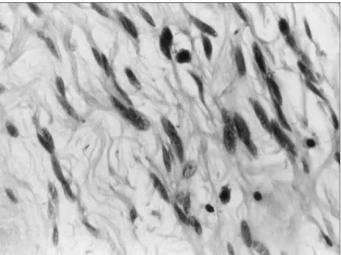
Figure 2. Small to medium size vessels with hyalinized walls, showing fusiform cells with bland nuclei and clear cytoplasm in an angiofibroma tumor (hematoxylin-eosin; 20 X). Figure 1.Photomicrograph of the angiofibroma showing its three characteristic com-ponents: fusiform cells, blood vessels and adipose tissue (hematoxylin-eosin; 4 X).
252
RESUMO Angiofibroma celular da vulva: relato de caso com estudo clinicopatológico e imunoistoquímico
CONTEXTO:O angiofibroma celular da vulva é um tumor raro que foi inicialmente descrito em 1997. Ocorre em mulheres de meia-idade (média de idade: 47 anos), apresentar pequeno tamanho (< 3 cm) e margem bem circunscrita.
RELATO DE CASO: Descrevemos um caso em mulher de 51 anos de idade cujo diagnóstico pré-operatório foi confundido com cisto de glândula de Bartholin. A neoplasia era bem delimitada e constituída por três componentes característicos: células fusiformes formando pequenos fascículos, numerosos vasos sangüíneos e tecido adiposo entremeado às células fusiformes. As células do estroma eram positivas para vimentina e negativas para CD34, proteína S-100, actina e desmina. O diagnóstico diferencial deste distinto tumor inclui angiomixoma agressivo, angiomiofibroblastoma, lipoma, tumor fibroso solitário, perineurioma, e leiomioma.
PALAVRAS-CHAVE: Angiofibroma. Tecido conjuntivo. Mesênquima. Neoplasia. Vulva.
AUTHOR INFORMATION
Adilha Misson Rua Micheletti, MD, PhD. Substitute Professor
of Discipline of Special Pathology, Universidade Federal do Triângulo Mineiro, Uberaba, Minas Gerais, Brazil.
Ana Cristina Araújo Lemos da Silva, MD. Resident doctor of
Pathology, Universidade Federal do Triângulo Mineiro,Universidade Federal do Triângulo Mineiro, do Triângulo Mineiro, Uberaba, Minas Gerais, Brazil.
Antonio Geraldo Nascimento, MD. Pathologist, specialist in
Soft Tissue, Mayo Clinic, Rochester, Minnesota, United States.
Cléber Sérgio Da Silva, MD. Substitute professor of Discipline
of Gynecology and Obstetrics, Universidade Federal doUniversidade Federal do do Triângulo Mineiro, Uberaba, Minas Gerais, Brazil.
Eddie Fernando Candido Murta, MD, PhD. Titular professor
of Discipline of Gynecology and Obstetrics, UniversidadeUniversidade Federal do Triângulo Mineiro, Minas Gerais, Brazil. do Triângulo Mineiro, Minas Gerais, Brazil.
Sheila Jorge Adad, MD, PhD. Associate professor of Discipline
of Special Pathology, Universidade Federal do TriânguloUniversidade Federal do Triângulo do Triângulo Mineiro, Uberaba, Minas Gerais, Brazil.
Address for correspondence: Eddie Fernando Candido Murta
Rua Getúlio Guarita, s/no — Abadia
Uberaba (MG) — Brasil — CEP 38025-440 Tel. (+55 34) 3318-5326
Fax. (+55 34) 3318-5342 E-mail: [email protected]
Copyright © 2005, Associação Paulista de Medicina
1. Nucci MR, Granter SR, Fletcher CD. Cellular angiofi-broma: a benign neoplasm distinct from angiomyofi-broblastoma and spindle cell lipoma. Am J Surg Pathol. 1997;21(6):636-44.
2. Nucci MR, Fletcher CD. Vulvovaginal soft tissue tumours: update and review. Histopathology. 2000;36(2):97-108.
3. Garijo MF, Val-Bernal JF. Extravulvar subcutaneous cellular angiofibroma. J Cutan Pathol. 1998;25(6):327-32. 4. Laskin WB, Fetsch JF, Mostofi FK.
Angiomyofibroblastoma-like tumor of the male genital tract: analysis of 11 cases with comparison to female angiomyofibroblastoma and spindle cell lipoma. Am J Surg Pathol. 1998;22(1):6-16.
Sources of funding: Not declared
Conflict of interest: Not declared
Date of first submission: May 11, 2004
Last received: July 9, 2004
Accepted: July 5, 2005
REFERENCES