Use of transfer factor in immunosuppressed surgical patients
Avaliação do uso de fator de transferência na resposta imunológica de pacientes
cirúrgicos imunodeprimidos
Celia regina oliveira garritano, tCBCrJ1; FranCesCodi nUBila1; renata M. CoUto1; rossano Kepler alviM Fiorelli, tCBC-rJ1;
lUCiana Berti aUn1.
INTRODUCTION
D
iscovered by Henry Sherwood Lawrence in 1955, the Transfer Factor (TF) is an extract obtained from calf splenic cells, consisting of a conjugated polypep-tide with molecular weight around 6,000 Daltons and structure similar to the RNA1-3. TF has an importantimmune stimulatory function, promoting the matura-tion and differentiamatura-tion of thymocytes in T lymphocy-tes, the restoration of function of malfunctioning peri-pheral lymphocytes, the recovery of humoral immunity through differentiation of B lymphocytes, forming plas-mocytes and synthesizing specific humoral antibodies, the increase in allogeneic graft rejection capacity, the in vitro activation of T lymphocytes through cytotoxic action, lymphokine production and increased activity of the mononuclear phagocytic system. When admi-nistered orally, it establishes direct contact with the Peyer’s plaques and lymph nodes, where it exerts a se-lective action on lymphocytes and antigen-presenting cells. Digestive enzymes and hydrochloric acid do not
influence its stability2-4.
The first evidence that cancer arises due to so-matic genetic changes came from studies on Burkitt’s lymphoma4. Since then, several malignancies have been
associated with oncogens, with the possibility of using immunomodulators as a complementary treatment to surgery, chemotherapy and radiotherapy5-10. The best
re-sults of neoplastic disease treatment are achieved when surgery accomplishes the reduction of tumor load, com-plemented by chemotherapy and radiotherapy. Howe-ver, these procedures affect the immune system, and even temporarily, influence the respective therapeutic regimens, which sometimes have to be interrupted due to the low number of leukocytes, lymphocytes and the important side effects resulting from impaired immune response. Immunostimulatory agents have contributed to avoid or minimize these collateral damages, among them the Transfer Factor, which was first used in the treatment of cancer by Fudenberg et al.11 in 1976, and
which has also been used in the treatment of non neo-plastic diseases2,3,11.
1 - Federal University of the State of Rio de Janeiro (UNIRIO), Department of General and Specialized Surgery / Master’s Degree in Medicine, Rio de Janeiro, RJ, Brazil.
A B S T R A C T
Objective : to evaluate the action of Transfer Factor on the immune response of patients with malignant neoplasm submitted to surgery, chemotherapy and radiotherapy. Method: we analyzed the variations of leukocytes, total lymphocytes, T-lymphocytes and CD4 counts in 60 patients submitted to immunostimulation with a single, daily dose of 0.5mg sublingual Transfer Factor, started simultaneously with che-motherapy and/or radiotherapy. Results: there were statistically significant increases in the counts of all cell lines studied, more pronounced after 12 months of use of the medication. Conclusion: the Transfer Factor restored immune response and showed no side effects.
METHODS
We carried out this study at the Gaffrée and Guinle University Hospital of the Federal University of the State of Rio de Janeiro – UNIRIO. We included 60 pa-tients, both men and women, aged over 30 years, with malignant neoplasms, confirmed by histopathological examination of the surgical specimen, submitted to Che-motherapy (QT) and/or radiotherapy (RT) after surgery, and followed as outpatients for 12 months. We applied the immunostimulation with TF provided by the Labora-tório de Extratos Alergênicos Ltda., registered with the Ministry of Health with the number 1729.0011.001-4. We administered the substance as a single dose of 0.5 mg sublingually daily and started concurrently with che-motherapy and/or radiotherapy. All patients underwent immunological evaluation prior to initiation of treatment by laboratory tests (leukocyte count, total lymphocytes, T lymphocytes, and CD4 lymphocyte subpopulation), whi-ch were repeated six and 12 months after initiation of therapy. We then compared those with the exams results
before the beginning of treatment.
We present results as mean and standard de-viation. We performed the data analysis using tables and graphs using the Microsft Office Excell7® software. For statistical analysis, we used Graph Pad Instat software version 3.0, San Diego California® and, for the purpose of interpretation, the type I error limit was up to 5% (p <0.05). We tested the variables through the Kolmogorov--Smirnov (KS) method, inference through the Wilcoxon’s test for non-parametric samples and the Student’s t-test for parametric samples.
The study was evaluated and approved by the Ethics in Research Committee, in accordance with Reso-lution 196/96.
RESULTS
In the statistical analysis, all the samples evalua-ted had a normal distribution by the Kolmogorov and Smirnov (KS) method. The characteristics of the patients analyzed are contained in table 1.
Table 1. Characteristics of the analyzed group.
Gender Age group – cases (%) Tumor location – cases (%)
Male Female 30-39 years 96.7%) 4 Breast 20 (33.3%)
14 (23.3%)
60 (76.7%)
40-49 years
15 (25%)
Intestine
18 (30%)
50-59 years 8 (13.3%) Stomach 11 (18.3%)
60-69 years 20 (33.3%) Pancreas 5 (8.3%)
+ 70 years 13 (21.7%) Uterus 3 (5%)
Lung 1 (1.7%)
Liposarcoma 1 (1.7%)
Kidney 1 (1.7%)
Regarding the total leukocyte count, 39 (65%) patients presented a 6-month increase in values and 50 (83.3%) in 12 months compared with the counts befo-re the beginning of therapy. This incbefo-rease ranged from 1.9% to 103% in six months, and from 2.1% to 170% in 12 months. We observed that of the 21 cases (35%) that had a reduction of the leukocyte values in six ths, 18 (85.7%) were able to recover them in 12 mon-ths, and 12 (57.1%) achieved rates higher than before
treatment.
The analysis of T lymphocytes revealed that 38 (63.3%) patients presented an increase in counts at six months and 46 (76.7%) at 12 months in comparison with the result before the start of therapy. This increase ranged from 0.4% to 320% in six months and from 0.5% to 160% in 12 months. We observed that of the 22 cases (36.7%) that had a reduction in the T lym-phocyte counts in six months, 17 (77.3%) were able to recover them in 12 months, and 11 (64.7%) achieved rates higher than before treatment.
As for the subpopulation of CD4 lympho-cytes, there were also increases, in 35 (58.3%) cases when comparing the time treatment with six months, and in 51 (85%) between the time before treatment and 12 months. This increase ranged from 0.6% to 162.1% in six months and from 0.5% to 337.1% in 12 months. We observed that of the 25 cases (41.7%)
that had CD4 subpopulation reduction in six months, 19 (76%) were able to recover them in 12 months, and 16 (84.2%) were able to obtain rates higher than befo-re tbefo-reatment.
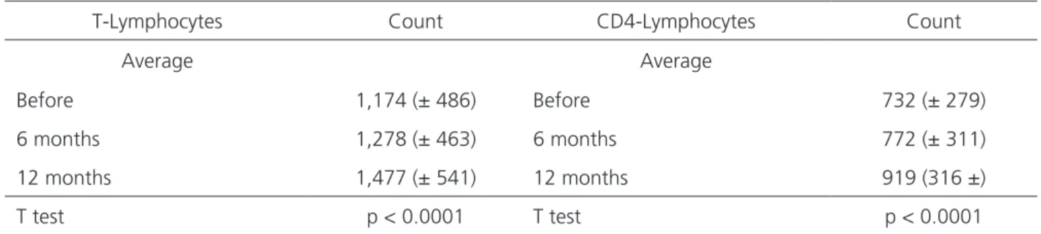
When we evaluated the T-lymphocyte avera-ges of the sample, we observed an increase of 8.8% when comparing the values before the start of treat-ment with those after six months, of 33.4% between pre-treatment and 12 months, and 15.6% between six and 12 months of treatment. The same was true for the CD4 subpopulation, with an increase of 5.5%, 20.6% and 19%, respectively. Statistical analysis of these varia-tions was very significant (Table 3).
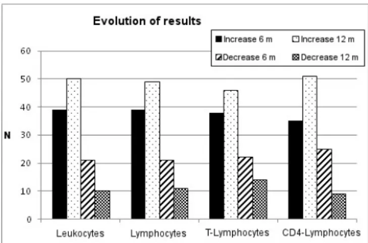
When we evaluated the means of the results in the studied period, we observed that in all of them there was an increase in values, which was more expres-sive after 12 months of treatment, as shown in figure 1. When analyzing the means of the leukocyte
counts, we observed an increase of 5.6% when compa-ring the rates before treatment with those after six mon-ths, of 20.1% between pre-treatment and 12 monmon-ths,
and of 12.4% between six and 12 months of treatment. Regarding total lymphocyte means, this increase was 5%, 24.8% and 14.9%, respectively. Statistical analysis of these variations was very significant (Table 2).
Table 2. Changes in the counts of leukocytes and lymphocytes.
Leukocytes Count Lymphocytes Count
Average Average
Before 5,073 (± 1281) Before 1,642 (± 537)
6 months 5,356 (± 1522) 6 months 1,742 (± 580)
12 months 6,019 (± 1341) 12 months 1,980 (± 594)
T test p < 0.0001 T test p < 0.0001
Values expressed as mean ± standard deviation.
Table 3. Changes in the counts of T and CD4 lymphocytes.
T-Lymphocytes Count CD4-Lymphocytes Count
Average Average
Before 1,174 (± 486) Before 732 (± 279)
6 months 1,278 (± 463) 6 months 772 (± 311)
12 months 1,477 (± 541) 12 months 919 (316 ±)
T test p < 0.0001 T test p < 0.0001
REFERENCES
1. Al-Askari S. Henry Sherwood Lawrence. In: Biographical Memoirs. Washington, D.C.: National Academy of Sciences; 2009. p. 237-55.
2. Gibson J, Basten A, Van Der Brink C. Clinical use of transfer factor 25 years. Immunol Allergy Clin N A. 1983;3(2):331-57.
3. Kirkpatrick CH. Therapeutic potential of transfer factor. N Engl J Med. 1980;303(7):390-1.
4. Cosme K, González M, Gorovaya L, Soria Y, Barcelona S, Quintana M, et al. Determinación de la actividad biológica in vivo del Factor de Transferencia. Técnica alternativa. Biotecnol Apl. 2001;18( Espec.):E16. 5. Croce CM. Oncogenes and cancer. N Engl J Med.
2008;358(5):502-11.
DISCUSSION
A deficient immune response favors the appea-rance of several diseases of viral, bacterial and neoplastic origin. When it comes to cancer, this becomes more se-rious because the tumor itself, as well as the use of QT, RT and corticosteroids, also affects the immune system, fur-ther accentuating immunosuppression. Several immuno-modulators have been used to reverse this situation with the aim not only of improving the immune response, mi-nimizing the side effects of QT and RT, but of preventing Figure 1. Comparison of the results of the exams performed.
the schedules used to be interrupted, which compromises treatment results10-20.
The lymphocytes and their T subclasses are fun-damental for the immune response, especially regarding solid tumors. Therefore, the combats to this type of tu-mor have the objective of making T-lymphocytes active and competent16,17,19,20. In this study, we observed that
the total lymphocytes and their subclasses showed an in-crease in counts, which was more pronounced with TF use for 12 months, and even when counts fell in the first six months of treatment, these were recovered after 12 months. The best response was evidenced by the subpo-pulation of CD4-lymphocytes, with an increase of 80% at the end of the study, and among those who had an initial decrease, 76% of presented an increase with 12 months of treatment.
We also sought to analyze the effect of TF on total leukocytes, and we also observed an increase in cou-nts in 83.3% of the cases with 12 months of therapy and, similar to lymphocytes, the 85.7% that had an initial re-duction displayed higher rates after 12 months of TF use. We conclude that TF promoted the activation of leukocytes, total lymphocytes and their subclasses, re-sulting in a stimulation of the immune response, specially when used for a period of 12 months.
Objetivo: avaliar a ação do Fator de Transferência na resposta imunológica de pacientes portadores de neoplasia maligna submetidos à cirurgia, quimioterapia e radioterapia. Método: análise das variações dos valores dos leucócitos, linfócitos totais, linfócitos T e CD4 em 60 pacientes submetidos à imunoestimulação com Fator de Transferência administrado em dose única de 0,5mg por via sublingual, diariamente e iniciada simultaneamente à quimioterapia e/ou radioterapia. Resultados: houve um aumento no número de todas as linhagens celulares estudadas que foi mais acentuada após 12 meses de uso da medicação. A análise estatística realizada com o software
Graph Pad Instat, testadas pelo método Kolmogorov and Smirnov, mostrou que os resultados foram significativos. Conclusão: o Fator de Transferência restabeleceu a resposta imune e não apresentou efeitos colaterais.
Descritores: Fator de Transferência. Imunidade Celular. Invasividade Neoplásica.
6. Cook JA, Taylor D, Cohen C, Hoffmann EO, Rodrigue J, Malshet V, et al. Evaluation of effector cells mediating the antitumor action of glucan. J Reticuloendothel Soc. 1977;22(1):21-34.
7. Diluzio NR. Macrophage glucan-activated macrophages and neoplasia. In: Altura BM, Saba TM, editors. Pathophysiology of the reticuloendothelial system. New York: Raven Press; 1981. p. 209-24. 8. Feng H, Shuda M, Chang Y, Moore PS. Clonal
integration of a polyomavirus in human Merkel cell carcinoma. Science. 2008;319(5866):1096-100. 9. Mantovani A, Allavena P, Sica A, Balkwill F.
Cancer-related inflammation. Nature. 2008;454(7203):436-44.
10. Garritano CRO, Gomes JCG, Pimenteira CAP. ß1-3 Glucan no tratamento do câncer de intestino. J Bras Med. 2010;98(4):22-4.
11. Levine PH, Pizza G, Ajmera K, De Vinci C, Viza D. Transfer factor in virus-associated malignancies: an underestimated weapon in prevention and treatment of cancer. Adv Tumor Virol. 2002;2:7-20.
12. Souza CA, Vigorito AC, Aranha FJP, Oliveira GB, Eid KAB, Ruiz MA. Terapêutica citoprotetora em pacientes tratados com quimio e/ou radioterapia anti neoplásica. Rev Bras Hematol.Hemoter. 2000;22(2):123-8.
13. Patchen ML, MacVittie TJ, Souza LM. Postirradiation treatment with granulocyte colony-stimulating factor and preirradiation WR-2721 administration synergize to enhance hemopoietic reconstitution and increase survival. Int J Radiat Oncol Biol Phys. 1992;22(4):773-9. 14. De Souza CA, Santini G, Marino G, Nati S,
Congiu AM, Vigorito AC, et al. Amifostine (WR-2712), a cytoprotective agent during high-dose cyclophosphamide treatment of non-Hodgkin’s
lymphomas: a phase II study. Braz J Med Biol Res. 2000;33(7):791-8.
15. Gattinoni L, Powell DJ Jr, Rosenberg SA, Restifo NP. Adoptive immunotherapy for cancer: building on success. Nat Rev Immunol. 2006;6(5):383-93.
16. Rosenberg SA, Restifo NP, Yang JC, Morgan RA, Dudley ME. Adoptive cell transfer: a clinical path to effective cancer immunotherapy. Nat Rev Cancer. 2008;8(4)299-308.
17. Guinn BA, Kasahara N, Farzaneh F, Habib NA, Norris JS, Deisseroth, AB. Recent advances and current challenges in tumor immunology and immunotherapy. Mol Ther. 2007;15(6):1065-71.
18. Morgan RA, Dudley ME, Wunderlich JR, Hughes MS, Yang JC, Sherry RM, et al. Cancer regression in patients after transfer of genetically engineered lymphocytes. Science. 2006;314(5796):126-9.
19. Ribeiro-Santos G. Quimioterapia do câncer: imunossupressão x imunoestimulação. Rev Inter Toxicol Risco Ambiental Soc. 2009;2(3):51-4.
20. Yoon TJ, Kim TJ, Lee H, Shin KS, Yun YP, Moon WK, et al. Anti-tumor metastatic activity of beta-glucan purified from mutated Saccharomyces cereviase. Int Immunoparmacol. 2008;8(1):36-42.
Received in: 16/03/2017
Accepted for publication: 01/06/2017 Conflict of interest: none.
Source of funding: none.
Mailing address:
Rossano Kepler Alvim Fiorelli