Christiane Aires Teixeira1, José Ernesto Dos Santos2, Gerusa Alves Silva3,
Elisa Sebba Tosta de Souza4, José Antônio Baddini Martinez5
Abstract
Objective: To investigate dyspnea in individuals with Class II or III obesity and look for correlations among the respiratory data related to
such individuals. Methods: This study involved 49 subjects with a body mass index > 35 kg/m2, divided into two groups (those with dyspnea
and those without). The baseline dyspnea index was evaluated, as were spirometry findings, maximal respiratory pressures and arterial blood
gas analysis. Results: Of the 49 subjects evaluated, 37 reported dyspnea and 12 reported no dyspnea. The baseline dyspnea index differed
between the two groups. The mean values were within the range of normality for all subjects and all parameters, except for the following: ratio of residual volume to total lung capacity; expiratory reserve volume; and the alveolar-arterial oxygen gradient. The subjects with dyspnea presented significantly lower values for expiratory reserve volume, maximal expiratory pressure and arterial pH. In all subjects, body mass index correlated significantly with the following: baseline dyspnea index; the residual volume/total lung capacity ratio; the forced expiratory volume in one second/forced vital capacity ratio; forced expiratory flow between 25 and 75% of forced vital capacity; arterial oxygen tension; the alveolar-arterial oxygen gradient; and arterial carbon dioxide tension. The baseline dyspnea index was found to correlate significantly with the following parameters: residual volume/total lung capacity ratio; expiratory reserve volume; arterial oxygen tension;
the alveolar-arterial oxygen gradient; and arterial carbon dioxide tension. Conclusion: Dyspnea is a common complaint in individuals with
class II or III obesity. Such individuals present a pronounced reduction in expiratory reserve volume and an increase in the alveolar-arterial
oxygen gradient. The correlations found suggest that obese individuals present dysfunction of the lower airways, and that obesity itself plays a role in the genesis of dyspnea.
Keywords: Obesity/complications; Dyspnea/epidemiology; Dyspnea/physiopathology; Respiratory function tests.
*Study conducted at the Faculdade de Medicina de Ribeirao Preto da Universidade de São Paulo (FMRP - USP, University of São Paulo at Ribeirão Preto School of Medicine) – Ribeirão Preto (SP) Brazil
1. Graduate student in the Department of Clinical Medicine of the University of São Paulo at Ribeirão Preto School of Medicine – FMRPUSP, Ribeirão Preto (SP) Brazil.
2. PhD Professor in the Division of Nutrology of the Department of Clinical Medicine of the Faculdade de Medicina de Ribeirao Preto da Universidade de São Paulo (FMRP - USP, University of São Paulo at Ribeirão Preto School of Medicine) – Ribeirão Preto (SP) Brazil.
3. PhD Professor in the Division of Pulmonology of the Department of Clinical Medicine of the Faculdade de Medicina de Ribeirao Preto da Universidade de São Paulo (FMRP - USP, University of São Paulo at Ribeirão Preto School of Medicine) – Ribeirão Preto (SP) Brazil.
4. Graduate student in the Department of Clinical Medicine of the Faculdade de Medicina de Ribeirao Preto da Universidade de São Paulo (FMRP - USP, University of São Paulo at Ribeirão Preto School of Medicine) – Ribeirão Preto (SP) Brazil.
5. PhD Professor in the Division of Pulmonology of the Department of Clinical Medicine Faculdade de Medicina de Ribeirao Preto da Universidade de São Paulo (FMRP - USP, University of São Paulo at Ribeirão Preto School of Medicine) – Ribeirão Preto (SP) Brazil.
Correspondence to: Christiane Aires Teixeira. Departamento de Clínica Médica, Hospital das Clínicas da Faculdade de Medicina de Ribeirão Preto da Universidade de São Paulo. Av. Bandeirantes, 3.900, CEP 14048-900, Ribeirão Preto, SP, Brasil. Phone 55 16 3602-2631. E-mail: [email protected]
Introduction
Numerous studies have called attention to the increasing prevalence of obesity in the world popu-lation.(1-3) The number of deaths due to diseases related to excess weight also increases annu-ally.(4) Therefore, more comprehensive study of this morbidity and its consequences has become imper-ative. Obesity is defined as a body mass index (BMI) of 30 kg/m2 or greater. The severity of the condi-tion, however, is evaluated in classes. Based on BMI values, we recognize the following levels of obesity: class I (BMI between 30 and 34.9 kg/m2); class
II
(BMI between 35 and 39.9 kg/m2); and class
III
(BMI greater than 40 kg/m2).
Obesity is correlated with various abnormali-ties of respiratory mechanics and with impaired gas exchange. Reduced strength and endurance of respi-ratory muscles, as well as pulmonary dysfunction and lower exercise capacity, have been described.(5-9) Alterations of this nature can contribute to the onset of dyspnea,(10,11) a symptom described as being more prevalent in obese individuals.(12,13) Some obese individuals can also develop alveolar hypoventilation syndrome.(14) In addition, there is an increased risk of obstructive sleep apnea.(15) In most cases, these two conditions are concomitant. Such patients frequently develop respiratory failure and cor pulmonale. Asthma and gastroesophageal reflux have also been described as being more common in obese individuals.(16-18)
In clinical practice, obesity is rarely recognized as cause of dyspnea, and the mechanisms involved in its genesis are not well known. In addition, it remains unclear how altered respiratory function corre-lates with the intensity of the sensation of dyspnea in these individuals. In view of this, the objective of this study was to investigate the occurrence of dyspnea in patients with pronounced obesity and without accompanying diseases or a significant smoking habit. An additional aim was to compare individuals complaining of dyspnea to those without such complaints in terms of the respiratory function data. Finally, we attempted to determine whether the intensity of dyspnea correlates with respiratory func-tion parameters and with BMI.
Methods
The study involved 49 obese patients evalu-ated at the Obesity Outpatient Clinic and Nutrition
Ward of the University of São Paulo at Ribeirão Preto School of Medicine Hospital das Clínicas. The sample comprised males and females, all with a BMI greater than 35 kg/m2. All of the patients were advised of the objectives of the study and the procedures involved, and all gave written informed consent. The study design was approved by the Hospital das Clínicas Ethics in Research Committee (Process 5464/99).
Patients who were current or former smokers with a history of tobacco consumption greater than 10 pack-years were excluded from the study. Patients currently under treatment for diabetes, as well as those with severe or poorly-controlled hypertension were also excluded, as were those suffering from any pulmonary, cardiac or systemic disease. Additional exclusion criteria were having a history of bronchoconstriction (even in childhood) and presenting symptoms that could be attributable to sleep apnea.
In the pulmonary function laboratory, the patients were initially questioned as to whether they experienced shortness of breath while performing any activity of their daily routine. The interviewers used questions such as: “Do you feel shortness of breath or fatigue of the muscles in your chest?” The answers obtained were simply classified as “yes” or “no”. Subsequently, all the individuals were asked questions that permitted the calculation of Mahler baseline dyspnea index (BDI), adapted by Stoller et al.(19) The degree of dyspnea evaluated by this instrument ranges from 0 to 12, the higher scores indicating less intensity of the symptom.
(ERV). The results are expressed as percentages of the values of normality and were calculated based on the equations devised by Pereira et al.(21) and Neder et al.(22) for the Brazilian population.
Maximal respiratory pressures were measured using an MPG vacuum manometer (OEM Medical, Marshalltown, IA, USA). Maximal inspiratory pres-sure (MIP) was meapres-sured based on the functional residual capacity, and maximal expiratory pressure (MEP) was based on TLC. The measurement of both (MIP and MEP) was made using at least three maneuvers and calculating the arithmetic mean of the selected values. Values are expressed as the percentage of normality according to the equations devised by Neder et al.(23)
The samples of arterial blood for the blood gas analysis were drawn from the radial artery while the individual was at rest and breathing room air. After collection, the material was promptly analyzed using a gas system analyzer (model 178; Corning, Medfield, MA, USA). The alveolar-arterial oxygen gradient (P(A-a)O2) was calculated using the clas-sical equation from the literature.(24) The equations used for the calculation of the predicted values for arterial oxygen tension (PaO2)(20) and P(A-a)O
2 were, respectively:
P(A-a)O2 sitting = (0.27 x age) + (1) 7 PaO2 = 143.6 − (0.39 x age) −
(0.56 x BMI) − (0.57 x PaCO2) where PaCO2 is arterial carbon dioxide tension.
The results obtained are expressed as means and standard deviations. The comparisons between the groups with and without dyspnea were performed using Student’s t-test, and the correlations between two variables were determined using Pearson’s correlation coefficient. The level of significance was set at p = 0.05.
Results
A total of 49 individuals were evaluated. There were 41 females and 8 males, the mean age was 38.1 ± 9.5 years, and the mean BMI was 52.9 ± 9.2 kg/m2. When questioned regarding the occur-rence of dyspnea, 37 individuals (75.5%) answered “yes”, and 12 (24.5%) answered “no” (24.5%). These were designated Group 1 and Group 2, respectively. As can be seen in Table 1, the mean BDI values differed significantly between these two groups
(p = 0.005). However, there were no significant differences regarding gender, age, BMI values or smoking history.
The mean values of the spirometric data obtained for all of the patients are shown in Table 1. Establishing a value of 80% of predicted as the lower limit of normality, we can consider that all the mean values of the parameters analyzed are within the expected range, with the exception of ERV (mean, 44.6 ± 23.1% of predicted). Adopting 120% as the upper limit of normality, the overall RV/TLC ratio for the sample as a whole could be considered high.
Regarding the spirometric data, the only signifi-cant difference observed between Groups 1 and 2 was related to ERV, which was lower in Group 1 than in Group 2 (40.8 ± 23.3% vs. 55.8 ± 19.5%).
The mean MEP and MIP values for the sample as a whole were 139.2 ± 34.5% and 106.7 ± 30.4% of predicted, respectively. Group 1 presented a mean MEP value significantly lower than that found for Group 2 (133.2 ± 34.6% vs. 157.6 ± 27.8%).
Table 1 also shows the mean arterial blood gas analysis values for the individuals studied. The mean P(A-a)O2 for the sample as a whole (22.5 ± 8.6 mmHg) was slightly higher than the expected value of 17.28 mmHg. Thirty-six individ-uals presented P(A-a)O2 values below the predicted value (73.5%). The mean PaO2 of the patients in the sample as a whole (75.9 ± 9.6 mmHg) was very close to the 77.64 ± 8.99 mmHg expected for their age. Nevertheless, 29 patients presented PaO2 values lower than that calculated (59.2%). Only 2 patients presented PaCO2 values greater than 45 mmHg, and the sample as a whole presented normocapnia. When comparing Group 1 and Group 2 in terms of the gas exchange data, we notice that the pH was significantly lower in Group 1 than in Group 2 (7.43 ± 0.03 vs. 7.48 ± 0.10).
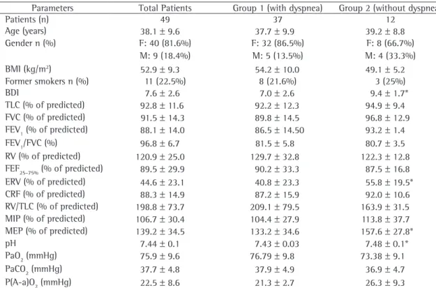
Table 1 - Clinical data and functional variables of the patients studied.
Parameters Total Patients Group 1 (with dyspnea) Group 2 (without dyspnea)
Patients (n) 49 37 12
Age (years) 38.1 ± 9.6 37.7 ± 9.9 39.2 ± 8.8
Gender n (%) F: 40 (81.6%) F: 32 (86.5%) F: 8 (66.7%)
M: 9 (18.4%) M: 5 (13.5%) M: 4 (33.3%)
BMI (kg/m2) 52.9 ± 9.3 54.2 ± 10.0 49.1 ± 5.2
Former smokers n (%) 11 (22.5%) 8 (21.6%) 3 (25%)
BDI 7.6 ± 2.6 7.0 ± 2.6 9.4 ± 1.7*
TLC (% of predicted) 92.8 ± 11.6 92.2 ± 12.3 94.9 ± 9.4
FVC (% of predicted) 91.5 ± 14.3 89.8 ± 14.5 96.8 ± 12.9
FEV1 (% of predicted) 88.1 ± 14.0 86.5 ± 14.50 93.2 ± 1.4
FEV1/FVC (%) 96.8 ± 6.7 81.5 ± 5.8 80.7 ± 3.5
RV (% of predicted) 120.9 ± 25.0 129.7 ± 32.8 122.3 ± 12.8
FEF25–75% (% of predicted) 89.5 ± 29.9 90.2 ± 33.3 87.5 ± 16.8
ERV (% of predicted) 44.6 ± 23.1 40.8 ± 23.3 55.8 ± 19.5*
CRF (% of predicted) 88.3 ± 14.9 87.2 ± 15.9 92.0 ± 10.6
RV/TLC (% of predicted) 198.8 ± 73.7 209.1 ± 79.5 163.9 ± 31.5
MIP (% of predicted) 106.7 ± 30.4 104.4 ± 27.9 113.8 ± 37.7
MEP (% of predicted) 139.2 ± 34.5 133.2 ± 34.6 157.6 ± 27.8*
pH 7.44 ± 0.1 7.43 ± 0.03 7.48 ± 0.1*
PaO2 (mmHg) 75.9 ± 9.6 76.79 ± 9.8 73.38 ± 9.1
PaCO2 (mmHg) 37.7 ± 4.8 37.9 ± 4.9 36.9 ± 4.7
P(A-a)O2 (mmHg) 22.5 ± 8.6 21.3 ± 2.7 26.3 ± 9.3
Data presented in numbers and percentages, mean and standard deviation; *p = 0.05 using Student's t-test; BMI: body mass index; BDI: baseline dyspnea index; TLC: total lung capacity; FVC: forced vital capacity; FEV1: forced expiratory volume in one second; RV:
residual volume; FEF25–75%: forced expiratory flow between 25 and 75% of FVC; ERV: expiratory reserve volume; FRC: functional residual capacity; MIP: maximal inspiratory pressure; MEP: maximal expiratory pressure; PaO2: arterial oxygen tension; PaCO2: arterial
carbon dioxide tension; and P(A-a)O2: alveolar-arterial oxygen gradient.
Discussion
In the present study, we investigated the preva-lence of dyspnea, as well as the correlations between dyspnea and respiratory function parameters, in a highly select group of obese individuals. All of the individuals studied presented advanced obesity (class II or III). In addition, none of the volunteers selected had a relevant history of smoking or signif-icant comorbidities that might cause respiratory complaints. Our results show that the prevalence of dyspnea was extremely high in these obese patients. In epidemiological studies, the prevalence of dyspnea in the general population has been reported to be from 3 to 5% in young individuals, approximately 10% in elderly men and 20% in elderly women.(12) Although the females outnumbered the males in the present study, 75.5% of the patients in the sample as a whole complained of dyspnea. This is a much
higher prevalence than that reported for the general population.
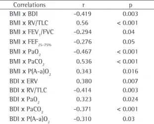
Table 2 - Pearson’s correlation coefficients for selected correlations.
Correlations r p
BMI x BDI -0.419 0.003
BMI x RV/TLC 0.56 < 0.001
BMI x FEV1/FVC -0.294 0.04
BMI x FEF25–75% -0.276 0.05
BMI x PaO2 -0.467 < 0.001
BMI x PaCO2 0.536 < 0.001
BMI x P(A-a)O2 0.343 0.016
BDI x ERV 0.380 0.007
BDI x RV/TLC -0.414 0.003
BDI x PaO2 0.323 0.024
BDI x PaCO2 -0.371 < 0.001
BDI x P(A-a)O2 -0.310 0.03
BMI: body mass index; BDI: baseline dyspnea index; RV: residual volume; TLC: total lung capacity; FEV1: forced expiratory volume in one second; FVC: forced vital capacity; FEF25–75%: forced expiratory flow between 25 and 75% of FVC; PaO2:
arte-rial oxygen tension; and PaCO2: arterial carbon dioxide tension; P(A-a)O2: alveolar-arterial oxygen gradient; ERV: expiratory
reserve volume.
which is the idea that the pulmonary blood volume increases, resulting in congestion of the vessels of the bronchial submucosa.(27) Another explanation is that the constriction of the lower airways results from excessive concentrations of circulating low density lipoproteins, which stimulates local hista-mine release by basophils.(28)
In contrast with the findings of other studies,(5-7,29) we observed no decrease in the mean TLC and FVC for our sample as a whole. Our finding suggests that drops in these pulmonary capacities are not as common in morbidly obese individuals as has been thought.(13,26) Another factor that could explain our results is the fact that we did not include a control group. In this context, an analysis based solely on percentage of predicted data would not be suffi-ciently sensitive to detect possible altered respiratory functions in obese individuals. In addition, few of the equations used in the calculation of the values of normality include weight as a component, since only age and height have shown predictive impor-tance in regression analyses. It must be stressed that such equations were devised based on data obtained in healthy reference populations of nonobese indi-viduals. Therefore, the real contribution of obesity
0 0 2 4 6 8 10 12
20 40 60 80 100
r = -0.419
BDI
BMI (kg/m2)
0 0 2 4 6 8 10 12
2 4 6 8 10 12
r = 0.380
ERV (% of predicted)
BDI
0 0 60 100 260 360 600
2 4 6 8 10 12
r = -0.414
RV/TLC (% of predicted)
BDI 200
160 300 400 460
0 20 40 60 80 100
BMI (kg/m2)
0 60 100 260 360 600
r = 0.560
RV/TLC (% of predicted)
200 160 300 400 460
as an independent variable for the pulmonary func-tion could not be adequately tested.(26)
The obese patients evaluated presented higher than average P(A-a)O2, if analyzed in relation to the value predicted for their age. We believe that the appearance of alterations in the ventilation/perfusion ratio or of micro-atelectasis, particularly in the lung bases, secondary to the compression created by the adipose tissue, can be the cause of these gas exchange disorders. It should be emphasized that the present study did not include patients with clinical manifestations that could be attributable to clin-ical profiles related to sleep disordered-breathing, although none of the subjects were submitted to polysomnography. Despite the fact that alveolar hypoventilation is known to be common in obese individuals,(14) only 2 of the patients in our study presented hypercapnia. Those patients presented quite low ERVs and high RV/TLC ratios, which seem sufficient to explain the gas exchange disorder.
The maximal respiratory pressures are generally described as normal in obese individuals.(5) However, decreased MEP has also been demonstrated and was found to be associated with reduced maximal volun-tary ventilation, with dyspnea and with reduced FEF75%.(13) Nevertheless, the results of this last study might have been influenced by the presence of smokers in the sample. The mean MIP and MEP found for the group of patients evaluated in the present study can be considered normal. The mean MEP value was 139.2% of predicted, which seems excessive. The reasons for this last finding are not clear. However, again, it is possible that the equation used to determine the expected values was inappro-priate. We can also assume that the obesity-related probable increase of the elastic recoil capacity of the chest cavity could have influenced this finding.
When we compared the patients divided into groups, based on the presence or absence of dyspnea, we observed that the BDI was, as expected, significantly lower in Group 1 than in Group 2. No significantly differences were detected regarding other clinical data or smoking history, although the mean BMI was considerably higher in Group 1 than in Group 2. Therefore, these factors, in isolation, do not seem to explain the occurrence of dyspnea. Regarding the functional data, mean ERV, MEP and arterial pH were significantly lower in Group 1 than in Group 2. Notwithstanding the fact that a totally acceptable explanation for the last two findings has
yet to be found, the lowest value of ERV in the group that presented the most dyspnea draws our attention to the importance of this pathophysiolog-ical finding in the genesis of the symptom.
We found that BMI and BDI both correlated with respiratory functional parameters, although the strength of those correlations was, in general, of a modest nature. Nevertheless, the study of the correlations between BMI and the remaining variables revealed the existence of a significant correlation between the intensity of dyspnea and the degree (class) of obesity. The BMI increased in parallel with a decrease in the BDI, the latter trans-lating to a greater intensity of dyspnea. In addition, as the excess of weight increases, the RV/TLC ratio increases, whereas the FEF25–75% and the FEV1/FVC ratio decrease. Similarly, increases in the degree of obesity are accompanied by decreases in the PaO2, as well as by increases in PaCO2 and P(A-a)O2. We believe that the explanation for these findings could reside in the presence of obstructive phenomena in the airways, which are associated with weight gain. In addition to the hypotheses mentioned above, we believe that obesity can also lead to the compression and narrowing of the small airways, principally at the level of the lung bases. Within this context, the presence of an excessive quantity of adipose tissue, especially at the upper abdominal level, could lead to an increase in the baseline peribronchial pres-sures, with subsequent impairment of airflow and increased closing volume. This last alteration can explain the drop in the mean ERV, as well as the tendencies toward the increase in RV and in the RV/TLC ratio, observed in the total group of patients. This same type of phenomenon can potentially lead to the appearance of alterations in the ventila-tion/perfusion ratio and shunt areas, which would explain the correlations observed between BMI and the intensity of the gas exchange disorders.
In recent years, it has been reported that the incidence of asthma is high in obese patients.(16-18,30) Based on the results obtained in the present study, we can suppose that the obesity-induced early occlu-sion of the small airways contributed to this finding. The clinical profile of asthma would appear in those patients with favorable genetic conditions and more accentuated bronchial hyperresponsiveness.
latter contribute to the appearance of dyspnea in obese patients. Naturally, the mechanisms involved in the pathogenesis of the last two alterations would be the same as those mentioned previously. The intensity of the dyspnea also correlated with the degree of arterial blood gas alterations. Decreases in PaO2 and increases in P(A-a)O2 parallel decreases in the BDI (increases in the intensity of the dyspnea). Similarly, increases in PaCO2 are accompanied by increases in the intensity of the dyspnea reported by the patients. It is known that hypoxemia, as well as, and principally, the hypercapnic stimulus, can contribute to the occurrence of unpleasant respi-ratory sensations.(10,11) However, the finding of a significant correlation between BDI and the wors-ening of gas exchange does not necessarily imply a cause-and-effect relationship.
In conclusion, dyspnea has proven to be a quite common symptom in individuals with class II or III obesity. Although the mechanisms related to the occurrence of dyspnea are complex and are not completely understood, the findings of the present study suggest that obstruction of the small airways and worsening of gas exchange plays a role in the genesis of the symptom in such patients. Additional studies, focused on the investigation of the genesis of dyspnea in individuals with class II or III obesity, will, of necessity, have to evaluate these aspects.
Acknowledgments
We gratefully acknowledge Luciana Straccia and Elizabeth Sobrani for their technical assistance, and Prof. Dr. João Terra Filho for his impeccable admin-istration of the Pulmonary Function Laboratory.
References
1. Sturm R. Increases in clinically severe obesity in the United States, 1986-2000. Arch Intern Med. 2003;163(18): 2146-8.
2. Flegal KM, Carrol MD, Ogden CL, Johnson CL. Prevalence and trends in obesity among US adults 1999-2000. JAMA. 2002;288(14):1723-7.
3. World Health Organization. Obesity: preventing and managing the global epidemic. Geneva, Switzerland.: WHO; 2000. (Technical Report Series, 894).
4. Allison DB, Fontaine KR, Manson JE, Stevens J, VanItallie TB. Annual deaths attributable to obesity in the United States. JAMA. 1999;282(16):1530-8.
5. Koenig, SM. Pulmonary complications of obesity. Am J Med Sci. 2001;321(4):249-79.
6. Jubber AS. Respiratory complications of obesity. Int J Clin Pract. 2004;58(6):573-80.
7. Li AM, Chan D, Wong E, Yin J, Nelson EA, Fok TF. The effects of obesity on pulmonary function. Arch Dis Child. 2003;88(4):361-3.
8. Ahmad D, Morgan WK. Obesity and lung function. Thorax. 2001;56(9):740-1
9. Milani RV, Lavie CJ, Mehra MR. Cardiopulmonary exercise testing: how do we differentiate the cause of dyspnea? Circulation. 2004;110(4):e27-31.
10. Martinez JAB, Padua AI. Dispnéia: novos conhecimentos sobre um velho problema. In: Terra Filho M, Fernandes ALG, Stirbulov R, editores. Pneumologia. Atualização e reciclagem. São Paulo: Vivali; 2001. vol. 2, p. 1-12.
11. Dyspnea. mechanisms, assessment, and management: a consensus statement. American Thoracic Society. Am J Respir Crit Care Med. 1999;159(1):321-40.
12. Curley FJ. Dyspnea. In: Irwin RS, Curley FJ, Grossman RF, editors. Diagnosis and treatment of symptoms of the respiratory tract. New York: Futura; 1997. p. 55-115. 13. Sahebjami H. Dyspnea in obese healthy men. Chest.
1998;114(5):1373-7.
14. Kessler R, Chaouat A, Schinkewitch P, Faller M, Casel S, Krieger J et al. The obesity-hypoventilation syndrome revisited. Chest. 2001;120(2):369-76.
15. Patel, SR. Shared genetic risk factors for obstructive sleep apnea and obesity. J Appl Physiol. 2005;99(4):1600-6. 16. Weiss ST, Shore S. Obesity and asthma. Directions for
research. Am J Respir Crit Care Med. 2004; 169(8): 963-8. 17. Luder E, Ehrlich RI, Lou WYW, Melnik TA, Kattan M. Body
mass index and risk of asthma in adults. Respir Med. 2004;98(1):29-37
18. Gunnbjornsdottir MI, Omenaas E, Gislason T, Norrman E, Olin AC, Jogi R, Jensen EJ, Lindberg E, Bjornsson E, Franklin K, Janson C, Gulsvik A, Laerum B, Svanes C, Toren K, Tunsater A, Lillienberg L, Gislason D, Blondal T, Bjornsdottir US, Jorundsdottir KB, Talvik R, Forsberg B, Franklin K, Lundback B, Soderberg M, Ledin MC, Boman G, Norback D, Wieslander G, Spetz-Nystrom U, Cashelunge KS, Ryden E; RHINE Study Group. Obesity and nocturnal gastro-oesophageal reflux are related to onset of asthma and respiratory symptoms. Eur Respir J. 2004;24(1):116-21.
19. Stoller JK, Ferranti R, Feinstein AR. Further specification and evaluation of a new clinical index for dyspnea. Am RevAm Rev Respir Dis. 1986;134(6):1129-34.
20. Sociedade Brasileira de Pneumologia e Tisiologia. III Consenso Brasileiro no Manejo da Asma 2002. J Pneumol. 2002;28(Supl 1):S3.
21. Pereira CAC, Barreto SP, Simões JG, Pereira FWL, Gerstler JG, Nakatani J, et al. Valores de referência para espirometria em uma amostra da população brasileira adulta. J Bras Pneumol. 1992;18(1):10-22.
22. Neder JA, Andreoni S, Castelo-Filho A, Nery LE. Reference values for lung function tests. I. Static Volumes. Braz J Med Biol Res 1999;32(6):703-17.
23. Neder JA, Andreoni S, Lerario MC, Nery LE. References values for lung function tests. II. Maximal respiratory pressures and voluntary ventilation. Braz J Med Biol Res 1999;32(6):719-27.
24. Miller WF, Scacci R, Gasti LR. Laboratory evaluation of pulmonary function. Philadelphia: Lippincott; 1987. 25. Rasslan Z, Saad Jr R, Stirbulov R, Fabbri RMA, Lima CAC.
26. Rubinstein I, Zamel N, DuBarry L, Hoffstein V. Airflow limitation in morbidly obese, nonsmoking men. Ann Intern Med. 1990;112(11):828-32. Erratum in: Ann Intern Med 1990;113(4):334. Erratum in: Ann Intern Med 1990;113(4):334.
27. Hogg JC, Pare PD, Moreno R. The effects of submucosal edema on airways resistance. Am Rev Respir Dis 1987;135(6 Pt 2):S54-6.
28. Gonen B, O’Donnell P, Post TJ, Quinn TJ, Schulman ES. Very low lipoproteins (VLDL) trigger the release of histamine from human basophils. Biochim Biophys Acta. 1987;917(3):418-24.
29. Canoy D, Luben R, Welch A, Bingham S, Wareham N, Day N, et al.. Abdominal obesity and respiratory function in men and women in EPIC-Norfolk Study, United Kingdom. Am J Epidemiol. 2004;159(12):1140-9.